A multiple drug loaded, functionalized pH sensitive nanocarrier as therapeutic and epigenetic modulator for osteosarcoma - Nature
←
→
Page content transcription
If your browser does not render page correctly, please read the page content below
www.nature.com/scientificreports
OPEN A multiple drug loaded,
functionalized pH‑sensitive
nanocarrier as therapeutic
and epigenetic modulator
for osteosarcoma
Ye Yuan, Jia‑Xing Song, Mei‑Na Zhang & Bao‑Shan Yuan*
Osteosarcoma is a malignant condition affecting adolescents and children more than adults.
Nanobiomedicine has opened up several avenues which have increased therapeutic efficiencies
than the conventional treatment for the same. In the current study, a novel organic nanoparticle
was devised conjugated with bisphosphonate zoledronic acid which has an affinity for bone tissues.
Moreover, the nanoparticle was loaded with multiple anti-cancer drugs like gemcitabine and
epirubicin. The nanoparticles were characterized by microscopic analysis, entrapment and loading
efficiencies, bone affinity studies, in-vitro release studies, cytotoxicity studies and finally in-vivo
tumor regression studies. Bone affinity studies depicted a high affinity of zoledronic acid towards
bone powder. The nanoparticle exhibited a nanosize dimension, high entrapment and loading
efficiencies with uniform symmetry devoid of agglomeration. The in-vitro release experiments
showed a measured release of drugs over a longer time without any hint of burst release. However,
the release was comparatively for a longer duration in acidic pH and normal physiological pH which
may be excellent for therapeutic efficiency. The cytotoxicity studies revealed enhanced cytotoxic
effect for MG-63 cell lines in comparison of free drug or single drug combinations. Nonetheless, they
proved to be cytocompatible with primary bone cells. Additionally, cellular uptake of nanoparticle
was appreciably improved. Significant tumor (250%) regression was seen upon treatment with
multiple drug loaded zoledronic acid conjugated nanoparticle, along with epigenetic changes
affecting microRNA expressions. The increased cytotoxicity and increased cellular uptake may be of
greater advantage in systemic osteosarcoma therapy. Combining all results, our study demonstrated
substantial potential towards management of osteosarcoma.
Nanobiomedicine, in today’s world, offers new techniques in developing novel therapeutics and diagnostic pro-
cedures for all known pathologies and c omplications1,2. Additionally, it also helps creating new drug/therapeutic
delivery vehicles to targeted tissue for achieving better t herapeutics3–6. This has genuinely revolutionized the field
of medicine for treatments of carcinomas. The advantages of nanoscience are manifold: increased therapeutic effi-
cacy, small quantity requisite, increased uptake of the drug by specific cells, non-clearance of drug from the cir-
culation owing to small size, better balance between the side effects & treatment efficacy and a targeted delivery.
Bone defects and tumours include a huge range of several skeletal disorders which impairs mobility in
humans in addition to causing d eath1. Significant amongst these is osteosarcoma, a very common malignant
bone tumor which mostly affects the adolescents and c hildren7. Although the last decade has witnessed great
changes in the management and treatment of osteosarcoma, the recovery rate is still 65–70%8. Unfortunately for
the rest 30–35%, the treatment is associated with toxic side effects9. Biphosphonates are a cluster of drugs which
are reported to lessen bone erosion and the hazards of bone osteoporotic impediments safely and e ffectively10,11.
They have also been known to reinstate bone mass and homeostasis12. Encompassing all these unique features,
The Department of Medicine Laboratory, The First Hospital, Jilin University, No.1 Xinmin Street, Changchun 130021,
Jilin, People’s Republic of China. *email: baoshanyuan@yahoo.com
Scientific Reports | (2020) 10:15497 | https://doi.org/10.1038/s41598-020-72552-z 1
Vol.:(0123456789)
www.nature.com/scientificreports/
bisphosphonates were attempted to conjugate with nanoparticles in several s tudies10,13 to gain osteotropicity for
treatment of osteoporosis. Amongst them, zoledronic acid is a bisphosphonate that contains nitrogen which was
demonstrated to have the potential to act as an anticancer agent14.
Therefore, in our current study, we conjugated zoledronic acid with PLGA nanoparticles which were loaded
with multiple anticancer drugs namely, gemcitabine and epirubicin. PLGA was the choice of organic nanocarrier
owing to its good biocompatibility and d egradability15,16. Gemcitabine (2′, 2′-difluorodeoxycytidine) is used as
first line treatment for cancer f requently17. Epirubicin, on the other hand, is an anthracycline anti-cancer drug
which was chosen because of its lower toxicity p rofile18,19. It has been viewed that nanotherapeutics for cancer
with single drug was not able to produce satisfactory results20. Therefore loading multiple chemotherapeutic
drugs with similar properties could act synergistically21 to lead to development of a novel therapy for osteo-
sarcoma. The study aimed at investigating the synthesis and characterization of a novel nanocarrier along with
its cytotoxicity caused by multiple drug regimen loaded in PLGA nanoparticle as well as effects of conjugating
zoledronic acid with the PLGA nanoparticles. Additionally, it also aimed to evaluate the novel nanocarrier for
its pH sensitivity through the in-vitro release kinetics studies too.
Materials and methods
Materials. PLGA 502H, ratio of lactide/glycolide being 50:50 while the inherent viscosity was 0.22 dl/g was
obtained from Boheringer Ingelheim, Germany. Poloxamer 188, HCl, NaOH, SDS, DMF and DMSO were pro-
cured from Sigma Aldrich, China. DMEM and RPMI 1640 media, FBS, PBS, penicillin, streptomycin, trypsin,
EDTA, HBSS were acquired from GIBCO, China. 3-(4,5-Dimethylthiazol-2-yl)-2,5-diphenyltetrazolium bro-
mide (MTT) and Propidium Iodide (PI) were acquired from Sigma Aldrich, China. The anti-cancer drugs gem-
citabine and epirubicin along with zoledronic acid were from Sun Pharma advanced research centre, India. All
the tissue culture and cell plates were procured from NUNC, Finland. Human bone particles were obtained
from Merck research laboratories for research use and no approval/clearance was needed for use of this research
material, as suggested by the Jilin University Committee on use of Human subjects/materials. No information
identifying the subjects was provided by the vendor and no consent was needed for our research study as this
was commercially available product without such requirements/limitations.
Synthesis of PLGA‑zoledronic acid. The procedure of ZOL conjugation with PLGA was executed uti-
lizing a conjugation linker, N, N′-Carbonyldiimidazole (CDI) in this case. The procedure was adapted from
Chaudhuri et al.10 with definite modifications. Briefly, zoledronic acid (ZOL, 100 mg) and distilled DMF were
mixed together with triethylamine (TEA). 90 g of moisture free CDI was further mixed with the resultant solu-
tion in firmly shut vessel with nitrogen atmosphere at 60 °C for 24 h continuously. Acetonitrile was utilized to
wash the precipitates twice after TEA was evaporated. Following this procedure, PLGA and activated ZOL were
dissolved in the ratio of 1:22 (w/w) in DMSO firmly secured vessel in nitrogen atmosphere with TEA and reac-
tion was allowed for 12 h. Dialysis of the reaction mixture was carried out with distilled water for removal of
surplus activated ZOL.
Synthesis of multiple drug loaded PLGA‑ZOL nanoparticles (NPs). Solvent diffusion or nano-pre-
cipitation was the preferred method for the synthesis of multiple drug loaded PLGA-ZOL NPs. The procedure
followed was adapted from Fessi et al.22 with minor changes. Acetone was the chosen organic phase with gemcit-
abine and epirubicin (7.5 mg each) and PLGA-ZOL (100 mg) were gradually added into 20 ml of aqueous phase
at the rate of 0.5 ml/min. The aqueous phase comprised of Poloxamer 188 (0.5% w/v) as stabilizer with a mag-
netic stirrer. The recovery of the NPs was carried out by centrifugation at 25,000 rpm for half an hour followed
by lyophilization with trehalose as a cryoprotectant for 48 h. The NPs hence prepared with dual drug was termed
PZ-3. The PLGA-ZOL NPs were also prepared loaded with single drug, ones prepared with only gemcitabine
was PZ-1, ones with only epirubicin was PZ-2. Blank PLGA-ZOL NPs without any drugs were prepared too.
In‑vitro bone binding studies. Zoledronic acid solution and PLGA-ZOL NPs were studied for their in-
vitro bone affinity. Human bone particles (125 mg) with sizes from 160 to 185 μm were washed two times
with binding buffer, 0.2 M Tris–formate at pH 7.4. Equal amount of concentration of both zoledronic acid and
PLGA-ZOL NPs were diluted with PBS and were continuous stirred with human bone powder for 6 h in binding
buffer, the final volume of which was adjusted to 100 μl at 22 °C. Every reaction was terminated by application
of vacuum. The resultant solution was washed three times with binding buffer and once with ethanol, each of
100 μl. Bound bone particles were then exposed to scintillant. At regular time interval of 1, 3 and 6 h, binding
with human bone powder was calculated by Microbeta JET by Perkin Elmer. The procedure was adapted from
an earlier report23.
Morphological analysis of NPs. The morphology of PLGA-ZOL NPs PZ-1, PZ-2, PZ-3 was observed
through transmission electron microscope (JEOL JEM-200CX). Concisely, samples of NPs were retained in a
copper grid coated with carbon and counter-stained with phosphotungistic acid along with air drying the whole
thing for 2.5 h. The NPs were studied in predictable TEM mode under acceleration voltage of 100 kV.
Size distribution and stability of NPs. The average size distribution of PLGA-ZOL-NPs, PZ-1, PZ-2,
PZ-3 was determined using Malvern zeta sizer (Malvern Instrument Inc). The dilution of the samples of NPs was
carried out with PBS followed by uniform dispersion with ultra-purified water and the analysis was performed
such that the mean count rate stayed near 200. Zeta potential measurements of PLGA-ZOL-NPs, PZ-1, PZ-2,
Scientific Reports | (2020) 10:15497 | https://doi.org/10.1038/s41598-020-72552-z 2
Vol:.(1234567890)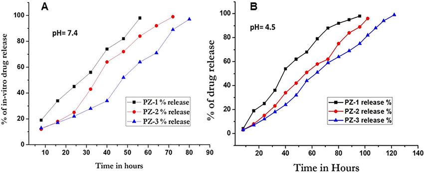
www.nature.com/scientificreports/
PZ-3 were done. The procedure involved utilizing an aqueous dip cell in an automatic manner where the diluted
NPs were inserted in the capillary measurement cell and locus of cell was attuned. The synthesized NPs were
deliberated for distribution of particle size and electrical properties as viewed by zeta potential measurements
and polydispersity index. The studies were done thrice to maintain reproducibility and the results were deliber-
ated as mean ± SD. The activities of NPs under changed external pH were also studied where they were diluted in
PBS (10 mM phosphate and 150 mM NaCl) where the pH ranged from 3.5 to 8.0. The studies were carried out
thrice to maintain reproducibility and the data attained was analyzed using the Zetasizer 6.01 software that was
delivered with the nano zeta sizer (Malvern Instrument, Inc).
Encapsulation and loading efficiency. The encapsulation efficiency (EE%) and loading efficiency (LE%)
of the drug into the NPs were determined using HPLC method. Filtration of PZ-1, PZ-2 and PZ-3 was done with
Amicon centrifugal tube whose molecular cut-off was 3000. The amount of free drug or non-entrapped drug
from the remnants of the total drug added was determined from the filtrate. The filtrate (10–12 µl) was inserted
into the HPLC column. Previously, separate calibration curves for epirubicin and gemcitabine were determined.
Calculations were made by the following equations to define the quantity of drugs in the filtrate
LE (%) = Weight of drug loaded ÷ Weight of the drug loaded NPs × 100
EE (%) = Weight of drug loaded ÷ Total amount of drug added × 100
In‑vitro release of drugs. The procedure was followed from Wang et al.24 with necessary modifications
for suiting our studies. Dialysis method was the chosen one to evaluate the drug release from PZ-1, PZ-2, PZ-3.
Concisely, PZ-1, PZ-2, PZ-3 NPs were freeze dried. The release study was performed in both at pH 7.4 (physi-
ological media; phosphate buffered saline) and pH 4.5 (tumor microenvironment) at room temperature. 1 ml
of ultra-purified water was used to disperse the freeze dried NPs. Then they were sealed in the tubing of dialysis
membrane, the molecular cut off of which was 3000. The sealed dialysis membrane was retained in 10 ml of
release media. This entire assemblage then was submerged in shaking water bath retained at room tempera-
ture. At particular intervals of time, 1 ml of the media was removed and renewed with identical volume of new
medium. Using HPLC technique, released drug in the media was quantified. The instruments used were Agilent
1100 alongside G1311A pump, a G1314A programmable diode array detector (DAD) and a G1313A auto-injec-
tor. Analytical column C18 (250 mm × 4.6 mm ID, 5 μm) was employed for the study. For characterization of the
concentration of individual drug released, different mobile phase was engaged.
Cytotoxicity studies. Cell culture. For 2D culture, the MG-63 cell line was obtained from ATCC (China).
Cell culture was dne in DMEM (Gibco™, Beijing, China) comprising of 10% fetal bovine serum (FBS) alongside
1% penicillin and streptomycin. MG-63 OS cells were seeded in 12-welled culture plate at 40,000 cells/well
2 days prior to incubation with free drugs and NPs. The incubation of cells was done at room temperature in a
saturated environment with 5% CO2.
Primary bone cells were obtained from Wistar rats which were used for some experiments in other laboratory.
However along with the in-vivo tumor study, permission for the work was obtained from University Committee
on Animal Ethics. The procedure was followed from Baker et al.25. The long bones of the rats were obtained and
harvested after which the epiphyses were removed followed by flushing out bone marrow with a syringe and
needle. Small fragments of the diaphysis were made which were washed with PBS followed by incubation in
2 mg/ml collagenase II in DMEM at room temperature in a pulsating waterbath for removal of adhering cells.
The fragments so obtained were washed with 10% fetal calf serum and cultured in DMEM augmented with 100
U/ml penicillin, 50 mg/ml streptomycin sulphate and gentamycin, 1.25 mg/ml fungizone along with 100 mg/ml
of ascorbate. On attaining confluency, 0.25% trypsin and 0.1% EDTA in PBS was used for cell harvesting. They
were then plated at 25 × 103 cells per well in 6-well cell culture plates and repeatedly cultured in 3 ml medium
mentioned above until confluency.
MTT assay. 3-(4,5-dimethythiazol-2-yl)-2,5-diphenyl tetrazolium bromide (MTT) assay was employed for
measurement of cytotoxicity of the free drugs and NPs. The principle of the assay is utilizing mitochondrial
succinate dehydrogenase for reduction of yellow MTT. Insoluble formazan complex was formed after the entry
of MTT in the live cells after which it was being reduced. For this, MG-63 and primary bone cells were seeded
at a density of 1 × 10⁴ in a 96-well plate. The cells after attaining confluency were treated with PLGA-ZOL NPs,
free epirubicin, free gemcitabine, PZ-1, PZ-2, PZ-3 NPs. The drug concentration of the PZ-1, PZ-2, PZ-3 were
adjusted to the free drug concentration which was 2.5 µg/ml. The cells were gestated for 24 and 48 h conse-
quently. After a certain time, the cell culture plates were treated with 100 μl of MTT solution (5 mg/ml) and
incubation was done for 4 h as per Wang et al.24. Addition of DMSO led to the extraction of the formed formazan
crystals and were incubated for 30 min further. Absorbance at 570 nm was read using a microplate reader (Ther-
moFischer, China) for each plate. The experiments were repeated for 6 times to maintain reproducibility.
Alamar blue assay. Cytotoxicity of the PLGA-ZOL NPs, free epirubicin, free gemcitabine, PZ-1, PZ-2, PZ-3
NPs were also assessed by Alamar blue a ssay13. MG-63 cell culture was done with DMEM medium at density of
104 cells in each well of 96-well plates. This was augmented with 10% FBS in a 5% CO2 humidified atmosphere at
37 °C within an incubator for 24 h. Replacement and renewal with fresh medium was carried out. The medium
was incubated with following samples: PLGA-ZOL NPs, free epirubicin, free gemcitabine, PZ-1, PZ-2, PZ-3
Scientific Reports | (2020) 10:15497 | https://doi.org/10.1038/s41598-020-72552-z 3
Vol.:(0123456789)www.nature.com/scientificreports/
NPs. The drug concentration of the PZ-1, PZ-2, PZ-3 were adjusted to the free drug concentration which was
2.5 µg/ml. After 72 h, removal of medium and rinsing with PBS was done followed by the addition of 250 µl of
Alamar blue solution (the ratio being 10:10:80 Alamar blue: FBS: medium 199 v/v) and further incubation was
carried out for 3 h. 300 µl of Alamar blue solution was removed into a new 96-well plate and reading was noted
with automated microplate spectrophotometer (EPOCH Microplate Spectrophotometer, USA) at 530 nm and
590 nm (excitation and emission wavelength respectively. the same process was repeated with primary bone cell
culture.
IC50 detection. The concentration of sample ensuing 50% cell death is I C50 of the sample which was deliberated
from concentration-effect curves, in view of the optical density of the control well (cells treated with PBS) as
100%.
IC50 is determined using the following formula:
Inhibition of 50% growth of cells (IC50) = OD control − OD treated cell × 100 ÷ OD control
The measurements were repeated thrice.
Cellular uptake of the NPs. The technique was modified from an earlier report26 with appropriate changes
to suit our study. 5 × 10⁴ cells/ml concentrations of MG-63 were harvested in 100 mm tissue culture plates. Incu-
bation of cells were done at room temperature for 48 h with free epirubicin, free gemcitabine, PZ-1, PZ-2, PZ-3
NPs at the earlier calculated IC50 concentration. Following this, washing of the cells was done with PBS twice
followed by trypsinization and resuspension in PBS. The amounts of drug amassed in the cells were determined
with an inductively coupled plasma mass spectrometer (ICP-MS, ELAN DRC-II, PerkinElmer). In brief, 23%
nitric acid treatment of the cell suspensions were carried out followed by 8% H2O2 and wet autoclaving of cell
suspension was done for 1 h. This was then inducted in the ICP-MS for quantification of amount of drug uptaken
by the cells.
In‑vivo anti‑tumor studies. Animals. Male Sprague–Dawley rats weighing upto 320–380 g were utilized
in the current study. All experiments were undertaken in 7–10 week old male rats. The Jilin University Commit-
tee on Animal Ethics granted permission for the animal experiments via its approval letter (6134, dated Jan 14,
2019). All experiments were performed as approved by the committee and all methods were carried out in ac-
cordance with relevant guidelines and regulations. 3 rats were used for a single group for all experimental work.
Anti‑tumor studies. Determination of better therapeutic efficacy of the PZ-1, PZ-2, PZ-3 NPs compared to free
drugs was done by the in-vivo studies. Tumors were grown in Sprague–Dawley rats by injecting MG-63 OS cells
(1 × 10⁵) in them. The conventions were appropriately modified from Eloy et al.27, Kempf et al.28 and Tomoda
et al.29. Tumors would grow to a noticeable size after 10–14 days of beginning of the treatment. Dilution of PZ-1,
PZ-2, PZ-3 NPs, PBS being negative control and epirubicin long with gemcitabine being positive controls were
done in PBS buffer at pH 7.4 before administration. The dose of epirubicin and gemcitabine administered are
2.0 mg/kg/day twice a week for 4 weeks. The drug concentration of the PZ-1, PZ-2, PZ-3 were adjusted to the
free drug concentration which was 2.0 mg/kg/day. The groups made were as follows: (I) PBS (negative control);
(II) Free epirubicin; (III) free gemcitabine; (IV) PZ-1; (V) PZ-2; (VI) PZ-3 (n = 5 in each group). Until the end
of the study, tumor volume and body weight were noted for all tumor-bearing mice following which they were
euthanized by anaesthetics overdose. The length and width of the tumors were measured with a calliper and
documented every 2 days. Calculation of tumor volume was done according to the equation: (width2 × length)/2.
For ease of comparison within groups, at each measurement point, relative tumor volume was deliberated.
Quantitative RT‑PCR. RNAs were extracted from tumor using Qiagen kits (Qiagen, Shanghai, China) and
suspended in nuclease-free water. Quantitative PCR was performed using an ABI7300 machine (Applied Bio-
systems, China). miRNA levels were determined, relative to U6, using the SYBR Green PCR kit (Qiagen, China).
Fold change in expression was determined by the method of cycle threshold (CT) using the formula of 2 −ΔΔCT.
Statistical analysis. Results are expressed as mean ± standard deviation. We used Student’s t-test to com-
pare groups.
Results
Physical characterization of the NPs. Electron micrographic images, as seen in Fig. 1A–D depicted that
PLGA-ZOL NPs, PZ-1, PZ-2, PZ-3 NPs have maintained intact shape. They were spherical in shape, not impec-
cably circular and a little edgier around the corners which may be due to the co-encapsulation of the two anti-
cancer drugs. There were no agglomeration of the particles and they seemed uniformly distributed. The sizes of
the NPs varied from 142 to 245 nm duly (elaborated in Table 1 along with the zeta potential and PDI). The zeta
potential was positive ranging from 22.4 to 26.5 mV. The approximate PDI was in the range from 0.23 ± 0.03,
indicating a stable NP formulation.
Under a varied range of pH, the maintenance of structural integrity of multiple drugs loaded PZ-3 NPs was
evaluated by calculating their particle size distribution as a function of external pH. PZ-3 NPs demonstrated
unimodal regularity in size distribution all through the pH range (3.5–8.0). The diameter of NPs was calculated
to be 197 ± 7 nm (PDI = 0.408 ± 0.042) at pH 7.0 and not much variation was seen in other pH solutions. The
information on the diameter of the NPs is provided in Table 2.
Scientific Reports | (2020) 10:15497 | https://doi.org/10.1038/s41598-020-72552-z 4
Vol:.(1234567890)www.nature.com/scientificreports/
Figure 1. A–D TEM photomicrographs of NPs; (A) PLGA-ZOL NPs; (B) PZ-1; (C) PZ-2; (D) PZ-3. (E)
Percentage of bone binding exhibited by PLGA-ZOL NPs and zoledronic acid.
Sl. no. Size of nanoparticles (nm) Zeta potential (mV) PDI
1 PLGA-ZOL NPs 142.92 ± 1.2 17.6 ± 0.1 0.211 ± 0.03
2 PZ-1 191.13 ± 1.2 22.4 ± 0.3 0.231 ± 0.04
3 PZ-2 195.13 ± 1.6 23.3 ± 0.2 0.262 ± 0.03
4 PZ-3 245.45 ± 1.5 26.5 ± 0.3 0.323 ± 0.01
Table 1. Size distribution, zeta potential and polydispersity index (PDI) of the NPs.
pH Hydrodynamic diameter PDI
3.5 245 ± 5 0.522 ± 0.041
4.0 257 ± 6 0.517 ± 0.024
5.0 220 ± 3 0.463 ± 0.021
6.0 212 ± 5 0.422 ± 0.036
7.0 197 ± 7 0.408 ± 0.042
8.0 150 ± 6 0.373 ± 0.034
Table 2. Hydrodynamic diameter and polydispersity index (PDI) values of PZ-3.
Sl. no. NP formulations EE (%) LE (%)
2 PZ-1 80.2 ± 5.3 27.3 ± 3.2
3 PZ-2 82.4 ± 2.3 29.2 ± 2.4
4 PZ-3 89.8 ± 4.5 33.2 ± 4.4
Table 3. EE (%) and LE (%) for all the NP formulations.
In‑vitro bone affinity studies. The binding affinity of zoledronic acid solution and PLGA-ZOL NPs was
high. The results depicted in Fig. 1E shows high affinity of both of them towards bone powder. Zoledronic acid
after 6 h was 89% absorbed in bone powder whereas 84% of PLGA-ZOL NPs was found localized in bone pow-
der.
Encapsulation and loading efficiency. The EE of PZ-1 was 80.2 ± 5.3% whereas the LE was 27.3 ± 3.2%.
For PZ-2 the EE was 82 ± 2.3% and LE was 29.2 ± 2.4%. For PZ-3, the EE and LE were 89 ± 4.5% and 33.2 ± 4.4%
respectively. The results presented in Table 3 clearly demonstrated high LE and EE for all the NP formulations.
Scientific Reports | (2020) 10:15497 | https://doi.org/10.1038/s41598-020-72552-z 5
Vol.:(0123456789)www.nature.com/scientificreports/
Figure 2. Percentage of in-vitro drug release; (A) pH 7.4 and (B) pH 4.5.
Figure 3. (A) Cytotoxicity of MG-63 cells on treatment with PBS, PLGA-ZOL NPs, free epirubicin, free
gemcitabine, PZ-1, PZ-2, PZ-3 as exhibited by MTT and Alamar Blue assay. (B) Cytotoxicity of primary bone
cells on treatment with PBS, PLGA-ZOL NPs, free epirubicin, free gemcitabine, PZ-1, PZ-2, PZ-3 as exhibited
by MTT and Alamar Blue assay.
In‑vitro release of drugs. In PZ-3, gemcitabine and epirubicin were encapsulated with the PLGA-ZOL
NP in the ratio of 1:1. However, it was seen that the release profile of gemcitabine and epirubicin in individual
NPs or in PZ-1 and PZ-2 are significantly different. In pH 7.4 or physiological milieu, epirubicin diffused faster
than gemcitabine at the end of 24 h. It was seen that about 45% of epirubicin (PZ-2) had diffused in comparison
of 25% of gemcitabine (PZ-1). The release of drugs from PZ-3 was steady, which lasted upto 80 h. However, in
acidic pH (pH 4.5) or tumor microenvironment milieu, the diffusion was slower and sustained. After 24 h, the
release of epirubicin (PZ-2) was 25% and gemcitabine (PZ-1) was 15% respectively. The drugs released from
PZ-3 continued upto 120 h. This is clearly depicted in Fig. 2.
Cytotoxicity studies. There was noteworthy alteration in cell toxicity between the MG-63 cells treated with
PLGA-ZOL NPs, free epirubicin, free gemcitabine, PZ-1, PZ-2, PZ-3 NPs. There was an increase in cell death
when the two drugs were co-encapsulated in PZ-3 than alone as indicated in Fig. 3A (MTT and Alamar blue
assay). It was seen that after 24 h the decrease in cell proliferation was more in PZ-1, PZ-2, PZ-3 NPs the free
drugs. The cell viability percentage in control was 91% and 94% (MTT and Alamar blue respectively) whereas
treatment with PLZA-ZOL NPs resulted in 64% and 69% viable cells after 72 h of incubation. The cell viabilities
were 33, 34 and 22% for PZ-1, PZ-2, PZ-3 respectively in MTT assay whereas it was 39, 40 and 27% for PZ-1,
PZ-2, PZ-3 respectively in Alamar blue assay. The cell viabilities were comparatively much lower than free epi-
rubicin and gemcitabine with 44% and 42% respectively for MTT assay & 45% and 49% for Alamar blue assay.
It was calculated that compared to free drugs, the cell viabilities were lessened by 1.33 times in both assays by
PZ-1, PZ-2, PZ-3 NPs (Fig. 3A).
With the primary bone cells, the cytotoxicity studies depicted entirely different results, as shown in Fig. 3B.
It was seen that PBS as control had the highest number of viable cells (94%) in MTT assay. However, the other
formulations (PLGA-ZOL NPs, PZ-1, PZ-2 and PZ-3) had comparable cytocompatibility of 84%, 75%, 77% and
82% respectively. The free epirubicin and gemcitabine had low cytocompatibility of 54% and 52% respectively. In
Scientific Reports | (2020) 10:15497 | https://doi.org/10.1038/s41598-020-72552-z 6
Vol:.(1234567890)www.nature.com/scientificreports/
Figure 4. (A–G) Cytotoxicity of MG-63 cells as seen by the live dead cell assay. (H) Cellular uptake of free
epirubicin, free gemcitabine, PZ-1, PZ-2, PZ-3 by MG-63 cells.
IC50 value ± S.D
Epirubicin 0.58 µg/ml ± 0.02
Gemcitabine 0.79 µg/ml ± 0.01
PZ-1 1.9 µg/ml ± 0.01
PZ-2 1.67 µg/ml ± 0.02
PZ-3 2.45 µg/ml ± 0.03
Table 4. IC50 concentration of the NPs in MG-63 cells.
Alamar blue assay of primary bone cells, a similar kind of cytocompatibility was seen (Fig. 3B). It was seen that
in MG-63 and primary bone cells, the MTT assay when compared to Alamar blue, was the more sensitive one.
The live dead cell assay shown in Fig. 4A–G reinforced the MTT assay results. An important point to be
noted is that the cells treated with PZ-1, PZ-2 and PZ-3 (Fig. 4E, F and G) exhibited damaged and broken cells
along with dead cells. It was seen in Fig. 4B that the blank PLGA-ZOL NPs did not inhibit cell growth but also
did not increase their proliferation. The I C50 concentration of the NPs in MG-63 cells is presented in Table 4.
Cellular uptake studies. Figure 4H shows the cellular uptake efficiency of free epirubicin, gemcitabine,
PZ-1, PZ-2, PZ-3 NPs. 2.5 µg/ml was the dosage of the free drug concentration which was attuned with the
concentration of the drug encapsulated in the NPs. It was seen that cellular uptake of free drugs was significantly
less than that of NPs by MG-63 osteosarcoma cell line. The cellular uptake of the PZ-1, PZ-2, PZ-3 NPs were
calculated in ng/mol. Accelerated cellular uptake of PZ-1, PZ-2, PZ-3 NPs was evidenced by the MG-63 uptake
when compared to free epirubicin and gemcitabine. It was calculated that there was an approximate 6 times
increase in the cellular uptake of the NPs as compared to free drugs.
In‑vivo studies. The anti-tumor studies, as seen in Fig. 5, revealed that the epirubicin solution had less effect
in decreasing the size of the tumors than gemcitabine. But the free drugs were still effective in reducing tumor
sizes, compared to PBS (control). On the contrary, the tumors were extremely sensitive towards PZ-3 displaying
statistically noteworthy reduction in tumor size when treated at 2.5 µg/ml twice a week for 4 weeks. The signifi-
cance of the PZ-3 where loading of multiple drugs was a tactic to increase the in-vivo anticancer effect was actu-
ally in view of the fact that there was almost 250% increased tumor volume on treating with PBS when compared
to PZ-3 and the tumor volume decreased by 130% with PZ-1, 115% with PZ-2. This is clearly seen in Fig. 5A,
B. It may be important to know that in-vivo multiple drugs loading in PLGA-ZOL NPs enhanced the efficacy of
drugs as exhibited by reduction in tumor volume and cellular uptake studies. Lastly, we checked for the miRNA
expressions as evidence for epigenetic regulations by PZ-3 and found that oncomiRs miR-21 and miR-10b were
downregulated by PZ-3 in the tumors while the tumor suppressor miR-34a was upregulated (Supplementary
Fig. 1). Thus, nanoparticles affect epigenetic regulation, which could serve as the possible mechanism of action.
Discussion
Owing to the perfusion and isolation of bone tissue, the chemotherapeutic advances were never able to attain
the concentration required for tumor obliteration. This has paved the pathways for designing of several nano-
drug carriers for therapy of osteosarcoma. To name a few important ones, Wang et al. formulated drug loaded
magnetic liposomes24, Ta et al. made orthophosphate hydrogel from chitosan–dipotassium for doxorubicin
delivery for osteosarcoma t reatment30 and Susa et al. prepared dextran-centred lipid-modified polymeric nano-
particle conjugate for the same9. Doxorubicin decorated magnetic liposomes were also tested for osteosarcoma
therapeutics13,31. However, all of these advances were either associated with cytotoxic effects or were extremely
expensive. Therefore, an effective nanoparticle therapeutic moiety with minimal side effects is an unmet and
Scientific Reports | (2020) 10:15497 | https://doi.org/10.1038/s41598-020-72552-z 7
Vol.:(0123456789)www.nature.com/scientificreports/
Figure 5. (A) Tumor volume obtained from rats treated with PBS, PLGA-ZOL NPs, free epirubicin, free
gemcitabine, PZ-1, PZ-2, PZ-3. (B) Size of the tumors dissected from rat models treated with PBS, PLGA-ZOL
NPs, free epirubicin, free gemcitabine, PZ-1, PZ-2, PZ-3. Note the decreased and restricted tumor size in rats
treated with PZ-3.
extremely imperative prerequisite of the hour. In our study, zoledronic acid conjugated PLGA nanoparticle would
have to be loaded with multiple anticancer drugs and targeted to the specific tissue. There have been instances
where it has been demonstrated that combination regimen of drugs that worked well in-vitro does not provide
adequate results in-vivo because of the complexity of cancer microenvironment. Therefore, in-vivo studies in rats
were carried out too. Interestingly, our data supports epigenetic regulation by the nanoparticles, as determined
by miRNA levels in the tumors after treatment with PZ-3, compared to controls. The oncomirs, miR-10b32, miR-
2133 were downregulated by PZ-3 and the tumor suppressor miR-34a34 was upregulated. This is in agreement
with epigenetic regulations in o steosarcoma35–37 that need further evaluations.
It had been already reported earlier that single chemotherapeutic drugs loaded in a nanocarrier may yield
unsatisfactory results for osteosarcoma therapeutics due to several f actors20. Therefore, in our study, a combi-
nation of drugs epirubicin and gemcitabine was loaded into an organic nanoparticle synthesised from PLGA.
This was done so that after successful drug delivery to the targeted tissue, the nanoparticle may be biodegraded
without any toxic side effects. PLGA NPs were of ideal sizes that avoided being cleared by reticuloendothelial
system38. It was also observed that despite the combinatorial drug theory, there wasn’t a significant improvement
in the osteosarcoma therapeutics due to early clearance of drugs from circulation, poor tissue targeting resulting
in non-significant accumulation of drugs in the affected bone c ells39. Therefore we hypothesized that a probable
conjugation with a bisphosphonate would possibly increase the chances of targeted drug delivery. Our current
strategy was to conjugate zoledronic acid with multiple drugs loaded PLGA NPs to focus on the state-of-the-
art development of active targeting of drug to bone tissue. From the bone binding assay, it was evident that the
bone binding was time dependant and that bisphosphonates binding to the bone was more since osteoclasts
were lacking in the human bone powder that may possibly interfere with the binding. The co-encapsulation was
challenging since epirubicin is mildly hydrophobic in nature. But the lipophilicity of gemcitabine made up for
their stability. The sizes of the PZ-1, PZ-2, PZ-3 NPs were in the range that may potentially avoid clearance from
the bloodstream and the positive zeta potential values indicated towards a stable colloidal solution with uniform
dispersion. Therefor what is attained is enhanced synergism. The micrographic images also hinted towards a
uniform NP with no leakage or disrupted membrane. As seen from the sizes of PZ-3 under the influence of
changing external pH, there were significant variations. T lower pH (3.5–5), the size of the PZ-3 did not show
significant variations whereas at higher pH (6–8) the sizes drastically reduced. This may be attributed to the fact
that at lower pH there is slow/sustained release of drugs whereas in higher pH an increased release of drugs.
The rigidity of lipophilic structure of the NPs at lower pH makes it more stable, hence the controlled release of
drugs. At higher pH the mobility of the drug molecules increased due to augmented hydrophilicity which causes
drug release and decrease in size. Hence PZ-3 may be correctly assumed as pH-sensitive. This promises a very
encouraging approach since the tumor microenvironment generally does not exceed pH 5. The high entrapment
efficiency (91%) of PZ-3 was may be a result of the lipophilicity of gemcitabine which overcame the mild hydro-
phobicity of epirubicin and allowed slow diffusion of the drugs across the membrane of PLGA-ZOL NPs. This
may be excellent for slow and sustained targeted release of drugs as may be seen from the in-vitro studies. It was
clearly noticed from the in-vitro release readings that the measured release of drugs from NPs was over a longer
time length in acidic pH than normal physiological pH. This may be better for future therapeutic applications
since tumor pH in majority of cases rarely exceed 5. Furthermore this progressive release of drugs may also be
attributed to their hydrophobicity and interaction within the NPs.
Scientific Reports | (2020) 10:15497 | https://doi.org/10.1038/s41598-020-72552-z 8
Vol:.(1234567890)www.nature.com/scientificreports/
The achievement of nanoparticle drug delivery along with conjugation of a bisphosphonate would entirely
depend upon the cytotoxicity of the drug delivery system within the cancerous cellular environment. Hence,
cytotoxicity studies formed a crucial part of our study where we performed the MTT assay, live dead assay and
Alamar blue assay. Alamar blue assay was chosen since it is a simple one-step assay, may be utilized for a large
screening and the values in absence of fluorescence microscopy may be colorimetrically measured. All the
studies revealed that the MG-63cell growth was inhibited with PZ-1, PZ-2 and PZ-3. However, the cell growth
was least on treatment with PZ-3 owing to the synergistic effects of multiple drugs. It was noted that the drug
concentration around the IC50 dosage resulted in maximum cell death. Too dilute samples hardly resulted in
any cytotoxicity while too much concentration resulted in cell toxicity in the surrounding cellular environment
which is not desirable. The treatment of primary bone cells with PZ-1, PZ-2 and PZ-3 exhibited cytocompatibility
comparable to the PBS since the multiple drugs encapsulation within an organic nanoparticle may have resulted
in a cytocompatible product than free drugs. Increased tumoricidal activity of the PZ-3 indicated towards better
encapsulation and targetivity of the nanocarriers as compared to the standard formulations. This interesting find
was consistent with that of the findings of the in-vitro studies. Reversion of the well-grown tumors may be due to
the effect of the release of tumoricidal drugs which were co-loaded in the NP vehicle. There was a criterion where
the best results were acquired with treatment of PZ-1, PZ-2 and PZ-3 within 10–14 days of development of tumor
since they weren’t particularly large. The pathway for the anti-tumoral action of the PZ NPs remains unclear
but diminished size of the tumors may be related to the possible instigation of macrophages and cytokines40.
Cellular uptake studies indicated increased uptake of NPs rather than the free drugs. This may be attributed
to conjugated zoledronic acid which has affinities towards bone cells. This conjugation made for ideal nanocar-
rier for targeted delivery of drug to the bone tumor tissues. The increased uptake of drugs by MG-63 cells may
escalate the therapeutic efficiency by causing increased cell apoptosis as a result of DNA damage. The progression
of increased cellular uptake in case of NPs may be due to the small size which led to improved endocytosis. This
benefits easy internalization of NPs within the cells thereby increasing cellular uptake efficiency. Nonetheless,
to oversimplify the process of internalization of NPs by cells is foolhardy when the procedure is almost non-
existent. But NPs having a size < 100 nm may be taken up by the cells via endocytosis. The surface charge on the
NPs also has a significant role in the process. Rapid internalization of NPs with a positive charge was generally
the accepted fact.
Conclusion
Our study presents the synthesis of nanoparticle drug delivery systems which resulted in a more encouraging
pharmacokinetic profile with increased circulation time that could possibly augment the drug uptake and accu-
mulation via passive diffusion method. The PLGA NPs were chosen since they already possessed significant fea-
tures such as tremendous cytocompatibility and biodegradability. The novel feature of the multiple drugs loaded
NPs synthesized was the conjugation with zoledronic acid which had a high affinity towards the bone cells which
caused enhanced cellular uptake of the NPs by MG-63 cells. Another important point was the co-encapsulation
of multiple drugs within the NPs which resulted in increased tumor regression in mice. Therefore, we can safely
conclude that a novel targeted biocompatible NP has been created which through the initial studies has been
proven to be extremely successful. However the transition of the PZ-3 NPs from laboratory bench to clinical
trials may be subjected to still more rigorous research which may be undertaken. The evaluation of a natural/
organic substance as a major component of NPs has been previously done. Nonetheless, its conjugation with a
bisphosphonate along with multiple drugs as a potential treatment for osteosarcoma may be furthered in future.
Received: 3 August 2020; Accepted: 31 August 2020
References
1. Gu, W., Wu, C., Chen, J. & Xiao, Y. Nanotechnology in the targeted drug delivery for bone diseases and bone regeneration. Int. J.
Nanomed. 8, 2305–2317. https://doi.org/10.2147/IJN.S44393 (2013).
2. Giljohann, D. A. & Mirkin, C. A. Drivers of biodiagnostic development. Nature 462, 461–464. https://doi.org/10.1038/nature0860
5 (2009).
3. Cheng, Z., Al Zaki, A., Hui, J. Z., Muzykantov, V. R. & Tsourkas, A. Multifunctional nanoparticles: cost versus benefit of adding
targeting and imaging capabilities. Science 338, 903–910. https://doi.org/10.1126/science.1226338 (2012).
4. Tang, F., Li, L. & Chen, D. Mesoporous silica nanoparticles: synthesis, biocompatibility and drug delivery. Adv. Mater. 24, 1504–
1534. https://doi.org/10.1002/adma.201104763 (2012).
5. Papasani, M. R., Wang, G. & Hill, R. A. Gold nanoparticles: the importance of physiological principles to devise strategies for
targeted drug delivery. Nanomedicine 8, 804–814. https://doi.org/10.1016/j.nano.2012.01.008 (2012).
6. Colson, Y. L. & Grinstaff, M. W. Biologically responsive polymeric nanoparticles for drug delivery. Adv. Mater. 24, 3878–3886.
https://doi.org/10.1002/adma.201200420 (2012).
7. Longhi, A., Errani, C., De Paolis, M., Mercuri, M. & Bacci, G. Primary bone osteosarcoma in the pediatric age: state of the art.
Cancer Treat. Rev. 32, 423–436. https://doi.org/10.1016/j.ctrv.2006.05.005 (2006).
8. Schwartz, C. L. et al. Multiple drug resistance in osteogenic sarcoma: INT0133 from the Children’s Oncology Group. J. Clin. Oncol.
25, 2057–2062. https://doi.org/10.1200/JCO.2006.07.7776 (2007).
9. Susa, M. et al. Doxorubicin loaded polymeric nanoparticulate delivery system to overcome drug resistance in osteosarcoma. BMC
Cancer 9, 399. https://doi.org/10.1186/1471-2407-9-399 (2009).
10. Chaudhari, K. R. et al. Bone metastasis targeting: a novel approach to reach bone using zoledronate anchored PLGA nanoparticle
as carrier system loaded with docetaxel. J. Control Release 158, 470–478. https://doi.org/10.1016/j.jconrel.2011.11.020 (2012).
11. Shane, E. Evolving data about subtrochanteric fractures and bisphosphonates. N. Engl. J. Med. 362, 1825–1827. https://doi.
org/10.1056/NEJMe1003064 (2010).
12. Chen, T. et al. Pharmacokinetics and pharmacodynamics of zoledronic acid in cancer patients with bone metastases. J. Clin.
Pharmacol. 42, 1228–1236. https://doi.org/10.1177/009127002762491316 (2002).
Scientific Reports | (2020) 10:15497 | https://doi.org/10.1038/s41598-020-72552-z 9
Vol.:(0123456789)www.nature.com/scientificreports/
13. Haghiralsadat, F. et al. New liposomal doxorubicin nanoformulation for osteosarcoma: drug release kinetic study based on thermo
and pH sensitivity. Chem. Biol. Drug Des. 90, 368–379. https://doi.org/10.1111/cbdd.12953 (2017).
14. Lipton, A. Emerging role of bisphosphonates in the clinic—antitumor activity and prevention of metastasis to bone. Cancer Treat.
Rev. 34(Suppl 1), S25-30. https://doi.org/10.1016/j.ctrv.2008.03.008 (2008).
15. Doppalapudi, S., Jain, A., Domb, A. J. & Khan, W. Biodegradable polymers for targeted delivery of anti-cancer drugs. Expert Opin.
Drug Deliv. 13, 891–909. https://doi.org/10.1517/17425247.2016.1156671 (2016).
16. Calzoni, E. et al. Biocompatible polymer nanoparticles for drug delivery applications in cancer and neurodegenerative disorder
therapies. J. Funct. Biomater. https://doi.org/10.3390/jfb10010004 (2019).
17. Caliskan, Y. et al. A new therapeutic combination for osteosarcoma: gemcitabine and clofazimine co-loaded liposomal formulation.
Int. J. Pharm. 557, 97–104. https://doi.org/10.1016/j.ijpharm.2018.12.041 (2019).
18. Basaran, M. et al. A phase II study of cisplatin, ifosfamide and epirubicin combination chemotherapy in adults with nonmetastatic
and extremity osteosarcomas. Oncology 72, 255–260. https://doi.org/10.1159/000113017 (2007).
19. Liu, Z. L. et al. Enhanced antitumor activity of epirubicin combined with cerulenin in osteosarcoma. Mol. Med. Rep. 5, 326–330.
https://doi.org/10.3892/mmr.2011.661 (2012).
20. Woodcock, J., Griffin, J. P. & Behrman, R. E. Development of novel combination therapies. N. Engl. J. Med. 364, 985–987. https://
doi.org/10.1056/NEJMp1101548 (2011).
21. Sriraman, S. K., Salzano, G., Sarisozen, C. & Torchilin, V. Anti-cancer activity of doxorubicin-loaded liposomes co-modified with
transferrin and folic acid. Eur. J. Pharm. Biopharm. 105, 40–49. https://doi.org/10.1016/j.ejpb.2016.05.023 (2016).
22. Fessi, H., Puisieux, F., Devissaguet, J. P., Ammoury, N. & Benita, S. Nanocapsule formation by interfacial polymer deposition fol-
lowing solvent displacement. Int. J. Pharm. 55, R1–R4. https://doi.org/10.1016/0378-5173(89)90281-0 (1989).
23. Chaudhari, K. R. et al. Opsonization, biodistribution, cellular uptake and apoptosis study of PEGylated PBCA nanoparticle as
potential drug delivery carrier. Pharm. Res. 29, 53–68. https://doi.org/10.1007/s11095-011-0510-x (2012).
24. Wang, B., Yu, X. C., Xu, S. F. & Xu, M. Paclitaxel and etoposide co-loaded polymeric nanoparticles for the effective combination
therapy against human osteosarcoma. J. Nanobiotechnol. 13, 22. https://doi.org/10.1186/s12951-015-0086-4 (2015).
25. Bakker, A. D., Soejima, K., Klein-Nulend, J. & Burger, E. H. The production of nitric oxide and prostaglandin E(2) by primary
bone cells is shear stress dependent. J. Biomech. 34, 671–677. https://doi.org/10.1016/s0021-9290(00)00231-1 (2001).
26. Tippayamontri, T., Kotb, R., Paquette, B. & Sanche, L. Cellular uptake and cytoplasm/DNA distribution of cisplatin and oxalipl-
atin and their liposomal formulation in human colorectal cancer cell HCT116. Investig. New Drugs 29, 1321–1327. https://doi.
org/10.1007/s10637-010-9494-3 (2011).
27. Eloy, J. O. et al. Liposomes as carriers of hydrophilic small molecule drugs: strategies to enhance encapsulation and delivery. Col‑
loids Surf. B Biointerfaces 123, 345–363. https://doi.org/10.1016/j.colsurfb.2014.09.029 (2014).
28. Kempf, R. A., Cebul, R. D. & Mitchell, M. S. Antitumor effects of doxorubicin against a virally-induced rat osteosarcoma with
minimal immunosuppression. J. Immunopharmacol. 2, 509–525. https://doi.org/10.3109/08923978009026409 (1980).
29. Tomoda, K., Kubota, Y. & Kato, J. Degradation of the cyclin-dependent-kinase inhibitor p27Kip1 is instigated by Jab1. Nature 398,
160–165. https://doi.org/10.1038/18230 (1999).
30. Ta, H. T., Dass, C. R., Larson, I., Choong, P. F. & Dunstan, D. E. A chitosan hydrogel delivery system for osteosarcoma gene therapy
with pigment epithelium-derived factor combined with chemotherapy. Biomaterials 30, 4815–4823. https://doi.org/10.1016/j.
biomaterials.2009.05.035 (2009).
31. Kubo, T. et al. Targeted delivery of anticancer drugs with intravenously administered magnetic liposomes in osteosarcoma-bearing
hamsters. Int. J. Oncol. 17, 309–315. https://doi.org/10.3892/ijo.17.2.309 (2000).
32. Ahmad, A. et al. Functional role of miR-10b in tamoxifen resistance of ER-positive breast cancer cells through down-regulation
of HDAC4. BMC Cancer 15, 540. https://doi.org/10.1186/s12885-015-1561-x (2015).
33. Zhou, L. et al. The role of miR-21/RECK in the inhibition of osteosarcoma by curcumin. Mol. Cell. Probes 51, 101534. https://doi.
org/10.1016/j.mcp.2020.101534 (2020).
34. Zhang, L., Liao, Y. & Tang, L. MicroRNA-34 family: a potential tumor suppressor and therapeutic candidate in cancer. J. Exp. Clin.
Cancer Res. 38, 53. https://doi.org/10.1186/s13046-019-1059-5 (2019).
35. Patil, S. L. et al. MicroRNA-509-3p inhibits cellular migration, invasion, and proliferation, and sensitizes osteosarcoma to cisplatin.
Sci. Rep. 9, 19089. https://doi.org/10.1038/s41598-019-55170-2 (2019).
36. Xie, Y. et al. MiR-302b Suppresses osteosarcoma cell migration and invasion by targeting Runx2. Sci. Rep. 7, 13388. https://doi.
org/10.1038/s41598-017-13353-9 (2017).
37. Sun, X. et al. miR-143-3p inhibits the proliferation, migration and invasion in osteosarcoma by targeting FOSL2. Sci. Rep. 8, 606.
https://doi.org/10.1038/s41598-017-18739-3 (2018).
38. Seju, U., Kumar, A. & Sawant, K. K. Development and evaluation of olanzapine-loaded PLGA nanoparticles for nose-to-brain
delivery: in vitro and in vivo studies. Acta Biomater. 7, 4169–4176. https://doi.org/10.1016/j.actbio.2011.07.025 (2011).
39. Lehar, J. et al. Synergistic drug combinations tend to improve therapeutically relevant selectivity. Nat. Biotechnol. 27, 659–666.
https://doi.org/10.1038/nbt.1549 (2009).
40. Dieter, P. et al. Prostaglandin E2 affects differently the release of inflammatory mediators from resident macrophages by LPS and
muramyl tripeptides. Mediat. Inflamm. 8, 295–303. https://doi.org/10.1080/09629359990306 (1999).
Author contributions
Y.Y., J.-X.S. and M.-N.Z. performed experiments and analysed data to prepare figures. B.-S.Y. cross-checked
data and wrote manuscript. Y.Y., J.-X.S., M.-N.Z. and B.-S.Y. proofread manuscript and approved submission.
Competing interests
The authors declare no competing interests.
Additional information
Supplementary information is available for this paper at https://doi.org/10.1038/s41598-020-72552-z.
Correspondence and requests for materials should be addressed to B.-S.Y.
Reprints and permissions information is available at www.nature.com/reprints.
Publisher’s note Springer Nature remains neutral with regard to jurisdictional claims in published maps and
institutional affiliations.
Scientific Reports | (2020) 10:15497 | https://doi.org/10.1038/s41598-020-72552-z 10
Vol:.(1234567890)www.nature.com/scientificreports/
Open Access This article is licensed under a Creative Commons Attribution 4.0 International
License, which permits use, sharing, adaptation, distribution and reproduction in any medium or
format, as long as you give appropriate credit to the original author(s) and the source, provide a link to the
Creative Commons licence, and indicate if changes were made. The images or other third party material in this
article are included in the article’s Creative Commons licence, unless indicated otherwise in a credit line to the
material. If material is not included in the article’s Creative Commons licence and your intended use is not
permitted by statutory regulation or exceeds the permitted use, you will need to obtain permission directly from
the copyright holder. To view a copy of this licence, visit http://creativecommons.org/licenses/by/4.0/.
© The Author(s) 2020
Scientific Reports | (2020) 10:15497 | https://doi.org/10.1038/s41598-020-72552-z 11
Vol.:(0123456789)You can also read