The comparative aspects of hystricomorph subplacenta: potential endocrine organ - BMC Zoology
←
→
Page content transcription
If your browser does not render page correctly, please read the page content below
Miglino et al. BMC Zoology      (2021) 6:16
https://doi.org/10.1186/s40850-021-00074-w
                                                                                                                                                BMC Zoology

 RESEARCH ARTICLE                                                                                                                                   Open Access

The comparative aspects of hystricomorph
subplacenta: potential endocrine organ
Maria Angelica Miglino1*, Gustavo de Sá Schiavo Matias1, Nathia Nathaly Rigoglio1, Jessica Borghesi1,
Taís Harumi de Castro Sasahara1, Maria Josephina Illera del Portal2, Juan Carlos Illera del Portal2,
Gema Silván Granado2, Sara Cristina Caceres Ramos2, Moacir Franco de Oliveira3 and Alan James Conley4

  Abstract
  Background: The placenta of hystricomorph rodents, lagomorphs and some primates includes an unusual
  structure, termed a subplacenta, which essentially consists of trophoblastic cells located deep to the central
  implantation site within the area of decidualization. It has been suggested that the subplacenta is functionally
  important, although considerable controversy remains on the issue. In this context, our objective was to compare
  the architecture and structure of the subplacentas of different hystricomorph species, to investigate the possibility
  that it is active in hormone synthesis.
  Methods: In total, the placentas of 3 capybaras (Hydrochaeris hydrochaeris), 2 pacas (Agouti paca), 5 agoutis
  (Dasyprocta leporina), 5 rock cavies (Kerodon rupestris) and 3 guinea pigs (Cavia porcellus) at different stages of
  pregnancy (early, middle and near term) were used for gross and microscopic examination. This included the
  preparation of latex injection casts, immunohistochemistry for steroidogenic enzymes, scanning and transmission
  electron microscopy. Tissue steroid concentrations were also determined.
  Results: The gross morphology and microvascular arrangement of the subplacentas were similar among the
  hystricomorphs studied including ultra-structural verification of cytotrophoblast and syncytiotrophoblast in all
  species. In guinea pigs, trophoblast cells exhibited characteristics consistent with intense metabolic and secretory
  activity in general. However, immuno-histochemical evidence also indicated that subplacental trophoblast
  expressed key steroidogenic enzymes, mainly in the chorionic villus region, consistent with tissue steroid
  concentrations.
  Conclusions: The subplacentas within placentas of hystricomorph rodent species are structurally similar and, in
  guinea pigs, have potential for steroid hormone secretion from, at least the early stages of pregnancy.
  Keywords: Rodents, Placentation, Hormones, Capybara, Agouti

* Correspondence: miglino@usp.br
1
 Department of Surgery, School of Veterinary Medicine and Animal Science
(FMVZ-USP), University of São Paulo- SP, Ave. Prof. Dr. Orlando Marques de
Paiva, 87, São Paulo 05508270, São Paulo, Brazil
Full list of author information is available at the end of the article

                                        © The Author(s). 2021 Open Access This article is licensed under a Creative Commons Attribution 4.0 International License,
                                        which permits use, sharing, adaptation, distribution and reproduction in any medium or format, as long as you give
                                        appropriate credit to the original author(s) and the source, provide a link to the Creative Commons licence, and indicate if
                                        changes were made. The images or other third party material in this article are included in the article's Creative Commons
                                        licence, unless indicated otherwise in a credit line to the material. If material is not included in the article's Creative Commons
                                        licence and your intended use is not permitted by statutory regulation or exceeds the permitted use, you will need to obtain
                                        permission directly from the copyright holder. To view a copy of this licence, visit http://creativecommons.org/licenses/by/4.0/.
                                        The Creative Commons Public Domain Dedication waiver (http://creativecommons.org/publicdomain/zero/1.0/) applies to the
                                        data made available in this article, unless otherwise stated in a credit line to the data.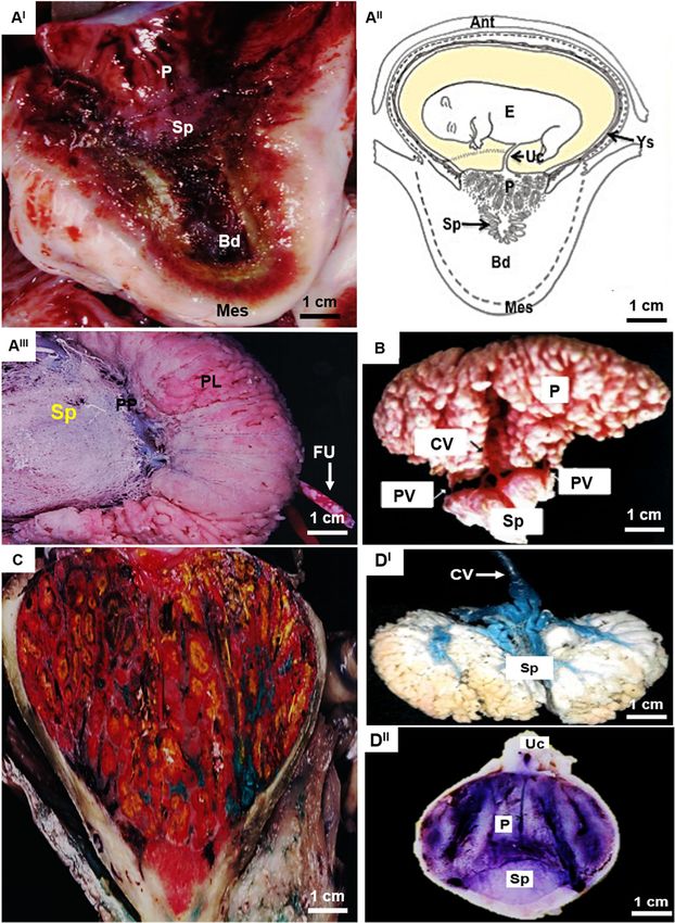
Miglino et al. BMC Zoology         (2021) 6:16                                                                                       Page 2 of 12

Background                                                                       to describe and compare the subplacentas from different
The success of viviparity in eutherian mammals is                                hystricomorph species using morphological techniques.
dependent on the establishment of a fully functional                             In addition, other approaches including latex injections,
chorioallantoic placenta in order to sustain successful                          immunohistochemistry, scanning and transmission elec-
embryonic and fetal development. In this context, pla-                           tron microscopy and analysis of tissue steroid concentra-
cental development from trophoblast is mediated in part                          tions were also employed in order to determine if
by interactions between maternal endometrial and extra-                          hormone synthesis was likely. These new data were dis-
embryonic tissues during the implantation process [1–                            cussed along with existing literature to better define
5]. The placenta is responsible for maternal-fetal meta-                         functional correlates that might provide insight into the
bolic exchange supporting the nutrition and respiration                          evolution of the subplacenta of hystricomorph rodents.
of, and excretion by, the fetus [6–8] and in some species,
it elaborates hormones providing endocrine support for
the pregnancy [9]. The structures associated with the                            Methods
placenta are complex and, depending on species, may                              Animals and gross morphology
consist of the amnion, chorion, allantois, yolk sac, and                         In total, placentae were analyzed from 3 capybaras
decidual elements [10–13]. In addition, these extra-                             (Hydrochaeris hydrochaeris), 2 pacas (Agouti paca), 5
embryonic membranes have substantive importance in                               agoutis (Dasyprocta leporina), 5 rock cavies (Kerodon
ontogeny.                                                                        rupestris), and 3 guinea pigs (Cavia porcellus) in differ-
   Some species within the Hystricomorph and Lago-                               ent stages of pregnancy (early, middle and near term).
morph sub-orders of the Rodentia have a specialized                              The samples were collected from multiple mothers. The
structure within their placentas, called a subplacenta                           guinea pig pregnancy age estimation was performed
[14–18], that is unusual among mammals. The structure                            based in [27, 28]. The animals were submitted to vaginal
is represented by zone of chorionic ectoderm, which is                           cytology to detect the onset of pregnancy, and ultra-
located in the central excavation of the placenta and is                         sound examination to monitor gestational development.
limited to the area of decidualization [19]. The functions                       The placentas were recovered after specific days of preg-
of this placental specialization remain unclear [1]. While                       nancy (Table 1) and the euthanasia was carry out by
some have speculated that the subplacenta might play                             ketamine (300 mg/kg) and xylazine (30 mg/kg) over-
an endocrine role, possibly gonadotrophin secretion [15,                         doses by intraperitonial administration. Samples were
20, 21], no definitive evidence exists to date. Uptake of                        obtained from the Center for Experimental Breeding of
histotroph [20, 22] or lactogen secretion [21] have also                         Capybara, at São Paulo State University, Araçatuba-SP;
been suggested as possible functions. However, accord-                           Center for Experimental Breeding of Paca, at São Paulo
ing to [23] none of these hypotheses has been resolved                           State University, Jaboticabal-SP; Center for Experimental
to a sufficient degree.                                                          Breeding of Agouti, at Federal University of Piaui,
   It is presumed that the subplacenta plays an import-                          Teresina-PI; and Center for the Breeding of Wild
ant, specialized role during pregnancy in hystricomorph                          Animals, at Federal Rural do Semi-Árido University,
rodents [18, 24–26]. However, despite the fact that this                         Mossoró-RN, Brazil.
structure has been investigated in numerous morpho-                                Material for data analysis was collected following pri-
logical studies, there is a dearth of information in the lit-                    mary fixation with 4 % paraformaldehyde injected in situ
erature in regard to the functions of this unusual                               via the maternal and fetal systems for further analysis
placental structure. Therefore, the aim of this study was                        and samples preservation. (Table 1). Gross aspects of the

Table 1 Summary of species and the different techniques utilized on the tissue samples collected
Species                             NS           TG                                                              GM        LI   IM          SEM
capybara (H. hydrochaeris)           3           end of gestation (90–140 days)                                  X         X                X
(150 days)
paca (A. paca)                       2           middle third of gestation (45–70 days)                          X         X
(150 days)
agouti (D. leporine)                5            middle of gestation (35–50 days)                                X         X                X
(106 days)
rock cavy (K. rupestris)            5            end of gestation (60–75 days)                                   X         X
(76 days)
guinea pig (C. porcellus)           3            early (around 10 days), middle (around 35 days) and             X              X
(72 days)                                        late (around 70 days) gestation
TG Time of gestation, GM Gross Morphology, LI Latex Injection, IM Immunohistochemistry, SEM Scanning Electron Microscopy
Miglino et al. BMC Zoology   (2021) 6:16                                                                    Page 3 of 12

placentae were photographed, and schematic diagrams           catalogue number K6068) and stained with DAB (Dako,
were drawn for documentation.                                 catalogue number K3468) according to the manufac-
                                                              turer’s instructions, and the slides were counterstained
Latex injections                                              with hematoxylin. Between each step prior to antibody
As a further aid to understanding vessel distribution,        incubation, slides were rinsed in PBS containing 0.2 %
some placentae in early/middle gestation were injected        BSA. Negative controls included no primary antiserum.
with coloured latex (Neoprene 650, DuPont, Brazil;              Slides were then mounted, viewed, and photographed
Latex Stain, Suvinil, Glassurit do Brazil S/A, São Ber-       on the Nikon Eclipse 80I microscope.
nardo do Campo, S.P., Brazil). Different colours were
injected in a uterine vein, a uterine artery, an umbilical    Scanning Electron Microscopy (SEM)
artery and the umbilical vein. The placentae were then        To study the microvasculature, term placentae were
immersion fixed in 10 % formalin, dissected and               injected with Mercox™ CL-2R resin (Okenshoji Co.,
analyzed.                                                     Ltd, Tokyo, Japan) [30]. The procedures were similar
                                                              to those applied by others to a wide range of placen-
Microscopic anatomy                                           tal types [31, 32]. A maternal and/or fetal vessel was
Preparations of histological sections for light microscopy    cannulated and the resin injected under gentle pres-
were performed at the University of São Paulo’s School        sure. Tissues were digested by immersion of the prep-
of Veterinary Medicine and Animal Science. Fragments          aration in several changes of 20 % NaOH solution at
of the placentas collected at different stages of gestation   50–60 °C. The casts were rinsed thoroughly in dis-
were fixed in 4 % paraformaldehyde solution. After fix-       tilled water, dried in an oven at 37 °C, and stored in
ation the material was washed in phosphate–buffered sa-       20 % gelatin at 4 °C. For scanning electron micros-
line (PBS), followed by dehydration in a series of            copy, pieces of the casts were rinsed in distilled water
increasing concentrations of ethanol (from 70 to 100 %),      to remove the gelatin, dried, and mounted on stubs
followed by diaphanization in xylol and paraffin embed-       with conductive carbon cement (Neubauer, Münster,
ding (Histosec) [29]. Paraffin blocks were sectioned at       Germany). They were then coated with gold using a
5 μm in an automatic microtome (Leica, RM2165,                sputter coater (Model K550, Emitech Products Inc.,
Germany). Sections were left to adhere overnight to           Houston TX, USA) and examined in a scanning elec-
glass slides in an incubator at 40 °C. After being deparaf-   tron microscope (Model 435 VP, Leo Electron Mi-
finized, the sections were stained with hematoxylin and       croscopy Ltd, Cambridge, UK).
eosin, as well as toluidine blue using routine methods.
                                                              Tissue steroid concentrations
Immunohistochemistry (IM)                                     Fragments of guinea pig placental tissue were col-
Tissues collected for immunohistochemistry were               lected at different stages of gestation (middle and
immersion fixed in 4 % paraformaldehyde in 70 mM              late). Samples of subplacenta and labyrinthine zones
phosphate buffer for 48 h, and then washed in cold PBS        were frozen at -20 °C for hormonal analysis. Steroid
for 72 h. The samples were embedded in paraffin, sec-         hormone concentration were measured using enzyme-
tioned to 5 μm in a microtome (Leica RM2265) and              immunoassays (EIA), previously validated by [33].
transferred to silanized slides (Sigma, # p8920). The sec-    Steroid hormones were detected using specific pri-
tions were rehydrated and washed in citrate buffer (1.83      mary antisera (estradiol, E2: C6E91; testosterone, T:
mM citric acid hydrate and 8.9 mM tribasic sodium cit-        R156 and progesterone, P4: C914) developed in the
rate, pH 6.0) for antigen recovery. Endogenous peroxid-       Department of Animal Physiology (UCM, Spain) and
ase was blocked with 3 % hydrogen peroxide diluted in         the Clinical Endocrinology Laboratory (University of
distilled water for 30 min in a darkroom. Tissue sections     California, Davis).
were blocked with 2 % bovine serum albumin (BSA) di-
luted in PBS for 30 min. Primary antibodies: cytokeratin      Results
(catalogue number RGE53, Novus, 1:100), vimentin              Morphology and immuno-histochemistry
(GTX35160, GeneTex, 1:100), 17β-hydroxysteroid de-            The gross morphology and microvascular arrangements
hydrogenase (17β-HSD, abcam, catalogue number                 of the subplacenta of these hystricomorph rodents were
ab97971, 1:100), 3β-hydroxysteroid dehydrogenase (3β-         observed during pregnancy (Fig. 1A-D). The subplacen-
HSD, Invitrogen, catalogue number FDO66Q 1:100),              tas in pacas appeared to regress in size from the middle
and aromatase cytochrome P450 (aromatase, Invitrogen,         of pregnancy to full term (Fig. 1C) and the subplacenta
catalogue number PA1-21398 1:100) were incubated              of the rock cavy was entirely absent by full term
overnight in a dark, humid chamber at 4 °C. The reac-         (Fig. 1DII). However, a major gross vasculature structure
tion was revealed using Dako Advance HRP (Dako,               was formed by maternal veins (Fig. 1DII).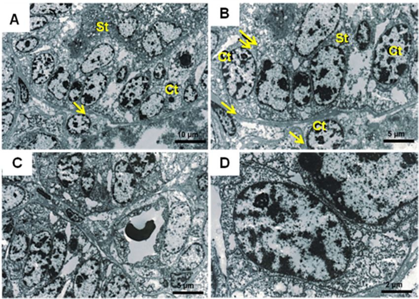
Miglino et al. BMC Zoology       (2021) 6:16                                                                                            Page 4 of 12

 Fig. 1 Gross morphology and microvascular architecture of the hystricomorph subplacenta. AI – AIII Capybara (Hydrochaeris hydrochaeris) around
 90–140 days of gestation. AI Sagittal section through the placenta showing the relationship among the chorioallantoic placenta (P), the
 subplacenta (Sp) and the basal deciduous (Bd) and the mesometrial side (Mes); AII Schematic drawing illustrating the relationship between the
 fetus (F), umbilical cord (Uc), yolk sac (Ys), Sp and Bd, and anti-mesometrial (Ant) of the uterus; AIII Corrosion model of the capybara which the
 different types of microvascularization of the placental lobes (PL) and Sp are observed. Placental vessels were injected with methacrylate and
 subjected to the corroding process. The relationship between Sp microvessels and larger vessels is verified (arrow), which possibly carry products
 produced by the subplacenta during this gestation phase - PP (placental peduncle) FU (umbilical funiculum). B Rock cavy (Kerodon rupestris)
 around 76 days: Placenta injected with methacrylate submitted to corrosion showing the central vessels (CV) and peripheral vessels (PV) in
 relation to the chorioallantoic placenta (P) and Sp. C Paca (Agouti paca) around 45–70 days: In C, the paca placenta injected with latex, the
 maternal vein injected with green, maternal artery with white, fetal vein with yellow and fetal artery with red. Note, the Sp appeared to have
 regressed in size at term compared with mid-gestation. DI – DII Agouti (Dasyprocta leporine) around 35–50 days: In DI, model of
 microvascularization of the agouti subplacenta, showing the central vessels injected in blue latex and the subplacenta in white latex. DII Showing
 the P and Sp and also the umbilical cord (Uc)
Miglino et al. BMC Zoology      (2021) 6:16   Page 5 of 12

 Fig. 2 (See legend on next page.)
Miglino et al. BMC Zoology (2021) 6:16 Page 6 of 12
(See figure on previous page.)
Fig. 2 Capybara subplacenta (H. hydrochaeris) around 90–140 days of gestation. AI Section through the subplacenta showing the junctional zone
(Jz) with fetal (arrow) and maternal (*) vessels in the labyrinth layer. AII Subplacenta showing the cytotrophoblast (Ct, arrow) interspersed by the
cells of the syncytiotrophoblast (*St) surrounded by vessels (vs.). BI Subplacenta capybara cytokeratin 20X. Observe a positive reaction present in
giant trophoblastic cells in the cytotrophoblast region (arrow) and negative reaction in the fetal mesenchyme. BII Subplacenta capybara
cytokeratin 60X. Observe a positive reaction present in giant trophoblastic cells in the cytotrophoblast region (arrow) and negative reaction in the
fetal mesenchyme (Mf). CI Subplacenta capybara actin 20X. CII Subplacenta capybara actin 60X. Positive marking on the muscular layer of the wall
of fetal vessels (Fv) of Capybara Subplacenta (arrow) and cytotrophoblast (Ct-asterisk) cells and negative reaction in the region of the
syncytiotrophoblast (St). DI Subplacenta capybara vimentin 20X - Observe the deciduous region (Dc) of the placenta of the capybara and
junctional zone (Jz). DII Subplacenta capybara vimentin 60X - Observe positive marking for vimentin in the wall of vessels (arrow) and giant cells
(asterisks) surrounded by cytotrophoblast (Ct) cells around the vessels, interspersed by the syncytiotrophoblast (St) region of the
Capybara placenta
connective tissue lamellae and often close to the fetal
Fetal and maternal blood vessels were prominent in vessels. The syncytiotrophoblast enclosed many lacunae
the labyrinth layer and near the junctional zone of the into which microvilli projected from the plasma mem-
capybara subplacenta (Fig. 2A). Giant cytotrophoblast brane and, as gestation advanced, these lacunae formed
cells were interspersed with syncytotroblast cells, which an extensive inter-connected system.
were in turn surrounded by vessels (Fig. 2AII). The giant
cells of the cytotrophoblastic region showed a positive Tissue steroid content
reaction to cytokeratin, however there was no immuno- To verify the capacity for hormonal production by the
staining in the fetal mesenchyme (Fig. 2BI − II). There subplacenta, samples of guinea pig subplacenta were an-
was positive actin labeling in the cells of the muscle alyzed for progesterone, testosterone and estradiol con-
layer of the fetal vessels, but no evidence of actin tent and compared with the labyrinth region. In general,
immuno-staining in the syncytotrophlast region (Fig. 2CI for all steroids, samples from mid-gestation had higher
− II
). Vimentin-positive labeling was observed on vessel steroid concentrations when compared to samples col-
walls and giant cells (Fig. 2DI − II). lected from late gestation. In addition, the subplacenta
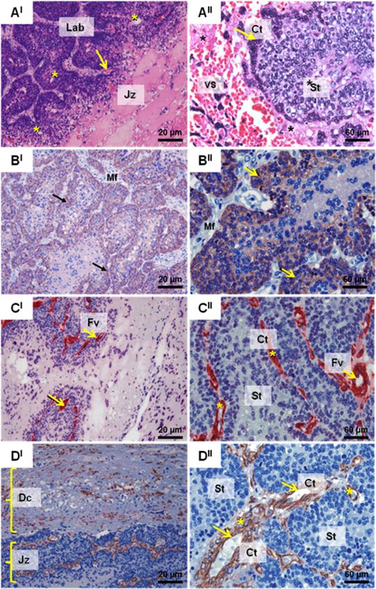
In guinea pig placentae, there was positive immuno- region showed higher hormone levels than labyrinthine
reactivity consistent with steroidogenesis, mainly in the samples (Table 2).
region of chorionic villi and, in the labyrinth region, only
in the blood vessel region. Among the enzymes tested, Discussion
17β-HSD expression was especially evident in the The subplacenta, as a structure found only on hystrico-
trophoblastic cells of the villi (Fig. 3A, AI). Evidence of morph rodents, lagomorphs, and some primates, has
expression of 3β-HSD was also evident in the same re- attracted interest, especially in terms of comparative
gion but was not as extensive expression (Fig. 3B, BI). morphology. However, to the best of our knowledge, no
The expression of aromatase (P450) was positive around studies have yet established an endocrine function for
the peripheral membrane protein of the labyrinth region the subplacenta during conceptus development. The re-
(Fig. 3C, CI). No labeling was observed in the absence of sults of early studies investigating the potential for pro-
the primary antisera, the negative controls (Fig. 3D, DI), gesterone synthesis by the guinea pig placenta indicated
in any of the sections. that the spongy trophoblast was a likely source [34, 35].
However, these investigators did not examine the sub-
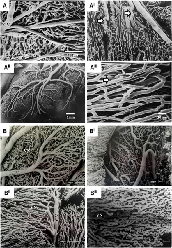
Microvascular arrangements placenta. To explore the potential for endocrine func-
Vascular molds examined by the scanning electron mi- tion, we investigated the comparative morphology of the
croscopy technique demonstrated evidence of larger subplacenta of several Hystricomorph rodents, with a
branches supplying the subplacenta. The division of particular focus on cell structures typical of secretory ac-
these larger branches allows the branching of smaller tivities, as well as the presence of steroid hormones and
vessels, which become tortuous with sinus dilations and enzymes involved in their synthesis.
constrictions in the vessel lumen as they course through The subplacenta has been described in several species
the tissue (Fig. 4). among the Hystricomorph rodents [1, 14, 15, 24, 25,
36–40]. In these species, the development of subplacenta
Ultra-structure is marked by an increase in the thickness of the cytotro-
In capybara the nuclei of the cytotrophoblast cells were phoblast lamellae forming the floor of the central exca-
large in relation to the amount of cytoplasm and nucle- vation. This occurs during the early development of the
oli were prominent (Fig. 5). The basal membrane of the viscacha (Lagostomus maximus) subplacenta [41] and
cytotrophoblast layer was seen to be in contact with the was observed at later stages in the chinchilla (ChinchillaMiglino et al. BMC Zoology (2021) 6:16 Page 7 of 12 Fig. 3 Immunohistochemistry (IM) from guinea pig (C. porcellus) subplacenta (early gestation). In A, AIand B, BI, positive labeling of 17β-HSD and 3β-HSD respectively in the villi (arrows) and in the labyrinth region (asterisks). In C, CI positive expression of aromatase (P450). In D, DI, no labeling was observed in the negative control lanigera) and coypu (Myocastor coypus) [42]. All cavio- stages of development. Certain aspects related to the morph rodents exhibit a subplacenta located deep within vascularization of the subplacenta remain to be defined. the disc of the main placenta [43], which appears at the In nutria (Myocastor coypus) [42], the majority of mater- beginning of pregnancy and regresses during the later nal arteries are diverted from the subplacenta. Some of
Miglino et al. BMC Zoology        (2021) 6:16                                                                                                Page 8 of 12

 Fig. 4 Scanning Electron Microscope (SEM) images of Capybara (H. hydrochaeris) subplacenta vascular molds around 90–140 days (A-AIII) and
 Agouti (D. leporine) around 35–50 days (B-BIII). In mid to late gestation, the subplacenta is supplied exclusively by fetal vessels. A large branch of
 the umbilical artery follows the central band of fetal mesenchyme to the base of the main placenta and then large central vessel (CV) insert to
 supply the subplacenta. The subplacental vessels pursue a tortuous course forming a vascular network (VN) with sinusoidal dilatations and
 constrictions (full arrow) in both species at different stages of pregnancy, which demonstrates that the vascular supply remains for the
 transportation of the products necessary for organ development

these arteries were observed to traverse the lobes of                          of rodent placentation [23, 43, 44] have provided
the subplacenta, but most were located outside of                              scant evidence of the function of the subplacenta,
the subplacental area and thus between the adjacent                            suggesting only that it is a region contributing to
lobes of the chorioallantoic placenta toward the                               cytotrophoblast growth and possible endocrine
central zone of each lobe. However, previous studies                           secretion.
Miglino et al. BMC Zoology      (2021) 6:16                                                                                           Page 9 of 12

 Fig. 5 Transmission electron microscope (TEM) images of Subplacenta of the Paca (a-d). Observe the presence of cytotrophoblast (Ct) giant cells
 interspersed with the syncytiotrophoblast (St), basal lamina (arrow) and intercellular spaces (double arrow) are present

   Prior indirect evidence supports a possible endocrine                   aspects including the central vasculature of the labyrinth
role for the subplacenta. Recent work resulted in the                      and junctional zones, and subplacental vasculature, were
creation of a library of differentially expressed genes                    comparable with small capillaries, around larger vessels,
(DEGs) from the subplacenta of the beaver (Castor fiber)                   where it was possible to identify sinusal dilatations and
carrying twin versus triplet fetuses. The transcripts iden-                anastomotic channels, like those described previously
tified were consistent with functions related to cellular                  [1]. Furthermore, others reported that the intercellular
processes, biological regulation, response to stimuli and                  space was marked by the presence of microvilli as evi-
metabolism but, compared to other parts of the placenta                    dence of communication between the syncytium and the
or even compared to other species, there appeared to be                    cytotrophoblast that make up the subplacenta in the
little difference in functional terms [45]. The subplacenta                cane rat [47]. While their content remains unknown,
is known to be responsive to the amount of oxygen                          there is a temporal concordance between the appearance
available, and its area is increased when oxygen is re-                    of these secretory granules in the subplacenta and the
stricted [46]. The fact that we have observed vesicles and                 increase in progesterone binding globulins during gesta-
secretory granules in addition to microvilli in the subpla-                tion. In addition, the peroxisomes found in the subpla-
centa cells of the paca and the guinea pig also suggests                   centa are consistent with lipid metabolism. These
that there may be local effects of secreted products                       organelles contain enzymes related to the beta-oxidation
within the placenta itself, suggestive of paracrine effects.               of fatty acids. Peroxisomes breakdown fatty acids into
   In addition to sub-cellular specializations, the subpla-                acetyl-CoA, which supports the biosynthesis of choles-
centa of several species examined here (capybara, paca,                    terol, the ultimate precursor of all steroid hormones
agouti and rock cavy) presents a distinct pattern of                       [48]. Burgess and Tam [34] reported that lipid droplets
vascularization, typical of endocrinal glands. Different                   which were first apparent in the spongy zone and other

Table 2 Median concentrations (ng/mg of tissue) of progesterone, testosterone and estradiol extracted and measured in 3
differentes samples from guinea pig labyrinth and subplacenta tissue samples
                       Progesterone                                 Testosterone                                  Estradiol
                       Middle                 Late                  Middle                 Late                   Middle                Late
Labyrinth              15.15 ± 1.74           8.87 ± 3.66           10.01 ± 4.09           5.52 ± 1.82            0.71 ± 0.05           0.55 ± 0.05
Subplacenta            24.88 ± 5.19           16.77 ± 2.87          15.59 ± 2.99           11.14 ± 0.17           0.95 ± 0.09           0.71 ± 0.02
Miglino et al. BMC Zoology (2021) 6:16 Page 10 of 12
areas of the placenta after day 25 of gestation were con- (FAPESP, grant number 2014/50844-3) providing financial assistance for
sistent with steroidogenic activity in these cells. Mitochon- sample collection, as well as for histological and data analysis.
dria, especially those derived from syncytiotrophoblast, are
Availability of data and materials
known to have the capacity to initiate steroid hormone The data that support the findings of this study are openly available. If
synthesis, being the sub-cellular site of expression of cyto- necessary, contact Maria Angelica Miglino (miglino@usp.br).
chrome P450 cholesterol side chain cleavage [49, 50].
More direct evidence for endocrine function was Declarations
provided by the detection of steroid hormones in subpla- Ethics approval and consent to participate
cental tissue and the expression of steroidogenic The project was conducted under rules of the Bioethics Commission of the
enzymes including 17β-HSD and 3β-HSD by immuno- School of Veterinary Medicine and Animal Science, University of Sao Paulo,
Brazil and by the Brazilian Institute of the Environment and Renewable
histochemistry which is consistent with the synthesis of Natural Resources (IBAMA) (Protocols Nr.77/2002; 927/2006). All Animal Ethics
progesterone and of testosterone and estradiol. The procedures are in accordance with BMC Zoology criteria.
catalytic activity of 17β-HSD is required for the reduc-
tion of DHEA to androstenediol, androstenedione to Consent for publication
Not applicable.
testosterone and estrone to estradiol. The presence of
progesterone in subplacental tissue is also consistent Competing interests
with expression of 3β-HSD and the oxidation of preg- The authors declare that they have no competing interests.
nenolone and 17α-hydroxypregnenolone to progesterone
Author details
and 17α-hydroxyprogesterone, respectively [51, 52]. The 1
Department of Surgery, School of Veterinary Medicine and Animal Science
failure to detect aromatase labeling is consistent with (FMVZ-USP), University of São Paulo- SP, Ave. Prof. Dr. Orlando Marques de
the low amount of estradiol found in the tissues com- Paiva, 87, São Paulo 05508270, São Paulo, Brazil. 2Department of Animal
Physiology, School of Veterinary Medicine, Complutense University of Madrid
pared to progesterone and testosterone. This is also (UCM), Madrid, Spain. 3Department of Animal Science, Federal University of
consistent with the low levels of expression of aromatase the Semi-Arid Region (UFERSA), Mossoro, Brazil. 4Department of Population
compared with cytochrome P450 17α-hydroxylase/1720- Health and Reproduction, School of Veterinary Medicine School of Veterinary
Medicine, University of California UC-Davis, 3223 VM3B, Sacramento,
lyase in ovarian [53] and testicular tissues [54]. Finally, California, USA.
immunohistochemistry is not a very sensitive technique
and low levels of enzyme activity together with poor Received: 29 July 2020 Accepted: 12 April 2021
cross-reactivity of antisera across species can present
challenges. References
1. Bonatelli M, Carter AM, Fernandes Machado MR, De Oliveira MF, de Lima
Conclusions MC, Miglino MA. Placentation in the paca (Agouti paca L). Reprod Biol
Endocrinol. 2005;3:9. https://doi.org/10.1186/1477-7827-3-9.
The subplacenta does not differ greatly in terms of the 2. Carter AM, Mess AM. Conservation of placentation during the tertiary
structural aspects among the hystricomorphs investi- radiation of mammals in South America. J Morphol. 2013;274:557–69.
gated, and it is has potential for hormonal secretion dur- https://doi.org/10.1002/jmor.20120.
3. Stewart JR, Thompson MB. Evolution of placentation among squamate
ing the early stages of pregnancy based on evidence reptiles: Recent research and future directions. Com Biochem And Physiol
consistent with steroidogenic enzyme activity. Part A: Mol & Integr Physiol. 2000;127:411–31. https://doi.org/10.1016/S1095-
6433(00)00273-7.
Abbreviations 4. Stewart JR, Thompson MB. Evolutionary Transformations of the Fetal
PBS - phosphate: Buffered saline; IM: Immunohistochemistry; mM: Millimolar; Membranes of Viviparous Reptiles: A Case Study of Two Lineages. J Exp
BSA: Bovine serum albumin; 17β-HSD: 17β-hydroxysteroid dehydrogenase; Zool. 2003;299A:13–32. https://doi.org/10.1002/jez.a.10288.
3β-HSD: 3β-hydroxysteroid dehydrogenase; P450: Aromatase cytochrome; 5. Carter AM, Enders AC, Pijnenborg R. The role of invasive trophoblast in
SEM: Scanning Electron Microscopy; EIA: Enzyme-immunoassays; implantation and placentation of primates. Philos Trans R Soc B Biol Sci.
DHEA: Dehydroepiandrosterone 2015;370:20140070. https://doi.org/10.1098/rstb.2014.0070.
6. Paavola LG, Furth EE, Delgado V, Boyd CO, Jacobs CC, Lei H, et al. Striking
Acknowledgements Changes in the Structure and Organization of Rat Fetal Membranes Precede
To Maria Acelina Martins Carvalho, Franceliusa Delys de Oliveira, Carlos Parturition1. Biol Reprod. 1995;53:321–38. https://doi.org/10.1095/
Eduardo Ambrósio and Rosangela Felipe Rodrigues for support in collected biolreprod53.2.321.
samples and Antonio Chaves de Assis Neto for assigning the antibody 7. Valdés G, Erices R, Chacón C, Corthorn J. Angiogenic, hyperpermeability and
aliquots of the study. vasodilator network in utero-placental units along pregnancy in the guinea-
pig (Cavia porcellus). Reprod Biol Endocrinol. 2008;6:13. https://doi.org/10.11
Authors’ contributions 86/1477-7827-6-13.
MAM, GSSM, NNR, JB and THCS: experimental conduction / MFO: animal 8. Schuler G, Fürbass R, Klisch K. Placental contribution to the endocrinology
sample collection / MAM, MJIP, JCIP and AJC: project idealization / MAM, of gestation and parturition. Anim Reprod. 2018;15(Suppl. 1):822–42. https://
GSSM, NNR, JB and AJC: writing / MJIP, JCIP, GSG and SCCR: hormonal doi.org/10.21451/1984-3143-AR2018-0015.
analysis / All authors: data interpretation / MAM: funding. All authors read 9. Geisert RD, Conley AJ. Secretion and Metabolism of Steroids in Subprimate
and approved the final manuscript. Mammals During Pregnancy. In: Endocrinology of Pregnancy. Totowa: Humana
Press; 1998. p. 291–318. https://doi.org/10.1007/978-1-4612-1804-3_10.
Funding 10. Oyen ML, Cook RF, Calvin SE. Mechanical failure of human fetal membrane
This work was supported by Coordination for the Improvement of Higher tissues. J Mater Sci Mater Med. 2004;15:651–8. https://doi.org/10.1023/B:
Education Personnel (CAPES), and by The São Paulo Research Foundation JMSM.0000030205.62668.90.Miglino et al. BMC Zoology (2021) 6:16 Page 11 of 12
11. Wolf E, Arnold GJ, Bauersachs S, Beier HM, Blum H, Einspanier R, et al. 33. Caceres S, Peña L, Silvan G, Illera MJ, Woodward WA, Reuben JM, et al.
Embryo-maternal communication in bovine - Strategies for deciphering a Steroid Tumor Environment in Male and Female Mice Model of Canine and
complex cross-talk. Reprod Domest Anim. 2003;38:276–89. https://doi.org/1 Human Inflammatory Breast Cancer. BioMed Res Int. 2016;2016:1–7. https://
0.1046/j.1439-0531.2003.00435.x. doi.org/10.1155/2016/8909878.
12. Stewart JR. Yolk sac placentation in reptiles: Structural innovation in a 34. Burgess SM, Tam WH. Ultrastructural changes in the guinea-pig placenta,
fundamental vertebrate fetal nutritional system. J Exp Zool. 1993;266:431– with special reference to organelles associated with steroidogenesis. J Anat.
49. https://doi.org/10.1002/jez.1402660509. 1978;126 Pt 2:319–27. http://www.ncbi.nlm.nih.gov/pubmed/670066.
13. Kot K, Kosik-Bogacka D, Łanocha-Arendarczyk N, Malinowski W, Szymański S, 35. Tam WH. Steroid synthesis in vitro by the placenta of the guinea pig, and
Mularczyk M, et al. Interactions between 14 elements in the human progesterone concentrations in systemic and uterine plasma. J Endocrinol.
placenta, fetal membrane and umbilical cord. Int J Environ Res Public 1977;73:483–9.
Health. 2019;16:1615. https://doi.org/10.3390/ijerph16091615. 36. Vasconcelos BG, Favaron PO, Miglino MA, Mess AM. Development and
14. Bonatelli M, Machado MRF, Cruz C, Miglino MA. Análise morfológica da morphology of the inverted yolk sac in the guinea pig (Cavia porcellus).
placenta da paca (Agouti paca, Linnaeus, 1766: Estudo ao microscópio de Theriogenology. 2013;80:636–41. https://doi.org/10.1016/j.theriogenology.2
luz e à microscopia eletrônica de transmissão. Braz J Vet Res Ani Sci. 2001; 013.06.007.
38:224–8. https://doi.org/10.1590/s1413-95962001000500005. 37. Flamini MA, Barreto RSN, Matias GSS, Birbrair A, Harumi de Castro Sasahara T,
15. Kaufmann P, Davidoff M. The Guinea-Pig Placenta. Berlin: Springer Berlin Barbeito CG, et al. Key characteristics of the ovary and uterus for reproduction
Heidelberg; 1977. https://doi.org/10.1007/978-3-642-66618-6. with particular reference to poly ovulation in the plains viscacha (Lagostomus
16. Leiser R, Kaufmann P. Placental structure: In a comparative aspect. Exp Clin maximus, chinchillidae). Theriogenology. 2020;142:184–95. https://doi.org/10.1
Endocrinol Diabetes. 1994;102:122–34. 016/j.theriogenology.2019.09.043.
17. Hradecký P, Mossman HW. Vertebrate Fetal Membranes: Comparative 38. Oliveira MF, Mess A, Ambrósio CE, Dantas CAG, Favaron PO, Miglino MA.
Ontogeny and Morphology; Evolution; Phylogenetic significance; Basic Chorioallantoic placentation in Galea spixii (Rodentia, Caviomorpha,
Functions; Research Opportunities. J Zoo Anim Med. 1987;18(2/3):55. https:// Caviidae). Reprod Biol Endocrinol. 2008;6:39. https://doi.org/10.1186/1477-
doi.org/10.2307/20460238. 7827-6-39.
18. Bezerra FVF, Favaron PO, Mess AM, Araújo Júnior HN, Oliveira GB, Pereira AF, 39. Miglino MA, Franciolli ALR, de Oliveira MF, Ambrósio CE, Bonatelli M,
et al. Subplacental development in Galea spixii. Pesqui Vet Bras. 2018;38: Machado MRF, et al. Development of the Inverted Visceral Yolk Sac in Three
2175–82. https://doi.org/10.1590/1678-5150-pvb-5527. Species of Caviids (Rodentia, Caviomorpha, Caviidae). Placenta. 2008;29:748–
19. Fadl S, Moshiri M, Fligner CL, Katz DS, Dighe M. Placental imaging: Normal 52. https://doi.org/10.1016/j.placenta.2008.05.007.
appearance with review of pathologic findings1. Radiographics. 2017;37:979–98. 40. Roberts CT, Sohlstrom A, Kind KL, Earl RA, Khong TY, Robinson JS, et al.
20. Davies J, Dempsey EW, Amoroso EC. The subplacenta of the guinea-pig Maternal food restriction reduces the exchange surface area and increases
development, histology and histochemistry. J Anat. 1961;95 Pt 3:311-324.13. the barrier thickness of the placenta in the guinea-pig. Placenta. 2001;22:
http://www.ncbi.nlm.nih.gov/pubmed/17105123. Accessed 7 Jan 2020. 177–85.
21. Rodrigues RF, Carter AM, Ambrosio CE, dos Santos TC, Miglino MA. The 41. Flamini MA, Portiansky EL, Favaron PO, Martins DS, Ambrósio CE, Mess AM,
subplacenta of the red-rumped agouti (Dasyprocta leporina L). Reprod Biol et al. Chorioallantoic and yolk sac placentation in the plains viscacha
Endocrinol. RB&E. 2006;4:31. https://doi.org/10.1186/1477-7827-4-31. (Lagostomus maximus) - A caviomorph rodent with natural polyovulation.
22. J.S. RCM & P. Hystricomorph embryology. Symp Zool Soc London. 1974;34: Placenta. 2011;32:963–8.
333–60. 42. Hillemann HH, Gaynor AI. The definitive architecture of the placentae of
23. Mess A. Development of the chorioallantoic placenta inOctodon degus—a nutria,Myocastor coypus (Molina). Am J Anat. 1961;109:299–317. https://doi.
model for growth processes in caviomorph rodents? J Exp Zool Part B Mol org/10.1002/aja.1001090306.
Dev Evol. 2007;308B:371–83. https://doi.org/10.1002/jez.b.21160. 43. Mess A, Blackburn DG, Zeller U. Evolutionary transformations of fetal
24. Miglino MA, Carter AM, Ambrosio CE, Bonatelli M, De Oliveira MF, Dos membranes and reproductive strategies. J Exp Zool. 2003;299A:3–12.
Santos Ferraz RH, et al. Vascular Organization of the Hystricomorph https://doi.org/10.1002/jez.a.10287.
Placenta: a Comparative Study in the Agouti, Capybara, Guinea Pig, Paca 44. Mess A. The subplacenta inOctodon degus andpetromus typicus—two
and Rock Cavy. Placenta. 2004;25:438–48. https://doi.org/10.1016/j.placenta.2 hystricognath rodents without significant placental lobulation. J Exp
003.11.002. Zool Part B Mol Dev Evol. 2007;308B:172–88. https://doi.org/10.1002/jez.
25. Oliveira MF, Carter AM, Bonatelli M, Ambrosio CE, Miglino MA. Placentation b.21126.
in the rock cavy, Kerodon rupestris (Wied). Placenta. 2006;27:87–97. https:// 45. Lipka A, Majewska M, Panasiewicz G, Bieniek-Kobuszewska M, Szafranska
doi.org/10.1016/j.placenta.2004.11.012. B. Gene structure of the pregnancy-associated glycoprotein-like (PAG-L)
26. Springer MS, Murphy WJ. Mammalian evolution and biomedicine: New in the Eurasian beaver (Castor fiber L.). Funct Integr Genomics. 2017;17:
views from phylogeny. Biol Rev. 2007;82:375–92. https://doi.org/10.1111/j.14 599–605.
69-185X.2007.00016.x. 46. Thompson LP, Pence L, Pinkas G, Song H, Telugu BP. Placental Hypoxia
27. Santos J, Fonseca E, van Melis J, Miglino MA. Morphometric analysis of fetal During Early Pregnancy Causes Maternal Hypertension and Placental
development of Cavia porcellus (Linnaeus, 1758) by ultrasonography—Pilot Insufficiency in the Hypoxic Guinea Pig Model. Biol Reprod. 2016;95:128–8.
study. Theriogenology. 2014;81:896–900. https://doi.org/10.1016/j. https://doi.org/10.1095/biolreprod.116.142273.
theriogenology.2014.01.004. 47. Oduor-Okelo D. An electron microscopic study of the chorioallantoic placenta
28. Schumann K, Guenther A, Göritz F, Jewgenow K. Characterization of fetal and the subplacenta of the cane rat (Thryonomys swinderianus Temminck).
growth by repeated ultrasound measurements in the wild guinea pig Placenta. 5:433–42. https://doi.org/10.1016/s0143-4004(84)80024-7.
(Cavia aperea). Theriogenology. 2014;82:490–4. https://doi.org/10.1016/j. 48. Cerqueira NMFSA, Oliveira EF, Gesto DS, Santos-Martins D, Moreira C,
theriogenology.2014.05.007. Moorthy HN, et al. Cholesterol Biosynthesis: A Mechanistic Overview.
29. Tolosa, Erasmo Magalhães Castro de - Rodrigues, Consuelo Junqueira - Biochemistry. 2016;55:5483–506. https://doi.org/10.1021/acs.biochem.
Behmer, Oswaldo Arruda - Freitas Neto A. Manual de Técnicas Para 6b00342.
Histologia Normal e Patológica. Manole. 2003;219–33. 49. Miller WL. Steroid hormone synthesis in mitochondria. Mol Cell Endocrinol.
30. Hodde KC, Nowell JA. SEM of micro-corrosion casts. Scanning Electron 2013;379:62–73. https://doi.org/10.1016/j.mce.2013.04.014.
Microsc. 1980; Pt 2:89–106. 50. Martinez F, Olvera-Sanchez S, Esparza-Perusquia M, Gomez-Chang E, Flores-
31. Leiser R, Krebs C, Ebert B, Dantzer V. Placental vascular corrosion cast Herrera O. Multiple functions of syncytiotrophoblast mitochondria. Steroids.
studies: A comparison between ruminants and humans. Microsc Res Tech. 2015;103:11–22.
1997;38:76–87. https://doi.org/10.1002/(SICI)1097-0029(19970701/15)38:1/2< 51. Nguyen PTT, Lee RSF, Conley AJ, Sneyd J, Soboleva TK. Variation in 3β-
76::AID-JEMT9>3.0.CO;2-S. hydroxysteroid dehydrogenase activity and in pregnenolone supply rate
32. Krebs C, Winther H, Dantzer V, Leiser R. Vascular interrelationships of near-term can paradoxically alter androstenedione synthesis. J Steroid Biochem Mol
mink placenta: Light microscopy combined with scanning electron Biol. 2012;128:12–20. https://doi.org/10.1016/j.jsbmb.2011.10.003.
microscopy of corrosion casts. Microsc Res Tech. 1997;38:125–36. https://doi. 52. Prough RA, Clark BJ, Klinge CM. Novel mechanisms for DHEA action. J Mol
org/10.1002/(SICI)1097-0029(19970701/15)38:1/23.0.CO;2-R. Endocrinol. 2016;56:R139–55. https://doi.org/10.1530/JME-16-0013.Miglino et al. BMC Zoology (2021) 6:16 Page 12 of 12
53. Corbin CJ, Moran FM, Vidal JD, Ford JJ, Wise T, Mapes SM, et al. Biochemical
assessment of limits to estrogen synthesis in porcine follicles. Biol Reprod.
2003;69:390–7. https://doi.org/10.1095/biolreprod.103.015578.
54. Moran FM, Ford JJ, Corbin CJ, Mapes SM, Njar VC, Brodie AM, et al. Regulation
of microsomal P450, redox partner proteins, and steroidogenesis in the
developing testes of the neonatal pig. Endocrinology. 2002;143:3361–9.
Publisher’s Note
Springer Nature remains neutral with regard to jurisdictional claims in
published maps and institutional affiliations.You can also read