Low Level Antibodies Against Alpha-Tropomyosin Are Associated With Increased Risk of Coronary Heart Disease - Frontiers
←
→
Page content transcription
If your browser does not render page correctly, please read the page content below

ORIGINAL RESEARCH
                                                                                                                                      published: 27 February 2020
                                                                                                                                    doi: 10.3389/fphar.2020.00195

                                           Low Level Antibodies Against
                                           Alpha-Tropomyosin Are Associated
                                           With Increased Risk of Coronary
                                           Heart Disease
                                           Yin Zhang 1,2 , Heru Zhao 1,2 , Bin Liu 2 , Li Li 2 , Lulu Zhang 1,2 , Mei Bao 1,2 , Xinyu Ji 2 ,
                                           Xiaojuan He 2 , Jianfeng Yi 1* , Peng Chen 3* , Cheng Lu 2* and Aiping Lu 4*
                                           1
                                             Key Laboratory for Research on Active Ingredients in Natural Medicine of Jiangxi Province, Yichun University, Yichun,
                                           China, 2 Institute of Basic Research in Clinical Medicine, China Academy of Chinese Medical Sciences, Beijing, China,
                                           3
                                             Beijing Key Laboratory of Traditional Chinese Medicine Basic Research on Prevention and Treatment for Major Diseases,
                                           Experimental Research Center, China Academy of Chinese Medical Sciences, Beijing, China, 4 School of Chinese Medicine,
                                           Hong Kong Baptist University, Hong Kong, China

                                           Objective: Natural autoantibodies have been implicated to play a key role in the
                         Edited by:        pathogenesis of coronary heart disease (CHD) because they augment autoimmune
                   Changhua Wang,
              Wuhan University, China
                                           activation. The aim of this study was to identify novel specific autoantibodies of CHD,
                     Reviewed by:
                                           and analyze the relationship between their levels and CHD risk indicators.
                 Takashi Hashimoto,
                                           Approach and Results: First, clinical data and sera from CHD patients were
         Osaka City University, Japan
               Baosheng Boris Guo,         collected. Then, one protein microarray containing 37 proteins that represent candidate
            Nanjing University, China      autoantigens was developed. The arrays were used to profile autoantibodies in
                  *Correspondence:         randomly selected sera from 35 samples (20 CHD patients, and 15 healthy
                           Jianfeng Yi
                    ycxyyjf@163.com        controls). After that, microarray data were analyzed and autoantibodies for CHD were
                          Peng Chen        screened out. Then, ELISA detection was conducted to validate the differentiable
              sdzpchenpeng@qq.com
                                           autoantibodies using larger numbers of serum samples (131 CHD patients, and
                            Cheng Lu
             lv_cheng0816@163.com          131 healthy controls). Finally, the associations of antibodies with CHD risk indicator
                             Aiping Lu     parameters were assessed. Inter-group comparison by microarray indicated that three
               AipingLu@hkbu.edu.hk
                                           CHD novel autoantibodies, including glucose-6-phosphate isomerase (G6PI), alpha-
                  Specialty section:       tropomyosin (TPM1), and heterogeneous nuclear ribonucleoprotein D-like (HnRNPDL),
        This article was submitted to      were significantly (P < 0.05) increased when compared with the healthy controls.
  Cardiovascular and Smooth Muscle
                       Pharmacology,       Moreover, a significant increase of IgG autoantibodies for these three autoantigens was
              a section of the journal     confirmed in CHD patients by ELISA (P < 0.0001). The correction analysis revealed a
           Frontiers in Pharmacology
                                           negative correlation of anti-TPM1 antibody levels and total cholesterol (P = 0.0034), and
             Received: 13 May 2019
        Accepted: 11 February 2020
                                           low-density lipoprotein cholesterol (P = 0.0086), respectively.
        Published: 27 February 2020
                                           Conclusion: G6PI, TPM1, and HnRNPDL were CHD natural autoantigens, and
                             Citation:
                                           serum anti-TPM1 antibody could be used as a potential marker to predict the risk
         Zhang Y, Zhao H, Liu B, Li L,
      Zhang L, Bao M, Ji X, He X, Yi J,    for CHD patients.
   Chen P, Lu C and Lu A (2020) Low
              Level Antibodies Against     Keywords: coronary heart disease, autoantigens, autoantibodies, risk factors, protein microarrays
   Alpha-Tropomyosin Are Associated
With Increased Risk of Coronary Heart      Abbreviations: CHD, coronary heart disease; ELISA, enzyme linked immunosorbent assay; G6PI, glucose-6-phosphate
  Disease. Front. Pharmacol. 11:195.       dehydrogenase; HnRNPDL, heterogeneous nuclear ribonucleoprotein D-like; IgG, immunoglobulin G; LDL, low-density
       doi: 10.3389/fphar.2020.00195       lipoprotein cholesterol; TC, total cholesterol; TPM1, alpha-tropomyosin.

Frontiers in Pharmacology | www.frontiersin.org                                  1                                          February 2020 | Volume 11 | Article 195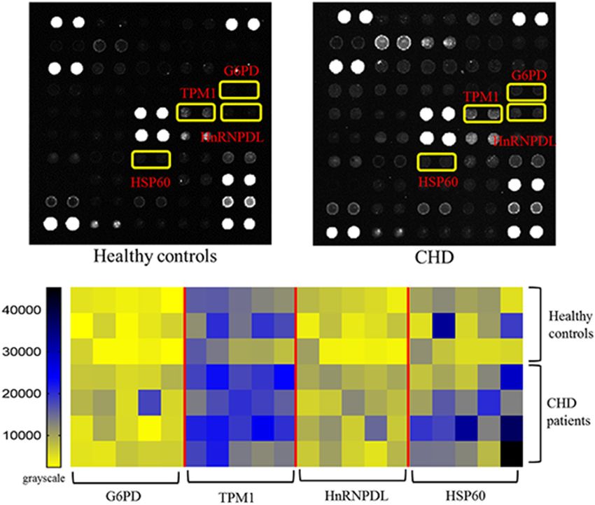
Zhang et al.                                                                                                       TPM1 Negatively With CHD Risk

INTRODUCTION                                                               elevation MI, were excluded from the evaluation. Patients with
                                                                           severe diseases other than stable CHD were also excluded. In
Coronary heart disease, also known as atherosclerotic vascular             summary, patients with stable CHD, defined as prior MI, prior
disease, is a common cardiovascular disease that is associated             coronary revascularization, or multivessel CHD confirmed by
with an increased risk of mortality and morbidity (White                   coronary angiography, were eligible. In addition, patients had
and Chew, 2008). Globally, CHD affected 110 million people                 to meet at least one of the following cardiovascular risk criteria:
and caused about one 10th of total deaths in 2015 (World                   aged ≥ 60 years, diabetes mellitus requiring pharmacotherapy,
Health Organization [WHO], 2014, May). The presence of                     high-density lipoprotein cholesterol level < 1.03 mmol/L, current
autoantibodies is a hallmark of autoimmune diseases (Fritzler,             or previous smoker (defined a ≥ 5 cigarettes per day on
2008). The signature autoantibodies of most autoimmune                     average), significant renal dysfunction (estimated glomerular
diseases have now been identified. Autoantibodies that are                 filtration rate ≥ 30 and < 60 mL/min per 1.73 m2 , or urine
directed against specific proteins can reveal the stages of disease,       albumin/creatinine ratio ≥ 30 mg albumin/g creatinine), or
and predict disease progression (Leeansyah et al., 2013).                  polyvascular disease (CHD and cerebrovascular disease or CHD
   Autoimmune activation, a fundamental mechanism of CHD,                  and peripheral arterial disease). In total, 131 individuals with
has been widely confirmed (Bjorkbacka et al., 2016). Current               stable CHD were included. All patients denied that they had a
studies have shown that there are two types of autoantibodies              typical history of rheumatism. In addition, serum samples were
for CHD. One type are pathological autoantibodies, and their               collected from 131 healthy controls, which was a convenience
elevation indicates a high-risk prognosis for CHD patients. The            sample of patients who reported good to excellent health
other types are protective autoantibodies from the immune                  and had no diagnoses or treatment of atherosclerotic disease,
system to the body. These natural autoantibodies are often                 hypertension, hyperlipidemia, or diabetes. Serum samples were
produced after myocardial infarction, cardiac transplantation              stored in central repositories at −80◦ C until biochemical
and pathogenic microorganism infection (Dunn et al., 1992).                analysis was performed.
Although the total number of CHD autoantibodies is increasing,
such as anti-Apolipoprotein B-100 antibody, anti-HSP60/65/70               Expression and Purification of
a few antibodies were found in these years (Ghayour-Mobarhan
et al., 2007; Zhang et al., 2010; Bjorkbacka et al., 2016).
                                                                           Recombinant Proteins
However, specific CHD antibodies have not yet been discovered              To express the proteins, the expression plasmids were
(Bjorkbacka et al., 2016; Ma et al., 2017). Many circulating protein       transformed into E. coli BL21 competent cells, respectively.
markers have been incorporated into clinical practice and have             The strains containing the expression plasmids were inoculated
been shown to have value as CHD risk factors, but few antibody             in germfree Luria-Bertani (LB) solid medium containing
markers have been applied in clinical application as a result of           kanamycin 30 µg/mL. Petri dishes were left overnight upside-
a lack of basic experimental support (Keller et al., 2009). Thus,          down at 37◦ C. The recombinant colonies were transformed into
further data are still required to identify novel autoantibodies and       LB liquid medium with kanamycin 30 µg/mL and shaken for
examine their effects on CHD (Leu et al., 2017).                           12–14 h at 37◦ C. The cultures were diluted 1/100 and grown
   Conventional methods such as ELISA that are used to evaluate            in the same medium to OD600 of 0.4–0.6, then isopropyl-D-
CHD autoantibodies are inefficient, and systematic screening               thiogalactopyranoside (IPTG) was added to a final concentration
is impossible (Miyashita et al., 2018). Functional protein                 of 1.0 mmol/L. After 5 h growth at 37◦ C, cells were harvested by
microarrays have been designed to perform high-throughput                  centrifugation at 4◦ C and 10,000 × rpm for 20 min. Centrifuged
target screening in one experiment (Song et al., 2010). It has             cells were resuspended in 20 mL equilibration buffer (PBS with
been widely used for autoantibody detection and identification             8M urea, pH 7.4), disrupted by sonication (Scientz, Ningpo,
as autoimmune disease biomarkers in many biological fluids                 China) for 1 h on ice, and the homogenate was centrifuged
(Lueking et al., 2005; Kalbas et al., 2006). Overall, the aim of           at 4◦ C and 8,000 × g for 20 min. Then, the supernatants of
this study was to identify novel autoantibodies and measure                proteins were collected and purified. Recombinant proteins were
the relationship between their titers with the prognosis of CHD            purified using Ni-NTA resin (Qiagen, Hilden, Germany). The
autoantigen microarrays generated in our lab.                              eluted proteins were stored at 4◦ C for use within 1 week, or at
                                                                           −80◦ C for use after a longer time. The purified proteins were
                                                                           analyzed on a 10% SDS-PAGE gel, and the resulting protein
MATERIALS AND METHODS                                                      bands were stained with Coomassie brilliant blue R250. Purified
                                                                           recombinant protein was confirmed by proteomics analyzer AB
Serum Samples                                                              4700 mass spectrometry (Applied Biosystems, Foster City, CA,
All human serum samples were collected at Beijing Hangxing                 United States).
Hospital. This study was approved by the Ethics Committee at the
Institute of Basic Research in Clinical Medicine, China Academy            Production of Protein Microarrays
of Chinese Medical Sciences, and was conducted according to                Protein expression and purification were conducted as previously
the standards of the Declaration of Helsinki. Written informed             described. Printed autoantigens included 10 expressed proteins
consent was obtained from participants. Patients presenting with           and 27 purchased antigen proteins (including classic specific
unstable angina, non-ST-segment elevation MI, or ST-segment                in collagen diseases/rheumatic diseases antigens: SSB, dsDNA,

Frontiers in Pharmacology | www.frontiersin.org                        2                                     February 2020 | Volume 11 | Article 195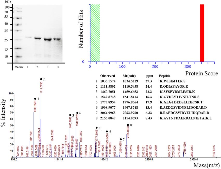
Zhang et al.                                                                                                                  TPM1 Negatively With CHD Risk

Rib-P-1, SmD2, Rib-P-2, nucleosome antigen, nRNP, Jo-1, Sm,                 stop buffer (2M, H2 SO4 ). Finally, the OD value of each well
CENP-B, SSA, candidate CHD antigens: aldehyde dehydrogenase                 was measured with an ELISA reader (BioTek, Winooski, VT,
1, fibrinogen, VEGF165, HSP70, Hsp60, prohibitin, vimentin,                 United States) at 450 nm.
myeloperoxidase, osteopontin, myoglobin, fatty acid-binding
protein, proBNP, S100B, Lp-PLA2, von Willebrand factor, brain-              Statistical Analysis
derived neurotrophic factor) (SinoBiological, Beijing, China),              Binding signals were acquired using a microarray scanner, and
(Diarect AG, Freiburg, Germany), (Arotec, São Paulo, Brazil).               intensity values (signal intensity – background intensity) were
These antigens are classic autoantigens and autoantigens                    normalized using the mean of the positive point signals. t-Test
associated with vascular lesions, and are associated with                   statistics, Spearman’s correlation coefficients, Fisher’s exact text
inflammation, thrombosis, and vascular lesions, and may be                  and cox proportional hazards regression were analyzed using SAS
associated with the occurrence and prognosis of CHD. These                  version 9.4 (SAS Institute, Inc., Cary, NC, United States). P-values
proteins represented candidate autoantigens in CHD. Proteins                of less than 0.05 were considered significant. The threshold for
were diluted to 1 mg/ml in PBS arrayed in 384-well titer                    defining a positive result was a value higher than that of the
dishes for printing. Purified human proteins and control                    healthy controls (mean + 2 SD).
proteins were spotted in duplicate onto polymer-Slide H-OP
(CapitalBio, Beijing, China) at high density using a Personal
Arrayer (CapitalBio, Beijing, China) (Schena et al., 1995). Every           RESULTS
autoantigen had an average diameter of 200 µm which was
printed twice. Each protein microarray included several control             Characteristics of the Study Groups
spots such as human IgG. Printing was performed in a cabinet                The study included 131 stable CHD patients (68.0 ± 7.0 years)
at 25◦ C and 55% humidity. These conditions were constantly                 and 131 healthy controls (66.5 ± 8.0 years), that consisted of
monitored by a thermohygrometer. The printed human protein                  66 males and 65 females, respectively. There was no statistical
chips were kept horizontal at room temperature (RT) for 1 h                 difference (P = 0.029) in age data between the two groups. The
before storage at 4◦ C. Protein sequences were derived from the             protein microarrays were incubated with a random selection
publicly available database UniProt.                                        of 20 patients and 15 healthy controls in the current serum
                                                                            samples. Table 1 shows the baseline characteristics of patients
Serum Profiling on Protein Microarrays                                      with stable CHD for protein microarrays and ELISA, with data
Arrays were circumscribed with hydrophobic fences and blocked               on sex, age, body mass index (BMI), waist circumference, systolic
with PBS containing 5% fetal calf serum for 1 h at RT. The protein          blood pressure, diastolic blood pressure, hypertension, TC, HDL,
microarrays were incubated with a random selection of 20 CHD                LDL, triglycerides (TG), glucose, and tobacco. The mean age of
patients and 15 healthy controls in a total of 262 samples. Then,           the men and women was 66.53 and 69.48 years, respectively.
these microarrays were probed with 1:20 dilution of CHD patient             The mean and median values for BMI, waist circumference, TC,
or healthy control serum for 1 h under the same condition,                  HDL, LDL, TG, and glucose were within normal limits. In total
followed by washing three times in PBST and incubation with                 52% of CHD patients had hypertension diagnosed, and 92.31%
a 1:400 dilution of Cy3-labeled anti-human IgG (Bioss, Beijing,
China) secondary antibody for 1 h at RT in the dark. The
microarray was blown dry with compressed air and scanned with               TABLE 1 | Baseline characteristics for protein microarrays and ELISA cohorts.
a microarray scanner (CapitalBio, Beijing, China). The binding
signals were acquired and analyzed using a microarray reader                Variable                           Protein microarrays             ELISA
(CapitalBio, Beijing, China).
                                                                            Number                                      20                       131
                                                                            Male, n (%)                               10 (50)                  66 (50.38)
Enzyme Linked Immunosorbent Assay                                           Age (year)                                64 (47–75)               70 (47–78)
Validation of CHD novel autoantigen was performed with
                                                                            BMI                                    26.33 (4.47)             24.87 (3.49)
ELISA. The 96-well microtiter plates were coated with 100 µL                Waist circumference (cm)               93.35 (14.38)               87 (66.5–123)
candidate proteins (500 ng/mL, dissolved in 0.05M carbonic                  Systolic blood pressure (mmHg)           143 (16.45)           143.48 (16.22)
buffer) overnight at 4◦ C. After removing the liquid phase, each            Diastolic blood pressure (mmHg)        82.15 (11.38)            80.63 (9.88)
well was blocked with 200 µL 5% fetal calf serum (diluted in PBS)           Hypertension, n (%)                       13 (65)                  52 (39.69)
for 2 h at 37◦ C. Then the liquid phase was removed again and the           On hypertensive treatment, n (%)          12 (60)                  48 (36.64)
sera of CHD patients and healthy controls (1:20, diluted in PBS)            Total cholesterol (mmol/L)              4.92 (1.04)              4.92 (0.93)
were used to incubate the 96-well microtiter plates for 1 h at 37◦ C,       HDL cholesterol (mmol/L)                1.22 (0.24)              1.26 (0.28)
followed by washing three times in PBST and incubation with                 LDL cholesterol (mmol/l)                2.90 (0.80)              2.86 (0.70)
100 µL 1:10,000 dilution of goat anti-human IgG-HRP (Bioss,                 Triglycerides (mmol/L)                  1.61 (0.81–6.86)         1.45 (0.42–6.86)
Beijing, China) for 45 min. Each was washed with 0.1% PBST                  Glucose (mmol/L)                        5.84 (0.68)               5.7 (4.5–7.9)
five times to elute the secondary autoantibodies. After that, 50 µL
tetramethylbenzidine (TMB) A and 50 µL TMB B was added to                   Values in the normal distribution are shown as the mean (SD). Values in the non-
                                                                            normal distribution are shown as the median (minimum–maximum). BMI, body
each well. After keeping the plates in a dark, room-temperature             mass index; HDL cholesterol, high density lipoprotein cholesterol; LDL cholesterol,
location for 15 min, the reaction was stopped by adding 50 mL               low density lipoprotein cholesterol.

Frontiers in Pharmacology | www.frontiersin.org                         3                                             February 2020 | Volume 11 | Article 195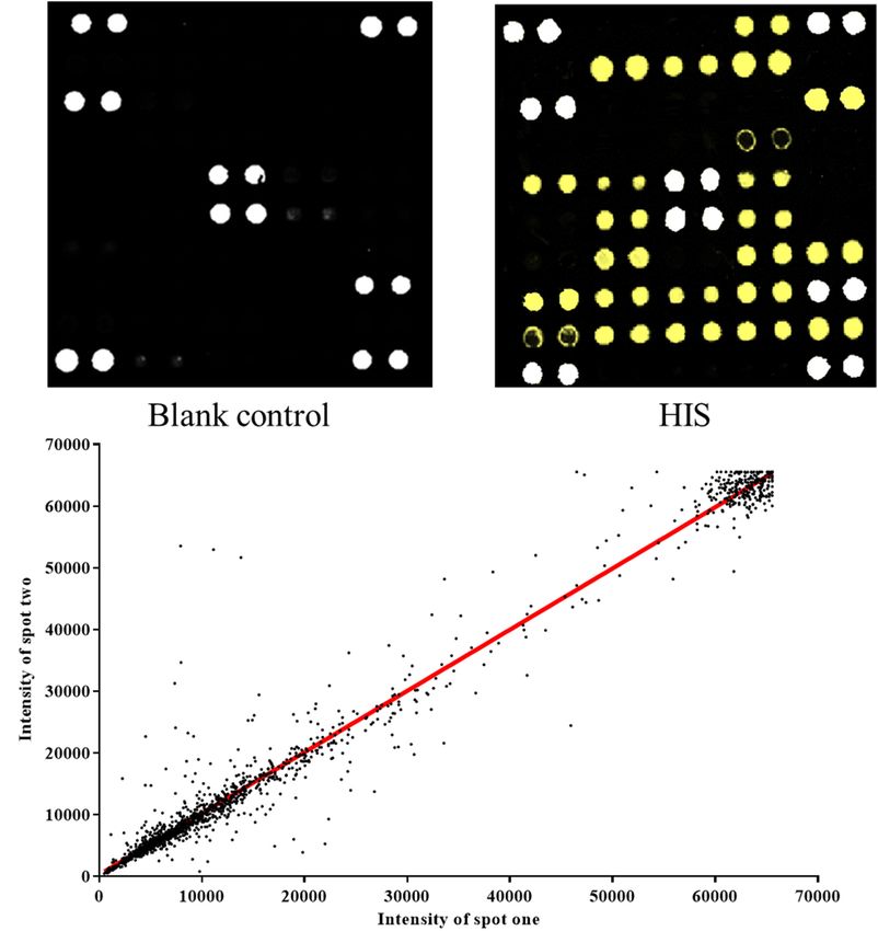
Zhang et al.                                                                                                                            TPM1 Negatively With CHD Risk

received treatment. The mean and median values for systolic                            were transformed into E. coli BL21(DE3) competent cells. The
and diastolic blood pressure were not confirmed as being within                        results demonstrated that the proteins were purified successfully
normal limits. Variables were expressed as the mean (SD) when                          as the specific bands corresponding to the expected molecular
they were normally distributed and as the median and minimum–                          weights of the proteins were detected by SDS-PAGE and Annexin
maximum range when they were not. No gender differences                                A2 as a representative protein was displayed (Figure 1A). The
were found in the data. Concurrently, 131 age-gender matched                           proteins obtained by Ni-NTA resin were confirmed by mass
healthy controls were also enrolled in this study to serve as a                        spectrometry. The target proteins were identified by spectrometry
blank control group.                                                                   analysis. The results showed that the peptide matching degree
                                                                                       score was high, which indicated that the putative protein has a
Expression and Purification of                                                         highly homologous identity (P < 0.05) (Figure 1B). Protein mass
                                                                                       spectrometry is shown in Figure 1C.
Recombinant Proteins for Protein
Microarrays
The pET expression system is one of the most widely used                               Quality Control of the Protein
for cloning and in vivo expression of recombinant proteins in                          Microarrays
E. coli (Liu and Naismith, 2009). In order to obtain the candidate                     The protein microarray contained 37 proteins which represent
proteins, the expression plasmids pET 28 a-Hsp27, pET 28                               candidate autoantigens in CHD. The printed autoantigens
a-Glucose-6-phosphate, pET 28 a-alpha-tropomyosin, pET 28                              included 10 expressed proteins from the lab and 27 CHD related
a-Annexin-A2, pET 28 a-JKTBP, pET 28 a-HnRNP-A2B1, pET                                 proteins that were purchased commercially. Each protein was
28 a-HnRNP-A1, pET 28 a-Moesin, and pET 28 a-Keratin 8                                 printed repeatedly in order to ensure the quality of protein

  FIGURE 1 | Expression, purification, and identification of recombinant proteins for protein microarrays. The recombinant proteins were expressed from the bacterial
  clones and purified, in parallel, under denaturing conditions. Annexin A2 was represented. (A) Result of scanning on SDS-PAGE gels. Lane 1–4 represented different
  elution times. (B) Ions score is –10∗ Log (P), where P is the probability that the observed match is a random event. Individual ions scores > 29 indicate identity or
  extensive homology (P < 0.05). Protein scores are derived from ions scores as a non-probabilistic basis for ranking protein hits. (C) Proteins mass spectrometry
  analysis.

Frontiers in Pharmacology | www.frontiersin.org                                    4                                             February 2020 | Volume 11 | Article 195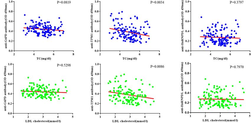
Zhang et al.                                                                                                                           TPM1 Negatively With CHD Risk

  FIGURE 2 | Construction of protein microarrays. (A) The blank control of protein microarray. BSA-Cy3 and recombinant human proteins were printed in duplicate on
  poly-L-lysine coated microscope slides. The BSA-Cy3 is represented in white (saturated intensity) to orient. (B) The protein microarray probed with anti-HIS
  monoclonal antibody. To monitor the quality and relative quantity of the printed proteins on glass slides, the human protein microarrays were probed with anti-HIS
  antibody, followed by Cy3-labeled secondary antibody to visualize the signals. The proteins positively detected by the anti-HIS antibody are represented in yellow
  (saturated intensity). (C) Correlation of spot intensities of all the duplicate pairs after experiment. The signal intensities of duplicate spots (Spot 1 versus its
  corresponding Spot 2) were plotted against each other. The resulting correlation coefficient was 0.9782, indicating high reproducibility of the protein spotting.

microarrays. Positive coordinate points and all other spots signal                     microarrays are showed in Figure 3A. The yellow boxes indicate
intensity in blank protein microarray were described (Figure 2A).                      positive candidate autoantigens between healthy controls and
The assay indicated that HIS-labeled proteins were detectable                          coronary heart disease patients, there was significant difference
with anti-HIS signals, which shows that these proteins work well                       between two groups (P < 0.05). The four autoantigens levels
in the chip system (Figure 2B). All duplicate pairs after done sera                    had a significant difference (P < 0.05) between the two groups;
experiments shows that there was a high correlation coefficient                        details of these differences are expressed in Figure 3B. For the
between the signal intensities of duplicate spots (Figure 2C).                         CHD autoantigens reported in the literature, HSP60 results were
                                                                                       similar to those previously reported (Zhang et al., 2008; Fust et al.,
Identification of CHD-Associated                                                       2012). This further illustrates the reliability of the chip method.
Autoantigens Using Protein Microarrays
The protein microarrays were incubated with a random selection                         Validation of Candidate Autoantibodies
of 20 CHD patients (Table 1) and 15 healthy controls in serum                          by ELISA
samples. Positive reactivity was detected in recombinant G6PI,                         Enzyme linked immunosorbent assay of three novel antigens was
HnRNPDL, TPM1 and HSP60 sera from 9, 5, 5, and 4 of 20                                 carried out with the sera of 131 CHD patients and 131 healthy
CHD patients, respectively. Representative results of protein                          controls to confirm the results of the microarrays. All ELISA

Frontiers in Pharmacology | www.frontiersin.org                                    5                                            February 2020 | Volume 11 | Article 195
Zhang et al. TPM1 Negatively With CHD Risk
FIGURE 3 | Results of protein microarrays. (A) Representative results of protein microarrays. The yellow boxes indicate positive candidate autoantigens between
healthy controls and coronary heart disease patients, there was significant difference between two groups (P < 0.05). (B) Autoantibody reactivity was determined by
15 healthy control and 20 CHD patient serum samples. Pairwise significance analysis of microarrays was performed to identify antigen features having statistically
significant.
results are shown in a heat map (Figure 4A). In large-scale rheumatoid arthritis or psoriasis, display an increased CHD risk
samples, positive reactivity was detected in recombinant G6PI, (Levy et al., 2008; Vizzardi et al., 2010; Lopez-Pedrera et al., 2012).
HnRNPDL, TPM1 and HSP60 sera from 36, 22, 17 of 131 CHD For patients without autoimmune diseases, but established CHD,
patients, respectively. The reactivity of CHD serum IgG against levels of natural autoantibodies against various endogenous
these three autoantigens was significantly higher than healthy epitopes, such as modified LDL, heat shock proteins have been
controls (P < 0.0001) in box plots (Figure 4B). shown to independently predict CHD outcome (Roux-Lombard
et al., 2013; Vuilleumier et al., 2014). Also, in vivo and in vitro
Correlation of Autoantibodies Levels evidence has demonstrated that some natural autoantibodies
Against TC and LDL in CHD Patients could directly influence atherogenesis and atherosclerotic plaque
The relationship between TC and candidate autoantibody levels vulnerability, mostly by activating innate immune receptors,
were analyzed, and there was a significant inverse correlation in thereby supporting a causal role of humoral autoimmunity in
anti-TPM1 antibodies (P = 0.0034, r = −0.03883) (Figures 5A– atherosclerosis (Carbone et al., 2013).
C). The same relationship was noticed for low density lipoprotein In this study, three novel autoantigens were found for CHD
cholesterol (Ueba et al., 2009; van den Berg et al., 2018) by analyzing microarray data; these included G6PI, HnRNPDL
(P = 0.0086, r = −0.04635) (Figures 5D–F). and TPM1. Some experimental studies confirm the hypothesis
of a significant protective effect against CHD resulting from the
genetic condition of G6PI deficiency (Meloni et al., 2008). G6PI
DISCUSSION also is an autoantigen widely present in rheumatoid arthritis.
A high concentration of G6PI was observed on the synovial
The pathogenesis of CHD is associated with autoimmune lining surface and the small artery endothelial cell surface.
activation, which can enhance atherosclerosis (Onat and Whether the same mechanism exists in CHD is worthy of further
Yuksel, 2013; Meurice et al., 2016). Patients suffering from study (van Boekel et al., 2001). However, this is enough to
an autoimmune disease, such as systemic lupus erythematosus, show that CHD has the property of autoimmunity. HnRNPL
Frontiers in Pharmacology | www.frontiersin.org 6 February 2020 | Volume 11 | Article 195Zhang et al. TPM1 Negatively With CHD Risk FIGURE 4 | Validation of candidate autoantibodies between CHD patients and controls by ELISA. (A) Autoantibody reactivity was determined by 131 CHD patients and 131 healthy controls serum samples by ELISA. The OD of each of the three autoantigens (G6PD, TPM1, HnRNPDL) reacting with the serum samples in each case are displayed. (B) Box plots of the ELISA OD for three CHD autoantigens in various serum groups. The rectangles define the interquartile range (IQR). The bar within the rectangle indicates the median value. The bars above and below the rectangles define the 1.5I QR outlier ranges (P < 0.0001). OD, optical density. proteins are related to a family of heterogeneous nuclear and RNA binding protein HnRNPL is a new member of hnRNPs ribonucleoproteins that function in mRNA biogenesis and that are involved in mRNA biogenesis (Akagi et al., 2000). The mRNA metabolism (Kawamura et al., 2002). The human DNA HnRNPs family are autoantigens of many autoimmune diseases. Frontiers in Pharmacology | www.frontiersin.org 7 February 2020 | Volume 11 | Article 195
Zhang et al.                                                                                                                          TPM1 Negatively With CHD Risk

  FIGURE 5 | Coronary heart disease risk factor correlation analysis. (A–C) Relationship between total cholesterol (TC) and candidate antibodies (G6PD, TPM1,
  HnRNPDL) OD. There was significant correlation in anti-TPM1 antibodies (P < 0.05). (D–F) Relationship between low density lipoprotein cholesterol (LDL cholesterol)
  and candidate antibodies OD. There was significant correlation in anti-TPM1 antibodies (P < 0.05). The result showed negative correlation between antibody
  concentration and CHD risk factor indicators.

Its release may be related to endothelial cell damage. At                             and/or inflammatory phospholipids (Frostegard, 2010). HSP-
the same time, its ectopic expression in the cell membrane                            70 antibodies have also been found to have the same trend in
may also participate in the early mechanism of autoimmunity                           CHD patients. The negative correlation between these natural
(Han et al., 2010).                                                                   antibodies and CHD is clearly not coincidental. We speculate
    Tropomyosin belongs to a family of actin-binding proteins                         that the two antibodies (G6PI and HnRNPDL) found in this
that are central to the control of calcium-regulated striated                         study belong to pathological autoantibodies, which reflect the
muscle contraction (Jagatheesan et al., 2003). The three primary                      autoimmune properties of CHD and may damage endothelial
tropomyosin isoforms, α-TM, β-TM and γ-TM, are alternatively                          cells and cardiac myocytes. But anti-TPM1 belongs to the
spliced products of the TPM1, TPM2, and TPM3 genes,                                   regulatory natural autoantibodies in CHD. Low abundance of
respectively. Also, they are highly homologous but exhibit                            this antibody may indicate a decline of the body’s ability to
distinct physiological properties (Jagatheesan et al., 2010). Alpha-                  restore homeostasis.
tropomyosin is a coiled-coil protein that associates with actin                          Current techniques to perform large-scale multiplex
filaments as a homodimer and plays a central role in the calcium                      characterization of autoantibody responses are extremely
dependent regulation of vertebrate striated muscle contraction                        limited. ELISA, Western-blot analysis and radio immune
(Janco et al., 2012). It is a major antigen in allergic responses                     assays are time-consuming and tedious processes. The
to invertebrates such as crustaceans, arachnids, insects, and                         conception and initial development of “multianalyte microspot
mollusks (Reese et al., 1999). Autoimmune responses to alpha-                         immunoassays” was initially proposed by Ekins (1989).
tropomyosin have been previously reported in the sera of patients                     MacBeath and Schreiber described spotted protein arrays
with ulcerative colitis, colonic intraepithelial lymphocytes and                      as a tool to detect protein and small-molecule interactions
Behçet’s syndrome (Biancone et al., 1998; Geng et al., 1998;                          (MacBeath and Schreiber, 2000). By enriching potential
Mor et al., 2002).                                                                    autoantigens and fabricating chips, the efficient screening
    A negative correlation of anti-TPM1 antibody concentrations                       of autoantigens of CHD was realized. We constructed and
and TC and LDL was found in our study. TPM1 is considered                             applied protein microarrays to perform simple, low-sample
to play an important part in the regulation of the smooth                             volume, fluorescence-based assays. By taking advantage
muscle contraction process (Perry, 2001). The titers of these                         of existing microarray-based data, we were able to readily
antibodies are dynamic and tend to balance with the gradual                           identify four potential autoantigens, and then further
recovery of homeostasis in the body. A negative correlation                           confirm three of these proteins as newly identified CHD-
between antibody titers and risk factors has been found in                            specific autoantigens. Although the present study provided
several studies. Low levels of anti-PC predict the development                        interesting findings about the association of novel autoantibody
of stroke and myocardial infarction, and represent a new model                        concentrations and the risk indicators of CHD, several potential
from chronic disease for the body to fight against oxidized                           limitations should be acknowledged. First, this pilot study and

Frontiers in Pharmacology | www.frontiersin.org                                   8                                            February 2020 | Volume 11 | Article 195
Zhang et al. TPM1 Negatively With CHD Risk
hypothesis-generating manuscript is limited by the group sample ETHICS STATEMENT
size. Second, the number of candidate autoantigens is relatively
small. Finally, although the relationship between autoantibodies This study was approved by the Ethics Committee at
concentration and CHD risk indicators parameters were the Institute of Basic Research in Clinical Medicine,
analyzed, more experiments are needed to understand to the China Academy of Chinese Medical Sciences, and was
mechanism of this phenomenon. conducted according to the standards of the Declaration
of Helsinki. Written informed consent was obtained
from participants.
CONCLUSION
We identified three novel autoantigens (G6PI, TPM1, AUTHOR CONTRIBUTIONS
HnRNPDL) for stable CHD and to verify the validity of HSP60.
The correction analysis revealed a negative correlation of CL, PC, AL, JY, and YZ conceived the project. PC,
anti-TPM1 antibody levels and TC, and LDL, respectively. YZ, and CL designed the experiments. YZ, PC, and HZ
carried out the research. YZ conducted the data analysis
and wrote the manuscript. YZ, HZ, BL, LL, LZ, MB, XJ,
XH, JY, PC, CL, and AL have read and approved the
SIGNIFICANCE final manuscript.
Immune responses play a key role in coronary heart
disease development. Previous studies have indicated inverse
associations between autoantibodies recognizing oxidized low-
FUNDING
density lipoprotein epitopes and cardiovascular disease. The This research has been supported by the National Science
strength of the present study is the discovery of novel antibodies and Technology Major Project (2018ZX10101001-005-003 and
that allow for a unique evaluation of anti-TPM1 autoantibodies 2018ZX10101001-005-004) and the Fundamental Research
to predict risks of future cardiovascular events. Analysis of Funds for the Central public welfare research institutes (ZZ11-
the autoantibody levels may offer an opportunity to identify 113 and Z0600). The funding agencies did not have any role in
individuals who are in need of immune-modulatory therapy. the design and conduct of the study, collection, management, and
interpretation of the data or preparation, review, or approval of
the manuscript.
DATA AVAILABILITY STATEMENT
The raw data supporting the conclusions of this article will be ACKNOWLEDGMENTS
made available by the authors, without undue reservation, to any
qualified researcher. We thank the reviewers for their critical comments.
REFERENCES Fritzler, M. J. (2008). Challenges to the use of autoantibodies as predictors of disease
onset, diagnosis and outcomes. Autoimmun. Rev. 7, 616–620. doi: 10.1016/j.
Akagi, T., Kamei, D., Tsuchiya, N., Nishina, Y., Horiguchi, H., Matsui, M., autrev.2008.06.007
et al. (2000). Molecular characterization of a mouse heterogeneous nuclear Frostegard, J. (2010). Low level natural antibodies against phosphorylcholine:
ribonucleoprotein D-like protein JKTBP and its tissue-specific expression. Gene a novel risk marker and potential mechanism in atherosclerosis and
245, 267–273. doi: 10.1016/s0378-1119(00)00032-9 cardiovascular disease. Clin. Immunol. 134, 47–54. doi: 10.1016/j.clim.2009.
Biancone, L., Monteleone, G., Marasco, R., and Pallone, F. (1998). Autoimmunity 08.013
to tropomyosin isoforms in ulcerative colitis (UC) patients and unaffected Fust, G., Uray, K., Bene, L., Hudecz, F., Karadi, I., and Prohaszka, Z. (2012).
relatives. Clin. Exp. Immunol. 113, 198–205. doi: 10.1046/j.1365-2249.1998. Comparison of epitope specificity of anti-heat shock protein 60/65 IgG type
00610.x antibodies in the sera of healthy subjects, patients with coronary heart disease
Bjorkbacka, H., Alm, R., Persson, M., Hedblad, B., Nilsson, J., and Fredrikson, G. N. and inflammatory bowel disease. Cell Stress Chaperones 17, 215–227. doi: 10.
(2016). Low levels of apolipoprotein B-100 autoantibodies are associated with 1007/s12192-011-0301-7
increased risk of coronary events. Arterioscler. Thromb. Vasc. Biol. 36, 765–771. Geng, X., Biancone, L., Dai, H. H., Lin, J. J., Yoshizaki, N., Dasgupta, A.,
doi: 10.1161/atvbaha.115.306938 et al. (1998). Tropomyosin isoforms in intestinal mucosa: production of
Carbone, F., Nencioni, A., Mach, F., Vuilleumier, N., and Montecucco, autoantibodies to tropomyosin isoforms in ulcerative colitis. Gastroenterology
F. (2013). Evidence on the pathogenic role of auto-antibodies in acute 114, 912–922. doi: 10.1016/s0016-5085(98)70310-5
cardiovascular diseases. Thromb. Haemost. 109, 854–868. doi: 10.1160/th12-10- Ghayour-Mobarhan, M., Taylor, A., Lamb, D. J., and Ferns, G. A. (2007).
0768 Association between indices of body mass and antibody titres to heat-shock
Dunn, M. J., Crisp, S. J., Rose, M. L., Taylor, P. M., and Yacoub, M. H. (1992). Anti- protein-60, -65 and -70 in healthy Caucasians. Int. J. Obes. 31, 197–200. doi:
endothelial antibodies and coronary artery disease after cardiac transplantation. 10.1038/sj.ijo.0803385
Lancet 339, 1566–1570. doi: 10.1016/0140-6736(92)91832-s Han, S. P., Tang, Y. H., and Smith, R. (2010). Functional diversity of the
Ekins, R. P. (1989). Multi-analyte immunoassay. J. Pharm. Biomed. Anal. 7, hnRNPs: past, present and perspectives. Biochem. J. 430, 379–392. doi: 10.1042/
155–168. doi: 10.1016/0731-7085(89)80079-2 bj20100396
Frontiers in Pharmacology | www.frontiersin.org 9 February 2020 | Volume 11 | Article 195Zhang et al. TPM1 Negatively With CHD Risk Jagatheesan, G., Rajan, S., Petrashevskaya, N., Schwartz, A., Boivin, G., Vahebi, Mor, F., Weinberger, A., and Cohen, I. R. (2002). Identification of alpha- S., et al. (2003). Functional importance of the carboxyl-terminal region of tropomyosin as a target self-antigen in Behcet’s syndrome. Eur. J. Immunol. 32, striated muscle tropomyosin. J. Biol. Chem. 278, 23204–23211. doi: 10.1074/jbc. 356–365. doi: 10.1002/1521-4141(200202)32:23.0.co;2-9 M303073200 Onat, A., and Yuksel, H. (2013). Autoimmune activation, a fundamental Jagatheesan, G., Rajan, S., and Wieczorek, D. F. (2010). Investigations into mechanism of coronary artery disease risk, missed by inadequate analysis. Turk tropomyosin function using mouse models. J. Mol. Cell Cardiol. 48, 893–898. Kardiyol. Dern. Ars. 41, 566–567. doi: 10.1016/j.yjmcc.2009.10.003 Perry, S. V. (2001). Vertebrate tropomyosin: distribution, properties and function. Janco, M., Kalyva, A., Scellini, B., Piroddi, N., Tesi, C., Poggesi, C., et al. (2012). J. Muscle Res. Cell Motil. 22, 5–49. alpha-Tropomyosin with a D175N or E180G mutation in only one chain differs Reese, G., Ayuso, R., and Lehrer, S. B. (1999). Tropomyosin: an invertebrate pan- from tropomyosin with mutations in both chains. Biochemistry 51, 9880–9890. allergen. Int. Arch. Allergy Immunol. 119, 247–258. doi: 10.1159/000024201 doi: 10.1021/bi301323n Roux-Lombard, P., Pagano, S., Montecucco, F., Satta, N., and Vuilleumier, Kalbas, M., Lueking, A., Kowald, A., and Muellner, S. (2006). New N. (2013). Auto-antibodies as emergent prognostic markers and possible analytical tools for studying autoimmune diseases. Curr. Pharm. Des. 12, mediators of ischemic cardiovascular diseases. Clin. Rev. Allergy Immunol. 44, 3735–3742. 84–97. doi: 10.1007/s12016-010-8233-z Kawamura, H., Tomozoe, Y., Akagi, T., Kamei, D., Ochiai, M., and Yamada, Schena, M., Shalon, D., Davis, R. W., and Brown, P. O. (1995). Quantitative M. (2002). Identification of the nucleocytoplasmic shuttling sequence of monitoring of gene expression patterns with a complementary DNA heterogeneous nuclear ribonucleoprotein D-like protein JKTBP and its microarray. Science 270, 467–470. doi: 10.1126/science.270.5235.467 interaction with mRNA. J. Biol. Chem. 277, 2732–2739. doi: 10.1074/jbc. Song, Q., Liu, G., Hu, S., Zhang, Y., Tao, Y., Han, Y., et al. (2010). Novel M108477200 autoimmune hepatitis-specific autoantigens identified using protein microarray Keller, T., Zeller, T., Peetz, D., Tzikas, S., Roth, A., Czyz, E., et al. (2009). Sensitive technology. J. Proteome Res. 9, 30–39. doi: 10.1021/pr900131e troponin I assay in early diagnosis of acute myocardial infarction. N. Engl. J. Ueba, T., Nomura, S., Nishikawa, T., Kajiwara, M., and Yamashita, K. (2009). Med. 361, 868–877. doi: 10.1056/NEJMoa0903515 Circulating oxidized LDL, measured with FOH1a/DLH3 antibody, is Leeansyah, E., Malone, D. F., Anthony, D. D., and Sandberg, J. K. (2013). Soluble associated with metabolic syndrome and the coronary heart disease biomarkers of HIV transmission, disease progression and comorbidities. risk score in healthy Japanese. Atherosclerosis 203, 243–248. doi: Curr. Opin. HIV AIDS 8, 117–124. doi: 10.1097/COH.0b013e32835c 10.1016/j.atherosclerosis.2008.05.048 7134 van Boekel, M. A., Vossenaar, E. R., van den Hoogen, F. H., and van Venrooij, W. J. Leu, H. B., Yin, W. H., Tseng, W. K., Wu, Y. W., Lin, T. H., Yeh, H. I., et al. (2001). Autoantibody systems in rheumatoid arthritis: specificity, sensitivity (2017). Identification of new biosignatures for clinical outcomes in stable and diagnostic value. Arthritis Res. Ther. 4, 87–93. doi: 10.1186/ar395 coronary artery disease - The study protocol and initial observations of a van den Berg, V. J., Haskard, D. O., Fedorowski, A., Hartley, A., Kardys, I., Caga- prospective follow-up study in Taiwan. BMC Cardiovasc. Disord. 17:42. doi: Anan, M., et al. (2018). TEMPORARY REMOVAL: IgM anti-malondialdehyde 10.1186/s12872-017-0471-z low density lipoprotein antibody levels indicate coronary heart disease and Levy, L., Fautrel, B., Barnetche, T., and Schaeverbeke, T. (2008). Incidence and necrotic core characteristics in the Nordic Diltiazem (NORDIL) study and the risk of fatal myocardial infarction and stroke events in rheumatoid arthritis Integrated Imaging and Biomarker Study 3 (IBIS-3). EBioMedicine 36, 63–72. patients. A systematic review of the literature. Clin. Exp. Rheumatol. 26, doi: 10.1016/j.ebiom.2018.08.023 673–679. Vizzardi, E., Raddino, R., Teli, M., Gorga, E., Brambilla, G., and Dei Cas, L. (2010). Liu, H., and Naismith, J. H. (2009). A simple and efficient expression and Psoriasis and cardiovascular diseases. Acta Cardiol. 65, 337–340. doi: 10.2143/ purification system using two newly constructed vectors. Protein Expr. Purif. ac.65.3.2050351 63, 102–111. doi: 10.1016/j.pep.2008.09.008 Vuilleumier, N., Montecucco, F., and Hartley, O. (2014). Autoantibodies to Lopez-Pedrera, C., Perez-Sanchez, C., Ramos-Casals, M., Santos-Gonzalez, apolipoprotein A-1 as a biomarker of cardiovascular autoimmunity. World J. M., Rodriguez-Ariza, A., and Cuadrado, M. J. (2012). Cardiovascular Cardiol. 6, 314–326. doi: 10.4330/wjc.v6.i5.314 risk in systemic autoimmune diseases: epigenetic mechanisms of immune White, H. D., and Chew, D. P. (2008). Acute myocardial infarction. Lancet 372, regulatory functions. Clin. Dev. Immunol. 2012:974648. doi: 10.1155/2012/ 570–584. doi: 10.1016/s0140-6736(08)61237-4 974648 World Health Organization [WHO] (2014). The Top 10 Causes of Death. Geneva: Lueking, A., Huber, O., Wirths, C., Schulte, K., Stieler, K. M., Blume-Peytavi, World Health Organization. U., et al. (2005). Profiling of alopecia areata autoantigens based on protein Zhang, X., He, M., Cheng, L., Chen, Y., Zhou, L., Zeng, H., et al. (2008). Elevated microarray technology. Mol. Cell. Proteomics 4, 1382–1390. doi: 10.1074/mcp. heat shock protein 60 levels are associated with higher risk of coronary heart T500004-MCP200 disease in Chinese. Circulation 118, 2687–2693. doi: 10.1161/circulationaha. Ma, W. T., Chang, C., Gershwin, M. E., and Lian, Z. X. (2017). Development 108.781856 of autoantibodies precedes clinical manifestations of autoimmune diseases: Zhang, X., Xu, Z., Zhou, L., Chen, Y., He, M., Cheng, L., et al. (2010). Plasma a comprehensive review. J. Autoimmun. 83, 95–112. doi: 10.1016/j.jaut.2017. levels of Hsp70 and anti-Hsp70 antibody predict risk of acute coronary 07.003 syndrome. Cell Stress Chaperones 15, 675–686. doi: 10.1007/s12192-010- MacBeath, G., and Schreiber, S. L. (2000). Printing proteins as microarrays for 0180-3 high-throughput function determination. Science 289, 1760–1763. Meloni, L., Manca, M. R., Loddo, I., Cioglia, G., Cocco, P., Schwartz, A., Conflict of Interest: The authors declare that the research was conducted in the et al. (2008). Glucose-6-phosphate dehydrogenase deficiency protects against absence of any commercial or financial relationships that could be construed as a coronary heart disease. J. Inherit. Metab. Dis. 31, 412–417. doi: 10.1007/s10545- potential conflict of interest. 008-0704-5 Meurice, C., Legrand, V., and Pierard, L. (2016). [Contribution of cardiac Copyright © 2020 Zhang, Zhao, Liu, Li, Zhang, Bao, Ji, He, Yi, Chen, Lu and Lu. catheterization to the diagnosis and treatment of coronary heart disease before This is an open-access article distributed under the terms of the Creative Commons age 40]. Rev. Med. Liege 71, 129–136. Attribution License (CC BY). The use, distribution or reproduction in other forums Miyashita, K., Fukamachi, I., Machida, T., Nakajima, K., Young, S. G., Murakami, is permitted, provided the original author(s) and the copyright owner(s) are credited M., et al. (2018). An ELISA for quantifying GPIHBP1 autoantibodies and and that the original publication in this journal is cited, in accordance with accepted making a diagnosis of the GPIHBP1 autoantibody syndrome. Clin. Chim. Acta academic practice. No use, distribution or reproduction is permitted which does not 487, 174–178. doi: 10.1016/j.cca.2018.09.039 comply with these terms. Frontiers in Pharmacology | www.frontiersin.org 10 February 2020 | Volume 11 | Article 195
You can also read