Trafficking of the EGFR ligand Spitz regulates its signaling activity in polarized tissues
←
→
Page content transcription
If your browser does not render page correctly, please read the page content below
Research Article                                                                                                                            4469

                          Trafficking of the EGFR ligand Spitz regulates its
                          signaling activity in polarized tissues
                          Josefa Steinhauer1,*, Hui Hua Liu2, Eli Miller1 and Jessica E. Treisman2,*
                          1
                           Yeshiva College, 2495 Amsterdam Ave., New York, NY 10033, USA
                          2
                           Skirball Institute for Biomolecular Medicine, 540 First Avenue, New York, NY 10016, USA
                          *Authors for correspondence (jsteinha@yu.edu; Jessica.Treisman@med.nyu.edu)

                          Accepted 15 July 2013
                          Journal of Cell Science 126, 4469–4478
                          ß 2013. Published by The Company of Biologists Ltd
                          doi: 10.1242/jcs.131169

                          Summary
                          Epidermal growth factor receptor (EGFR) ligands undergo a complex series of processing events during their maturation to active signaling
                          proteins. Like its mammalian homologs, the predominant Drosophila EGFR ligand Spitz is produced as a transmembrane pro-protein. In
                          the secretory pathway, Spitz is cleaved within its transmembrane domain to release the extracellular signaling domain. This domain is
                          modified with an N-terminal palmitate group that tethers it to the plasma membrane. We found that the pro-protein can reach the cell
                          surface in the absence of proteolysis, but that it fails to activate the EGFR. To address why the transmembrane pro-protein is inactive,
                          whereas membrane association through the palmitate group promotes activity, we generated a panel of chimeric constructs containing the
                          Spitz extracellular region fused to exogenous transmembrane proteins. Although the orientation of the EGF domain and its distance from
                          the plasma membrane varies in these chimeras, they are all active in vivo. Thus, tethering Spitz to the membrane via a transmembrane
Journal of Cell Science

                          domain at either terminus does not prevent activity. Conversely, removing the N-terminal palmitate group from the C-terminally tethered
                          pro-protein does not render it active. Furthermore, we show that the Spitz transmembrane pro-protein can activate the EGFR in a tissue
                          culture assay, indicating that its failure to signal in vivo is not due to structural features. In polarized imaginal disc cells, unprocessed Spitz
                          pro-protein localizes to apical puncta, whereas the active chimeric Spitz constructs are basolaterally localized. Taken together, our data
                          support the model that localized trafficking of the pro-protein restricts its ability to activate the receptor in polarized tissues.

                          Key words: Palmitoylation, Rhomboid, Imaginal disc, Spitz, EGFR

                          Introduction                                                                  a single downstream signal transduction pathway (Shilo, 2005).
                          The epidermal growth factor receptor (EGFR) family of                         Three of the ligands, Spitz (Spi), Keren (Krn) and Gurken (Grk),
                          transmembrane tyrosine kinase receptors is involved in diverse                resemble the canonical mammalian ligands and are produced as
                          developmental programs, and dysregulation of EGFR signaling                   transmembrane pro-proteins, whereas Vein (Vn) resembles the
                          drives numerous human cancers (Hynes and Lane, 2005).                         non-canonical mammalian neuregulin ligands and lacks a
                          Binding of an extracellular ligand to the EGFR induces a                      transmembrane domain (Schnepp et al., 1996). Spi is required
                          cascade of phosphorylation events that ultimately results in                  for numerous processes throughout development (Rutledge et al.,
                          changes in gene expression (Jorissen et al., 2003). Mammals                   1992), acting redundantly with Krn in the eye, ovary and gut
                          possess seven canonical EGFR ligands, all of which share a                    (Brown et al., 2007; Jiang and Edgar, 2009; McDonald et al.,
                          conserved domain organization including at least one receptor-                2006; Xu et al., 2011), whereas Grk expression is limited to the
                          binding EGF domain, a transmembrane domain and a C-terminal                   germline (Neuman-Silberberg and Schüpbach, 1993).
                          intracellular domain (Schneider and Wolf, 2009). Cleavage by                     Spi is synthesized as a type I transmembrane pro-protein
                          metalloproteinases in the juxtamembrane region produces                       (membrane Spi, mSpi) (Schweitzer et al., 1995). Transport of mSpi
                          diffusible growth factors that are capable of paracrine pathway               out of the endoplasmic reticulum (ER) requires the transmembrane
                          activation (Singh and Harris, 2005). Mammalian EGFR ligands                   chaperone Star (Lee et al., 2001; Tsruya et al., 2002). In later
                          have also been observed to induce juxtacrine pathway activation               secretory compartments, Spi is cleaved in its transmembrane
                          by binding to their receptors in their transmembrane pro-protein              domain by the multi-pass serine protease Rhomboid (Rho)
                          forms (Anklesaria et al., 1990; Brachmann et al., 1989; Wong                  (Strisovsky et al., 2009; Urban et al., 2001) and palmitoylated at
                          et al., 1989). Juxtacrine and paracrine activation of the receptor            its ultimate N-terminal cysteine by the membrane bound O-
                          can result in different outcomes, suggesting that regulation of               acyltransferase Rasp (Miura et al., 2006) (Fig. 1A). Both post-
                          ligand cleavage can determine the consequences of pathway                     translational modifications are necessary for Spi to activate the
                          activity (Iwamoto et al., 1999; Pan et al., 2002; Prince et al.,              EGFR in vivo (Lee et al., 2001; Miura et al., 2006; Tsruya et al.,
                          2010; Singh et al., 2004; Takemura et al., 1997).                             2002). As spi and rasp are ubiquitously expressed, the expression
                             Drosophila melanogaster has proven to be a valuable model                  pattern of rho determines the location of Spi activity. Rho and Rasp
                          for studying EGFR signaling. The EGFR pathway is conserved in                 appear to act independently; a form of Spi that is truncated at the
                          Drosophila, but it has much less complexity and redundancy than               Rho cleavage site and, therefore, lacks the transmembrane domain
                          the mammalian system, with only four ligands, one receptor and                and cytoplasmic C-terminus (secreted Spi, sSpi) (Schweitzer et al.,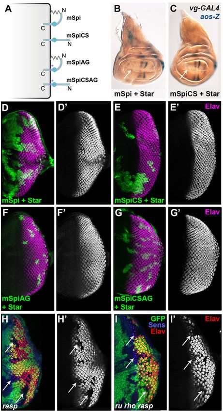
4470 Journal of Cell Science 126 (19)
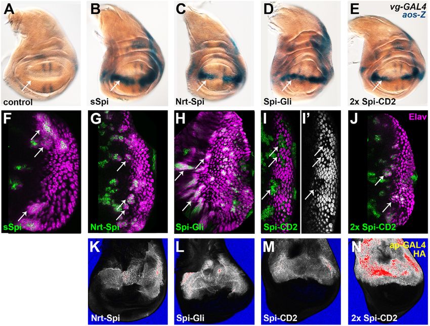
Fig. 1. Transmembrane Spi chimeras are trafficked to the
cell surface and can activate the EGFR in vitro. (A) Full-
length mSpi pro-protein has its receptor-binding EGF domain
located close to the transmembrane (TM) domain. Removal of
the signal peptide and cleavage in the transmembrane domain
by Rho generates active sSpi, which is palmitoylated at its N-
terminus. (B) Chimeric transmembrane proteins were created
by fusing sSpi to Nrt (light green), Gli (dark green) or the
transmembrane domain of CD2 (orange). All three chimeras
have an HA tag (purple). The EGF domain is represented by a
blue circle. (C–F) S2R+ cells expressing Nrt–Spi (D), Spi–Gli
(E), Spi–CD2 (F) or control cells expressing only intracellular
GFP (C) were stained for HA (magenta, D–F) or GFP (magenta,
C) in the absence of detergent. GFP fluorescence (green) is
visible in C. (G) S2 cells expressing intracellular GFP, sSpi,
Nrt–Spi, Spi–Gli or Spi-CD2 were co-cultured with EGFR-
expressing D2F cells and assayed by western blotting for
MAPK phosphorylation (p-MAPK) and total MAPK levels.
Journal of Cell Science
(H–L) FasIII expression (purple, arrowheads) in the embryonic
ventral ectoderm (H) was lost in spi zygotic mutant embryos
(I) and was rescued by expression of Nrt–Spi (J), Spi–Gli (K) or
two copies of Spi–CD2 (L)(stained with anti-HA antibody in
brown) driven in the ventral midline cells by sim-GAL4.
Yellow arrows indicate the ventral midline.
1995), is palmitoylated normally, and a non-palmitoylatable form that palmitoylation at the N-terminal cysteine residue tethers Spi
of Spi pro-protein (mSpiCS) is cleaved (Miura et al., 2006). to the cell membrane, restricting its diffusion (Miura et al., 2006).
Spi, like the mammalian EGFR ligands, has been suggested to If the palmitate group directly inserts into the plasma membrane,
act as a diffusible paracrine factor. Spi is secreted into the medium this would place the N-terminus of Spi proximal to the membrane
when expressed in cultured cells (Lee et al., 2001; Schweitzer et al., (Fig. 1A,B). In order to understand why the extracellular domain
1995), and can act at a distance of three to four cell diameters in of Spi (sSpi) is active when tethered to the membrane by a
vivo (Freeman et al., 1992; Golembo et al., 1996a). Palmitoylation palmitate group but not when anchored to the membrane by the
results in Spi having a strong membrane association and has been transmembrane domain of the pro-protein, we first tested the effect
proposed to concentrate it on the surface of secreting cells (Miura of substituting an exogenous transmembrane domain for the
et al., 2006), restricting its range of action. Although the Spi pro- palmitate group. We fused the transmembrane protein Neurotactin
protein and the cleaved ligand are both membrane-associated, only (Nrt) to the N-terminus of the extracellular domain of Spi,
the latter is thought to signal. Our results confirm previous reports replacing the palmitoylation site, to generate the chimeric protein
that, in contrast to mammalian EGFR ligands, Spi is incapable of Nrt–Spi. Because Nrt is a type II transmembrane protein (de la
signaling in its transmembrane pro-protein form (Freeman, 1996; Escalera et al., 1990; Hortsch et al., 1990), the Nrt–Spi chimera
Pickup and Banerjee, 1999; Schweitzer et al., 1995). We therefore would have its N-terminus proximal to the membrane and its C-
sought to understand the mechanism by which post-translational terminus distal, reproducing the predicted orientation of wild-type
processing controls Spi activity. We used a panel of chimeric palmitoylated Spi (Fig. 1B). Unlike cytoplasmic GFP, HA-tagged
proteins to show that structural features of the extracellular domain Nrt–Spi was detectable on the plasma membrane of cultured S2R+
do not prevent signaling by the pro-protein mSpi, and found that cells stained in the absence of detergent, indicating that
mSpi is in fact capable of activating the receptor in tissue culture. the chimeric protein is trafficked to the cell surface (Fig. 1C,D).
Our data suggest that signaling by mSpi is limited in vivo by When S2 cells expressing Nrt–Spi were co-cultured with EGFR-
polarized trafficking in epithelial cells. A similar trafficking expressing cells, the receptor was activated, leading to MAPK
mechanism has been proposed to underlie the basolateral secretion phosphorylation (Fig. 1G). Furthermore, Nrt–Spi was able to
of Hedgehog (Hh) (Callejo et al., 2011), suggesting that this is a substitute for Spi in vivo. Expression of Fasciclin III (Fas III) in the
common route for the secretion of palmitoylated proteins. embryonic ventral ectoderm requires Spi expression at the ventral
midline (Golembo et al., 1996a). When Nrt-–Spi was expressed at
Results the ventral midline using the single-minded (sim)-GAL4 driver
Membrane-tethered forms of Spi can activate the EGFR in spi mutant embryos, Fas III expression was restored (Fig. 1H–
Although Spi was originally thought to be a soluble diffusible J). Substitution of the palmitate group by an N-terminal
ligand (Schweitzer et al., 1995), our previous results suggested transmembrane domain thus allows Spi activity.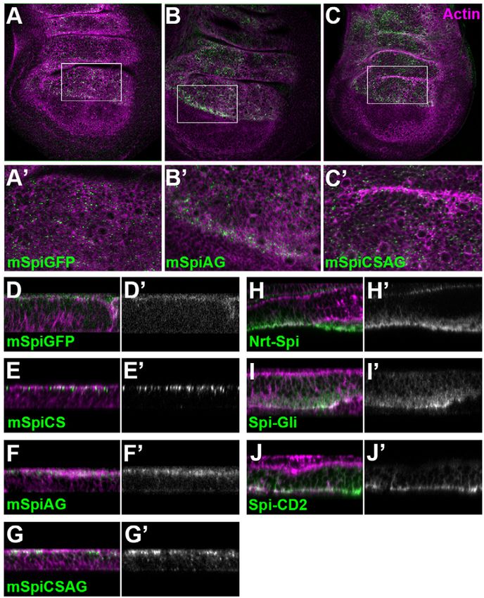
Trafficking alters Spitz activity               4471

                             Unlike Nrt–Spi, the transmembrane Spi precursor (mSpi) is              In vivo, overexpression of sSpi stimulates strong EGFR
                          unable to signal in vivo until it is cleaved by Rho (Golembo et al.,   activation, and palmitoylation is essential for its activity (Miura
                          1996a; Lee et al., 2001, see Fig. 3). mSpi differs from Nrt–Spi in     et al., 2006). To compare the activities of our transmembrane Spi
                          the orientation of the extracellular domain of Spi with respect to     chimeras in vivo, we assayed their ability to activate the EGFR in
                          the cell membrane. To assess whether orientation is important for      larval imaginal discs. Expression of sSpi, Nrt–Spi, Spi–Gli or Spi–
                          Spi activity, we generated the chimeric construct Spi–Gli, in          CD2 at the dorsal-ventral margin of the wing imaginal disc using
                          which the extracellular domain of Spi is fused at its C-terminus to    the vestigial (vg)-GAL4 driver induced ectopic expression of argos
                          the type I transmembrane protein Gliotactin (Gli; Fig. 1B) and         (aos), a transcriptional target of the EGFR pathway (Golembo
                          palmitoylation is prevented by mutating the N-terminal cysteine        et al., 1996b, Fig. 2A–E). Another transcriptional target of the
                          to serine (Miura et al., 2006). Aside from the difference in           pathway, kekkon1 (Ghiglione et al., 1999), was also ectopically
                          orientation, the extracellular domains of Gli and Nrt are similar in   expressed in response to expression of each of the three chimeras
                          size and sequence (Auld et al., 1995). Like Nrt–Spi, Spi–Gli is        in the wing disc (supplementary material Fig. S1). However, Spi–
                          transported to the plasma membrane of S2R+ cells, activates the        CD2 appeared to be a weaker activator of the pathway, as two
                          EGFR in the co-culture assay and rescues Fas III expression in         copies of the Spi–CD2 transgene were required to activate aos and
                          spi mutant embryos (Fig. 1E,G,K). Thus, Spi can activate the           kekkon1 to the same extent as a single copy of the other chimeras.
                          EGFR when it is tethered to the membrane in either the type I or       In the eye imaginal disc, ectopic expression of sSpi results in the
                          the type II orientation.                                               recruitment of extra photoreceptors (Freeman, 1996), which can be
                             In the Nrt–Spi and Spi–Gli chimeras, a large extracellular          detected by the neuronal marker Elav (Fig. 2F). Each of the
                          domain separates the receptor-binding EGF domain from the              three transmembrane chimeras induced ectopic photoreceptor
                          membrane, whereas in mSpi the EGF domain is located close to           differentiation when expressed in clones in the eye disc (Fig. 2G–
                          the membrane. To test whether the distance between the EGF             J). Spi–CD2 again appeared to be a weaker activator of the
                          domain and the membrane is important, we constructed the               pathway, as two copies of Spi–CD2 were required to induce
                          chimera Spi–CD2, in which the extracellular domain of Spi              ectopic photoreceptor formation (Fig. 2J). Expression of a single
Journal of Cell Science

                          carrying the palmitoylation-site mutation is fused at its C-terminus   Spi–CD2 transgene resulted in loss of R1–R7 photoreceptors
                          to the transmembrane domain of the rat type I cell surface protein     (Fig. 2I), a common consequence of weak pathway activation that
                          CD2 (Fig. 1B). Like the other chimeras, Spi–CD2 localized to the       is probably due to induction of Aos, a feedback inhibitor of the
                          plasma membrane, activated the receptor in cultured cells and          pathway (Golembo et al., 1996b; Lesokhin et al., 1999; Miura et al.,
                          rescued Fas III expression (Fig. 1F,G,L), indicating that separation   2006). Cell surface expression of Spi–CD2 was comparable to the
                          of the EGF domain from the membrane is not essential for Spi           other chimeric proteins in the wing disc (Fig. 2K–N), suggesting
                          signaling. However, all the chimeric forms of Spi were weaker          that its weaker activity is due to the proximity of the EGF domain
                          activators of MAPK phosphorylation in S2 cells than constitutively     to the membrane. Taken together, our tissue culture and in vivo
                          secreted and palmitoylated sSpi (Fig. 1G).                             data demonstrate that Spi does not require palmitoylation for its

                                                                                                                                 Fig. 2. Transmembrane Spi chimeras
                                                                                                                                 activate the EGFR in vivo. (A–E) Wing
                                                                                                                                 imaginal discs expressing sSpi (B), Nrt–
                                                                                                                                 Spi (C), Spi–Gli (D) or two copies of
                                                                                                                                 Spi–CD2 (E) at the dorsal-ventral
                                                                                                                                 midline driven by vg-GAL4 showed
                                                                                                                                 ectopic aos-lacZ expression (blue,
                                                                                                                                 arrows) compared to control discs (A).
                                                                                                                                 (F–J) GFP-marked spi mutant clones
                                                                                                                                 (green) expressing sSpi (F), Nrt–Spi (G),
                                                                                                                                 Spi–Gli (H) or two copies of Spi–CD2
                                                                                                                                 (J) in eye imaginal discs recruited
                                                                                                                                 ectopic photoreceptors (stained with
                                                                                                                                 Elav in magenta, arrows). Clones
                                                                                                                                 expressing only one copy of Spi–CD2
                                                                                                                                 (I) displayed loss of photoreceptors
                                                                                                                                 (arrows; Elav channel shown in white in
                                                                                                                                 I9). (K–N) Nrt–Spi (K), Spi–Gli (L) and
                                                                                                                                 Spi–CD2 (M) were expressed on the cell
                                                                                                                                 surface at similar levels. Expressing two
                                                                                                                                 copies of Spi–CD2 (N) resulted in much
                                                                                                                                 higher protein levels. Discs were
                                                                                                                                 processed in parallel by extracellular
                                                                                                                                 immunofluorescent staining with anti-
                                                                                                                                 HA antibody (pixel intensity heat map)
                                                                                                                                 following expression with apterous-
                                                                                                                                 GAL4. Images were taken with identical
                                                                                                                                 confocal settings.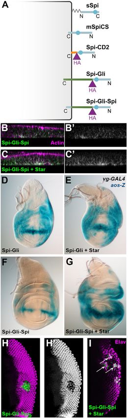
4472 Journal of Cell Science 126 (19)
The Spi pro-protein does not activate the EGFR in vivo,
even when it is not palmitoylated
Although overexpression of sSpi strongly activated the EGFR in
vivo, overexpression of the full-length pro-protein (mSpi) had no
effect, even when its chaperone Star was coexpressed
(Fig. 3B,D). These results are in agreement with previous
reports showing that mSpi is only active in cells expressing
endogenous Rho (Freeman, 1996; Pickup and Banerjee, 1999;
Schweitzer et al., 1995). We confirmed that an unprocessed form
of mSpi carrying mutations that disrupt the Rho cleavage site
(mSpiAG) is also inactive (Fig. 3F) (Strisovsky et al., 2009). In
contrast, Spi–CD2, which resembles mSpi in its type I orientation
and short extracellular domain, is able to activate the EGFR.
Because Spi–CD2 lacks the N-terminal palmitoylation site,
we hypothesized that the presence of membrane tethers at both
the N-terminus and C-terminus of mSpi might promote a
conformation that is unfavorable for signaling (Fig. 3A). To
test whether unpalmitoylated mSpi can activate the EGFR, we
ectopically expressed the palmitoylation site mutant form mSpiCS
in imaginal discs. Neither mSpiCS nor uncleavable and
unpalmitoylatable mSpiCSAG induced ectopic aos expression or
ectopic photoreceptor recruitment when coexpressed with Star
(Fig. 3C,E,G), indicating that unpalmitoylated mSpi remains
inactive.
Journal of Cell Science
In the eye disc, clones of cells lacking the Spi acyltransferase
Rasp show reduced differentiation of photoreceptors R1–R7
(Fig. 3H) (Miura et al., 2006). In rasp mutant clones, the
endogenous Spi pro-protein is processed by Rho and a second
protease specific to the eye disc, Roughoid (Ru) (Wasserman
et al., 2000), but fails to be palmitoylated (Miura et al., 2006).
We tested whether preventing the cleavage and release of
unpalmitoylated Spi by removing ru and rho function from rasp
mutant clones would restore photoreceptor differentiation. We
observed no rescue of photoreceptor differentiation in ru rho rasp
triple mutant clones, confirming that the endogenous uncleaved
and unpalmitoylated pro-protein is inactive (Fig. 3I). Therefore,
the inability of mSpi to activate the EGFR is independent of its
palmitoylation.
The Spi pro-protein accumulates on the cell surface and
activates the EGFR in tissue culture
The failure of mSpi to activate the EGFR in vivo led us to
consider the possibility that mSpi is not trafficked to the plasma
Fig. 3. The mSpi pro-protein does not activate the EGFR in vivo. membrane. In order to visualize only protein on the cell surface,
(A) Palmitoylation of mSpi would result in membrane association at both we expressed Flag-tagged mSpi in cultured S2 cells and stained
termini, whereas mSpiCS cannot be palmitoylated and remains free at its N- with anti-Flag antibody under non-permeabilizing conditions. We
terminus. mSpiAG and mSpiCSAG contain mutations that prevent cleavage were able to detect mSpi at the cell surface only in the presence
by Rho (purple asterisks) (Strisovsky et al., 2009). (B,C) Neither mSpi (B) nor
of co-expressed Star, which allows its export from the ER
mSpiCS (C) induced ectopic aos-lacZ (aos-Z) expression at the dorsal-ventral
midline (arrows) when coexpressed with Star in the wing disc using vg-
(Fig. 4A,B). The same was true for mSpiCS, the palmitoylation
GAL4. (D–G) Flip-out clones (marked with GFP, green) expressing mSpi (D), site mutant (Fig. 4C,D). To verify that cell surface staining
mSpiCS (E), mSpiAG (F) or mSpiCSAG (G), together with Star, did not represented full-length Spi pro-protein and not Spi processed by
recruit ectopic photoreceptors (Elav, magenta, shown alone in D9, E9, F9 and Rho and Rasp, we repeated the experiment using the mSpi
G9). (H–I) Mitotic clones (arrows) mutant for rasp (H) failed to differentiate variants lacking the Rho cleavage site (mSpiAG) (Strisovsky
photoreceptors R1–R7 (marked with Elav in red, shown alone in H9), et al., 2009) and both the cleavage and palmitoylation sites
although differentiation of the founder photoreceptor R8 (marked with (mSpiCSAG). These proteins were also detectable on the cell
Senseless in blue) was normal. Clones mutant for ru and rho in addition to surface, demonstrating that the mSpi pro-protein can be
rasp showed a similar loss of photoreceptor differentiation (I,I9). trafficked to the plasma membrane in cultured cells (Fig. 4E–H).
Surprisingly, when mSpi-expressing S2 cells were co-cultured
activity when it is tethered by a transmembrane domain. Moreover, with EGFR-expressing cells, the EGFR was activated and MAPK
its activity is not critically dependent on either the orientation or phosphorylation was induced (Fig. 4I–K). This effect was
the distance of the EGF domain from the cell membrane. dependent on co-expression of Star, confirming that the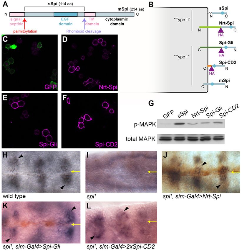
Trafficking alters Spitz activity                4473

                                                                                                     Fig. 4. mSpi pro-protein activates the EGFR in vitro. (A–H) Flag-
                                                                                                     tagged mSpi (A,B), mSpiCS (C,D), mSpiAG (E,F) or mSpiCSAG (G,H)
                                                                                                     was detected on the surface of S2 cells by anti-Flag staining (green) in
                                                                                                     the absence of detergent when co-expressed with Star (A,C,E,G). In the
                                                                                                     absence of Star, no cell-surface mSpi was detected (B,D,F,H). Staining
                                                                                                     for the transmembrane protein HA–Lar (magenta) marks the plasma
                                                                                                     membrane. (I–K) Co-culture of S2 cells expressing mSpi, mSpiCS,
                                                                                                     mSpiAG or mSpiCSAG with EGFR-expressing D2F cells resulted in
                                                                                                     MAPK phosphorylation. Quantification was performed using Li-Cor
Journal of Cell Science

                                                                                                     Odyssey (K). For each condition, the phospho-MAPK level was divided
                                                                                                     by total MAPK level (% dpERK) and this value was normalized to the
                                                                                                     percentage dpERK obtained for control D2F cells co-cultured with S2
                                                                                                     cells expressing intracellular GFP alone in that experiment. The
                                                                                                     mean6s.e.m. of three experiments is shown. sSpi-expressing cells
                                                                                                     induced more than a twofold increase in phospho-MAPK in co-cultured
                                                                                                     D2F cells compared to GFP-expressing cells. Each mSpi construct co-
                                                                                                     expressed with Star induced about a 1.5-fold increase in phospho-MAPK
                                                                                                     compared to GFP, whereas mSpi constructs without co-expressed Star
                                                                                                     did not increase phospho-MAPK above the level observed with GFP.
                                                                                                     *P,0.05 between the percentage dpERK induced by each Spi construct
                                                                                                     when co-expressed with Star compared to the percentage dpERK
                                                                                                     induced when expressed without Star (asterisks) as determined by paired
                                                                                                     Student’s t-tests.

                          observed activity was due to binding of mSpi to EGFR at the cell       the basolateral domain, and that localization of mSpi to the apical
                          surface. Mutant forms of mSpi lacking the palmitoylation site,         domain could render it inactive in vivo.
                          the Rhomboid cleavage site or both, were likewise capable of
                          activating EGFR in a Star-dependent manner (Fig. 4I–K). These          The Spi cytoplasmic domain is not sufficient for apical
                          data reveal that the unprocessed mSpi protein is able to activate      localization and inactivation
                          the EGFR in cultured cells.                                            Despite their similar structures and almost identical extracellular
                                                                                                 domains, Spi–CD2 and the unpalmitoylated pro-protein mSpiCS
                          Localization at the cell membrane correlates with signaling            show different activities and apical-basal localization. To test
                          activity                                                               whether the cytoplasmic domain of mSpi controls its trafficking
                          The results above suggest that mSpi is inherently capable of           and inhibits its signaling ability, we used it to replace the
                          activating the EGFR, but is prevented from doing so in vivo. In        cytoplasmic domain of Spi–Gli, creating Spi–Gli–Spi (Fig. 6A). In
                          order to investigate the nature of this inhibition, we used an         comparison with Spi-Gli, which was a strong activator of the
                          extracellular immunofluorescent staining protocol to detect cell       pathway, Spi–Gli–Spi only weakly activated the EGFR in vivo.
                          surface GFP- or Flag-tagged mSpi and its uncleavable and/or            Spi–Gli–Spi induced weak ectopic aos-lacZ expression in the wing
                          unpalmitoylatable variants in wing discs expressing these molecules    imaginal disc (Fig. 6F), and caused loss of R1–R7 photoreceptors
                          together with Star in the dorsal compartment under the control of      in the eye disc, consistent with aos induction by weak EGFR
                          apterous (ap)-GAL4 (Strigini and Cohen, 2000). We were able to         activation (Fig. 6H). Nevertheless, when coexpressed with Star,
                          detect all the tagged forms of mSpi extracellularly, confirming that   Spi–Gli–Spi was able to strongly activate the pathway, inducing
                          mSpi can reach the cell surface in vivo (Fig. 5A–G). mSpi staining     strong ectopic aos-lacZ expression (Fig. 6G) and ectopic
                          was punctate (Fig. 5A–C) and localized to the apical region of the     photoreceptor differentiation (Fig. 6I). Thus, adding the Spi
                          plasma membrane in imaginal disc cells (Fig. 5D–G). In contrast,       cytoplasmic domain partially inhibits the activity of Spi–Gli, but
                          the active Spi chimeras were confined to the basolateral domain        this inhibition can be overcome by the presence of Star, suggesting
                          (Fig. 5H–J). These data suggest that productive signaling occurs in    that its effect is to sequester Spi–Gli–Spi in the ER (Lee et al.,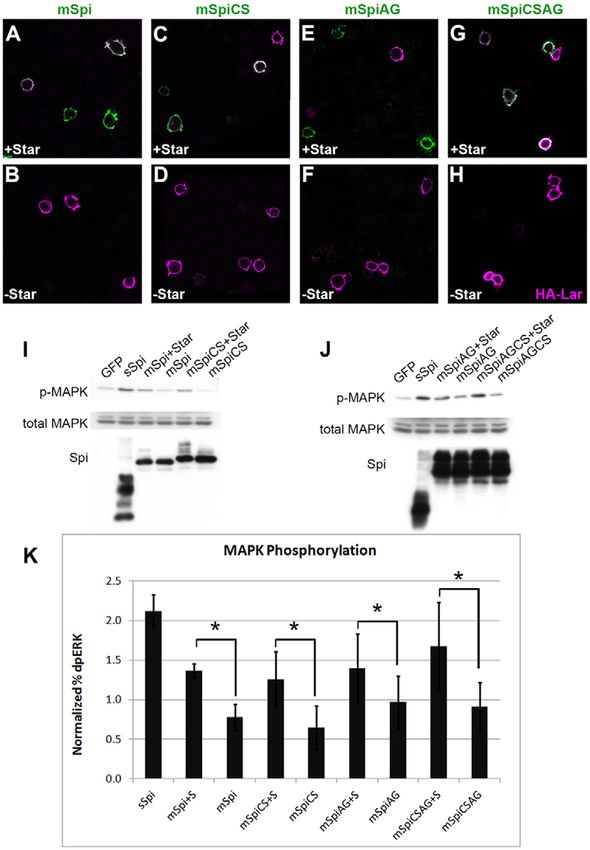
4474      Journal of Cell Science 126 (19)
Journal of Cell Science

                                                                                                              Fig. 5. mSpi localizes to apical puncta at the cell surface in
                                                                                                              vivo. Imaginal discs were stained for extracellular antigen (green,
                                                                                                              shown alone in D9, E9, F9, G9, H9, I9, J9) and counterstained
                                                                                                              following fixation and permeabilization with TRITC–phalloidin
                                                                                                              (magenta) to mark cell membranes. mSpi–GFP (anti-GFP; A,D),
                                                                                                              mSpiAG (anti-Flag; B,F), mSpiCSAG (anti-Flag; C,G) and
                                                                                                              mSpiCS (anti-Flag; E) were detected on the cell surface in apical
                                                                                                              puncta. In contrast, Nrt–Spi (H), Spi–Gli (I) and Spi–CD2 (J), all
                                                                                                              detected with anti-HA antibody, were observed basolaterally.

                          2001). Consistent with this hypothesis, we observed greater cell           surface, suggesting a possible role for the transmembrane domain
                          surface expression of Spi–Gli–Spi in the presence of coexpressed           in this process. Taken together, our results suggest that the mSpi
                          Star (Fig. 6B,C), and Spi–Gli activity was not enhanced by Star            pro-protein is structurally capable of signaling, but is prevented
                          (Fig. 6D,E). With or without Star coexpression, Spi–Gli–Spi                from activating the EGFR in vivo by directed trafficking.
                          localized basolaterally (Fig. 6B,C), consistent with its ability to
                          activate EGFR. Thus the cytoplasmic domain of Spi is not                   Determinants of mSpi localization
                          sufficient to drive its apical localization.                               Although Spi–CD2 and mSpi differ only in their transmembrane
                                                                                                     and cytoplasmic domains, Spi–CD2 localizes to the basolateral
                          Discussion                                                                 surface of epithelial cells and activates the EGFR in vivo,
                          EGFR ligands are diffusible paracrine molecules that are produced          whereas mSpi does not. Therefore, we expected the Spi
                          by cleavage of transmembrane pro-proteins. Some pro-proteins are           cytoplasmic domain to contain sequences that control
                          also capable of activating the receptor, but they often elicit different   localization and signaling. The cytoplasmic domain of TGF-a
                          responses from the processed ligands. We investigated why the              is sufficient for basolateral trafficking in MDCK cells (Dempsey
                          Drosophila EGFR ligand Spi is inactive in its pro-protein form             et al., 2003), and the transmembrane and cytoplasmic domains of
                          (mSpi), even though the active processed form (sSpi) is associated         human EGF prevent it from engaging in juxtacrine signaling
                          with the membrane through a palmitate adduct. Our results show             (Dong et al., 2005). Indeed, replacing the cytoplasmic domain of
                          that sSpi is capable of activating the receptor when tethered to the       the strong activator Spi–Gli with the mSpi cytoplasmic domain
                          membrane by an exogenous transmembrane domain in either                    significantly weakens its signaling capacity. Nonetheless, Spi–
                          orientation. Increased spacing between the receptor-binding EGF            Gli–Spi is localized basolaterally, and coexpression of Star with
                          domain and the membrane appears favorable but not essential for            Spi–Gli–Spi restores strong signaling activity, suggesting that the
                          signaling. The mSpi pro-protein is trafficked to the cell surface and      inhibitory effect of the cytoplasmic domain is due to ER
                          can activate the EGFR in cultured cells, but not in vivo. mSpi             retention. Previous studies have shown roles for both the
                          appears largely confined to the apical domain of imaginal disc cells       cytoplasmic and lumenal domains of mSpi in mediating Star
                          and is observed in puncta, whereas active Spi forms are uniformly          dependence (Lee et al., 2001; Reich and Shilo, 2002). Thus, the
                          distributed in the basolateral domain (Fig. 7A). The intracellular         Spi cytoplasmic domain is not sufficient to drive apical
                          domain alone is not sufficient to direct a Spi chimera to the apical       trafficking or prevent signaling. The Spi transmembrane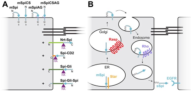
Trafficking alters Spitz activity 4475
domain might contribute to apical trafficking, or the Gliotactin Sorting of receptor and ligand to apical and basolateral
transmembrane and extracellular domains, which promote domains
basolateral trafficking (Schulte et al., 2003), might override the Although the mSpi pro-protein is inactive in vivo, we found that
Spi trafficking signal in Spi-Gli-Spi. it is capable of activating EGFR in cultured S2 cells. One
possible explanation for this difference is that S2 cells, unlike
epithelial imaginal disc cells, lack polarity. In wing disc cells, the
active Spi chimeras localize to a different membrane domain than
the inactive pro-protein, potentially allowing them preferential
access to the receptor. Previous studies have reported an apical
localization of Drosophila EGFR in some tissues (Alvarado et al.,
2004; Zak and Shilo, 1992); however, our results strongly suggest
that, at least in the imaginal discs, active signaling can occur in
the basolateral domain. We have observed extracellular EGFR
staining throughout the apical-basal axis in wing imaginal discs
(data not shown).
Spatial segregation of the ligand and receptor has been
demonstrated in cultured human airway epithelia, where the
EGFR ligand heregulin a (also known as neuregulin-1) is apically
localized and its receptors are restricted basolaterally. In this
context, signaling is prevented when the epithelial sheet is intact,
and only upon wounding can the ligand access the receptor,
thereby initiating the signaling that induces cell proliferation for
wound repair (Vermeer et al., 2003). In contrast, localization of
the C. elegans EGFR homolog LET-23 to the basolateral surface
Journal of Cell Science
of vulval precursor cells is necessary to allow its interaction with
LIN-3 ligand secreted from the anchor cell (Stetak et al., 2006)
(Kaech et al., 1998). Our study suggests that localization of the
ligand as well as the receptor regulates EGFR signaling in vivo. A
recent study of Spi production by socket cells in mechanosensory
organs reached a similar conclusion; polarized protrusions from
the basolateral surface of socket cells appear to deliver Spi to the
prospective bract cell (Peng et al., 2012).
A striking feature of mSpi localization is its appearance in
puncta detectable with an extracellular staining protocol (Strigini
and Cohen, 2000). One possibility is that these puncta represent
aggregates at the cell surface, similar to those observed for
another palmitoylated ligand, Hh (Vyas et al., 2008).
Alternatively, they might represent endocytosing vesicles
trapped at the cell surface by the cold temperature of the
staining protocol. Rapid endocytosis of the pro-protein could
prevent the protein from residing at the cell surface long enough
to engage and activate the receptor. Wg receptors have been
reported to undergo endocytosis preferentially from the apical
domain (Marois et al., 2006). In addition, Hh is thought to
undergo a recycling process in which it is first secreted apically
from producing cells, then endocytosed and shuttled to the
Fig. 6. Spi–Gli–Spi activity depends on Star. (A) The Gliotactin
cytoplasmic domain in Spi–Gli was replaced with the Spi cytoplasmic domain
to generate Spi–Gli–Spi. (B,C) Spi–Gli–Spi, detected with anti-HA
extracellular staining (green, white in B9 and C9; TRITC–phalloidin shown in
magenta) localized basolaterally in the absence (B) and presence (C) of co-
expressed Star, but its cell surface expression was greater when co-expressed
with Star. B and C were imaged in parallel with identical confocal settings.
(D–G) Both Spi-Gli (D–E) and Spi-Gli-Spi (F–G) induced ectopic aos-lacZ in
wing imaginal discs when expressed with vg-GAL4 either alone (D,F) or with
co-expressed Star (E,G), but for Spi–Gli–Spi the induction was stronger when
it was co-expressed with Star. D–G were stained and imaged in parallel.
(H,I) Flip-out clones (green) expressing Spi–Gli–Spi and Star (I) induced
ectopic photoreceptor recruitment (marked with Elav in magenta), whereas
clones expressing Spi–Gli–Spi without Star caused photoreceptor loss
(H, Elav shown alone in H9).4476 Journal of Cell Science 126 (19)
Fig. 7. Models for Spi trafficking and activation. (A) Cartoon
representing the localization and activity of all the Spi variants tested.
Black lines indicate the adherens junctions; mSpi, mSpiCS, mSpiAG
and mSpiCSAG localize to the apical surface and are inactive (2),
whereas Nrt–Spi, Spi–CD2, Spi–Gli and Spi–Gli–Spi localize to the
basolateral surface and are active (+). (B) A model for Spi trafficking.
mSpi is exported from the ER with the help of the chaperone protein
Star. It is palmitoylated by Rasp in the Golgi and trafficked to the
apical cell surface. It might be endocytosed from this location, cleaved
in late endosomes by the protease Rho and secreted basolaterally. The
palmitate group would tether sSpi to the basolateral plasma membrane.
basolateral domain by Dispatched. Hh released from the signaling molecules, perhaps by altering their localization or
basolateral domain associates with Interference hedgehog, retention on the cell surface.
Dally-like protein and filopodia to signal over a long range
(Callejo et al., 2011). It is possible that a similar mechanism Materials and Methods
limits release of functional Spi to the basolateral domain. A role Drosophila stocks
Drosophila stocks used were: aosW11, kek101433 and ap-GAL4 (Bloomington
for endocytosis in Spi release has already been proposed based on Drosophila Stock Center, Bloomington, IN, USA), UAS-sSpi, UAS-Rasp-HA,
the colocalization of Rho with endosomal markers and the UAS-Star, UAS-rho, FRT40 spiSC1, FRT2A raspT802, UAS-mSpi, and UAS-mSpiCS
requirement for the recycling endosome component Rab11 for (Miura et al., 2006), vg-GAL4 (Simmonds et al., 1995) and sim-GAL4 (Golembo
et al., 1996a). The triple ru rho rasp mutant stock was made by recombining
secretion of functional Spi from photoreceptors (Tsruya et al., FRT2A raspT802 with ru1 rho7M43 h th st cu sr e ca (Bloomington Drosophila
2007; Yogev et al., 2010). Our results suggest a model wherein Stock Center). Transgenic lines were made by Genetic Services Inc. (Cambridge,
Journal of Cell Science
mSpi is exported to the apical surface and rapidly endocytosed, MA, USA) or GenetiVision (Houston, TX, USA).
processed by Rho in the endocytic pathway, and subsequently MARCM clones were made using hs-FLP, UAS-GFP; FRT40, tub-GAL80; tub-
GAL4/TM6B. Flip-out clones were made using actin.y+.GAL4, UAS-GFP; hs-
recycled to the basolateral domain (Fig. 7B). This model predicts FLP, MKRS/TM6B or actin.CD2.GAL4, hs-FLP; UAS-GFP/TM6B.
that endocytosis would promote release of the active ligand, and
might also prevent signaling by the pro-protein. Alternatively, Molecular biology
mSpi might be exported from the Golgi to Rho-containing The neurotactin coding region was amplified from genomic DNA using primers
endosomes and localized directly to the basolateral surface. In 59Nrt_EcoRI 59-TTCTCGCAGAATTCTCTAAACGAT-39 and 39Nrt_BsiWIHA
59-CGTACGGGCGTAATCTGGCACATCATACGGGTAGCGCGCATACCGCG-
this case, the presence of mSpi on the apical plasma membrane GCACAAT-39 and cloned into pUASt-sSpiCS (Miura et al., 2006) using EcoRI and
would result from its inappropriate accumulation when BsiWI to make UAS-Nrt-Spi.
overexpressed in cells that lack Rho. sSpiCS, which contains the signal peptide, has the cysteine residue following the
signal peptide mutated to serine and is truncated at the Rho cleavage site (Miura
et al., 2006), was cloned into Gateway pENTR/D-TOPO (Invitrogen/Life
Pro-protein signaling Technologies, Grand Island, NY, USA) using primers Spi up TOPO 59-
The signaling ability of mammalian EGFR ligand pro-proteins has CACCATGCATTCCACAATGAGTGTACAAC-39 and Spi down TOPO 59-
been controversial. Studies in vitro showed that a TGF-a pro- CATATGCCGGTAAAGCTTGGCGTAATCTGGCACATCATACGGGTACGG-
CCTCTTGGGCAGGTAAG-39. The downstream primer introduced an HA tag as
protein carrying mutations in the metalloproteinase cleavage site well as HindIII and NdeI sites. Gliotactin was amplified from cDNA RE15719
could signal, but in vivo, mutation of the metalloproteinase itself (Drosophila Genomics Resource Center, Bloomington, IN, USA) using primers
appeared to recapitulate loss of TGF-a (Brachmann et al., 1989; Gli up 59-ATGGGCAAGCTTCGGCCAGGCGTTGACTACCA-39 and Gli down
59-CGGGTACATATGTCACCGACTGCTGCCCGAGACACTG-39 and cloned
Peschon et al., 1998; Sanderson et al., 2006; Wong et al., 1989). In into pENTR-sSpiCS using HindIII and NdeI. Spi–Gli was then recombined into
recent years, mounting evidence has been provided in support of pTWG (DGRC) to make UAS-Spi-Gli.
pro-protein signaling, both by TGF-a and by HB-EGF, which has The transmembrane domain from rat CD2 was amplified from cDNA (Invitrogen
opposing effects on cell survival in its membrane-bound and I.M.A.G.E. clone 7374969) using primers CD2 up 59-ATGGGCAAGCTTGT-
CAACTGTCCAGAGAAA-39 and CD2 down 59-CGGGTACATATGTCATTTT-
secreted forms (Pan et al., 2002; Prince et al., 2010; Shi et al., ATCTCCAGCTCTTC-39 and cloned into pENTR-sSpiCS using HindIII and NdeI.
2000; Singh et al., 2007; Yang et al., 2000). Some pro-proteins, Spi-CD2 was then recombined into pTWG (DGRC) to make UAS-Spi-CD2.
such as amphiregulin, might be released on exosomes, allowing pUASt-mSpi-Flag was a gift from B. Shilo (Tsruya et al., 2002). Flag-tagged
mSpiAG was a gift from M. Freeman (Strisovsky et al., 2009). mSpiAG-Flag
them to act at a distance (Higginbotham et al., 2011). However, was subcloned into pUASt using BglII and KpnI sites introduced by the following
EGF appears to signal only in its soluble cleaved form (Dong et al., primers: mSpi_FBglII 59-GCATAGATCTGCCGATACGCTACGCCAAAAAG-
2005; Dong et al., 1999). Pro-protein signaling often induces 39 and mSpi_RKpn 59-GCATGGTACCCGCTCACAGCTTGTTGCTGCGT-39.
different outcomes from signaling by the mature ligands, making A Flag tag was introduced into the BsiWI site of pUASt-mSpiCS by annealing
the following oligos: Bsi_FlagF 59-GTACGGATTACAAGGATGACGATGACA-
ligand processing a critical step in determining pathway effects. AGC-39 and Bsi_FlagR 59-GTACGCTTGTCATCGTCATCCTTGTAATCC-39.
We found that mSpi can signal in a tissue culture assay but not The CS mutation was introduced into pUASt-mSpiAG using overlapping
in imaginal discs. This seems to be the result of trafficking to PCR products made with the following primers: mSpi_FBglII 59-
GCATAGAT\CTGCCGATACGCTACGCCAAAAAG-39 and mSpiCS_XhoIR
different regions of the cell surface in polarized cells. Notably, 59-GTCCGTACGGCTGCTCGAGGCCTCGACGTGCCAC-39, mSpiCS_XhoIF
sSpi is a stronger activator of the pathway than mSpi in S2 cells 59-GTGGCACGTCGAGGCCTCGAGCAGCCGTACGGAC-39 and mSpiCS_SapI
(Fig. 4), indicating that the pro-protein and processed ligand are 59-GTGCCGGACATGGACGAGGAGA-39. The mutated fragment was cloned into
pUASt-mSpiAG using the BglII and SapI sites.
not equivalent even in this context. Our model suggests that pro-
UAS-Spi-Gli-Spi was cloned by overlap PCR, using the primers Spi-Gli-Kpn59
proteins are not inherently inactive and that certain mammalian 59-AGAAGGTACCCGTTTATATGTACGTAC-39 and Spi-Gli-overlap39 59-AG-
contexts might have evolved to utilize the pro-proteins as active CCCGCTGCTCGAAGCGCCACATGATGCAGCAGATGAC-39 to amplify aTrafficking alters Spitz activity 4477
region of Spi-Gli ending in the transmembrane domain, and the primers Spi-Gli- use of her confocal microscope. The manuscript was improved by the
overlap59 59-GTCATCTGCTGCATCATGTGGCGCTTCGAGCAGCGGGCT-39 critical comments of Sergio Astigarraga, Cheuk Hei Ho, Kevin
and Spi-AscI39 59-ATTCGGCGCGCCGCATGCGGTAGGTAAATAGGAGTTA-
TC-39 to amplify the cytoplasmic domain of Spi and introduce an AscI site.
Legent, Justine Oyallon, and Annabelle Suisse.
The two PCR products were mixed and amplified with Spi-Gli-Kpn59 and Spi-
AscI39 to generate a product that was cloned into the KpnI and AscI sites of Author contributions
UAS-Spi-Gli. J.S. performed the majority of the experiments. H.H.L. performed
tissue culture experiments shown in Fig. 4. E.M. performed staining
Tissue culture and phospho-MAPK assay shown in Fig. 6. J.E.T. generated the Spi-Gli-Spi construct. J.S. and
S2 and S2R+ cells were maintained in Schneider’s Drosophila medium (Invitrogen/Life
J.E.T. designed the experiments and wrote the manuscript.
Technologies) supplemented with 10% fetal calf serum and penicillin-streptomycin. All
UAS constructs were co-transfected with actin-Gal4 using Effectene Transfection
Reagent (Qiagen, Germantown, MD, USA). Constructs used for transfection were UAS- Funding
GAP-GFP (Ritzenthaler et al., 2000), UAS-sSpi (Miura et al., 2006), UAS-mSpi-Flag This work was supported by the March of Dimes Birth Defects
(Tsruya et al., 2002), UAS-mSpiCS-Flag, UAS-mSpiAG-Flag, UAS-mSpiCSAG-Flag, Foundation; and the National Institutes of Health [grant number
pRM-Star-Myc (a gift from M. Freeman; Lee et al., 2001), UAS-Nrt-Spi, UAS-Spi-Gli,
UAS-Spi-CD2 and UAS-Spi-Gli-Spi.
EY13777]. Deposited in PMC for release after 12 months.
For the phospho-MAPK assay, on day 1, S2 cells stably expressing the EGFR
(D2F, (Schweitzer et al., 1995)) were plated and S2 cells were plated and Supplementary material available online at
transfected with pRM-Star-Myc, actin-GAL4 and a UAS-Spi construct. On day http://jcs.biologists.org/lookup/suppl/doi:10.1242/jcs.131169/-/DC1
2, transfected cells were treated with 700 mM CuSO4 to induce Star expression.
On day 3, EGFR expression was induced in the D2F cells with 60 mM CuSO4.
After 3 hours, cells were collected, washed in PBS, resuspended in half the References
volume of Schneider’s medium, and co-cultured for 1–3 hours with rocking at Alvarado, D., Rice, A. H. and Duffy, J. B. (2004). Knockouts of Kekkon1 define
room temperature. Co-cultures were resuspended in RIPA buffer and sequence elements essential for Drosophila epidermal growth factor receptor
immediately centrifuged, diluted into Laemmli buffer, boiled for 5 minutes inhibition. Genetics 166, 201-211.
Anklesaria, P., Teixidó, J., Laiho, M., Pierce, J. H., Greenberger, J. S. and
and frozen.
Massagué, J. (1990). Cell-cell adhesion mediated by binding of membrane-anchored
transforming growth factor alpha to epidermal growth factor receptors promotes cell
Antibody staining and Western blot proliferation. Proc. Natl. Acad. Sci. USA 87, 3289-3293.
Chamber slides (LabTek, Electron Microscopy Sciences, Hatfield, PA, USA) were Auld, V. J., Fetter, R. D., Broadie, K. and Goodman, C. S. (1995). Gliotactin, a novel
Journal of Cell Science
treated with poly-L-lysine overnight. Cultured cells were stained under non- transmembrane protein on peripheral glia, is required to form the blood-nerve barrier
permeabilizing conditions in the absence of detergent as follows. Cells were in Drosophila. Cell 81, 757-767.
allowed 30 minutes to adhere following plating. Cells were fixed in 4% Brachmann, R., Lindquist, P. B., Nagashima, M., Kohr, W., Lipari, T., Napier,
formaldehyde-PBS, washed for 15 minutes to 1 hour in PBS, and stained M. and Derynck, R. (1989). Transmembrane TGF-alpha precursors activate EGF/
overnight in primary antibody in PBS at 4 ˚C. Cells were washed in PBS, stained TGF-alpha receptors. Cell 56, 691-700.
for 2 hours in secondary antibody and imaged. Brown, K. E., Kerr, M. and Freeman, M. (2007). The EGFR ligands Spitz and Keren
Tissues were stained using previously described protocols (Lee and Treisman, act cooperatively in the Drosophila eye. Dev. Biol. 307, 105-113.
2001). Extracellular staining was performed following the protocol of Strigini and Callejo, A., Bilioni, A., Mollica, E., Gorfinkiel, N., Andrés, G., Ibáñez, C., Torroja,
C., Doglio, L., Sierra, J. and Guerrero, I. (2011). Dispatched mediates Hedgehog
Cohen (Strigini and Cohen, 2000). Imaginal discs were dissected in complete
basolateral release to form the long-range morphogenetic gradient in the Drosophila
Schneider’s medium and incubated for 30–60 minutes in primary antibody diluted
wing disk epithelium. Proc. Natl. Acad. Sci. USA 108, 12591-12598.
in complete Schneider’s medium with rocking at 4 ˚C. Discs were rinsed in cold de la Escalera, S., Bockamp, E. O., Moya, F., Piovant, M. and Jiménez, F. (1990).
PBS and fixed at 4 ˚C in 4% formaldehyde for 30 minutes, washed in PBS plus Characterization and gene cloning of neurotactin, a Drosophila transmembrane
0.1% Triton X-100, incubated with secondary antibody, and counterstained with protein related to cholinesterases. EMBO J. 9, 3593-3601.
TRITC–phalloidin (Sigma, St Louis, MO, USA). Discs were mounted in Dempsey, P. J., Meise, K. S. and Coffey, R. J. (2003). Basolateral sorting of transforming
Fluoromount G (Southern Biotech, Birmingham, AL, USA). All fluorescence growth factor-alpha precursor in polarized epithelial cells: characterization of
images were taken with a Zeiss LSM510 Confocal (Carl Zeiss, Oberkochen, cytoplasmic domain determinants. Exp. Cell Res. 285, 159-174.
Germany). X-gal staining was performed as described previously (Lee and Dong, J., Opresko, L. K., Dempsey, P. J., Lauffenburger, D. A., Coffey, R. J. and
Treisman, 2001). Wiley, H. S. (1999). Metalloprotease-mediated ligand release regulates autocrine
Primary antibodies for immunostaining were: rat anti-HA 3F10 (1:100 for signaling through the epidermal growth factor receptor. Proc. Natl. Acad. Sci. USA
standard immunofluorescence, 1:30 for extracellular staining, Roche, Basel, 96, 6235-6240.
Switzerland), mouse anti-Flag M2 (1:500 for standard immunofluorescence, 1:150 Dong, J., Opresko, L. K., Chrisler, W., Orr, G., Quesenberry, R. D., Lauffenburger,
for extracellular staining; Sigma), rat anti-Elav (1:100) and mouse anti-Fasciclin D. A. and Wiley, H. S. (2005). The membrane-anchoring domain of epidermal
III (1:1) (Developmental Studies Hybridoma Bank, Iowa City, IA, USA), guinea growth factor receptor ligands dictates their ability to operate in juxtacrine mode.
pig anti-Senseless (1:1000; Nolo et al., 2000), rabbit anti-b–galactosidase (1:5000, Mol. Biol. Cell 16, 2984-2998.
Cappel, MP Biomedicals, Santa Ana, CA, USA), mouse anti-GFP (1:16 for Freeman, M. (1996). Reiterative use of the EGF receptor triggers differentiation of all
extracellular staining, Santa Cruz Biotechnology, Dallas, TX, USA), mouse anti- cell types in the Drosophila eye. Cell 87, 651-660.
Freeman, M., Klämbt, C., Goodman, C. S. and Rubin, G. M. (1992). The argos gene
EGFR E2906 (1:50 for extracellular staining, Sigma-Aldrich, St. Louis, MO) or
encodes a diffusible factor that regulates cell fate decisions in the Drosophila eye. Cell
rabbit anti-EGFR (1:500)(Rodrigues et al., 2005). Secondary antibodies were from
69, 963-975.
Jackson Immunoresearch (1:200, Westgrove, PA, USA) or Molecular Probes Ghiglione, C., Carraway, K. L., III, Amundadottir, L. T., Boswell, R. E., Perrimon,
(1:1000). Embryo stainings were developed with DAB (Sigma) and Vectastain N. and Duffy, J. B. (1999). The transmembrane molecule kekkon 1 acts in a feedback
Elite ABC System (Vector Laboratories, Burlingame, CA, USA). loop to negatively regulate the activity of the Drosophila EGF receptor during
Western blots were performed as described previously (Miura et al., 2006), oogenesis. Cell 96, 847-856.
blocked in 5% evaporated milk, and developed either with ECL (Thermo Fisher Golembo, M., Raz, E. and Shilo, B. Z. (1996a). The Drosophila embryonic midline is
Scientific, Rockford, IL, USA) or with Odyssey (Li-Cor, Lincoln, NE, USA). the site of Spitz processing, and induces activation of the EGF receptor in the ventral
Quantification was performed using Odyssey. Primary antibodies for western ectoderm. Development 122, 3363-3370.
blotting were: mouse anti-diphosphorylated-ERK (1:2000, Sigma), rabbit anti- Golembo, M., Schweitzer, R., Freeman, M. and Shilo, B. Z. (1996b). Argos
MAPK (1:20,000, Sigma), rat anti-Spi (1:50) (Developmental Studies Hybridoma transcription is induced by the Drosophila EGF receptor pathway to form an
Bank), mouse anti-HA (1:1000; Covance, Princeton, NJ, USA). Secondary inhibitory feedback loop. Development 122, 223-230.
antibodies were HRP-conjugated antibodies from Jackson Immunoresearch Higginbotham, J. N., Demory Beckler, M., Gephart, J. D., Franklin, J. L.,
(1:4000) or Infrared-conjugated antibodies from Li-Cor (1:10,000). Bogatcheva, G., Kremers, G. J., Piston, D. W., Ayers, G. D., McConnell, R. E.,
Tyska, M. J. et al. (2011). Amphiregulin exosomes increase cancer cell invasion.
Curr. Biol. 21, 779-786.
Acknowledgements Hortsch, M., Patel, N. H., Bieber, A. J., Traquina, Z. R. and Goodman, C. S. (1990).
We thank Nick Baker, Hugo Bellen, Matthew Freeman, Justin Drosophila neurotactin, a surface glycoprotein with homology to serine esterases, is
Kumar, Mark Lemmon, Daniel Marenda, Benny Shilo, the dynamically expressed during embryogenesis. Development 110, 1327-1340.
Hynes, N. E. and Lane, H. A. (2005). ERBB receptors and cancer: the complexity of
Bloomington Drosophila Stock Center, the Drosophila Genomics targeted inhibitors. Nat. Rev. Cancer 5, 341-354.
Resource Center, and the Developmental Studies Hybridoma Bank Iwamoto, R., Handa, K. and Mekada, E. (1999). Contact-dependent growth inhibition
for fly stocks and reagents. We are grateful to Ruth Lehmann for the and apoptosis of epidermal growth factor (EGF) receptor-expressing cells by the4478 Journal of Cell Science 126 (19)
membrane-anchored form of heparin-binding EGF-like growth factor. J. Biol. Chem. Schulte, J., Tepass, U. and Auld, V. J. (2003). Gliotactin, a novel marker of tricellular
274, 25906-25912. junctions, is necessary for septate junction development in Drosophila. J. Cell Biol.
Jiang, H. and Edgar, B. A. (2009). EGFR signaling regulates the proliferation of 161, 991-1000.
Drosophila adult midgut progenitors. Development 136, 483-493. Schweitzer, R., Shaharabany, M., Seger, R. and Shilo, B. Z. (1995). Secreted Spitz
Jorissen, R. N., Walker, F., Pouliot, N., Garrett, T. P., Ward, C. W. and Burgess, triggers the DER signaling pathway and is a limiting component in embryonic ventral
A. W. (2003). Epidermal growth factor receptor: mechanisms of activation and ectoderm determination. Genes Dev. 9, 1518-1529.
signalling. Exp. Cell Res. 284, 31-53. Shi, W., Fan, H., Shum, L. and Derynck, R. (2000). The tetraspanin CD9 associates
Kaech, S. M., Whitfield, C. W. and Kim, S. K. (1998). The LIN-2/LIN-7/LIN-10 with transmembrane TGF-alpha and regulates TGF-alpha-induced EGF receptor
complex mediates basolateral membrane localization of the C. elegans EGF receptor activation and cell proliferation. J. Cell Biol. 148, 591-602.
LET-23 in vulval epithelial cells. Cell 94, 761-771. Shilo, B. Z. (2005). Regulating the dynamics of EGF receptor signaling in space and
Lee, J. D. and Treisman, J. E. (2001). Sightless has homology to transmembrane
time. Development 132, 4017-4027.
acyltransferases and is required to generate active Hedgehog protein. Curr. Biol. 11,
Simmonds, A. J., Brook, W. J., Cohen, S. M. and Bell, J. B. (1995). Distinguishable
1147-1152.
Lee, J. R., Urban, S., Garvey, C. F. and Freeman, M. (2001). Regulated intracellular functions for engrailed and invected in anterior-posterior patterning in the Drosophila
ligand transport and proteolysis control EGF signal activation in Drosophila. Cell 107, wing. Nature 376, 424-427.
161-171. Singh, A. B. and Harris, R. C. (2005). Autocrine, paracrine and juxtacrine signaling by
Lesokhin, A. M., Yu, S. Y., Katz, J. and Baker, N. E. (1999). Several levels of EGF EGFR ligands. Cell. Signal. 17, 1183-1193.
receptor signaling during photoreceptor specification in wild-type, Ellipse, and null Singh, A. B., Tsukada, T., Zent, R. and Harris, R. C. (2004). Membrane-associated
mutant Drosophila. Dev. Biol. 205, 129-144. HB-EGF modulates HGF-induced cellular responses in MDCK cells. J. Cell Sci. 117,
Marois, E., Mahmoud, A. and Eaton, S. (2006). The endocytic pathway and formation 1365-1379.
of the Wingless morphogen gradient. Development 133, 307-317. Singh, A. B., Sugimoto, K., Dhawan, P. and Harris, R. C. (2007). Juxtacrine
McDonald, J. A., Pinheiro, E. M., Kadlec, L., Schüpbach, T. and Montell, D. J. activation of EGFR regulates claudin expression and increases transepithelial
(2006). Multiple EGFR ligands participate in guiding migrating border cells. Dev. resistance. Am. J. Physiol. 293, C1660-C1668.
Biol. 296, 94-103. Stetak, A., Hoier, E. F., Croce, A., Cassata, G., Di Fiore, P. P. and Hajnal, A. (2006).
Miura, G. I., Buglino, J., Alvarado, D., Lemmon, M. A., Resh, M. D. and Treisman, Cell fate-specific regulation of EGF receptor trafficking during Caenorhabditis
J. E. (2006). Palmitoylation of the EGFR ligand Spitz by Rasp increases Spitz activity elegans vulval development. EMBO J. 25, 2347-2357.
by restricting its diffusion. Dev. Cell 10, 167-176. Strigini, M. and Cohen, S. M. (2000). Wingless gradient formation in the Drosophila
Neuman-Silberberg, F. S. and Schüpbach, T. (1993). The Drosophila dorsoventral wing. Curr. Biol. 10, 293-300.
patterning gene gurken produces a dorsally localized RNA and encodes a TGF alpha- Strisovsky, K., Sharpe, H. J. and Freeman, M. (2009). Sequence-specific
like protein. Cell 75, 165-174. intramembrane proteolysis: identification of a recognition motif in rhomboid
Nolo, R., Abbott, L. A. and Bellen, H. J. (2000). Senseless, a Zn finger transcription substrates. Mol. Cell 36, 1048-1059.
factor, is necessary and sufficient for sensory organ development in Drosophila. Cell Takemura, T., Kondo, S., Homma, T., Sakai, M. and Harris, R. C. (1997). The
102, 349-362.
Journal of Cell Science
membrane-bound form of heparin-binding epidermal growth factor-like growth factor
Pan, B., Sengoku, K., Goishi, K., Takuma, N., Yamashita, T., Wada, K. and
promotes survival of cultured renal epithelial cells. J. Biol. Chem. 272, 31036-31042.
Ishikawa, M. (2002). The soluble and membrane-anchored forms of heparin-binding
Tsruya, R., Schlesinger, A., Reich, A., Gabay, L., Sapir, A. and Shilo, B. Z. (2002).
epidermal growth factor-like growth factor appear to play opposing roles in the
survival and apoptosis of human luteinized granulosa cells. Mol. Hum. Reprod. 8, Intracellular trafficking by Star regulates cleavage of the Drosophila EGF receptor
734-741. ligand Spitz. Genes Dev. 16, 222-234.
Peng, Y., Han, C. and Axelrod, J. D. (2012). Planar polarized protrusions break the Tsruya, R., Wojtalla, A., Carmon, S., Yogev, S., Reich, A., Bibi, E., Merdes, G.,
symmetry of EGFR signaling during Drosophila bract cell fate induction. Dev. Cell Schejter, E. and Shilo, B. Z. (2007). Rhomboid cleaves Star to regulate the levels of
23, 507-518. secreted Spitz. EMBO J. 26, 1211-1220.
Peschon, J. J., Slack, J. L., Reddy, P., Stocking, K. L., Sunnarborg, S. W., Lee, Urban, S., Lee, J. R. and Freeman, M. (2001). Drosophila rhomboid-1 defines a family
D. C., Russell, W. E., Castner, B. J., Johnson, R. S., Fitzner, J. N. et al. (1998). An of putative intramembrane serine proteases. Cell 107, 173-182.
essential role for ectodomain shedding in mammalian development. Science 282, Vermeer, P. D., Einwalter, L. A., Moninger, T. O., Rokhlina, T., Kern, J. A.,
1281-1284. Zabner, J. and Welsh, M. J. (2003). Segregation of receptor and ligand regulates
Pickup, A. T. and Banerjee, U. (1999). The role of star in the production of an activated activation of epithelial growth factor receptor. Nature 422, 322-326.
ligand for the EGF receptor signaling pathway. Dev. Biol. 205, 254-259. Vyas, N., Goswami, D., Manonmani, A., Sharma, P., Ranganath, H. A.,
Prince, R. N., Schreiter, E. R., Zou, P., Wiley, H. S., Ting, A. Y., Lee, R. T. and VijayRaghavan, K., Shashidhara, L. S., Sowdhamini, R. and Mayor, S. (2008).
Lauffenburger, D. A. (2010). The heparin-binding domain of HB-EGF mediates Nanoscale organization of hedgehog is essential for long-range signaling. Cell 133,
localization to sites of cell-cell contact and prevents HB-EGF proteolytic release. 1214-1227.
J. Cell Sci. 123, 2308-2318. Wasserman, J. D., Urban, S. and Freeman, M. (2000). A family of rhomboid-like
Reich, A. and Shilo, B. Z. (2002). Keren, a new ligand of the Drosophila epidermal genes: Drosophila rhomboid-1 and roughoid/rhomboid-3 cooperate to activate EGF
growth factor receptor, undergoes two modes of cleavage. EMBO J. 21, 4287-4296. receptor signaling. Genes Dev. 14, 1651-1663.
Ritzenthaler, S., Suzuki, E. and Chiba, A. (2000). Postsynaptic filopodia in muscle Wong, S. T., Winchell, L. F., McCune, B. K., Earp, H. S., Teixidó, J., Massagué, J.,
cells interact with innervating motoneuron axons. Nat. Neurosci. 3, 1012-1017. Herman, B. and Lee, D. C. (1989). The TGF-alpha precursor expressed on the cell
Rodrigues, A. B., Werner, E. and Moses, K. (2005). Genetic and biochemical analysis
surface binds to the EGF receptor on adjacent cells, leading to signal transduction.
of the role of Egfr in the morphogenetic furrow of the developing Drosophila eye.
Cell 56, 495-506.
Development 132, 4697-4707.
Xu, N., Wang, S. Q., Tan, D., Gao, Y., Lin, G. and Xi, R. (2011). EGFR, Wingless and
Rutledge, B. J., Zhang, K., Bier, E., Jan, Y. N. and Perrimon, N. (1992). The
Drosophila spitz gene encodes a putative EGF-like growth factor involved in dorsal- JAK/STAT signaling cooperatively maintain Drosophila intestinal stem cells. Dev.
ventral axis formation and neurogenesis. Genes Dev. 6, 1503-1517. Biol. 354, 31-43.
Sanderson, M. P., Dempsey, P. J. and Dunbar, A. J. (2006). Control of ErbB signaling Yang, H., Jiang, D., Li, W., Liang, J., Gentry, L. E. and Brattain, M. G. (2000).
through metalloprotease mediated ectodomain shedding of EGF-like factors. Growth Defective cleavage of membrane bound TGFalpha leads to enhanced activation of the
Factors 24, 121-136. EGF receptor in malignant cells. Oncogene 19, 1901-1914.
Schneider, M. R. and Wolf, E. (2009). The epidermal growth factor receptor ligands at Yogev, S., Schejter, E. D. and Shilo, B. Z. (2010). Polarized secretion of Drosophila
a glance. J. Cell. Physiol. 218, 460-466. EGFR ligand from photoreceptor neurons is controlled by ER localization of the
Schnepp, B., Grumbling, G., Donaldson, T. and Simcox, A. (1996). Vein is a novel ligand-processing machinery. PLoS Biol. 8, e1000505.
component in the Drosophila epidermal growth factor receptor pathway with Zak, N. B. and Shilo, B. Z. (1992). Localization of DER and the pattern of cell divisions
similarity to the neuregulins. Genes Dev. 10, 2302-2313. in wild-type and Ellipse eye imaginal discs. Dev. Biol. 149, 448-456.You can also read