Growth Inhibitory Signaling of the Raf/MEK/ERK Pathway - MDPI
←
→
Page content transcription
If your browser does not render page correctly, please read the page content below
International Journal of
Molecular Sciences
Review
Growth Inhibitory Signaling of the
Raf/MEK/ERK Pathway
Pui-Kei Wu 1, * , Andrew Becker 1 and Jong-In Park 1,2, *
1 Department of Biochemistry, Medical College of Wisconsin, Milwaukee, WI 53226, USA; abecker@mcw.edu
2 Department of Surgery, Medical College of Wisconsin, Milwaukee, WI 53226, USA
* Correspondence: pkwu@mcw.edu (P.-K.W.); jipark@mcw.edu (J.-I.P.)
Received: 20 July 2020; Accepted: 28 July 2020; Published: 30 July 2020
Abstract: In response to extracellular stimuli, the Raf/MEK/extracellular signal-regulated kinase
(ERK) pathway regulates diverse cellular processes. While mainly known as a mitogenic signaling
pathway, the Raf/MEK/ERK pathway can mediate not only cell proliferation and survival but also cell
cycle arrest and death in different cell types. Growing evidence suggests that the cell fate toward
these paradoxical physiological outputs may be determined not only at downstream effector levels
but also at the pathway level, which involves the magnitude of pathway activity, spatial-temporal
regulation, and non-canonical functions of the molecular switches in this pathway. This review
discusses recent updates on the molecular mechanisms underlying the pathway-mediated growth
inhibitory signaling, with a major focus on the regulation mediated at the pathway level.
Keywords: Raf; MEK1/2; extracellular signal-regulated kinase 1 and 2 (ERK1/2); growth arrest;
cell death
1. Introduction
The mitogen activated protein kinase (MAPK) pathways allow cells to respond to various specific
extracellular stimuli. There are four major and distinct MAPK cascades: extracellular signal-regulated
kinase 1 and 2 (ERK1/2); c-Jun N-terminal kinase (1, 2, and 3); p38 MAPK (α, β, γ, and δ); and ERK5.
The MAPK signaling participates in various biological contexts, ranging from early development to
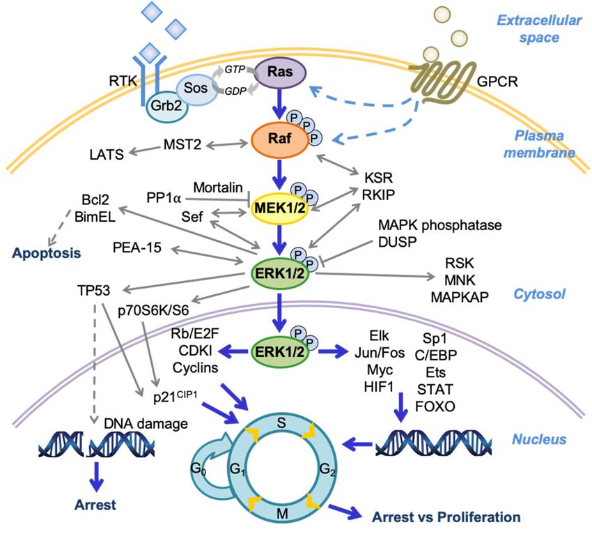
human diseases, with significant implications for cancer. As depicted in Figure 1, the Raf/MEK/ERK
pathway is mainly activated through ligand stimulation of a receptor tyrosine kinase (RTK) on the
plasma membrane, although this pathway can also be activated by a G-protein-coupled receptor
(GPCR) via as yet unclear mechanisms (reviewed in [1,2]). The RTK signals are then transmitted
by growth factor receptor-bound protein 2 (Grb2) and son of sevenless (Sos) to activate the small
guanosine triphosphatase (GTPase) Ras, recruit Ras and the Ser/Thr kinase Raf (i.e., A-Raf, B-Raf,
or C-Raf/Raf-1) to the plasma membrane followed by complex formation, and then activate Raf by
inducing phosphorylation/dephosphorylation of several serine residues on Raf. Active Raf in turn
phosphorylates and activates the dual-specificity kinases MEK1 and its homologue MEK2 (MEK1/2)
at Ser218/Ser222 of MEK1 and Ser222/Ser226 of MEK2. MEK1/2 then sequentially phosphorylate Tyr
and Thr residues in the TEY site (Thr202/Tyr204 of ERK1; Thr185/Tyr187 of ERK2) of the activation
loop of their only known physiological substrates, the serine/threonine kinases ERK1/2. ERK1/2 then
activate or inactivate a variety of proteins via phosphorylation in different subcellular compartments.
While a number of ERK1/2 substrates have been identified to date (reviewed in [3,4]), their catalogue is
likely to expand further due to continuing efforts in phosphoproteomics approaches that search for
candidate ERK1/2 substrates (reviewed in [5,6]). The Raf/MEK/ERK pathway is regulated by a complex
network of regulators, including additional small GTPases, phosphatases, scaffolds, and other kinases,
which affects the magnitude, duration, and subcellular compartmentalization of the pathway activity.
Int. J. Mol. Sci. 2020, 21, 5436; doi:10.3390/ijms21155436 www.mdpi.com/journal/ijmsInt. J. Mol. Sci. 2020, xx, x FOR PEER REVIEW 2 of 12
Int. J. Mol. Sci. 2020, 21, 5436 2 of 12
including additional small GTPases, phosphatases, scaffolds, and other kinases, which affects the
magnitude, duration, and subcellular compartmentalization of the pathway activity. For more
For more
general general information
information on molecularon activity
molecular
andactivity and of
regulation regulation of the Raf/MEK/ERK
the Raf/MEK/ERK pathway,
pathway, readers are
readers are directed to review articles elsewhere
directed to review articles elsewhere [7–10]. [7–10].
Figure 1.
Figure A schema
1. A schema of of the
the Raf/MEK/extracellular signal-regulated kinase
Raf/MEK/extracellular signal-regulated kinase (ERK)
(ERK) pathway
pathway and
and its
its
regulators and effectors. Growth factor receptor-bound protein 2 (Grb2); son of sevenless
regulators and effectors. Growth factor receptor-bound protein 2 (Grb2); son of sevenless (Sos); large (Sos);
large tumor
tumor suppressors
suppressors (LATS);
(LATS); dual-specificity
dual-specificity phosphatase
phosphatase (DUSP);
(DUSP); ribosomalS6S6kinase
ribosomal kinase (RSK);
(RSK);
mitogen activated protein kinase (MAPK)-activated protein kinase (MAPKAP);
mitogen activated protein kinase (MAPK)-activated protein kinase (MAPKAP); cyclin-dependent cyclin-dependent
kinase inhibitor
kinase inhibitor(CDKI);
(CDKI); Hypoxia-inducible
Hypoxia-inducible factor 1 (HIF1);
factor CCAAT-enhancer-binding
1 (HIF1); protein (C/EBP);
CCAAT-enhancer-binding protein
signal transducer and activator of transcription protein (STAT); forkhead box protein O
(C/EBP); signal transducer and activator of transcription protein (STAT); forkhead box protein(FOXO); protein
O
phosphorylation (P).
(FOXO); protein phosphorylation (P).
Other than mediating diverse cellular processes, the Raf/MEK/ERK pathway is mainly known for
Other than mediating diverse cellular processes, the Raf/MEK/ERK pathway is mainly known
its ability to promote cellular proliferation and survival and its deregulated activity is a hallmark of
for its ability to promote cellular proliferation and survival and its deregulated activity is a hallmark
many epithelial cancers, wherein highly selective small molecule inhibitors of B-Raf and MEK1/2 are
of many epithelial cancers, wherein highly selective small molecule inhibitors of B-Raf and MEK1/2
currently used for therapy [11–14]. Contrary to this, a significant body of evidence suggests that the
are currently used for therapy [11–14]. Contrary to this, a significant body of evidence suggests that
Raf/MEK/ERK pathway can also mediate growth inhibitory signaling (reviewed in [15–18]). In addition
the Raf/MEK/ERK pathway can also mediate growth inhibitory signaling (reviewed in [15–18]). In
to these in vitro evidences, growing evidence obtained from animal models and patient tumor tissue
addition to these in vitro evidences, growing evidence obtained from animal models and patient
specimens for different tumor types suggests that this growth inhibitory signaling may also occur
tumor tissue specimens for different tumor types suggests that this growth inhibitory signaling may
in vivo [19–25]. While this ability of Raf/MEK/ERK has significance in different biological contexts,
also occur in vivo [19–25]. While this ability of Raf/MEK/ERK has significance in different biological
including early development and neuronal differentiation, its significance in cancer is also noteworthy.
contexts, including early development and neuronal differentiation, its significance in cancer is also
For example, oncogenic mutants of RTK, Ras, or Raf can paradoxically induce growth inhibition in
noteworthy. For example, oncogenic mutants of RTK, Ras, or Raf can paradoxically induce growth
normal cells as well as in certain tumor cells, suggesting that this oncogene-induced growth inhibitory
inhibition in normal cells as well as in certain tumor cells, suggesting that this oncogene-induced
signaling may be an obstacle in the path of carcinogenesis [26–28]. Various mechanisms have been
growth inhibitory signaling may be an obstacle in the path of carcinogenesis [26–28]. Various
shown for their involvement in these phenomena, mainly including the cell cycle machinery that
mechanisms have been shown for their involvement in these phenomena, mainly including the cell
consist of Rb/E2F and cyclin-dependent kinase inhibitors p16INK4A and p21CIP1 , the tumor suppressor
cycle machinery that consist of Rb/E2F and cyclin-dependent kinase inhibitors p16INK4A and p21CIP1,
TP53 pathways, and different transcription factors and cell death machinery [15–17,29]. Indeed, genetic
the tumor suppressor TP53 pathways, and different transcription factors and cell death machinery
alterations, such as loss- or gain-of-function mutations and gene deletion that affect these mechanisms,
[15–17,29]. Indeed, genetic alterations, such as loss- or gain-of-function mutations and gene deletion
are frequently detected in tumors and address how tumor cells bypass the control of growth. In addition,
that affect these mechanisms, are frequently detected in tumors and address how tumor cells bypass
at the pathway level, spatio-temporal control can determine these paradoxical effects, as reviewed
the control of growth. In addition, at the pathway level, spatio-temporal control can determine these
in [7–10]. Whilst these mechanisms have been addressed at a variety of downstream effector levels,
paradoxical effects, as reviewed in [7–10]. Whilst these mechanisms have been addressed at a varietyInt. J. Mol. Sci. 2020, 21, 5436 3 of 12
our understanding of the mechanisms mediated at the Raf/MEK/ERK levels is relatively limited. In this
review, we will discuss recent updates in the molecular mechanisms underlying growth inhibitory
signaling of the Raf/MEK/ERK pathway with a major focus on its molecular switches and regulators.
2. Mechanisms of Raf/MEK/ERK Growth Inhibitory Signaling
2.1. Non-Canonical Effects of Raf Are Also Involved in Growth Inhibitory Signaling
A common hallmark of the growth inhibitory signaling mediated by the Raf/MEK/ERK pathway
is the sustained activation of ERK1/2, which contrasts with the transient nature of most other
ERK1/2-mediated cellular processes. As demonstrated by their constitutively active oncogenic mutants
or chimeras of kinase domains, Raf proteins can sufficiently induce growth inhibition through
their kinase function, which activates the MEK/ERK cascade (reviewed in [16,18]). Intriguingly,
recent evidences suggest that non-catalytic Raf functions are also implicated in cell proliferative
and growth inhibitory signaling. For example, a kinase-inactivating mutation (D594A) in B-Raf
was shown to cooperate with oncogenic K-Ras to drive tumor progression in a C-Raf-dependent
manner, as demonstrated in in vivo lung adenocarcinoma models [30–32]. Mechanistically, kinase-dead
B-Raf hetero-dimerized with catalytically competent C-Raf and promoted K-Ras-mediated C-Raf
activation [30,33]. Intriguingly, kinase-dead B-Raf was also shown to promote DNA damage, senescence,
and apoptotic cell death at an early stage of KRAS tumorigenesis in an animal model [32]. Recent studies
have revealed a co-regulatory mechanism involving C-Raf and A-Raf, wherein these Raf isoforms
physically bind to, sequester and inhibit the pro-apoptotic kinase mammalian sterile 20-like kinase
(MST2) independent of their kinase activity [34,35]. Intriguingly, A-Raf inactivated MST2 in the
mitochondria in a scaffold protein kinase suppressor of Ras 2 (KSR2)-dependent manner by actively
proliferating squamous epithelia and tumor cells [35]. Consistent with this, KSR2 depletion led to A-Raf
dissociation from MST2 and re-location to the plasma membrane in non-malignant and differentiated
squamous epithelia that underwent MST2-mediated apoptosis [35]. As such, C-Raf and A-Raf can
affect cell proliferation and survival by regulating not only MEK/ERK but also MST-large tumor
suppressor (LATS) pathways. Additional evidence supports kinase activity-independent effects of
A-Raf in growth inhibitory signaling. For example, a kinase domain-deficient A-Raf splicing variant
(DA-Raf) binds to Ras, suppresses MEK/ERK activities, and induces cell cycle arrest and myocyte
differentiation [36]. This truncated A-Raf is unable to regulate MST2, suppresses K-Ras-induced
transformation, and inhibits the proliferation of the human colon, head and neck, and lung cancer
cells [37,38]. These studies demonstrate that all three Raf proteins have kinase-independent functions in
addition to their canonical function as Ser/Thr kinases, indicating the complexity of pathway signaling
at Raf levels.
2.2. Differential Regulation of MEK1 and MEK2 Levels in Cells
Constitutively active mutants of MEK1 and MEK2, generated by replacing the serine residues in
activation loop with phosphomimetic aspartate (residues 218/222 for MEK1 and 222/226 for MEK2),
are sufficient to phenocopy most, if not all, growth inhibitory effects of Raf in different cell lines
(reviewed in [16,18]). It is known that signal amplification in the Raf/MEK/ERK pathway occurs more
at the Raf-MEK step due to the greater molar ratio between Raf and MEK1/2 than between MEK1/2
and ERK1/2 [39]. Because MEK1/2 activate only ERK1/2 in most, if not all, biological contexts, whereas
ERK1/2 serve as the focal points of the pathway signaling, thus no substantial contribution of MEK1/2
to signal amplification is expected, the importance of MEK1/2 in this ostensibly inefficient arrangement
of molecular switches has been unclear. Nevertheless, it appears that cellular activity and expression
levels of MEK1/2 are subject to dynamic feedback regulation. For example, we recently discovered
that, under a condition wherein activated Raf induces p21CIP1 expression and subsequently growth
arrest, MEK1 levels are transcriptionally upregulated, whereas MEK2 levels are downregulated due to
decreased protein stability via ERK1/2-mediated feedback mechanisms [40]. In line with this differentialInt. J. Mol. Sci. 2020, 21, 5436 4 of 12
regulation, knockdown of MEK1 blocked Raf-mediated p21CIP1 mRNA expression and growth arrest
more effectively than MEK2 knockdown [40]. Of note, similar differential feedback regulation of MEK1
and MEK2 levels were also detected in a subset of B-Raf mutated tumor cells [40]. MEK1 and MEK2
are >86% identical at the amino acid level, and evaluation of their constitutively active mutants has
revealed almost identical functional redundancy in various physiological contexts [41–44], although it
was also noted that MEK1 and MEK2 exhibit distinct physical interactions with A-Raf [45] and the
scaffold MEK Partner 1 (MP1) [46]. Further, many distinct effects reported in association with single
depletion of MEK1 and MEK2 were mostly due to their differential expression, as demonstrated in
different biological contexts, such as early development [47–49] and epidermal neoplasia [50]. As such,
the differential effects of MEK1 and MEK2 knockdown on p21CIP1 expression and growth arrest are
interpreted in a similar context. While the molecular mechanisms underlying these feedback MEK1/2
regulations require further elucidation, these findings add to the relatively short list of mechanisms
regulating cellular MEK1/2 levels, which include Hu antigen R (HuR) regulation of MEK1 mRNA
stability at its 30 untranslated region [51].
2.3. Intrinsic Properties of ERK1/2 Affecting the Cell Fate Toward Growth Arrest Versus Death
ERK1 and ERK2, the bona fide substrates of MEK1/2, are highly homologous and functionally
redundant. Although studies in mice have shown distinct effects of ERK1 and ERK2 ablation at
different stages of development, including stem cell lineage commitment [52,53], T cell development [54],
thymocyte maturation [55], and trophoblast development [49], studies also suggest that functional
redundancy of ERK1 and ERK2 is evolutionarily conserved [56] and that differentially regulated
expression of ERK1 and ERK2 mainly drive their biological differences [57]. ERK1 and ERK2 are
also functionally redundant and interchangeable in Raf/MEK/ERK-mediated growth inhibitory
signaling [43]. Intriguingly, we recently demonstrated that overexpression of ERK1 or ERK2 can switch
C-Raf-induced growth arrest responses to caspase-dependent apoptotic death responses in different
cell line models, which can then be reverted to growth arrest responses upon titrating the degree
of ERK1/2 activation using MEK1/2 inhibitors [58]. Consistent with our observation, other groups
showed that overexpression of ectopic ERK1/2 can induce robust cell death responses in a subset of
human B-RafV600E melanoma cells [59,60]. Kinase function of ERK1/2 is crucial for these death effects,
as catalytic site-disabled ERK2 mutants cannot induce cell death responses [58]. These phenomena
suggest that the magnitude of ERK1/2 catalytic activity should be higher than a certain threshold
to trigger cell death, while the availability of their death-specific substrates is also important in
determining the cell fate. As such, it is possible that different cellular responses in the face of aberrant
Raf/MEK activation, i.e., growth arrest vs. cell death observed in different cell types [16,18], might be
partly attributed to different molecular composition in cells that affect the magnitude of ERK1/2
activity and the availability of context-dependent ERK1/2 targets. Of note, cellular ERK1/2 levels are
subject to post-transcriptional mechanisms (reviewed in [61]), e.g., Pumilio2/PUM2 regulation of ERK2
translation [62]. It would be of interest to determine whether cellular ERK1/2 levels are correlated with
cellular ability to display different growth inhibitory responses.
Of note, these lethal effects caused by ERK1/2 overexpression are quite contrasted with the
effects of ERK1/2 mutants that contain a disabled active site but an intact activation loop. Although
unable to mediate death responses, these ERK1/2 mutants could selectively restore growth arrest
responses in Raf-activated but ERK1/2 knocked down cells, including p21CIP1 induction and E2F1
downregulation [43]. Therefore, it appears that different intrinsic properties of ERK1/2 in an active
conformation are implicated in cellular growth inhibitory responses and that non-kinase ERK1/2 effects
are involved in growth arrest signaling, while high magnitude ERK1/2 kinase activity is necessary for
death signaling. Since the discovery of the effects of the diphosphorylated kinase-dead ERK2 mutant
to activate topoisomerase II in vitro [63], kinase-independent effects of ERK1/2 have been reported in a
few different biological contexts (reviewed in [64]).Int. J. Mol. Sci. 2020, 21, 5436 5 of 12
2.4. Ectopic Expression of Autophosphorylating ERK2 Mutant Can Induce Cell Cycle Arrest
Given the strikingly high affinity between MEK1/2 and ERK1/2, relative to a typical enzyme-substrate
interaction [65], ERK1/2 have been supposed as the focal point of Raf/MEK/ERK pathway signaling.
Although the necessity of ERK1/2 for the pathway signaling has been demonstrated in various biological
contexts, including cell proliferation [42,57,66], as well as growth arrest [43,44], demonstration of their
sufficiency has been stymied, mainly due to the limit in deriving constitutively active ERK1/2 mutants
because modification of their activation loop by phosphomimetic approaches do not make ERK1/2
active. As such, alternative approaches have been attempted, e.g., exploiting the synergistic mutations
that facilitate autophosphorylation. Briefly, Tyr phosphorylation in the TEY site of the activation loop
is critical for ERK1/2 to switch into active conformation, while subsequent Thr phosphorylation locks
the kinase in the active conformation [67]. Therefore, phosphorylation of both residues is important to
achieve maximal ERK1/2 activity in cells [67,68]. ERK1/2 can autophosphorylate their TEY sites [69–71].
Natali Ahn’s group demonstrated that Lys73Pro and Ser151Asp switches in rat ERK2 and facilitates
the intramolecular interactions between Tyr on the TEY site and catalytic residues in the active site,
which promotes ERK2 autophosphorylation and catalytic activity, albeit mildly [72,73]. Because the
activation loop of this mutant, i.e., ERK2-L73P/S151D, is intact, its sufficiency for a physiological
effect should be determined only in cells exhibiting low MEK1/2 activity. Our laboratory recently
demonstrated that ectopic expression of ERK2-L73P/S151D mutant is indeed sufficient for inducing
growth arrest in LNCaP cells and neurite differentiation in PC12 cells, which exhibit relatively low
basal MEK1/2 activity [74]. Of note, this ERK2 mutant exhibits only mild kinase activity in cells and
its overexpression did not induce cell death responses, which is in agreement with the notion that
ERK1/2-mediated death signaling requires their high kinase activity.
The common docking (CD) site and F-recruitment site (FRS) are two major domains of ERK1/2 for
physical interactions [10], which are independent of each other with respect to ERK1/2 catalysis [75].
Intriguingly, mutations that impair the CD site did not affect the growth arrest responses induced by
ERK2-L73P/S151D or the aforementioned death responses induced by wild type ERK2 overexpression
(Section 2.3) in Raf/MEK-activated cells [58,74]. In contrast, the FRS (Y261N) mutation markedly
attenuated the death responses induced by wild-type ERK2 overexpression in Raf/MEK-activated
cells [58], suggesting the significance of FRS in ERK1/2-mediated growth inhibitory signaling; the effects
of Y261N could not be evaluated in ERK2-L73P/S151D because Y261N inhibited autophosphorylation
of this mutant [74]. The F-site signature “Phe-Xaa-Phe-Pro” is relatively less frequent than the D-site
signature and is found only in certain ERK1/2 substrates, including the cell-proliferative transcription
factors ELK1, c-Fos, Fra1, and c-Myc, as well as the anti-apoptotic BH3-only protein BimEL [76–78].
Nonetheless, although ELK1 was required for ERK2-L73P/S151D to mediate PC12 differentiation and
was not required for the ERK mutant to mediate growth arrest in LNCaP cells [74]. It is thus conceivable
that a failure in relaying the ERK1/2 signal to these ERK effectors may partly contribute to the onset
of growth inhibitory responses, while these responses would also be subject to cell type-specific
expression of ERK1/2 effectors.
2.5. Regulators for Fine Tuning of Pathway Activity in Growth Inhibitory Signaling
Subcellular compartmentalization and temporal regulation of active ERK1/2 plays an important
role in determining the physiological outputs of Raf/MEK/ERK signaling (reviewed in [9]) and are also
important in growth inhibitory effects of pathway signaling (reviewed in [15]). These regulations are
highly cell-type specific and mediated by a variety of proteins that directly interact with Raf, MEK1/2,
and/or ERK1/2, which include scaffolds, anchors, and phosphatases [79,80]. As such, pathway access
to an effector in a specific subcellular compartment or duration and magnitude of pathway activity
can be precisely regulated. Multiple spatial and temporal regulators of the Raf/MEK/ERK pathway
are known in the context of cell proliferative versus growth inhibitory signaling. A notable example
is phosphoprotein-enriched-in-astrocytes (PEA-15), a 15 kDa acidic serine-phosphorylated protein
that interacts with the common docking site of ERK1/2 via its C-terminal domain [81]. PEA-15 canInt. J. Mol. Sci. 2020, 21, 5436 6 of 12
promote ERK1/2 activation but sequesters ERK1/2 in the cytosol, limiting nuclear ERK1/2 activity and,
consequently, ERK1/2-dependent cell proliferative transcription [82]. Consistent with this, ERK1/2
are mainly localized in the cytosol of cells undergoing Ras-induced senescence, mainly by virtue
of PEA-15 [83]. Moreover, PEA-15-mediated sequestration of ERK1/2 in the cytosolic compartment
can promote cell survival via autophagy [84], while PEA-15 depletion increases nuclear ERK1/2
in association with apoptosis in mouse testis cells [85]. Therefore, PEA-15 exerts anti-proliferative
and anti-apoptotic effects via its ability to regulate ERK1/2 in a cell type-specific manner. Another
example is similar-expression-to-FGF-genes (Sef), which can prevent ERK1/2 nuclear import while
promoting cytoplasmic ERK1/2 activity by complexing with MEK1/2 and sequestering ERK1/2 in the
cytosol [86–88].
The cell fate between proliferation and growth inhibition can also be determined by the magnitude
of the signaling intensity, as demonstrated in a yeast model [89]. While PEA-15 and Sef regulate
subcellular compartmental ERK1/2 activity, there are notable examples for the scaffolds that affect the
magnitude of pathway activity. Kinase suppressor of Ras 1 (KSR1) is a well characterized molecular
scaffold that promotes Raf/MEK/ERK activation and was previously shown for its requirement for Ras
oncogene-induced senescence, as well as replicative senescence [90]. On the contrary, KSR1-mediated
ERK1/2 activation was also necessary for cells to escape from cell cycle arrest induced by mitomycin
C [91], suggesting that KSR1 regulation of ERK1/2 can induce different effects in opposing contexts.
Another example is the Raf-kinase-inhibitor-protein (RKIP), which inhibits MEK1/2 phosphorylation
by competitively interacting with C-Raf [92] and also suppresses MEK/ERK activity in BRAF-mutated
melanoma cells [93]. MEK/ERK activity is also determined by the regulators that are located at the
upstream tiers of the cascade, such as guanine nucleotide exchange factors (GEFs) and GTPase-activating
proteins (GAPs) [8,94]. GEFs and GAPs can be expressed at different levels, which may affect the
sensitivity of a cell to MEK/ERK. In support, loss of RasGAPs, via loss-of-function mutation or
epigenetic inactivation, is common in cancers and correlates with poor prognosis in patients [95].
Inactivation of the pathway via dephosphorylation is also an important mechanism to regulate
the pathway activity, and a number of phosphatases are known in this regard [80]. Nonetheless,
the mechanisms underlying this negative regulation are less well known. We recently identified
mortalin/HSPA9, a heat shock protein 70 paralog, as a negative regulator of Raf/MEK/ERK-mediated
growth inhibitory signaling that functions at MEK1/2 levels via direct physical interaction [96]. We then
demonstrated that mortalin can limit the pathway activity by promoting the physical interaction
between MEK1/2 and the protein phosphatase PP1α via direct physical interactions through its
C-terminal peptide binding domain, subsequently facilitating dephosphorylation of MEK1/2 [97].
These observations suggest that mortalin serves as a “rheostat”, which determines physiological output
of Raf/MEK/ERK signaling. Intriguingly, although well known for its ability to sequester and inactivate
the tumor suppressor TP53, mortalin depletion induced p21CIP1 transcription in TP53-deficient BRAF
tumor cells, for which activation of the transcription factor Sp1 by upregulated Raf/MEK/ERK activity
was necessary [98]. Hyperactivation of ERK1/2 can also lead to degradation of cell cycle regulators [99].
Currently, it is not known whether this mechanism is also involved in p21CIP1 regulation upon mortalin
depletion in BRAF tumor cells. Because mortalin depletion or inhibition can induce lethality associated
with altered mitochondrial permeability and bioenergetics in various tumor cells [100–104], it may
be possible that mortalin has a role in coordinating oncogenic MEK/ERK activity and mitochondrial
metabolism to facilitate tumor cell survival and proliferation.
3. Future Perspective
While a blockade of the activity of the Raf/MEK/ERK pathway is currently the mainstream
strategy to treat RTK/Ras/Raf-driven tumors [11–14], many tumors exhibit innate or adaptive resistance
to the therapies aimed at blocking the pathway activity. Although various mechanisms underlie
therapy resistance, most of the resistance mechanisms converge at reactivation of MEK/ERK [105–107],
indicating that this pathway remains as a key therapeutic target, even for therapy-resistant tumors.Int. J. Mol. Sci. 2020, 21, 5436 7 of 12
While various newer inhibitors are under development to continue the strategy of the pathway
blockade, the ability of tumor cells to develop resistance foretells the limit of this strategy. Growth
inhibitory signaling of the Raf/MEK/ERK pathway may indicate a potential for an additional strategy
in targeting this pathway for tumor suppression, as supported by recent intriguing observations of
tumor cell responses to hyper ERK1/2 activity. For example, discontinued drug treatment rendered
regression of drug-resistant tumors in correlation with rebounded ERK1/2 activity in tumors cells [108].
Hyperactivation of ERK1/2, mediated by overexpression of oncogenic B-Raf or by depletion of
dual-specificity phosphatase 6 (DUSP6), was also correlated with suppressed tumor cell growth in
different cancer contexts [109,110]. In addition, ERK1/2 overexpression was sufficient to induce death of
certain BRAF tumor cells [59,60]. A key question for future study is how to trigger this lethal potential
of hyper ERK1/2 activity in tumor cells and whether a regulator of the Raf/MEK/ERK pathway can be
exploited for that purpose. In this regard, close evaluation of known regulators of the pathway, as well
as identification of additional regulators, ideally druggable, will be important.
4. Conclusion
The presence of growth inhibitory signaling of Raf/MEK/ERK suggests that cells must constrain
the pathway activity within a desired range of signal intensity in order to achieve proper growth and
proliferation. As such, not only too low but also too high signal intensity would be interpreted as a
signal for cells to trigger anti-proliferative/survival responses. Further elucidation of the molecular
mechanisms underlying this relatively less-known aspect of the Raf/MEK/ERK pathway signaling
would broaden our understanding of this pathway and may offer an opportunity leading to the
development of a novel therapeutic strategy.
Funding: This work was supported by the National Cancer Institute (R01CA138441) to J.P.
Acknowledgments: We thank the members of the Park lab for critical reading of the manuscript.
Conflicts of Interest: The authors declare no conflict of interest.
References
1. McKay, M.M.; Morrison, D.K. Integrating signals from RTKs to ERK/MAPK. Oncogene 2007, 26, 3113–3121.
[CrossRef]
2. Jain, R.; Watson, U.; Vasudevan, L.; Saini, D.K. ERK Activation Pathways Downstream of GPCRs. Int. Rev.
Cell Mol. Biol. 2018, 338, 79–109.
3. Yoon, S.; Seger, R. The extracellular signal-regulated kinase: Multiple substrates regulate diverse cellular
functions. Growth Factors 2006, 24, 21–44. [CrossRef]
4. Eblen, S.T. Extracellular-Regulated Kinases: Signaling From Ras to ERK Substrates to Control Biological
Outcomes. Adv. Cancer Res. 2018, 138, 99–142. [PubMed]
5. Unal, E.B.; Uhlitz, F.; Bluthgen, N. A compendium of ERK targets. FEBS Lett. 2017, 591, 2607–2615. [CrossRef]
[PubMed]
6. Yang, L.; Zheng, L.; Chng, W.J.; Ding, J.L. Comprehensive Analysis of ERK1/2 Substrates for Potential
Combination Immunotherapies. Trends Pharmacol. Sci. 2019, 40, 897–910. [CrossRef] [PubMed]
7. Pearson, G.; Robinson, F.; Beers Gibson, T.; Xu, B.E.; Karandikar, M.; Berman, K.; Cobb, M.H. Mitogen-activated
protein (MAP) kinase pathways: Regulation and physiological functions. Endocr. Rev. 2001, 22, 153–183.
[PubMed]
8. Shaul, Y.D.; Seger, R. The MEK/ERK cascade: From signaling specificity to diverse functions. Biochim. Biophys.
Acta 2007, 1773, 1213–1226. [CrossRef] [PubMed]
9. Wortzel, I.; Seger, R. The ERK Cascade: Distinct Functions within Various Subcellular Organelles. Genes Cancer
2011, 2, 195–209. [CrossRef]
10. Roskoski, R., Jr. ERK1/2 MAP kinases: Structure, function, and regulation. Pharmacol. Res. 2012, 66, 105–143.
[CrossRef]
11. Yaeger, R.; Corcoran, R.B. Targeting Alterations in the RAF-MEK Pathway. Cancer Discov. 2019, 9, 329–341.
[CrossRef] [PubMed]Int. J. Mol. Sci. 2020, 21, 5436 8 of 12
12. Kidger, A.M.; Sipthorp, J.; Cook, S.J. ERK1/2 inhibitors: New weapons to inhibit the RAS-regulated
RAF-MEK1/2-ERK1/2 pathway. Pharmacol. Ther. 2018, 187, 45–60. [CrossRef] [PubMed]
13. Samatar, A.A.; Poulikakos, P.I. Targeting RAS-ERK signalling in cancer: Promises and challenges. Nat. Rev.
Drug Discov. 2014, 13, 928–942. [CrossRef]
14. Wu, P.K.; Park, J.I. MEK1/2 Inhibitors: Molecular Activity and Resistance Mechanisms. Semin. Oncol. 2015,
42, 849–862. [CrossRef]
15. Mebratu, Y.; Tesfaigzi, Y. How ERK1/2 activation controls cell proliferation and cell death: Is subcellular
localization the answer? Cell Cycle 2009, 8, 1168–1175. [CrossRef]
16. Cagnol, S.; Chambard, J.C. ERK and cell death: Mechanisms of ERK-induced cell death–apoptosis, autophagy
and senescence. FEBS J. 2010, 277, 2–21. [CrossRef]
17. Subramaniam, S.; Unsicker, K. ERK and cell death: ERK1/2 in neuronal death. FEBS J. 2010, 277, 22–29.
[CrossRef]
18. Park, J.I. Growth arrest signaling of the Raf/MEK/ERK pathway in cancer. Front. Biol. 2014, 9, 95–103.
[CrossRef]
19. Braig, M.; Lee, S.; Loddenkemper, C.; Rudolph, C.; Peters, A.H.; Schlegelberger, B.; Stein, H.; Dorken, B.;
Jenuwein, T.; Schmitt, C.A. Oncogene-induced senescence as an initial barrier in lymphoma development.
Nature 2005, 436, 660–665. [CrossRef]
20. Michaloglou, C.; Vredeveld, L.C.; Soengas, M.S.; Denoyelle, C.; Kuilman, T.; van der Horst, C.M.; Majoor, D.M.;
Shay, J.W.; Mooi, W.J.; Peeper, D.S. BRAFE600-associated senescence-like cell cycle arrest of human naevi.
Nature 2005, 436, 720–724. [CrossRef]
21. Thaler, S.; Hahnel, P.S.; Schad, A.; Dammann, R.; Schuler, M. RASSF1A mediates p21Cip1/Waf1-dependent
cell cycle arrest and senescence through modulation of the Raf-MEK-ERK pathway and inhibition of Akt.
Cancer Res. 2009, 69, 1748–1757. [CrossRef] [PubMed]
22. Collado, M.; Gil, J.; Efeyan, A.; Guerra, C.; Schuhmacher, A.J.; Barradas, M.; Benguria, A.; Zaballos, A.;
Flores, J.M.; Barbacid, M.; et al. Tumour biology: Senescence in premalignant tumours. Nature 2005, 436, 642.
[CrossRef]
23. Sarkisian, C.J.; Keister, B.A.; Stairs, D.B.; Boxer, R.B.; Moody, S.E.; Chodosh, L.A. Dose-dependent
oncogene-induced senescence in vivo and its evasion during mammary tumorigenesis. Nat. Cell Biol.
2007, 9, 493–505. [CrossRef] [PubMed]
24. Mo, L.; Zheng, X.; Huang, H.Y.; Shapiro, E.; Lepor, H.; Cordon-Cardo, C.; Sun, T.T.; Wu, X.R. Hyperactivation
of Ha-ras oncogene, but not Ink4a/Arf deficiency, triggers bladder tumorigenesis. J. Clin. Invest. 2007, 117,
314–325. [CrossRef] [PubMed]
25. Guerra, C.; Collado, M.; Navas, C.; Schuhmacher, A.J.; Hernandez-Porras, I.; Canamero, M.;
Rodriguez-Justo, M.; Serrano, M.; Barbacid, M. Pancreatitis-induced inflammation contributes to pancreatic
cancer by inhibiting oncogene-induced senescence. Cancer Cell 2011, 19, 728–739. [CrossRef]
26. Mooi, W.J.; Peeper, D.S. Oncogene-induced cell senescence–halting on the road to cancer. N. Engl. J. Med.
2006, 355, 1037–1046. [CrossRef]
27. Courtois-Cox, S.; Jones, S.L.; Cichowski, K. Many roads lead to oncogene-induced senescence. Oncogene
2008, 27, 2801–2809. [CrossRef]
28. Collado, M.; Serrano, M. Senescence in tumours: Evidence from mice and humans. Nat. Rev. Cancer 2010,
10, 51–57. [CrossRef]
29. McDuff, F.K.; Turner, S.D. Jailbreak: Oncogene-induced senescence and its evasion. Cell Signal. 2011, 23, 6–13.
[CrossRef]
30. Heidorn, S.J.; Milagre, C.; Whittaker, S.; Nourry, A.; Niculescu-Duvas, I.; Dhomen, N.; Hussain, J.;
Reis-Filho, J.S.; Springer, C.J.; Pritchard, C.; et al. Kinase-dead BRAF and oncogenic RAS cooperate
to drive tumor progression through CRAF. Cell 2010, 140, 209–221. [CrossRef]
31. Kamata, T.; Hussain, J.; Giblett, S.; Hayward, R.; Marais, R.; Pritchard, C. BRAF inactivation drives aneuploidy
by deregulating CRAF. Cancer Res. 2010, 70, 8475–8486. [CrossRef] [PubMed]
32. Nieto, P.; Ambrogio, C.; Esteban-Burgos, L.; Gomez-Lopez, G.; Blasco, M.T.; Yao, Z.; Marais, R.; Rosen, N.;
Chiarle, R.; Pisano, D.G.; et al. A Braf kinase-inactive mutant induces lung adenocarcinoma. Nature 2017,
548, 239–243. [CrossRef] [PubMed]
33. Poulikakos, P.I.; Zhang, C.; Bollag, G.; Shokat, K.M.; Rosen, N. RAF inhibitors transactivate RAF dimers and
ERK signalling in cells with wild-type BRAF. Nature 2010, 464, 427–430. [CrossRef]Int. J. Mol. Sci. 2020, 21, 5436 9 of 12
34. Romano, D.; Nguyen, L.K.; Matallanas, D.; Halasz, M.; Doherty, C.; Kholodenko, B.N.; Kolch, W. Protein
interaction switches coordinate Raf-1 and MST2/Hippo signalling. Nat. Cell Biol. 2014, 16, 673–684. [CrossRef]
35. Rauch, J.; Vandamme, D.; Mack, B.; McCann, B.; Volinsky, N.; Blanco, A.; Gires, O.; Kolch, W. Differential
localization of A-Raf regulates MST2-mediated apoptosis during epithelial differentiation. Cell Death Differ.
2016, 23, 1283–1295. [CrossRef] [PubMed]
36. Yokoyama, T.; Takano, K.; Yoshida, A.; Katada, F.; Sun, P.; Takenawa, T.; Andoh, T.; Endo, T. DA-Raf1,
a competent intrinsic dominant-negative antagonist of the Ras-ERK pathway, is required for myogenic
differentiation. J. Cell Biol. 2007, 177, 781–793. [CrossRef]
37. Rauch, J.; Moran-Jones, K.; Albrecht, V.; Schwarzl, T.; Hunter, K.; Gires, O.; Kolch, W. c-Myc regulates
RNA splicing of the A-Raf kinase and its activation of the ERK pathway. Cancer Res. 2011, 71, 4664–4674.
[CrossRef]
38. Kanno, E.; Kawasaki, O.; Takahashi, K.; Takano, K.; Endo, T. DA-Raf, a dominant-negative antagonist of the
Ras-ERK pathway, is a putative tumor suppressor. Exp. Cell Res. 2018, 362, 111–120. [CrossRef]
39. Ferrell, J.E., Jr. Tripping the switch fantastic: How a protein kinase cascade can convert graded inputs into
switch-like outputs. Trends Biochem. Sci. 1996, 21, 460–466. [CrossRef]
40. Hong, S.K.; Wu, P.K.; Karkhanis, M.; Park, J.I. ERK1/2 can feedback-regulate cellular MEK1/2 levels. Cell Signal.
2015, 27, 1939–1948. [CrossRef]
41. Mansour, S.J.; Candia, J.M.; Matsuura, J.E.; Manning, M.C.; Ahn, N.G. Interdependent domains controlling
the enzymatic activity of mitogen-activated protein kinase kinase 1. Biochemistry 1996, 35, 15529–15536.
[CrossRef] [PubMed]
42. Voisin, L.; Julien, C.; Duhamel, S.; Gopalbhai, K.; Claveau, I.; Saba-El-Leil, M.K.; Rodrigue-Gervais, I.G.;
Gaboury, L.; Lamarre, D.; Basik, M.; et al. Activation of MEK1 or MEK2 isoform is sufficient to fully transform
intestinal epithelial cells and induce the formation of metastatic tumors. BMC Cancer 2008, 8, 337. [CrossRef]
43. Hong, S.K.; Yoon, S.; Moelling, C.; Arthan, D.; Park, J.I. Noncatalytic function of ERK1/2 can promote
Raf/MEK/ERK-mediated growth arrest signaling. J. Biol. Chem. 2009, 284, 33006–33018. [CrossRef] [PubMed]
44. Guegan, J.P.; Ezan, F.; Gailhouste, L.; Langouet, S.; Baffet, G. MEK1/2 overactivation can promote growth
arrest by mediating ERK1/2-dependent phosphorylation of p70S6K. J. Cell Physiol. 2014, 229, 903–915.
[CrossRef] [PubMed]
45. Wu, X.; Noh, S.J.; Zhou, G.; Dixon, J.E.; Guan, K.L. Selective activation of MEK1 but not MEK2 by A-Raf from
epidermal growth factor-stimulated Hela cells. J. Biol. Chem. 1996, 271, 3265–3271. [CrossRef] [PubMed]
46. Schaeffer, H.J.; Catling, A.D.; Eblen, S.T.; Collier, L.S.; Krauss, A.; Weber, M.J. MP1: A MEK binding partner
that enhances enzymatic activation of the MAP kinase cascade. Science 1998, 281, 1668–1671. [CrossRef]
[PubMed]
47. Giroux, S.; Tremblay, M.; Bernard, D.; Cardin-Girard, J.F.; Aubry, S.; Larouche, L.; Rousseau, S.; Huot, J.;
Landry, J.; Jeannotte, L.; et al. Embryonic death of Mek1-deficient mice reveals a role for this kinase in
angiogenesis in the labyrinthine region of the placenta. Curr. Biol. 1999, 9, 369–372. [CrossRef]
48. Belanger, L.F.; Roy, S.; Tremblay, M.; Brott, B.; Steff, A.M.; Mourad, W.; Hugo, P.; Erikson, R.; Charron, J. Mek2
is dispensable for mouse growth and development. Mol. Cell Biol. 2003, 23, 4778–4787. [CrossRef]
49. Nadeau, V.; Guillemette, S.; Belanger, L.F.; Jacob, O.; Roy, S.; Charron, J. Map2k1 and Map2k2 genes contribute
to the normal development of syncytiotrophoblasts during placentation. Development 2009, 136, 1363–1374.
[CrossRef]
50. Scholl, F.A.; Dumesic, P.A.; Barragan, D.I.; Harada, K.; Charron, J.; Khavari, P.A. Selective role for Mek1 but
not Mek2 in the induction of epidermal neoplasia. Cancer Res. 2009, 69, 3772–3778. [CrossRef]
51. Wang, P.Y.; Rao, J.N.; Zou, T.; Liu, L.; Xiao, L.; Yu, T.X.; Turner, D.J.; Gorospe, M.; Wang, J.Y. Post-transcriptional
regulation of MEK-1 by polyamines through the RNA-binding protein HuR modulating intestinal epithelial
apoptosis. Biochem. J. 2010, 426, 293–306. [CrossRef] [PubMed]
52. Hamilton, W.B.; Kaji, K.; Kunath, T. ERK2 suppresses self-renewal capacity of embryonic stem cells, but is
not required for multi-lineage commitment. PLoS ONE 2013, 8, e60907. [CrossRef]
53. Saulnier, N.; Guihard, S.; Holy, X.; Decembre, E.; Jurdic, P.; Clay, D.; Feuillet, V.; Pages, G.; Pouyssegur, J.;
Porteu, F.; et al. ERK1 regulates the hematopoietic stem cell niches. PLoS ONE 2012, 7, e30788. [CrossRef]
[PubMed]
54. Fischer, A.M.; Katayama, C.D.; Pages, G.; Pouyssegur, J.; Hedrick, S.M. The role of erk1 and erk2 in multiple
stages of T cell development. Immunity 2005, 23, 431–443. [CrossRef]Int. J. Mol. Sci. 2020, 21, 5436 10 of 12
55. Pages, G.; Guerin, S.; Grall, D.; Bonino, F.; Smith, A.; Anjuere, F.; Auberger, P.; Pouyssegur, J. Defective
thymocyte maturation in p44 MAP kinase (Erk 1) knockout mice. Science 1999, 286, 1374–1377.
56. Busca, R.; Christen, R.; Lovern, M.; Clifford, A.M.; Yue, J.X.; Goss, G.G.; Pouyssegur, J.; Lenormand, P. ERK1
and ERK2 present functional redundancy in tetrapods despite higher evolution rate of ERK1. BMC Evol.
Biol. 2015, 15, 179. [CrossRef]
57. Lefloch, R.; Pouyssegur, J.; Lenormand, P. Single and combined silencing of ERK1 and ERK2 reveals their
positive contribution to growth signaling depending on their expression levels. Mol. Cell Biol. 2008, 28,
511–527. [CrossRef] [PubMed]
58. Hong, S.K.; Wu, P.K.; Park, J.I. A cellular threshold for active ERK1/2 levels determines Raf/MEK/ERK-
mediated growth arrest versus death responses. Cell Signal. 2018, 42, 11–20. [CrossRef]
59. Goetz, E.M.; Ghandi, M.; Treacy, D.J.; Wagle, N.; Garraway, L.A. ERK mutations confer resistance to
mitogen-activated protein kinase pathway inhibitors. Cancer Res. 2014, 74, 7079–7089. [CrossRef]
60. Leung, G.P.; Feng, T.; Sigoillot, F.D.; Geyer, F.C.; Shirley, M.D.; Ruddy, D.A.; Rakiec, D.P.; Freeman, A.K.;
Engelman, J.A.; Jaskelioff, M.; et al. Hyperactivation of MAPK Signaling Is Deleterious to RAS/RAF-mutant
Melanoma. Mol. Cancer Res. 2019, 17, 199–211. [CrossRef]
61. Whelan, J.T.; Hollis, S.E.; Cha, D.S.; Asch, A.S.; Lee, M.H. Post-transcriptional regulation of the
Ras-ERK/MAPK signaling pathway. J. Cell Physiol. 2012, 227, 1235–1241. [CrossRef]
62. Lee, M.H.; Hook, B.; Pan, G.; Kershner, A.M.; Merritt, C.; Seydoux, G.; Thomson, J.A.; Wickens, M.; Kimble, J.
Conserved regulation of MAP kinase expression by PUF RNA-binding proteins. PLoS Genet. 2007, 3, e233.
[CrossRef] [PubMed]
63. Shapiro, P.S.; Whalen, A.M.; Tolwinski, N.S.; Wilsbacher, J.; Froelich-Ammon, S.J.; Garcia, M.; Osheroff, N.;
Ahn, N.G. Extracellular signal-regulated kinase activates topoisomerase IIalpha through a mechanism
independent of phosphorylation. Mol. Cell Biol. 1999, 19, 3551–3560. [CrossRef] [PubMed]
64. Rodriguez, J.; Crespo, P. Working without kinase activity: Phosphotransfer-independent functions of
extracellular signal-regulated kinases. Sci. Signal. 2011, 4, re3. [CrossRef] [PubMed]
65. Fukuda, M.; Gotoh, Y.; Nishida, E. Interaction of MAP kinase with MAP kinase kinase: Its possible role in
the control of nucleocytoplasmic transport of MAP kinase. EMBO J. 1997, 16, 1901–1908. [CrossRef]
66. Vantaggiato, C.; Formentini, I.; Bondanza, A.; Bonini, C.; Naldini, L.; Brambilla, R. ERK1 and ERK2
mitogen-activated protein kinases affect Ras-dependent cell signaling differentially. J. Biol. 2006, 5, 14.
[CrossRef]
67. Barr, D.; Oashi, T.; Burkhard, K.; Lucius, S.; Samadani, R.; Zhang, J.; Shapiro, P.; MacKerell, A.D.; van der
Vaart, A. Importance of domain closure for the autoactivation of ERK2. Biochemistry 2011, 50, 8038–8048.
[CrossRef]
68. Zhang, F.; Strand, A.; Robbins, D.; Cobb, M.H.; Goldsmith, E.J. Atomic structure of the MAP kinase ERK2 at
2.3 A resolution. Nature 1994, 367, 704–711. [CrossRef]
69. Robbins, D.J.; Zhen, E.; Owaki, H.; Vanderbilt, C.A.; Ebert, D.; Geppert, T.D.; Cobb, M.H. Regulation and
properties of extracellular signal-regulated protein kinases 1 and 2 in vitro. J. Biol. Chem. 1993, 268, 5097–5106.
70. Rossomando, A.J.; Wu, J.; Michel, H.; Shabanowitz, J.; Hunt, D.F.; Weber, M.J.; Sturgill, T.W. Identification
of Tyr-185 as the site of tyrosine autophosphorylation of recombinant mitogen-activated protein kinase
p42mapk. Proc. Natl. Acad. Sci. USA 1992, 89, 5779–5783. [CrossRef]
71. Seger, R.; Ahn, N.G.; Boulton, T.G.; Yancopoulos, G.D.; Panayotatos, N.; Radziejewska, E.; Ericsson, L.;
Bratlien, R.L.; Cobb, M.H.; Krebs, E.G. Microtubule-associated protein 2 kinases, ERK1 and ERK2, undergo
autophosphorylation on both tyrosine and threonine residues: Implications for their mechanism of activation.
Proc. Natl. Acad. Sci. USA 1991, 88, 6142–6146. [CrossRef] [PubMed]
72. Emrick, M.A.; Hoofnagle, A.N.; Miller, A.S.; Ten Eyck, L.F.; Ahn, N.G. Constitutive activation of extracellular
signal-regulated kinase 2 by synergistic point mutations. J. Biol. Chem. 2001, 276, 46469–46479. [CrossRef]
[PubMed]
73. Emrick, M.A.; Lee, T.; Starkey, P.J.; Mumby, M.C.; Resing, K.A.; Ahn, N.G. The gatekeeper residue controls
autoactivation of ERK2 via a pathway of intramolecular connectivity. Proc. Natl. Acad. Sci. USA 2006, 103,
18101–18106. [CrossRef] [PubMed]
74. Wu, P.K.; Hong, S.K.; Yoon, S.H.; Park, J.I. Active ERK2 is sufficient to mediate growth arrest and differentiation
signaling. FEBS J. 2015, 282, 1017–1030. [CrossRef]Int. J. Mol. Sci. 2020, 21, 5436 11 of 12
75. Lee, S.; Warthaka, M.; Yan, C.; Kaoud, T.S.; Ren, P.; Dalby, K.N. Examining docking interactions on ERK2
with modular peptide substrates. Biochemistry 2011, 50, 9500–9510. [CrossRef]
76. Murphy, L.O.; Smith, S.; Chen, R.H.; Fingar, D.C.; Blenis, J. Molecular interpretation of ERK signal duration
by immediate early gene products. Nat. Cell Biol. 2002, 4, 556–564. [CrossRef]
77. Murphy, L.O.; MacKeigan, J.P.; Blenis, J. A network of immediate early gene products propagates subtle
differences in mitogen-activated protein kinase signal amplitude and duration. Mol. Cell Biol. 2004,
24, 144–153. [CrossRef]
78. Ley, R.; Hadfield, K.; Howes, E.; Cook, S.J. Identification of a DEF-type docking domain for extracellular
signal-regulated kinases 1/2 that directs phosphorylation and turnover of the BH3-only protein BimEL. J. Biol.
Chem. 2005, 280, 17657–17663. [CrossRef]
79. Brown, M.D.; Sacks, D.B. Protein scaffolds in MAP kinase signalling. Cell Signal. 2009, 21, 462–469. [CrossRef]
80. Owens, D.M.; Keyse, S.M. Differential regulation of MAP kinase signalling by dual-specificity protein
phosphatases. Oncogene 2007, 26, 3203–3213. [CrossRef]
81. Callaway, K.; Abramczyk, O.; Martin, L.; Dalby, K.N. The anti-apoptotic protein PEA-15 is a tight binding
inhibitor of ERK1 and ERK2, which blocks docking interactions at the D-recruitment site. Biochemistry 2007,
46, 9187–9198. [PubMed]
82. Formstecher, E.; Ramos, J.W.; Fauquet, M.; Calderwood, D.A.; Hsieh, J.C.; Canton, B.; Nguyen, X.T.;
Barnier, J.V.; Camonis, J.; Ginsberg, M.H.; et al. PEA-15 mediates cytoplasmic sequestration of ERK MAP
kinase. Dev. Cell 2001, 1, 239–250. [CrossRef]
83. Gaumont-Leclerc, M.F.; Mukhopadhyay, U.K.; Goumard, S.; Ferbeyre, G. PEA-15 is inhibited by adenovirus
E1A and plays a role in ERK nuclear export and Ras-induced senescence. J. Biol. Chem. 2004, 279, 46802–46809.
[CrossRef] [PubMed]
84. Bartholomeusz, C.; Rosen, D.; Wei, C.; Kazansky, A.; Yamasaki, F.; Takahashi, T.; Itamochi, H.; Kondo, S.;
Liu, J.; Ueno, N.T. PEA-15 induces autophagy in human ovarian cancer cells and is associated with prolonged
overall survival. Cancer Res. 2008, 68, 9302–9310. [CrossRef] [PubMed]
85. Mizrak, S.C.; Renault-Mihara, F.; Parraga, M.; Bogerd, J.; van de Kant, H.J.; Lopez-Casas, P.P.; Paz, M.;
del Mazo, J.; de Rooij, D.G. Phosphoprotein enriched in astrocytes-15 is expressed in mouse testis and
protects spermatocytes from apoptosis. Reproduction 2007, 133, 743–751. [CrossRef]
86. Furthauer, M.; Lin, W.; Ang, S.L.; Thisse, B.; Thisse, C. Sef is a feedback-induced antagonist of
Ras/MAPK-mediated FGF signalling. Nat. Cell Biol. 2002, 4, 170–174. [CrossRef]
87. Tsang, M.; Friesel, R.; Kudoh, T.; Dawid, I.B. Identification of Sef, a novel modulator of FGF signalling.
Nat. Cell Biol. 2002, 4, 165–169. [CrossRef]
88. Preger, E.; Ziv, I.; Shabtay, A.; Sher, I.; Tsang, M.; Dawid, I.B.; Altuvia, Y.; Ron, D. Alternative splicing
generates an isoform of the human Sef gene with altered subcellular localization and specificity. Proc. Natl.
Acad. Sci. USA 2004, 101, 1229–1234. [CrossRef]
89. Conlon, P.; Gelin-Licht, R.; Ganesan, A.; Zhang, J.; Levchenko, A. Single-cell dynamics and variability of
MAPK activity in a yeast differentiation pathway. Proc. Natl. Acad. Sci. USA 2016, 113, E5896–E5905.
[CrossRef]
90. Kortum, R.L.; Johnson, H.J.; Costanzo, D.L.; Volle, D.J.; Razidlo, G.L.; Fusello, A.M.; Shaw, A.S.; Lewis, R.E.
The molecular scaffold kinase suppressor of Ras 1 is a modifier of RasV12-induced and replicative senescence.
Mol. Cell Biol. 2006, 26, 2202–2214. [CrossRef]
91. Razidlo, G.L.; Johnson, H.J.; Stoeger, S.M.; Cowan, K.H.; Bessho, T.; Lewis, R.E. KSR1 is required for cell
cycle reinitiation following DNA damage. J. Biol. Chem. 2009, 284, 6705–6715. [CrossRef] [PubMed]
92. Yeung, K.; Seitz, T.; Li, S.; Janosch, P.; McFerran, B.; Kaiser, C.; Fee, F.; Katsanakis, K.D.; Rose, D.W.;
Mischak, H.; et al. Suppression of Raf-1 kinase activity and MAP kinase signalling by RKIP. Nature 1999, 401,
173–177. [CrossRef] [PubMed]
93. Schuierer, M.M.; Bataille, F.; Hagan, S.; Kolch, W.; Bosserhoff, A.K. Reduction in Raf kinase inhibitor protein
expression is associated with increased Ras-extracellular signal-regulated kinase signaling in melanoma cell
lines. Cancer Res. 2004, 64, 5186–5192. [CrossRef] [PubMed]
94. Vigil, D.; Cherfils, J.; Rossman, K.L.; Der, C.J. Ras superfamily GEFs and GAPs: Validated and tractable
targets for cancer therapy? Nat. Rev. Cancer 2010, 10, 842–857. [CrossRef] [PubMed]
95. Harrell Stewart, D.R.; Clark, G.J. Pumping the brakes on RAS - negative regulators and death effectors of
RAS. J. Cell Sci. 2020, 133, jcs238865. [CrossRef]Int. J. Mol. Sci. 2020, 21, 5436 12 of 12
96. Wu, P.K.; Hong, S.K.; Veeranki, S.; Karkhanis, M.; Starenki, D.; Plaza, J.A.; Park, J.I. A mortalin/HSPA9-
mediated switch in tumor-suppressive signaling of Raf/MEK/extracellular signal-regulated kinase. Mol. Cell
Biol. 2013, 33, 4051–4067. [CrossRef] [PubMed]
97. Wu, P.K.; Hong, S.K.; Park, J.I. Steady-State Levels of Phosphorylated Mitogen-Activated Protein Kinase
Kinase 1/2 Determined by Mortalin/HSPA9 and Protein Phosphatase 1 Alpha in KRAS and BRAF Tumor
Cells. Mol. Cell Biol. 2017, 37, e00061-17. [CrossRef]
98. Karkhanis, M.; Park, J.I. Sp1 regulates Raf/MEK/ERK-induced p21(CIP1) transcription in TP53-mutated
cancer cells. Cell Signal. 2015, 27, 479–486. [CrossRef]
99. Deschenes-Simard, X.; Gaumont-Leclerc, M.F.; Bourdeau, V.; Lessard, F.; Moiseeva, O.; Forest, V.; Igelmann, S.;
Mallette, F.A.; Saba-El-Leil, M.K.; Meloche, S.; et al. Tumor suppressor activity of the ERK/MAPK pathway
by promoting selective protein degradation. Genes Dev. 2013, 27, 900–915. [CrossRef]
100. Wu, P.K.; Hong, S.K.; Chen, W.; Becker, A.E.; Gundry, R.L.; Lin, C.W.; Shao, H.; Gestwicki, J.E.; Park, J.I.
Mortalin (HSPA9) facilitates BRAF-mutant tumor cell survival by suppressing ANT3-mediated mitochondrial
membrane permeability. Sci. Signal. 2020, 13, eaay1478. [CrossRef]
101. Wu, P.K.; Hong, S.K.; Starenki, D.; Oshima, K.; Shao, H.; Gestwicki, J.E.; Tsai, S.; Park, J.I. Mortalin/HSPA9
targeting selectively induces KRAS tumor cell death by perturbing mitochondrial membrane permeability.
Oncogene 2020, 39, 4257–4270. [CrossRef] [PubMed]
102. Starenki, D.; Hong, S.K.; Lloyd, R.V.; Park, J.I. Mortalin (GRP75/HSPA9) upregulation promotes survival and
proliferation of medullary thyroid carcinoma cells. Oncogene 2015, 34, 4624–4634. [CrossRef] [PubMed]
103. Starenki, D.; Park, J.I. Selective Mitochondrial Uptake of MKT-077 Can Suppress Medullary Thyroid
Carcinoma Cell Survival In Vitro and In Vivo. Endocrinol Metab 2015, 30, 593–603. [CrossRef]
104. Starenki, D.; Sosonkina, N.; Hong, S.K.; Lloyd, R.V.; Park, J.I. Mortalin (GRP75/HSPA9) Promotes Survival
and Proliferation of Thyroid Carcinoma Cells. Int. J. Mol. Sci. 2019, 20, 2069. [CrossRef]
105. Konieczkowski, D.J.; Johannessen, C.M.; Garraway, L.A. A Convergence-Based Framework for Cancer Drug
Resistance. Cancer Cell 2018, 33, 801–815. [CrossRef]
106. Moriceau, G.; Hugo, W.; Hong, A.; Shi, H.; Kong, X.; Yu, C.C.; Koya, R.C.; Samatar, A.A.; Khanlou, N.;
Braun, J.; et al. Tunable-combinatorial mechanisms of acquired resistance limit the efficacy of BRAF/MEK
cotargeting but result in melanoma drug addiction. Cancer Cell 2015, 27, 240–256. [CrossRef] [PubMed]
107. Kong, X.; Kuilman, T.; Shahrabi, A.; Boshuizen, J.; Kemper, K.; Song, J.Y.; Niessen, H.W.M.; Rozeman, E.A.;
Geukes Foppen, M.H.; Blank, C.U.; et al. Cancer drug addiction is relayed by an ERK2-dependent phenotype
switch. Nature 2017, 550, 270–274. [CrossRef] [PubMed]
108. Das Thakur, M.; Salangsang, F.; Landman, A.S.; Sellers, W.R.; Pryer, N.K.; Levesque, M.P.; Dummer, R.;
McMahon, M.; Stuart, D.D. Modelling vemurafenib resistance in melanoma reveals a strategy to forestall
drug resistance. Nature 2013, 494, 251–255. [CrossRef] [PubMed]
109. Maddodi, N.; Huang, W.; Havighurst, T.; Kim, K.; Longley, B.J.; Setaluri, V. Induction of autophagy and
inhibition of melanoma growth in vitro and in vivo by hyperactivation of oncogenic BRAF. J. Invest. Dermatol.
2010, 130, 1657–1667. [CrossRef]
110. Unni, A.M.; Harbourne, B.; Oh, M.H.; Wild, S.; Ferrarone, J.R.; Lockwood, W.W.; Varmus, H. Hyperactivation
of ERK by multiple mechanisms is toxic to RTK-RAS mutation-driven lung adenocarcinoma cells. Elife 2018,
7, e33718. [CrossRef]
© 2020 by the authors. Licensee MDPI, Basel, Switzerland. This article is an open access
article distributed under the terms and conditions of the Creative Commons Attribution
(CC BY) license (http://creativecommons.org/licenses/by/4.0/).You can also read