Intravital live cell triggered imaging system reveals monocyte patrolling and macrophage migration in atherosclerotic arteries
←
→
Page content transcription
If your browser does not render page correctly, please read the page content below
Intravital live cell triggered imaging
                 system reveals monocyte patrolling
                 and macrophage migration in
                 atherosclerotic arteries

                 Sara McArdle
                 Grzegorz Chodaczek
                 Nilanjan Ray
                 Klaus Ley

Downloaded From: https://www.spiedigitallibrary.org/journals/Journal-of-Biomedical-Optics on 30 Oct 2021
Terms of Use: https://www.spiedigitallibrary.org/terms-of-use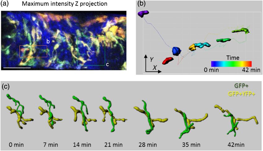
Journal of Biomedical Optics 20(2), 026005 (February 2015)

                Intravital live cell triggered imaging system reveals
                monocyte patrolling and macrophage migration in
                atherosclerotic arteries
                Sara McArdle,a,b Grzegorz Chodaczek,a,c Nilanjan Ray,d and Klaus Leya,*
                a
                  La Jolla Institute, 9420 Athena Circle, La Jolla, California 92037, United States
                b
                  University of California, Department of Bioengineering, San Diego, 9500 Gilman Drive, La Jolla, California 92093, United States
                c
                  Wroclaw Research Centre EIT+, Stabłowicka 147, 54-066 Wroclaw, Poland
                d
                  University of Alberta, Department of Computing Science, 8900 114 Street Northwest, Edmonton, Alberta T6G 2S4, Canada

                              Abstract. Intravital multiphoton imaging of arteries is technically challenging because the artery expands with
                              every heartbeat, causing severe motion artifacts. To study leukocyte activity in atherosclerosis, we developed the
                              intravital live cell triggered imaging system (ILTIS). This system implements cardiac triggered acquisition as well
                              as frame selection and image registration algorithms to produce stable movies of myeloid cell movement in ath-
                              erosclerotic arteries in live mice. To minimize tissue damage, no mechanical stabilization is used and the artery is
                              allowed to expand freely. ILTIS performs multicolor high frame-rate two-dimensional imaging and full-thickness
                              three-dimensional imaging of beating arteries in live mice. The external carotid artery and its branches (superior
                              thyroid and ascending pharyngeal arteries) were developed as a surgically accessible and reliable model of ath-
                              erosclerosis. We use ILTIS to demonstrate Cx3cr1GFP monocytes patrolling the lumen of atherosclerotic arteries.
                              Additionally, we developed a new reporter mouse (Apoe−/−Cx3cr1GFP/+Cd11cYFP) to image GFP+ and GFP+YFP
                              + macrophages “dancing on the spot” and YFP+ macrophages migrating within intimal plaque. ILTIS will be help-
                              ful to answer pertinent open questions in the field, including monocyte recruitment and transmigration, macro-
                              phage and dendritic cell activity, and motion of other immune cells. © 2015 Society of Photo-Optical Instrumentation Engineers
                              (SPIE) [DOI: 10.1117/1.JBO.20.2.026005]

                              Keywords: intravital microscopy; multiphoton microscopy; triggering; registration; atherosclerosis; monocytes.
                              Paper 140757R received Nov. 14, 2014; accepted for publication Jan. 12, 2015; published online Feb. 24, 2015.

                1     Introduction                                                                 bifurcation, were not suitable for these purposes, we developed
                Intravital optical imaging is an invaluable tool for accurately                    a new model based on a more accessible artery, a branch of
                studying biological and disease processes. However, imaging                        the external carotid (EC) artery.
                live animals introduces technical challenges due to motion arti-                       There are three commonly used methods for minimizing
                facts, especially in cardiovascular organs that are inherently sub-                motion artifacts in intravital laser scanning microscopy of tis-
                ject to vigorous motion. One such tissue with significant medical                  sues with innate motion, such as the heart or lungs: mechanical
                relevance and that is still largely unexplored is atherosclerotic                  stabilization, triggering, and image postprocessing.2 Various
                arteries. In the course of atherosclerosis artery walls become                     techniques have been successfully used to physically restrain
                infiltrated over time by various immune cells such as macro-                       tissue movement, such as tissue glue,3–5 compression,6,7 or suc-
                phages, dendritic cells, T lymphocytes, and other inflammatory                     tion8–10 to attach a stabilizer. However, these methods are not
                cells.1 Dynamic intravital visualization of these processes at the                 suitable for arterial preparations. The cylindrical geometry of
                cellular and subcellular levels still awaits its realization due to                the artery makes a suction ring approach infeasible. Also, the
                difficulties with access to the vessel luminal side and image sta-                 artery wall, especially the adventitia, is a delicate tissue that
                bility during the acquisition. Two-photon microscopy is the only                   may be damaged by tissue glue. In contrast, triggered acquisi-
                type of fluorescent microscopy that can image through the depth                    tion enables high-resolution imaging while allowing the tissue
                of the atherosclerotic plaque, which can be 100- to 200-μm                         to move freely by only acquiring data when the tissue is in
                thick. Additionally, in a living mouse, large arteries expand                      a quasisteady position. Alternatively, triggering can also be
                and contract with every heartbeat, moving the wall vertically                      done in a retrospective manner, where data are acquired contin-
                and laterally, which severely compromises the image quality.                       uously while the heartbeat and breathing are recorded and then
                Motivated by the need to visualize recruitment of inflammatory                     only images or image fractions that were acquired within pre-
                cells from the blood and monitor the movement of macrophages                       determined windows in both cycles are kept. Triggering based
                inside the plaque we developed and refined a new intravital live                   on heartbeat and respiration has been used to image healthy
                cell triggered imaging system (ILTIS), which has the potential                     arteries; however, to date the use of this technique alone
                to study the process of atherosclerosis in vivo with high quality                  has been limited to image a single plane in animals with
                and subcellular resolution. Because commonly studied areas of                      low heart rates, such as rats or deeply anesthetized mice.11
                plaque development, including the aortic arch and carotid artery                   Triggering has also been used to improve two-dimensional

                *Address all correspondence to: Klaus Ley, E-mail: klaus@liai.org                  1083-3668/2015/$25.00 © 2015 SPIE

                Journal of Biomedical Optics                                               026005-1                                       February 2015   •   Vol. 20(2)

Downloaded From: https://www.spiedigitallibrary.org/journals/Journal-of-Biomedical-Optics on 30 Oct 2021
Terms of Use: https://www.spiedigitallibrary.org/terms-of-use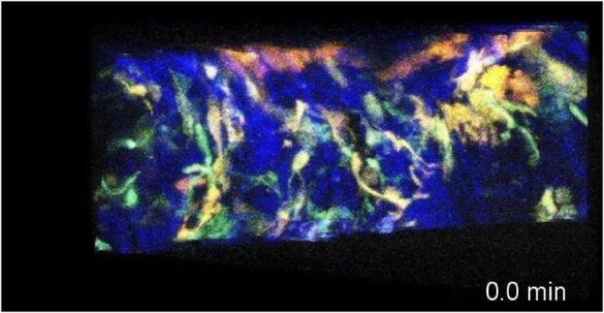
McArdle et al.: Intravital live cell triggered imaging system reveals monocyte patrolling. . .

                (2-D)4,10 and three-dimensional (3-D) imaging of the heart3,6 and                  2       Materials and Methods
                carotid artery7 that was predominantly stabilized mechanically.
                The third method, postprocessing, is applied after data collec-                    2.1      Mice
                tion to remove motion artifacts by selecting for further analysis                          −/−
                                                                                                   Apoe       mice were obtained from Jackson Laboratories.
                only a fraction of the acquired images based on apparent motion
                                                                                                   Cd11cYFP mice were provided by M. Nussenzweig (Rockefeller
                in the images themselves.12 Just as with retrospective triggering,
                                                                                                   University, New York).17 Cx3cr1GFP mice were provided by S.
                relying on postprocessing alone can lead to phototoxicity and
                                                                                                   Jung (Weizmann Institute of Science, Israel).16 Mice were kept in
                photobleaching because the tissue is exposed to extra excitation
                                                                                                   specific pathogen-free conditions in an AAALAC-approved
                light that will never be used for analysis; however, it has been
                                                                                                   barrier facility, and all experiments were performed in accor-
                successfully combined with mechanical stabilization to produce
                                                                                                   dance with IACUC standards. Cd11cYFP mice and Cx3cr1GFP
                high-quality movies of various organs.13                                           mice were both bred onto the Apoe−/− background. The indepen-
                    Multiphoton imaging of leukocytes in mouse arteries was                        dent strains were then bred to each other to create the Cx3cr1GFP/+
                pioneered by the Weber/Sohnlein group.12 The authors used                          Cd11cYFP Apoe−/− strain used in this study. For these experiments,
                an instrument (LaVision TrimScope) that was state-of-the-art                       only the Cx3cr1GFP/+ genotype was used to exclude any potential
                at the time and detected LysmGFP-labeled neutrophils adherent                      effects associated with deletion of Cx3cr1 gene in homozygous
                to the lumen of the common carotid artery. Some neutrophils                        mice.18 All three combinations of genotypes were used in the
                appeared to be crawling into what may be plaque tissue.                            study: Cx3cr1GFP/+ Cd11cYFP, Cx3cr1GFP/+, and Cx3cr1WT/WT
                However, no other cells or structures were labeled except col-                     Cd11cYFP. The Cd11cYFP transgene was screened using the follow-
                lagen by second-harmonic generation (SHG) and elastin by                           ing primers for CD11c and YFP, respectively: 5′-TGC TGG TTG
                autofluorescence, so the location of these neutrophils remains                     TTG TGC TGT CTC ATC-3′ and 5′-GGG GGT GTT CTG CTG
                unclear. Statistical analysis was not possible because only a sin-                 GTA GTG GTC-3′.The Cx3cr1 wild type allele was screened for
                gle experiment was reported in their study. Frames were man-                       using the following primers: 5′-TTC ACG TTC GGT CTG GTG
                ually selected without proper registration, making it difficult to                 GG-3′ and 5′-CGT CTG GAT GAT TCG GAA GTA GC-3′. The
                distinguish cell motion apart from motion artifacts due to focal                   GFP knock-in construct was screened with the following pri-
                plane changes.                                                                     mers: 5′-TAA ACG GCC ACA AGT TCA GCG-3′ and 5′-
                    The same group together with Hidalgo and Andres published                      TAC TCC AGC TTG TGC CCC AGG ATG TT-3′. RT-PCR test-
                a refined method of imaging neutrophils in atherosclerosis.7                       ing was conducted by Transnetyx. All mice were fed WD start-
                Mechanical stabilization achieved by restraining the carotid                       ing at 6 to 8 weeks of age.
                artery between a metal holder and a coverslip limited the vessel
                wall excursions due to the heartbeat. Respiratory motion was
                removed by triggered acquisition. The authors reported one                         2.2      Histology and Immunofluorescence
                experiment with LysmGFP mice in which a Leica SP5 II micro-                        The right EC artery from an Apoe−/− mouse fed WD for 9 weeks
                scope was used to image neutrophils, collagen, and the plasma                      was dissected from the branch point toward the internal carotid
                volume. In a shallow stack (total thickness 18 μm), the authors                    artery to the edge of the visible plaque of the ST, AP, and EC
                showed what appeared to be neutrophil crawling on the endo-                        arteries and embedded in optimal cutting temperature
                thelial surface, similar to what was previously reported in micro-                 compound. Serial 5-μm cross sections were cut on a cryostat.
                vessels.14 However, it is unclear what effect the mechanical                       Sections, every 30 μm throughout the length of the visible
                stabilization had on the cell behavior.                                            plaque, were stained with Oil Red O, Trichrome and H&E
                    To image live cells in a large volume of the freely beating                    using standard procedures. Sections were stained for CD11b
                atherosclerotic artery, we developed ILTIS, a novel technique                      (biotinylated rat anti-mouse, BD Pharmingen, M1/70, 1∶100)
                that relies predominantly on cardiac triggering followed by                        and CD11c (hamster anti-mouse, BD Pharmingen, HL3, 1∶20),
                image selection and registration to remove residual artifacts,                     or α-smooth muscle actin (αSMA) (polyclonal rabbit anti-
                without any mechanical stabilization. This system was used                         mouse, Abcam, 1∶1000) overnight. The secondary antibodies
                to image cells in the EC, superior thyroid (ST), and ascending                     were: streptavidin-Alexa Fluor 555 (Invitrogen, 1∶500), anti-
                pharyngeal (AP) arteries of Apoe−/− mice fed a high-fat                            hamster-Alexa Fluor 633 (Jackson IR, 1∶250), and anti-rab-
                “Western diet” (WD), a commonly used model of atherosclero-                        bit-Alexa Fluor 594 (Invitrogen, 1∶500). Directly conjugated
                sis.15 This method can be applied in two modes: high time res-                     APC-rat anti-mouse CD31 (BD Pharmingen, Mec13.3, 1∶100)
                olution 2-D, or full wall thickness 3-D imaging. Taking                            was added to the slides stained for αSMA. Nuclei were stained
                advantage of Cx3cr1GFP mice,16 in which all nonclassical mono-                     with Yoyo-1 (Invitrogen).
                cytes are labeled with GFP, we were repeatedly able to demon-
                strate monocytes crawling on the endothelium in the lumen of
                                                                                                   2.3      Surgery
                atherosclerotic arteries. To image macrophages in the vessel
                associated with the plaque, we generated double transgenic                         Mice were anesthetized with a standard mixture of ketamine/
                mice that express YFP under the CD11c promoter17 and GFP                           xylazine/atropine (100∕10∕0.4 mg∕kg intraperitoneally). Fur
                under the Cx3cr1 promoter. Macrophages accumulated in ath-                         on the thigh and anterior neck was removed with depilatory
                erosclerotic lesion showed two types of behaviors: some YFP+                       cream and then the skin was washed with 70% ethanol to
                cells moved substantial distances (up to 100 μm), whereas most                     remove the cream. Saline was injected intraperitoneally
                GFP+ and GFP+YFP+ cells within the artery wall extended and                        (0.5 mL) to protect the mouse from dehydration. A 2 cm cra-
                retracted dendritic processes and showed a “dancing on the                         niocaudal opening was cut in the anterior neck. The salivary
                spot” behavior. This and other phenomena revealed through                          glands were separated, and then moved to each side and
                ILTIS will definitely help in further investigations and in our                    weighed down with tissue clamps. The muscle surrounding
                understanding of atherosclerosis.                                                  the trachea was cut away, and a tracheal cannula (PE90) was

                Journal of Biomedical Optics                                               026005-2                                        February 2015   •   Vol. 20(2)

Downloaded From: https://www.spiedigitallibrary.org/journals/Journal-of-Biomedical-Optics on 30 Oct 2021
Terms of Use: https://www.spiedigitallibrary.org/terms-of-use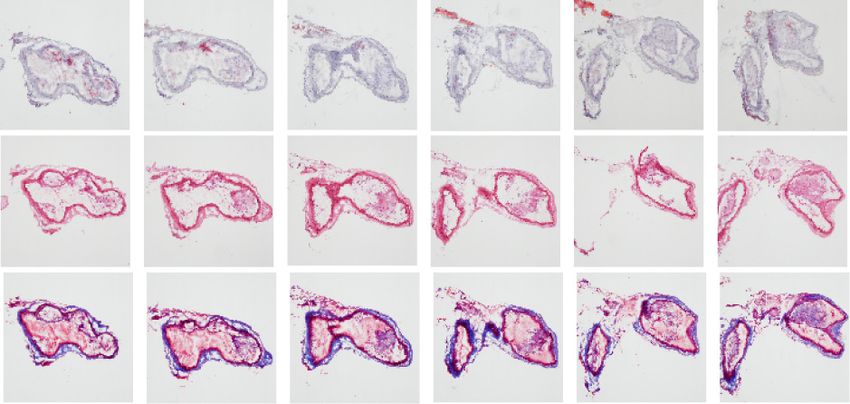
McArdle et al.: Intravital live cell triggered imaging system reveals monocyte patrolling. . .

                placed and tied securely. The left jugular vein was cleaned of fat,
                and a cannula (PE10 with a 28G needle) was inserted and tied.
                For extra security, the jugular vein cannula was secured with
                tissue glue. Supplemental anesthetic was applied at 20- to
                30-min intervals via the jugular vein catheter as needed. The
                ligament of the right sternomastoideus muscle was cut and the
                proximal end of the muscle was pulled away from the right
                carotid artery and secured with a tissue clamp. Extraneous tissue
                around the EC artery was removed without touching the thin
                fascia covering the artery.

                2.4     Multiphoton Microscopy
                All imaging experiments were conducted on a Leica SP5 with
                a water-dipping objective (Olympus XLUMPLFL 20X NA
                0.95). The system is composed of a DM6000 microscope,
                a Ti-Sapphire laser (Chameleon Ultra II, Coherent), tuned to
                920 nm for all experiments, a resonant scanhead for fast scan-
                ning, and a Leica trigger box. The emitted light was split into
                three photomultiplier tube detectors by two dichroic mirrors
                (520 and 495 nm) and three filters (513∕17 nm for GFP,
                513∕22 nm for YFP, and 460/50 for collagen visualized by
                SHG).
                    Acquired images cover 512 pixels in x (the vessel axis) and
                200 to 256 pixels in y, depending on heart rate. All images used
                3-6x line scan averaging, for a total of 35 to 70 ms∕frame. The
                xy pixels were isotropic at 890 nm∕pixel. We chose to acquire
                a large field of view at the expense of maximum spatial
                resolution.
                    During the imaging, the imaged area was kept wet by with a
                slow drip (5 to 10 mL∕h) of superfusion fluid [Roswell Park
                Memorial Institute medium (RPMI) without phenol red] from
                a syringe pump [Fig. 1(a)]. The mouse was warmed to 37°C
                with a heating pad on a feedback loop while an objective heater
                (OW-2D, Warner) heats both the objective and the superfusion
                fluid [Fig. 1(a)]. Preliminary experiments showed that injectable
                anesthesia resulted in a reduction of the heart rate to ∼350 bpm,
                but also caused low tissue oxygen saturation and arrhythmias as
                measured by the pulse oximeter. Reducing the heart rate with                       Fig. 1 (a) Schematic diagram of intravital live cell triggered imaging
                anesthesia is desirable because it lengthens the diastolic phase,                  system (ILTIS) setup. The mouse is placed supine on a heating pad
                allowing for more time to acquire a frame. Oxygen saturation                       on the microscope stage, and is breathing freely with additional oxy-
                improved to 98% and arrhythmias were reduced when the mice                         gen provided. A syringe pump pumps RPMI into the space between
                were given supplemental oxygen next to the cannulated trachea.                     the carotid artery and the objective and both the liquid and the objec-
                                                                                                   tive are kept warm with an objective heater. The mouse’s heartbeat is
                                                                                                   monitored by a pulse oximeter on the mouse’s thigh, which is con-
                2.5     Triggered Imaging                                                          nected through the Arduino microcontroller circuit to the scanhead
                                                                                                   of the two-photon microscope. The position of the objective is con-
                In order to achieve triggered acquisition, the mouse heartbeat                     trolled by a piezo-actuator, which is in communication with MATLAB
                needs to “drive” the microscope. Figure 1 describes the setup                      and the heartbeat detection circuit. (b) Illustration of triggered image
                and workflow for achieving triggered acquisition. The trigger                      acquisition in response to the heartbeat, showing representative out-
                pulse is derived from a pulse oximeter (MouseOx, Starr Life                        put from the pulse oximeter (black line), detection of a heartbeat when
                                                                                                   pulse oximeter signal become negative (dot), adjustable delay time
                Sciences) placed on the thigh of the mouse [Fig. 1(a)], using                      after heartbeat detection (labeled δ, thin line), representation of trigger
                the output that reflects oxygenated hemoglobin [Fig. 1(b)].                        pulse sent to Leica trigger box (dotted line), and ∼50 ms image
                The analog pulse wave signal is conditioned in a simple cus-                       acquisition time during diastole (black bars above pulse oximeter
                tom-built heartbeat detection circuit, utilizing an Arduino micro-                 graph). (c) Control diagram of triggering system. Arrows show direc-
                controller (Duemilanove, Smartprojects) [Fig. 1(a)]. Systole is                    tion of information flow. The mouse’s heartbeat is monitored by the
                detected when the pulse oximeter output goes from positive                         pulse oximeter (1), which outputs an analog signal (2). The waveform
                                                                                                   is digitized (3) and sent to an Arduino microcontroller, which after an
                to negative [Fig. 1(b)]. Since the heartbeat signal is acquired
                                                                                                   adjustable delay (4) outputs a trigger pulse (5) to the microscope to
                at a different location from the imaged area, an adjustable                        acquire an image (6). This repeats three times (7), and then the
                delay circuit is implemented to ensure that image acquisition                      Arduino suppresses all trigger signals for 1 s (8) to reduce photo-
                proceeds during effective diastole [Figs. 1(b) and 1(c)]. The                      bleaching. For three-dimensional (3-D) imaging, the Arduino also sig-
                delay (typically 0 to 30 ms) is adjusted manually until the                        nals through MATLAB to the piezo-actuator (9) to change the focal
                apparent motion between frames is minimal. After the delay,                        plane (10). Red lines (2,4)—analog signals, blue lines (3,5)—digital
                the microcontroller produces a trigger pulse that is fed into                      signals, and green lines (9)—serial communication.

                Journal of Biomedical Optics                                               026005-3                                             February 2015    •   Vol. 20(2)

Downloaded From: https://www.spiedigitallibrary.org/journals/Journal-of-Biomedical-Optics on 30 Oct 2021
Terms of Use: https://www.spiedigitallibrary.org/terms-of-use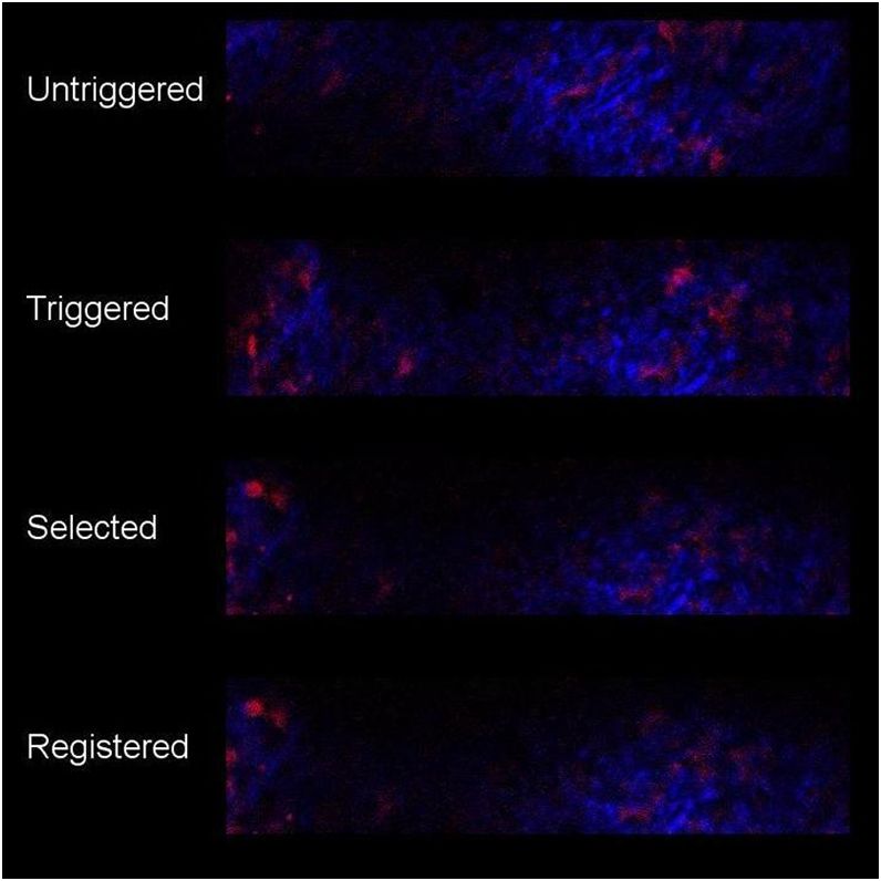
McArdle et al.: Intravital live cell triggered imaging system reveals monocyte patrolling. . .

                the microscope scanhead using a Leica trigger box and initiates                         image at every location. Minimization of the above cost function
                acquisition of a single frame [Fig. 1(b)]. The process is then                          happens to be a quadratic maximization problem, which, in
                repeated at the next heartbeat.                                                         general, is nondeterministic polynomial-time hard.21 Thus,
                    Three (occasionally four) images in consecutive heartbeats                          even with only pairwise similarity, the image selection problem
                were acquired, and then a short pause was inserted to reduce                            seems notoriously hard to solve. Our rescue is to approximate
                photobleaching [Figs. 1(b) and 1(c)]. In postprocessing, one                            the cost and cast it to an optimization for which practical and fast
                out of each triplet of frames was automatically chosen to com-                          algorithms exist.
                pile the final movie (see below).19 For 2-D movies, a 1 s pause                            We make an approximation of the image selection problem
                was inserted. This time was chosen to be fast enough (average                           by selecting the images in stages: suppose all the images in
                final rate of 0.66 Hz) to accurately track cells between frames                         j’th Z-stack have been selected, we proceed to select images
                but still allow for 30 to 45 min movies with minimal photo-                             in the ðj þ 1Þ’th Z-stack by minimizing the following cost
                bleaching. For 3-D movies, in between each triplet of frames,                           function:
                the Arduino microcontroller sent a signal through MATLAB
                to the nosepiece piezo-controller (Piezosystem Jena, NV 40/1                                   X X
                                                                                                                 K X
                                                                                                                   K
                CLE and MIPOS 500 SG) on the microscope objective to                                     min                       ai;jþ1;n aiþ1;jþ1;m ð0.5kI i;j − I i;jþ1;m k
                change the imaging focal plane [Figs. 1(a) and 1(c)]. The                                       i     n¼1    m¼1
                inserted pause in this case was only 300 ms (approximately 1                                 þ kI i;jþ1;m − I iþ1;jþ1;n k þ 0.5kI iþ1;j − I iþ1;jþ1;n kÞ;
                heartbeat). Stacks of 25 to 40 Z slices were acquired, with a
                final frame rate of approximately one stack every 42 s. After
                one out of each triplet of frames was selected, image registration                      where I i;j is the selected image at the (i; j) location. The
                was used to reduce residual motion. These postprocessing algo-                          above cost function has a one-dimensional grid structure,
                rithms effectively remove artifacts due to respiration along with                       as opposed to 2-D and dynamic programming (DP) can be
                those from other unknown sources.                                                       applied here with a polynomial complexity of OðMK 2 Þ to
                                                                                                        find out the global minimum.22 Because we have to run
                                                                                                        DP for each of N Z-stacks, the total computational complexity
                                                                                                        is OðMNK 2 Þ.
                2.6     Image Selection Method in 3-D Experiments
                The image selection problem is defined as follows. If there are                         2.7     Image Registration After Frame Selection
                M 2-D images in each Z-stack (spatial direction) and N Z-stacks
                (time direction) in total, with K images acquired at every space-                       One widely used method for the registration of temporal image
                time location, the exhaustive search space to select one image                          sequence is template matching, such as the ImageJ plug-in
                per location is enormous: K MN . To reduce this computational                           StackReg.23 Our recent study shows that when the image
                complexity, it is natural to impose similarity among images                             sequence contains periods of short subsequences at varying
                acquired at neighboring locations in the space-time grid, much                          focal planes, template matching results in inferior performances
                like Markov random field type modeling in image analysis.20                             with large registration errors.19 Periods with drift in the focal
                Thus, while selecting an image at i’th spatial location and                             plane exist in our experiments, due in part to the large number
                j’th time point, we would like to impose its similarity with                            of frames in the high-temporal resolution 2-D movies. Thus,
                images acquired at ði þ 1; jÞ’th, ði − 1; jÞ’th, ði; j þ 1Þ’th, and                     the template matching methods are unsuitable for this application.
                ði; j − 1Þ’th locations. To formulate this pairwise similarity                          To remedy this, we devised a novel image sequence method based
                within a sound mathematical framework, consider I i;j;n to                              on minimum spanning trees (MSTs) algorithm.19 The underlying
                be the n’th of the K images acquired at location ði; jÞ. The                            graph here consists of N nodes corresponding to the image frames
                image selection problem is then to select one out of K images                           selected at N time points. Two nodes are connected in this graph
                at every ði; jÞ location in the space-time grid. The aforemen-                          only if they are δ-distance apart along the time scale. δ is a design
                tioned image selection problem with pairwise similarity on                              parameter, the value of which has been experimentally deter-
                the 2-D space-time grid can now be formulated as a minimiza-                            mined as in our earlier study.19 The weight of the edge between
                tion of the following cost function with respect to binary vari-                        two graph nodes I i and I j is defined as the registration error:
                ables ai;j;n :                                                                          kI i − RðI i ; I j Þk þ kI j − RðI j ; I i Þk. Here, RðI i ; I j Þ represents a
                                                                                                        registration algorithm (e.g., rigid body registration) that registers
                                 X                  X
                                                    K X
                                                      K                                                 image I j to image I i and returns the registered image I j . The
                 min                                             ai;j;n ak;l;m kI i;j;n − I k;l;m k;    weighted MSTs algorithm24 determines an MST that can bypass
                       ði;jÞ;ðk;lÞ∶ji−kjþjj−lj¼1    n¼1    m¼1                                          the image frames with poor image quality and contain registration
                                                                                                        errors in a long image sequence.19
                                                                                                            This method was extended to 3-D movies. First, each Z-
                such that                                                                               stack is registered individually by our MST algorithm. Then,
                                                                                                        Z projections of these Z-stacks are computed to form a tempo-
                 X
                 K                                                                                      ral 2-D image sequence. The MST algorithm is now applied
                       ai;j;n ¼ 1; ∀ ði; jÞ; and ai;j;n ∈ f0;1g; ∀ ði; j; nÞ:                           to this 2-D temporal image sequence to compute the final
                 n¼1                                                                                    registration.
                                                                                                            Movie quality was quantified using the structural similarity
                    The norm k k measures the sum of absolute value of pixel                            (SSIM) score.25 A movie was acquired without the trigger,
                difference between two images. The binary variable encoding                             and then a second movie was acquired with the trigger in the
                here implies that image I i;j;n is selected only when ai;j;n ¼ 1.                       same location. The triggered movie was then processed using
                The equality constraint enforces selection of exactly one                               the image selection and registration algorithms. Two hundred

                Journal of Biomedical Optics                                                      026005-4                                             February 2015     •   Vol. 20(2)

Downloaded From: https://www.spiedigitallibrary.org/journals/Journal-of-Biomedical-Optics on 30 Oct 2021
Terms of Use: https://www.spiedigitallibrary.org/terms-of-use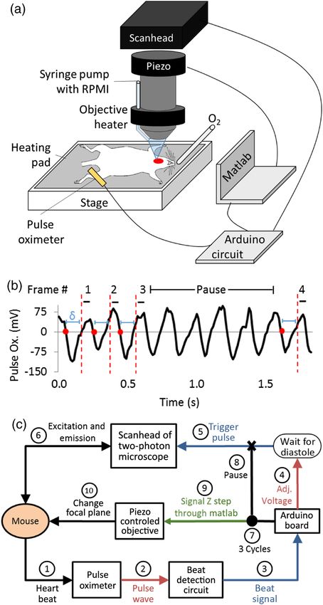
McArdle et al.: Intravital live cell triggered imaging system reveals monocyte patrolling. . .

                twenty frames from each image series (untriggered, triggered,                      3       Results
                triggered with image selection, triggered with image selection
                and registration) were selected. The SSIM score was calculated                     3.1      Plaque Develops Reliably in the External
                in MATLAB (Mathworks) using the m-file written by Wang                                      Carotid Artery
                et al.,25 for each sequential pair of images in each movie
                                                                                                   The EC artery, specifically near the branch points of the ST and
                (between frames 1 and 2, 2 and 3, etc.) to determine how differ-
                                                                                                   AP arteries, reliably develops plaque [Fig. 2(a)]. Serial section-
                ent each frame is from the previous one in each stage.
                                                                                                   ing revealed that plaque is present throughout all branches
                                                                                                   of this artery (including ST and AP), though with varying com-
                                                                                                   positions in different locations. We analyzed serial sections
                2.8     Cell Motion Analysis                                                       obtained at 30 μm intervals from an Apoe−/− mouse fed WD
                After image selection and registration, cells were tracked in                      9 weeks. Immunofluorescence revealed CD11b+, CD11c+,
                Imaris (Bitplane). In 2-D movies containing migrating mono-                        and double positive cells as expected [Fig. 2(f)].26 αSMA stain-
                cytes, small round cells were detected using an absolute inten-                    ing revealed the medial layers and CD31 stained the endothelial
                                                                                                   monolayer enclosing the lumen [Fig. 2(g)]. To visualize cellular
                sity threshold, and then the position of each cell’s centroid was
                                                                                                   composition by histology, we used H&E staining [Figs. 2(b) and
                tracked using Imaris’s autoregressive motion algorithm. Broken
                                                                                                   2(d)]. Neutral lipids were stained with Oil Red O [Figs. 2(b) and
                or inaccurate tracks were corrected manually. Only cells that
                                                                                                   2(c)] and collagen and elastin fibers were stained with trichrome
                showed visible net motion during the movie were tracked
                                                                                                   [Figs. 2(b) and 2(e)]. The classical histology staining methods
                to ensure the removal of GFP+ macrophages from analysis
                                                                                                   confirmed the presence of atherosclerotic lesions defined as
                (the slowest average velocity detected was 10 μm∕ min). Cells
                                                                                                   accumulation of lipids, infiltration of immune cells, and thick-
                were tracked for the length of the entire movie unless they left                   ening of the artery wall.
                the field-of-view or the focal plane. Occasionally, additional
                registration was performed in Imaris to remove sharp changes
                in the cell tracks. In 3-D movies, round macrophages were                          3.2      Cardiac Triggering and Image Postprocessing
                tracked automatically using Imaris’ surface algorithm, while                                Improves Movie Quality
                dendritic-shaped cells were analyzed manually using Imaris’                        The cardiac triggered imaging system was first tested using
                filament tracer algorithm.                                                         Apoe−/− Cd11cYFP mice, which have fluorescent myeloid cells

                                                                (a)                          ST        AP                (c)    Oil Red O

                                                                      CC
                                                                                        EC

                                                                                 IC

                                        (b)

                                        Oil Red O                                                                        (d)       H&E

                                          H&E

                                       Trichrome

                                                          (f)               CD11b            (g)              αSMA       (e)    Trichrome
                                                                             CD11c                            CD31
                                                                            Double

                                                                                                             200 µm                      200 µm

                                       Fig. 2 (a) Plaque is visible (opaque white) in the external carotid (EC) artery and superior thyroid artery
                                       branch of an Apoe−/− mouse fed WD for 12 weeks. Plaques can also develop in the ascending pharyn-
                                       geal artery and lower EC branches. CC, common carotid artery; IC, internal carotid artery. (b) Serial
                                       sections every 60 μm from an Apoe−/− mouse fed WD 9 weeks were stained for neutral lipids (Oil
                                       Red O), cell components (H&E), and collagen and elastin fibers (Trichrome.) Scale bar ¼ 200 μm.
                                       Every other stained section is shown. Larger examples are shown for visibility (c-e). Serial sections
                                       were also immunostained for (f) CD11b and CD11c and (g) α-smooth muscle actin and CD31.
                                       Autofluorescence and nuclei (stained with Yoyo-1) appear in blue. Brightness was increased by 40%
                                       on all fluorescence images for visibility.

                Journal of Biomedical Optics                                               026005-5                                         February 2015   •   Vol. 20(2)

Downloaded From: https://www.spiedigitallibrary.org/journals/Journal-of-Biomedical-Optics on 30 Oct 2021
Terms of Use: https://www.spiedigitallibrary.org/terms-of-use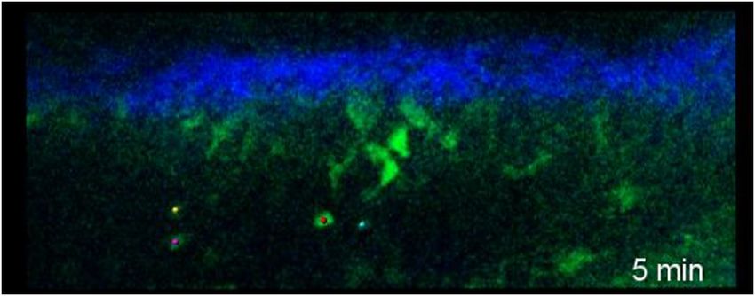
McArdle et al.: Intravital live cell triggered imaging system reveals monocyte patrolling. . .

                in their arterial walls.26 In addition to the YFP signal, collagen
                was visualized through SHG.27 Triggered acquisition greatly
                reduced motion artifacts compared to untriggered movies
                [Figs. 3(a) and 4]. However, even with cardiac triggering,
                residual motion of the artery was still visible. Movie quality
                was further improved by our automated frame-dropping algo-
                rithm. We acquired three frames in three consecutive heartbeats
                with a pause between sets of images. One “best” image from
                each set of frames was selected by minimizing a cost function
                based on the sum of absolute differences of pixel values between
                the images. Final image stabilization was accomplished by
                image registration correcting for translation and rotation
                (Fig. 4). The triggered acquisition, frame selection, and correc-
                tion for residual translation resulted in image series that were
                steady. Based on the SSIM score,25 the triggered acquisition pro-
                vided the most benefit, followed by the frame selection, with the
                final correction for translational movement adding further image
                stabilization [Fig. 3(c)].

                3.3     High Frame-Rate 2-D Imaging
                To investigate inflammatory cell recruitment from the lumen,
                high frame-rate 2-D imaging in a single plane is most suitable.12
                Monocytes are relevant to atherosclerosis and are known to roll
                in the carotid arteries of Apoe−/− mice in vivo and ex vivo.28–30
                Triggered multiphoton imaging of atherosclerotic live EC
                arteries of Apoe−/− mice resulted in movies with a frame rate
                of 0.66 Hz that were up to 45-min long. We imaged Apoe−/−
                Cx3cr1GFP/+ mice that express GFP on various cell types, includ-
                ing blood monocytes and tissue macrophages, as well as small
                subsets of other immune cells.16,18 In seven out of eight imaged
                mice, we found patrolling31 monocytes on the luminal side of
                the vessel. Figure 5(a) shows example frames acquired from
                an Apoe−/−Cx3cr1GFP/+ mouse with four patrolling monocytes,
                three moving with the direction of flow and one moving against
                the flow [Figs. 5(b) and 6]. Cells flowing in the blood adjacent
                to the tracked cells are visible, suggesting that the monocytes are
                on the endothelial surface. This patrolling behavior has previ-
                ously been described in microvessels31,32 but not large arteries.

                3.4     3-D Multicolor Imaging
                Macrophages are a common cell type in atherosclerotic plaque
                in Apoe−/− mice.26,33 To demonstrate 3-D imaging of these
                cells within atherosclerotic plaques, we generated Apoe−/
                −
                 Cx3cr1GFP/+Cd11cYFP mice. In the plaque, these mice have
                fluorescent subsets of macrophages and dendritic cells. Z-stacks
                were acquired covering one quarter of the circumference of the
                artery, imaging up to 200 μm deep [Fig. 7]. In one representative                  Fig. 3 (a, b) Example images showing the effect of triggered image
                example, stacks were collected in 42-s intervals, producing a                      acquisition on data quality. (a) Three sequential images freely
                four-dimensional representation of fluorescently labeled macro-                    acquired from an Apoe−/− Cd11cYFP mouse. (b) Three sequential
                                                                                                   images acquired using the trigger as described. Red—YFP+ cells
                phages moving within the plaque [Fig. 8]. The quality of 3-D                       and blue—collagen visualized through SHG. See Video 1 for corre-
                movies acquired from live mice [Fig. 9(a)] was comparable to                       sponding movies. (c) Pairwise structural similarity scores for image
                those previously obtained from explanted aortas.26 The visual-                     sequences taken from the same location of an Apoe−/− Cd11cYFP
                ized cells are interior to the collagen, but no blood flow is visible              mouse and processed in various ways. The image sets that were
                in the same focal plane, suggesting that these cells are within the                analyzed were: free, untriggered acquisition, triggered acquisition,
                intimal plaque. Some YFP+ fluorescent cells migrated within                        triggered images with the image selection algorithm described in
                                                                                                   the text, and triggered, selected images with additional registration.
                the plaque [Fig. 9(b)] while other GFP+ or GFP+YFP+ den-
                                                                                                   Data shows mean (standard deviation).
                dritic-shaped cells exhibited “dancing on the spot”-type behav-
                ior with extensions and contractions of cell processes and little
                centroid motion [Fig. 9(c)].

                Journal of Biomedical Optics                                               026005-6                                          February 2015   •   Vol. 20(2)

Downloaded From: https://www.spiedigitallibrary.org/journals/Journal-of-Biomedical-Optics on 30 Oct 2021
Terms of Use: https://www.spiedigitallibrary.org/terms-of-use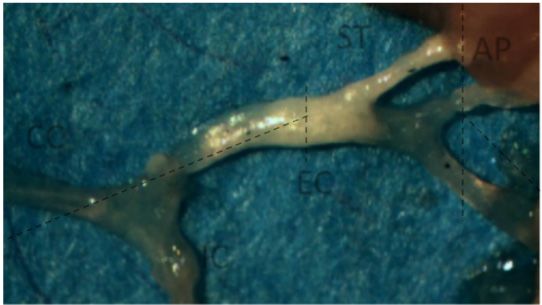
McArdle et al.: Intravital live cell triggered imaging system reveals monocyte patrolling. . .

                                                                                                             (a)

                                                                                                                         0 min

                                                                                                                                          Blood flow

                                                                                                                         3 min

                                                                                                                         6 min

                Fig. 4 Example movies taken from an Apoe−/− Cd11cYFP mouse. The
                first (untriggered) was freely acquired without use of the triggering
                system. Frames were acquired at 24.3 Hz, and are played at real
                time. The second (triggered) was acquired in the same location, using                                    9 min
                the triggering system described in Fig. 1. Acquisition rate matched the
                mouse’s heart rate at approximately 3.7 Hz, but is played sped up
                by 6.7×. The third (selected) is the result of using the image selection
                algorithm on the triggered movies, and is played at the same frame                                (b)
                rate as the triggered movie. The last (registered) is the result of using
                                                                                                           Circ. position (µ m)
                                                                                                                                    0
                the minimum spanning tree (MST) registration on the frame-selected
                movie, and is played at the same frame rate. (Video 1, MPEG,
                4.85 MB) [URL: http://dx.doi.org/10.1117/1.JBO.20.2.026005.1].                                                     50

                4     Discussion                                                                                                  100
                                                                                                                                    150                100              50                0
                The ILTIS system developed here successfully images leuko-                                                                             Axial position (µ m)
                cytes in the lumen and atherosclerotic plaques of arteries in
                live mice. Combining a cardiac trigger and image postprocess-                      Fig. 5 (a) Time series of monocytes moving along the lumen of the
                ing allows for high spatial- and time-resolution imaging of the                    EC artery of a live Cx3cr1+/GFP Apoe−/− mouse fed WD for 4 months.
                beating EC artery and its branches without risking tissue dam-                     Images were acquired at approximately 2 Hz as depicted in Fig. 1 and
                                                                                                   then selected and registered as described in the text. Four crawling
                age from mechanical stabilization or altering the artery’s native                  cells were tracked. Blood flow is left to right. Green—GFP+ mono-
                movement. Importantly, we were able to keep the thin fascia                        cytes and macrophages and blue—collagen in the adventitia visual-
                overlying this artery in place, thus protecting the vessel wall                    ized through SHG. (b) The positions of the four tracked cells over
                from surgical trauma. Squeezing the artery has been shown                          the 9 min movie. The dot shows the starting position of each cell,
                to cause cell death and matrix damage,11 which can be avoided                      color coded by the box they are marked with.
                by triggered acquisition without mechanical stabilization. The
                resolution with ILTIS allowed not only the tracking of cell
                movement but also detailed analysis of cell shapes over time.
                A large volume of tissue can be imaged (up to 200 μm deep)
                with minimal registration artifacts. Additionally, this technique
                is reproducible and can reliably produce stable movies. Simple
                custom circuitry was built, but no highly specialized equipment
                is needed.
                    ILTIS has two modes of operations, 2-D and 3-D, useful for
                studying different processes. The high time-resolution 2-D im-
                aging is ideal for studying fast cells on the endothelial surface.
                The number of frames per set and delays between sets can be
                optimized. Optimal time resolution can be achieved by acquir-
                ing one frame per heartbeat, resulting in 4 to 6 frames∕s, though
                this comes at a cost of photobleaching. Alternatively, photo-                      Fig. 6 Single Z plane movie of the EC artery of a live Cx3cr1+/GFP
                bleaching can be minimized by imaging less frequently.                             Apoe−/− mouse fed WD for 4 months. Images were acquired at
                Here, we used a net frame rate of 0.66 Hz, which can allow                         approximately 2 Hz (three triggered frames in 600 ms, followed by
                                                                                                   1 s pause). Image selection and registration were applied to minimize
                for up to 30 min of useful imaging time without detectable                         movement artifacts, reducing the frame rate to 0.66 Hz. Colored dot
                photobleaching. Using Apoe−/− Cx3cr1GFP/+ mice, we used 2-                         shows tracked cell centroid. Green—GFP+ cells and blue—collagen
                D imaging to demonstrate that monocytes actively crawl both                        visualized by SHG. (Video 2, MPEG, 2.34 MB) [URL: http://dx.doi.org/
                with and against blood flow in large, atherosclerotic arteries.                    10.1117/1.JBO.20.2.026005.2].

                Journal of Biomedical Optics                                               026005-7                                                                 February 2015   •   Vol. 20(2)

Downloaded From: https://www.spiedigitallibrary.org/journals/Journal-of-Biomedical-Optics on 30 Oct 2021
Terms of Use: https://www.spiedigitallibrary.org/terms-of-use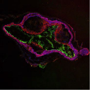
McArdle et al.: Intravital live cell triggered imaging system reveals monocyte patrolling. . .
Fig. 8 Maximum intensity Z projections of the 3-D movie of live
Cx3cr1+/GFP Cd11cYFP Apoe−/− mouse fed WD for 5 months.
Images were collected using triggered acquisition, and then image
selection and registration were applied for stabilization. Stacks
were acquired 42 s apart, video played at 15 Hz. Green—GFP+
cells; orange—YFP+ cells; and blue—collagen visualized by SHG.
Fig. 7 Typical size and location of imaged volume (yellow box), which (Video 3, MPEG, 1.41 MB) [URL: http://dx.doi.org/10.1117/1.JBO
captures 1∕4 of the circumference of the arterial wall. .20.2.026005.3].
pause after systole is detected before acquiring a frame, the
Three-dimensional imaging is suitable for imaging cells number of line scan averages, the number of lines scanned in
within the thick intimal plaque, which move slower but in all the Y-direction, the number of replicate frames taken in each
three directions. We used ILTIS to show that intravascular Z slice or between pauses, and the length of the pause. Our
YFP+ macrophages were able to migrate within the plaque goal was to maximize the signal-to-noise ratio and the size
and GFP+ and GFP+YFP+ cells “danced on the spot,” where of the field of view, while ensuring that all frames were entirely
there was motion of the cell’s dendrites but little net centroid taken during effective diastole. We found that our acquisition
motion. This behavior has previously been reported in aortic parameters were best suited for mice with a heart rate of 300
explants.26 Our work shows, for the first time, that this type to 350 bpm.
of cell movement occurs in atherosclerotic plaques in vivo. Intravital imaging of large arteries is an invaluable tool for
For all imaging, rectangular fields-of-view were chosen to studying leukocyte activity in atherosclerosis. ILTIS will be
minimize acquisition time of each frame, and the X-axis helpful for answering pertinent open questions in the field,
(scan direction) was aligned with the direction of blood flow. including monocyte recruitment and transmigration, macro-
There are multiple settings that can be optimized for ideal phage and dendritic cell activity, and motion of other immune
speed, spatial resolution, and image stability, including the cells.
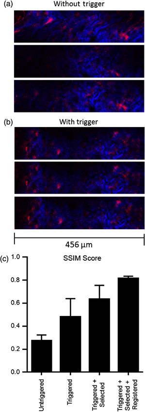
Fig. 9 (a) Maximum intensity Z projection of a single time point of the 3-D movie of the EC artery of a
live Cx3cr1+/GFP Cd11cYFP Apoe−/− mouse fed WD for 5 months with representative moving cells high-
lighted (boxes). Scale bar ¼ 100 μm. Green—GFP; orange—YFP; and blue—collagen visualized by
SHG. See Video 3 for corresponding time lapse recording. (b) Shape and location of an YFP+ cell
in orange box in (a) was tracked in Imaris over 42 min, color coded by time. Cell size is decreased
to enable visualization of cell shape at all time points. (c) Shape changes of two intertwining
dendritic-shaped GFP+ (green) and GFP+YFP+ (yellow) cells in green box in (a) was tracked in
Imaris over time.
Journal of Biomedical Optics 026005-8 February 2015 • Vol. 20(2)
Downloaded From: https://www.spiedigitallibrary.org/journals/Journal-of-Biomedical-Optics on 30 Oct 2021
Terms of Use: https://www.spiedigitallibrary.org/terms-of-use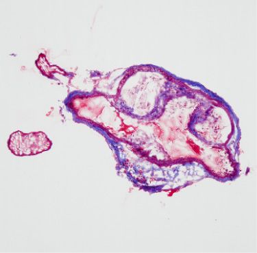
McArdle et al.: Intravital live cell triggered imaging system reveals monocyte patrolling. . .

                Acknowledgments                                                                    23. P. Thevenaz, U. E. Ruttimann, and M. Unser, “A pyramid approach to
                                                                                                       subpixel registration based on intensity,” IEEE Trans. Image Process.
                We thank M. Chadwell (La Jolla Institute) for expert sectioning                        7(1), 27–41 (1998).
                and histological staining. We thank H. Ouyang and J. Miller                        24. T. H. Cormen et al., Introduction to Algorithms, 2nd ed., MIT Press and
                (La Jolla Institute) for their outstanding technical help in main-                     McGraw-Hill, Cambridge, Massachusetts (2001).
                taining the mouse colony. This work was funded by NIH                              25. Z. Wang et al., “Image quality assessment: from error visibility to
                R01 (115232) to K.L. and AHA (#11PRE7580009), HHMI                                     structural similarity,” IEEE Trans. Image Process. 13(4), 600–612
                                                                                                       (2004).
                (56005681), and NHLBI (T32HL105373-03) to S.M.
                                                                                                   26. E. K. Koltsova et al., “Dynamic T cell-APC interactions sustain chronic
                                                                                                       inflammation in atherosclerosis,” J. Clin. Invest. 122(9), 3114–3126
                                                                                                       (2012).
                References                                                                         27. W. R. Zipfel et al., “Live tissue intrinsic emission microscopy using
                 1. H. Ait-Oufella et al., “Adaptive (T and B cells) immunity and control by           multiphoton-excited native fluorescence and second harmonic genera-
                    dendritic cells in atherosclerosis,” Circ. Res. 114(10), 1640–1660                 tion,” Proc. Natl. Acad. Sci. U. S. A. 100(12), 7075–7080 (2003).
                    (2014).                                                                        28. Y. Huo et al., “The chemokine KC, but not monocyte chemoattractant
                 2. C. Vinegoni et al., “Advanced motion compensation methods for intra-               protein-1, triggers monocyte arrest on early atherosclerotic endo-
                    vital optical microscopy,” IEEE J. Sel. Topics Quantum Electron. 20(2),            thelium,” J. Clin. Invest. 108(9), 1307–1314 (2001).
                    1–9 (2014).                                                                    29. Y. Huo, A. Hafezi-Moghadam, and K. Ley, “Role of vascular cell adhe-
                 3. S. Lee et al., “Real-time in vivo imaging of the beating mouse heart at            sion molecule-1 and fibronectin connecting segment-1 in monocyte
                    microscopic resolution,” Nat. Commun. 3, 1054 (2012).                              rolling and adhesion on early atherosclerotic lesions,” Circ. Res.
                 4. C. Vinegoni et al., “Sequential average segmented microscopy for high              87(2), 153–159 (2000).
                    signal-to-noise ratio motion-artifact-free in vivo heart imaging,”             30. Y. Huo et al., “Circulating activated platelets exacerbate atherosclerosis
                    Biomed. Opt. Express 4(10), 2095–2106 (2013).                                      in mice deficient in apolipoprotein E,” Nat. Med. 9(1), 61–67 (2003).
                 5. W. Li et al., “Intravital 2-photon imaging of leukocyte trafficking in         31. C. Auffray et al., “Monitoring of blood vessels and tissues by a pop-
                    beating heart,” J. Clin. Invest. 122(7), 2499–2508 (2012).                         ulation of monocytes with patrolling behavior,” Science 317(5838),
                 6. S. Lee et al., “Improved intravital microscopy via synchronization of              666–670 (2007).
                    respiration and holder stabilization,” J. Biomed. Opt. 17(9), 096018           32. L. M. Carlin et al., “Nr4a1-dependent Ly6C(low) monocytes monitor
                    (2012).                                                                            endothelial cells and orchestrate their disposal,” Cell 153(2), 362–375
                 7. R. Chevre et al., “High-resolution imaging of intravascular atherogenic            (2013).
                    inflammation in live mice,” Circ. Res. 114(5), 770–779 (2014).                 33. E. Galkina et al., “Lymphocyte recruitment into the aortic wall before
                 8. K. Jung et al., “Endoscopic time-lapse imaging of immune cells in                  and during development of atherosclerosis is partially L-selectin depen-
                    infracted mouse hearts,” Circ. Res. 112(6), 891–899 (2013).                        dent,” J. Exp. Med. 203(5), 1273–1282 (2006).
                 9. M. R. Looney et al., “Stabilized imaging of immune surveillance in the
                    mouse lung,” Nat. Methods 8(1), 91–96 (2011).                                  Sara McArdle received a BS degree in biomedical engineering from
                10. C. Vinegoni et al., “Motion compensation using a suctioning stabilizer         Columbia University in 2009. She is currently a PhD candidate at
                    for intravital microscopy,” Intravital 1(2), 115–121 (2012).                   the University of California, San Diego, in the Department of Bioen-
                11. R. T. Megens et al., “In vivo high-resolution structural imaging of large      gineering, with a specialization in multiscale biology. She is complet-
                    arteries in small rodents using two-photon laser scanning microscopy,”         ing her PhD research at the La Jolla Institute for Allergy and
                    J. Biomed. Opt. 15(1), 011108 (2010).                                          Immunology in the Division of Inflammation Biology with Dr. Klaus
                12. M. Drechsler et al., “Hyperlipidemia-triggered neutrophilia promotes           Ley.
                    early atherosclerosis,” Circulation 122(18), 1837–1845 (2010).
                13. D. Soulet et al., “Automated filtering of intrinsic movement artifacts         Grzegorz Chodaczek is a head of the Confocal Microscopy Labora-
                    during two-photon intravital microscopy,” PLoS One 8(1), e53942                tory at the Wroclaw Research Centre EIT+, Wroclaw, Poland. He
                    (2013).                                                                        received his PhD degree in immunology from the Institute of Immu-
                14. M. Phillipson et al., “Intraluminal crawling of neutrophils to emigration      nology and Experimental Therapy in Poland. Between 2007 and
                    sites: a molecularly distinct process from adhesion in the recruitment         2011, he was a postdoctoral fellow at MD Anderson Cancer Center
                    cascade,” J. Exp. Med. 203(12), 2569–2575 (2006).                              at Houston, Texas, USA, and until March 2014, he was a microscopy
                                                                                                   core manager at the La Jolla Institute. His research focuses on intra-
                15. Y. Nakashima et al., “ApoE-deficient mice develop lesions of all phases
                                                                                                   vital imaging of immunological processes.
                    of atherosclerosis throughout the arterial tree,” Arterioscler. Thromb.
                    14(1), 133–140 (1994).                                                         Nilanjan Ray received his B.M. Engg. from Jadavpur University,
                16. S. Jung et al., “Analysis of fractalkine receptor CX(3)CR1 function by         India, in 1995, his MTech degree from CS Indian Statistical Institute,
                    targeted deletion and green fluorescent protein reporter gene insertion,”      Calcutta, India, in 1997, and his PhD degree in E. Engg. from the Uni-
                    Mol. Cell. Biol. 20(11), 4106–4114 (2000).                                     versity of Virginia in 2003. He is an associate professor in the Depart-
                17. R. L. Lindquist et al., “Visualizing dendritic cell networks in vivo,” Nat.    ment of Computing Science, University of Alberta, Canada. His
                    Immunol. 5(12), 1243–1250 (2004).                                              research area is image and video analysis: image segmentation,
                18. L. Landsman et al., “Cx3cr1 is required for monocyte homeostasis and           registration, tracking, and motion analysis. He has coauthored two
                    atherogenesis by promoting cell survival,” Blood 113(4), 963–972               monographs—Biomedical Image Analysis: Tracking and Biomedical
                    (2009).                                                                        Image Analysis: Segmentation, Morgan & Claypool Publishers.
                19. S. McArdle et al., “Registering sequences of in vivo microscopy images
                    for cell tracking using dynamic programming and minimum spanning               Klaus Ley received his MD degree in 1982 followed by postdoctoral
                    trees,” in IEEE Int. Conf. on Image Processing, IEEE, Paris, France            training in Berlin and San Diego. In 1994, he became
                    (2014).                                                                        professor of biomedical engineering and later director of the Robert
                20. S. Z. Li, Markov Random Field Modeling in Image Analysis, Springer,            M. Berne Cardiovascular Research Center at the University
                    London, England (2009).                                                        of Virginia. In 2007, he became a professor and founding head of
                21. R. Horst and H. Tuy, Global Optimization Deterministic Approaches,             the Division of Inflammation Biology at the La Jolla Institute. He
                                                                                                   received the 2008 Bonazinga and the 2010 Malpighi awards.
                    Springer, Heidelberg, New York (1996).
                22. D. P. Bertsekas, Dynamic Programming and Optimal Control, 2nd ed.,
                    Athena Scientific, Belmont, Massachusetts (2000).

                Journal of Biomedical Optics                                                 026005-9                                            February 2015      •   Vol. 20(2)

Downloaded From: https://www.spiedigitallibrary.org/journals/Journal-of-Biomedical-Optics on 30 Oct 2021
Terms of Use: https://www.spiedigitallibrary.org/terms-of-use
You can also read