Peroxisomal Dysfunction in Neurological Diseases and Brain Aging - Frontiers
←
→
Page content transcription
If your browser does not render page correctly, please read the page content below
REVIEW
published: 10 March 2020
doi: 10.3389/fncel.2020.00044
Peroxisomal Dysfunction in
Neurological Diseases and Brain
Aging
Ndidi-Ese Uzor 1,2 , Louise D. McCullough 2,3,4 and Andrey S. Tsvetkov 1,2,4 *
1
Department of Neurobiology and Anatomy, University of Texas McGovern Medical School, Houston, TX, United States, 2 The
University of Texas Graduate School of Biomedical Sciences, Houston, TX, United States, 3 Department of Neurology,
University of Texas McGovern Medical School, Houston, TX, United States, 4 UTHealth Consortium on Aging, University of
Texas McGovern Medical School, Houston, TX, United States
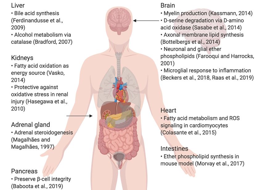
Peroxisomes exist in most cells, where they participate in lipid metabolism, as well as
scavenging the reactive oxygen species (ROS) that are produced as by-products of
their metabolic functions. In certain tissues such as the liver and kidneys, peroxisomes
have more specific roles, such as bile acid synthesis in the liver and steroidogenesis
in the adrenal glands. In the brain, peroxisomes are critically involved in creating and
maintaining the lipid content of cell membranes and the myelin sheath, highlighting
their importance in the central nervous system (CNS). This review summarizes the
peroxisomal lifecycle, then examines the literature that establishes a link between
peroxisomal dysfunction, cellular aging, and age-related disorders that affect the CNS.
This review also discusses the gap of knowledge in research on peroxisomes in the CNS.
Keywords: neuronal peroxisomes, peroxisome biogenesis disorders, aging peroxisomes, neurodegenerative
disease, peroxisomal dysfunction
INTRODUCTION
Edited by:
Chao Deng, Peroxisomes are small, nearly ubiquitous organelles found in almost all cell types, except mature red
University of Wollongong, Australia blood cells (Gronowicz et al., 1984). Their major functions include the beta-oxidation of very-long-
Reviewed by:
chain fatty acids and lipid peroxidation; as a result of this metabolism, they secrete reactive oxygen
Björn Spittau, species (ROS) as by-products (Reddy and Hashimoto, 2001; Poirier et al., 2006; Lodhi et al., 2015;
University Hospital Rostock, Germany Park et al., 2019). Peroxisomes also possess enzymes that break down ROS, such as catalase and
Lorenzo Di Cesare Mannelli, glutathione peroxidase, which breaks down hydrogen peroxide, and superoxide dismutase, which
University of Florence, Italy breaks down superoxide (Nordgren and Fransen, 2014). They degrade prostaglandins, amino acids,
*Correspondence: polyamines, and purines, and are commonly enriched in the kidneys, liver, pancreas and adrenal
Andrey S. Tsvetkov glands, which are involved in fat metabolism and detoxification (Magalhães and Magalhães, 1997;
andrey.s.tsvetkov@uth.tmc.edu Bradford, 2007; Ferdinandusse et al., 2009; Hasegawa et al., 2010; Smith and Aitchison, 2013;
Vasko, 2016; Baboota et al., 2019). Furthermore, they are implicated in lipogenic and ROS signaling
Received: 05 December 2019 roles in the heart and intestines (Colasante et al., 2015; Morvay et al., 2017). In the central nervous
Accepted: 18 February 2020
system (CNS) in particular, peroxisomes synthesize lipids that make up the myelin sheath and
Published: 10 March 2020
cellular membranes, as well as ether phospholipids in neurons and glia; peroxisome dysfunction
Citation: is also known to impair neuronal migration and membranes (Farooqui and Horrocks, 2001;
Uzor N-E, McCullough LD and
Powers, 2001; Bottelbergs et al., 2010; Kassmann, 2014). They also play a critical role in breaking
Tsvetkov AS (2020) Peroxisomal
Dysfunction in Neurological Diseases
down D-serine via D-amino acid oxidase (DAO), important in glutamatergic signaling (Sasabe
and Brain Aging. et al., 2014; Figure 1). Certain diseases, such as peroxisomal biogenesis disorders, underscore the
Front. Cell. Neurosci. 14:44. importance of functional peroxisomes in the CNS. Peroxisomal biogenesis disorders are a subset of
doi: 10.3389/fncel.2020.00044 diseases where: (1) peroxisomes are either not present, leading to severe neurological phenotypes
Frontiers in Cellular Neuroscience | www.frontiersin.org 1 March 2020 | Volume 14 | Article 44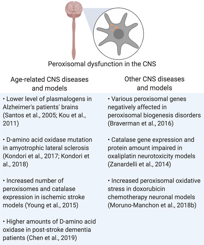
Uzor et al.                                                                                                                          Peroxisomal Dysfunction

  FIGURE 1 | Summary of specialized roles of peroxisomes in some organs, including the brain. Created using BioRender.

(as seen in neonatal adrenoleukodystrophy, where seizures,                          Jan et al., 2014). After this, peroxisomal proteins are inserted
hypotonia, and loss of vision and hearing occur) and a short                        into peroxisomal membranes and matrices by the peroxisomal
lifespan; or (2) genes coding for a single peroxisomal protein are                  protein Pex5 (Smith and Aitchison, 2013). Pex5 recognizes
defective, where the symptoms are not as severe (Fujiki et al.,                     the peroxisomal targeting sequence (PTS1) serine-lysine-leucine
2012; Aubourg et al., 2013). To conclude, peroxisomes are small,                    (SKL), which is found on the C-terminal of many peroxisomal
but important organelles that play supportive, yet critical roles in                proteins (Brocard and Hartig, 2006). After proteins are
maintaining cellular health, especially in the CNS.                                 inserted, peroxisomes are considered mature and functional. For
    This review summarizes peroxisomal biogenesis, and yeast                        peroxisomal maintenance, division and maturation, peroxisomes
and mammalian pexophagy, with an extended focus on                                  are known to make contact with the endoplasmic reticulum (Hua
peroxisomes in cellular senescence models, and the peroxisomal                      et al., 2017). To conclude, peroxisomal division and maintenance
dysfunction shared by both age-related neurodegenerative                            are modulated by the endoplasmic reticulum, and peroxisomes
diseases and peroxisomal biogenesis disorders. In all of these                      mature due to peroxisomal protein import into their matrices
conditions, functional peroxisomes move from understudied,                          and membranes.
secondary organelles to critical sustainers of cellular homeostasis
that are disrupted by disease. Future studies will elucidate the                    AUTOPHAGY AND PEXOPHAGY
role of peroxisomes in aging and CNS function in other diseases
and models.                                                                         The peroxisomal lifespan in mammalian cells lasts about
                                                                                    2 to 3 days (Poole et al., 1969; Huybrechts et al., 2009;
PEROXISOMAL BIOGENESIS                                                              Moruno-Manchon et al., 2018b). Peroxisomes are then degraded
                                                                                    by a selective form of macroautophagy: macropexophagy,
Peroxisomes begin their lifecycle by budding off the endoplasmic                    which specifically targets peroxisomes (Yang and Klionsky,
reticulum in response to peroxisome proliferator-activated                          2010; Bartoszewska et al., 2012; Cho et al., 2018). A lesser-
receptor (PPAR) activation due to signaling of the PPAR                             known form of pexophagy micropexophagy exists, but has
gamma coactivator-1α (PGC-1α) protein (Bagattin et al.,                             only been, so far, observed in yeast models (Strømhaug
2010). Unlike mitochondrial proteins, peroxisomal proteins are                      et al., 2001; Mukaiyama et al., 2004). In macroautophagy,
synthesized on free ribosomes in the cytosol (Koehler, 2000;                        targets for degradation are recognized by a phagophore,

Frontiers in Cellular Neuroscience | www.frontiersin.org                        2                                        March 2020 | Volume 14 | Article 44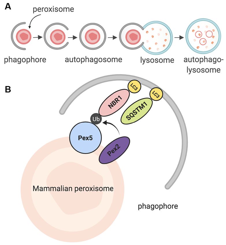
Uzor et al.                                                                                                                          Peroxisomal Dysfunction

  FIGURE 2 | Summary of pexophagy. (A) In macropexophagy, a form of
  macroautophagy selective for peroxisomes, a single membrane known as a
  phagophore engulfs a peroxisome for degradation. The phagophore matures        FIGURE 3 | Summary of peroxisomal dysfunction in age-related diseases in
  into an autophagosome, which then fuses with a lysosome. Their fusion          the CNS, and other diseases that affect the CNS. Created using BioRender.
  creates an autophagolysosome that degrades the target. (B) In mammalian
  systems, pexophagy occurs when Pex2 ubiquitinates Pex5. As a result,
  autophagy adaptor proteins NBR1 or SQSTM1 (p62) bind to ubiquitinated
  Pex5, and then eventually bind to LC3 on the phagophore. Due to this          (Lefevre et al., 2013; Williams and van der Klei, 2013; Kao
  process, pexophagy occurs. Created using BioRender.                           and Bartel, 2015; Young and Bartel, 2016). The Saccharomyces
                                                                                cerevisiae yeast homolog of PEX5, Pex5, recognizes cytosolic
which matures to form an autophagosome (Reggiori and                            peroxisomal matrix proteins and delivers them to the
Tooze, 2009; Mizushima et al., 2011; Feng et al., 2014;                         peroxisomal membrane (Carvalho et al., 2007). In this model,
Biazik et al., 2015; Moruno Manchon et al., 2015, 2016).                        pexophagy involves Pex3, which is recognized and bound by
The autophagosome envelops the targets and then fuses                           phosphorylated Atg36, which is itself recognized by Atg8 or
with an acidic structure known as the lysosome. Together,                       Atg11, which are bound to the phagophore (Motley et al.,
they form the autophagolysosome, which degrades the target                      2012a,b; Farré et al., 2013; Yamashita et al., 2014). In some
(Figure 2A; Nakamura and Yoshimori, 2017; Sasaki et al., 2017).                 cases, such as when mitochondria and peroxisomes interact,
Pexophagy itself uses the same process; however, peroxisomes                    peroxisomal fission occurs before pexophagy, modulated by
are targeted via particular proteins on their membrane (Jin                     Dnm1 and Vps1 (Mao et al., 2014).
et al., 2013; Cho et al., 2018). Once recognized, peroxisomes                      Further studies in mammalian models revealed that in
are enveloped by the phagophore, and eventually degraded by                     order for mammalian pexophagy to begin, PEX5 has to be
the autophagolysosome. Recently, a study in HeLa SH-SY5Ycells                   monoubiquitinated by PEX2, an E3 ubiquitin ligase (Nordgren
and mutant Drosophila flies unearthed a novel pexophagy                         et al., 2015; Sargent et al., 2016; Germain and Kim, 2020).
inducer: HSPA9, a heat shock protein which responds to                          Previously, it was not clear what directly induces pexophagy;
cellular changes such as glucose deprivation (Jo et al., 2020).                 however, recent evidence has shown that increased ROS in the
In summary, peroxisomes that have reached the end of                            cytosol can stimulate this monoubiquitination, allowing PEX5 to
their life cycle are degraded through a selective autophagic                    act as a ROS sensor, leading to an increase in peroxisome
process known as pexophagy, due to the enzymatic action of                      degradation (Kim et al., 2008; Zhang et al., 2015; Walton et al.,
the autophagolysosome.                                                          2017). After PEX5 is monoubiquitinated, it is recognized by
                                                                                one of two LC3 adaptor proteins: NBR1, or p62 (SQSTM1);
PEXOPHAGY IN MAMMALS                                                            these proteins are then bound to LC3, which is bound to the
                                                                                autophagosome (Figure 2B; Kabeya et al., 2000; Kirkin et al.,
The foundation of the pexophagy mechanism (and peroxisome                       2009; Deosaran et al., 2013). Together, the interaction of these
biology) began in studies using yeast and plants as models                      proteins induces pexophagy in the mammalian cell.

Frontiers in Cellular Neuroscience | www.frontiersin.org                    3                                           March 2020 | Volume 14 | Article 44
Uzor et al. Peroxisomal Dysfunction
TABLE 1 | Summary of neurological symptoms in neurological and peroxisomal disorders that arise as a result of peroxisomal dysfunction.
Neurological disorder Peroxisomal protein/function affected Neurological result
Alzheimer disease Plasmalogen production Lowered plasmalogens in the brain, increase in peroxisomal density and
VLCAS in gyrus frontalis; peroxisome loss correlated with tau (Santos et al.,
2005; Kou et al., 2011)
Amyotrophic lateral sclerosis (ALS) D-amino acid oxidase (DAO) enzyme DAO inactivity; increase in D-serine (Kondori et al., 2017, 2018)
Oxaliplatin neuropathy models Catalase expression and amount Lipid peroxidation; neuropathic phenotype in an animal model (Zanardelli
et al., 2014)
Post-stroke dementia D-amino acid oxidase (DAO) enzyme Increase in DAO in patient plasma levels (Chen et al., 2019)
Peroxisomal disorder Peroxisomal gene affected Neurological result
Adult Refsum disease PHYH Phytanic acid buildup, anosmia, polyneuropathy, hearing and vision loss
(Wanders et al., 2011; Wanders and Poll-The, 2017; Gettelfinger and Dahl,
2018)
Infantile Refsum disease PEX1, PEX3, PEX6, PEX12, PEX26 Phytanic acid buildup, hypomyelination, hearing and vision loss,
polyneuropathy (Warren et al., 2018)
Neonatal adrenoleukodystrophy PEX1, PEX2, PEX3, PEX5, PEX6, PEX10, Buildup of VLCFAs, seizures, hearing loss, neuropathy (Aubourg et al., 1986)
PEX11β, PEX12, PEX13, PEX14, PEX16,
PEX19, PEX26
Rhizomelic chondrodysplasia PEX7; PEX5 (short isoform) Epilepsy, seizures, cataracts, neuroregression (Purdue et al., 1999; Malheiro
punctata et al., 2015; Landino et al., 2017)
Zellweger syndrome PEX1, PEX2, PEX3, PEX5, PEX6, PEX10, Limited neuronal migration, issues with myelination and brain development
PEX11β, PEX12, PEX13, PEX14, PEX16, (Waterham and Ebberink, 2012; Klouwer et al., 2015)
PEX19, PEX26
THE KNOWN: DYSFUNCTIONAL the number of aged people has increased (Dorsey et al.,
2018). In PD, neurodegeneration occurs in the substantia nigra,
PEROXISOMES AND PEXOPHAGY IN leading to tremors, bradykinesia, postural instability, and rigidity
NEURODEGENERATIVE DISEASE, (Jagadeesan et al., 2017). Huntington disease (HD) occurs due
PEROXISOMAL DISORDERS, AND to the mutated huntingtin gene and affects the medium spiny
NEUROPATHIES neurons in the striatum as well as neurons in the cortex,
leading to symptoms such as chorea (jerky movements), rigidity
In the CNS, neurons rely on different forms of autophagy and progressive motor failure (Ehrlich, 2012; Wyant et al.,
(general and selective) to clear organelles and proteins that are 2017). In Parkinson disease and HD, damaged mitochondria
no longer of use; this use of autophagy is due to neurons being and causative proteins (alpha-synuclein and to a much smaller
post-mitotic and unable to divide, making them more vulnerable extent, tau in PD, and mutant huntingtin in HD) accumulate
than cells that can divide and dilute toxic protein build-up in affected neurons, indicating a problem with autophagy or
(Moore and Holzbaur, 2016; Evans and Holzbaur, 2019; Stavoe the ubiquitin/proteasome system (Bloom, 2014; Atik et al.,
and Holzbaur, 2019a,b). Neuronal autophagy is compartment- 2016; Zhao et al., 2016; Chiasseu et al., 2017; Zhang et al.,
specific: it begins at the distal axon, after which axonal 2018; Finkbeiner, 2019; Harrison et al., 2019). Amyotrophic
autophagosomes then move into the cell soma; the soma also lateral sclerosis (ALS) can be familial or sporadic, leading
contains its own autophagosomes (Maday and Holzbaur, 2016; to neurodegeneration of motor neurons in the CNS; a wide
Kulkarni et al., 2018; Moruno-Manchon et al., 2018a). Neurons range of genetic mutations can induce this neurodegeneration,
also respond to autophagy inducers differently than other neural including the SOD1 gene, which codes for superoxide dismutase
cells, underscoring the uniqueness of neuronal autophagy among (Peters et al., 2015). Inducing autophagy improves survival
other forms of autophagy (Ferguson et al., 2009; Pamenter et al., in neuronal ALS models (Barmada et al., 2014). In aging
2012; Bordi et al., 2016; Moruno Manchon et al., 2016; Kulkarni neurons, mitochondrial senescence is observed (Gilmer et al.,
et al., 2019; Sung and Jimenez-Sanchez, 2020). 2010; Menzies et al., 2017). However, not much is known
Interestingly, a common trait of neurodegenerative diseases about how pexophagy, or how peroxisomal proteins are affected
is the impairment of protein and organelle turnover. Alzheimer by these diseases. First, we will summarize the present data
disease (AD) is the most common form of dementia in elderly on peroxisomes and pexophagy in neurodegenerative disease
people, with patients exhibiting symptoms such as memory studies, then review cases where global peroxisomal disturbances
loss and mood changes; the disease eventually destroys neurons lead to neurodegenerative phenotypes.
in the hippocampus and the cortex (Liang et al., 2008; GBD In some neurodegenerative diseases, the amount and/or
2013 Mortality and Causes of Death Collaborators, 2015). In AD, function of peroxisomes may be compromised. In Alzheimer’s
beta-amyloid and tau accumulate, and senescent mitochondria disease, in which beta-amyloid and tau accumulate in neurons,
are also present (Zilka et al., 2006; Mitchell, 2009; Nilsson peroxisomes may be affected. In one study, rat hippocampal
et al., 2013; Shi et al., 2016; Harada et al., 2018). While cultures with beta-amyloid overexpression were treated with
Parkinson’s disease (PD) has a lower prevalence than AD, Wy-14.463, a peroxisomal proliferator. This treatment increased
the number of people with PD has increased over time, as peroxisomal number and catalase activity reduced ROS
Frontiers in Cellular Neuroscience | www.frontiersin.org 4 March 2020 | Volume 14 | Article 44Uzor et al. Peroxisomal Dysfunction
production, and overall, reduced the degenerative effects of symptoms such as epilepsy and age-related conditions such as
beta-amyloid such as the instability of beta-catenin and the cataracts (Purdue et al., 1999; Malheiro et al., 2015; Landino
increase of calcium (Santos et al., 2005). In a clinical study, et al., 2017). In conclusion, peroxisomal dysfunction in the CNS
plasmalogens (which peroxisomes synthesize) were negatively is shared by both neurodegenerative and peroxisomal disorders,
affected in post-mortem samples of Alzheimer patients’ brains, leading to disrupted cellular homeostasis that contributes to the
suggesting a reduction in peroxisomal activity, or a shorter pathogenesis of those diseases (Figure 3 and Table 1).
half-life of plamalogens (Goodenowe and Senanayake, 2019). Peroxisomal dysfunction also contributes to neuropathies.
ALS, a disease in which motor neurons degenerate, is linked to For instance, oxaliplatin, a chemotherapy drug for colorectal
peroxisome dysfunction through a genetic mutation that codes cancer, is known to cause peripheral neuropathies in patients
for DAO, a peroxisomal enzyme that specifically breaks down (Grothey, 2003; Banach et al., 2018). A study uncovered the
D-serine (Kondori et al., 2017, 2018). role of peroxisomes in this mechanism using primary rat
In other cases, peroxisome dysfunction, as seen in peroxisome astrocyte cultures, a human colon cancer cell line and ex vivo
biogenesis disorders, may lead to degenerative neurological analysis of an oxaliplatin neuropathy rat model: peroxisomal
symptoms. Peroxisome biogenesis disorders occur due to catalase expression and levels were impaired with oxaliplatin
peroxisome genetic defects, either resulting in single peroxisomal treatment of cell cultures, and in the dorsal root ganglia and
enzyme dysfunction, or in rare cases, the absence of peroxisomes spinal cords of treated animals; this change was also linked
themselves (Braverman et al., 2016). Two groupings of with lipid peroxidation in the spinal cord of treated animals
peroxisome biogenesis disorders exist under the Zellweger (Zanardelli et al., 2014). More recent research has strengthened
spectrum (neonatal adrenoleukodystrophy, Zellweger syndrome the role of peroxisome function in neuropathies: the peripheral
and infantile Refsum disease), and those outside of it. In nerves in peroxisomal mutation mouse models exhibited
Zellweger syndrome, which is inherited in an autosomal recessive various abnormalities, such as impaired lysosomal function,
manner, one of 13 peroxin (PEX) genes is mutated (PEX1, accumulation of ganglioside, and a changed redistribution of
PEX2, PEX3, PEX5, PEX6, PEX10, PEX11β, PEX12, PEX13, Kv1 channels and their anchoring proteins that may lead to
PEX14, PEX16, PEX19, PEX26), leading to issues with neuronal impaired signaling (Kleinecke et al., 2017). These studies, in
migration, myelination and brain development (Waterham and conclusion, highlight the important, but previously hidden role
Ebberink, 2012; Klouwer et al., 2015; Wang et al., 2015). A that peroxisomal function plays, not only in the CNS but in
cellular model of Zellweger syndrome, particularly of a Pex5 peripheral nerves as well.
mutation, has shown an increase in alpha-synuclein Lewy
bodies; alpha-synuclein is thought to be a causative agent in THE SOMEWHAT KNOWN: CELLULAR
Parkinson disease, particularly in familial cases (Yakunin et al., BIOLOGY OF PEROXISOMES IN NEURAL
2010; Riederer et al., 2019). In vivo, Pex5−/− mouse brain CELL TYPES
samples exhibited an increase in alpha-synuclein oligomers
in comparison to control, suggesting a correlation between As previously mentioned, peroxisomes are negatively affected
peroxisome dysfunction and PD (Yakunin et al., 2010). Neonatal by disorders that affect the CNS, leading to undesirable
adrenoleukodystrophy is also an autosomal recessive PBD, but consequences. Some characterization of basal peroxisomal
with multiple peroxisomal enzymes affected; infant patients pathways has been made in oligodendrocytes and astrocytes in
exhibit neurological symptoms such as hearing loss, neuropathy, the CNS (Chistyakov et al., 2014; Di Cesare Mannelli et al.,
and demyelination (Aubourg et al., 1986). The last PBD under 2014; Aguirre-Rueda et al., 2015; Nury et al., 2018). In the
the Zellweger spectrum is infantile Refsum disease, where a case of neurons, there has also been a focus on peroxisomes
build-up of phytanic acid and other very-long-chain fatty acids (Ballister et al., 2015; Olenick et al., 2016). In hippocampal
in the body (a result of mutated PEX genes) leads to neurological neurons, it was discovered that preventing tuberous sclerosis
symptoms such as mixed neuropathy and hearing loss (Warren complex 2 (TSC2; a regulator of mTORC1 activity) from
et al., 2018). Outside the Zellweger spectrum, adult Refsum localizing to peroxisomes led to several axons extending from
disease has similar symptoms to infantile Refsum disease, but the the neuronal body, indicating a change in morphology (Zhang
adult-onset disease is due to a mutation in the PHYH gene that et al., 2013). In studies of noise-induced hair loss, neurons in
codes for the peroxisomal enzyme phytanoyl-CoA dioxygenase, mice deficient in pevjakin (a protein associated with neuronal
which peroxisomes use to break down phytanic acid into peroxisomes in the auditory pathway), exhibited less peroxisomal
pristanic acid (Wanders et al., 2011; Wanders and Poll-The, 2017; proliferation in response to loud sounds in comparison to their
Gettelfinger and Dahl, 2018). wild-type counterparts; peroxisomal proliferation is protective
Rhizomelic chondrodysplasia punctata (RCDP) is a set against oxidative stress produced by loud sounds (Defourny
of peroxisome biogenesis disorders where peroxisomal genes et al., 2019). We recently discovered that in neuronal models
coding for proteins involved in plasmalogen synthesis are of doxorubicin treatment (a chemotherapy drug that leads to
mutated (Barøy et al., 2015). Of note is RCDP type 1, which chemobrain), peroxisomes exhibited increased oxidative stress,
is due to the mutation of PEX7, which codes for PEX7, a which eventually damaged neurons (Kesler, 2014; Wefel et al.,
peroxisomal receptor that inserts proteins into the peroxisomal 2015; Kesler and Blayney, 2016; Manchon et al., 2016; Moruno-
membrane that carries peroxisome targeting signal 2 (PTS2; Manchon et al., 2016, 2018b). A more positive link has been
Purdue et al., 1999). This mutation results in severe neurological found between peroxisomes and ischemic stroke; peroxisomal
Frontiers in Cellular Neuroscience | www.frontiersin.org 5 March 2020 | Volume 14 | Article 44Uzor et al. Peroxisomal Dysfunction
volume in in vitro and in vivo models of ischemia increased in senescent human fibroblasts, where there is a reported
after injury, leading to an increased number of peroxisomes, reduction in the import of PTS1-tagged proteins, an increase
as well as increased expression of peroxisomal catalase (Young in hydrogen peroxide and peroxisomal number, and changes
et al., 2015). Inhibiting catalase or dynamin-related protein in peroxisomal appearance (Legakis et al., 2002). Proteomic
1 (Drp1), a protein needed for peroxisomal fission, led to analysis of C. elegans also shows a reduction of peroxisomal
increased neuronal susceptibility to death from oxygen-glucose protein import, as well as a reduction in about 30 peroxisomal
deprivation (OGD), a cellular model of ischemic stroke (Young proteins, including PRX-5, the nematode homolog of PEX5;
et al., 2015). These findings inspired a clinical study, which PRX-5 was also found to be mislocalized in the aged animals,
investigated the link between post-stroke dementia (PSD) and suggesting that peroxisomal proteins were not properly localized
peroxisomal DAO, an enzyme that oxidizes D-serine; plasma (Narayan et al., 2016). Knocking it out reduced brood size,
levels of PSD patients had higher levels of DAO, indicating its implicating a potential role of PRX-5 in both development
role in stroke and stroke-related dementia (Chen et al., 2019). and aging (Narayan et al., 2016). Cell type-specific ribosome
In conclusion, these neuronal studies show that peroxisomal profiling of Drosophila melanogaster oenocytes (cells involved
dysfunction can contribute to changes in neuronal morphology, in liver-like processes) revealed that peroxisomal pathways were
increased oxidative stress, and even death in the CNS. Therefore, downregulated with aging (Huang et al., 2019). Some related
it is crucial to keep the negative side effects of treatments evidence exists in post-mortem Alzheimer’s studies, where there
on various metabolic pathways, including those that involve is an increase in peroxisomal density and very-long-chain fatty
peroxisomes, in mind. acids (but a reduction in plasmalogen levels) in neurons in
the gyrus frontalis of AD patients, and a loss of peroxisomes
THE SOMEWHAT KNOWN: CELLULAR in neuronal processes where phosphorylated tau is present
BIOLOGY OF PEROXISOMES IN (Kou et al., 2011). However, a search of the literature does not
MICROGLIA currently reveal evidence of peroxisomal perturbations in the
normal aging brain. Another gap in the literature is present when
The link between peroxisomal function and inflammation has investigating how sex, particularly in age-related neurological
been established in non-CNS models; however, a few microglial disease, affects peroxisomes. For instance, there is evidence that a
studies have shed light on potential peroxisomal dysfunction sex difference exists in response to cerebral ischemia, or ischemic
mechanisms in the brain (Di Cara et al., 2019). For one, stroke, but it is unknown how these sex-associated differences
deleting the MFP2 peroxisomal enzyme (which is responsible may affect peroxisomes specifically (Siegel and McCullough,
for β-oxidation) in mouse microglia, switched their state to a 2013; Mirza et al., 2015; Ritzel et al., 2017). Future studies
pro-inflammatory one, but this change did not affect neuronal on age-related neurological changes should investigate how
health or the microglial response to injury (Beckers et al., 2019). peroxisomal pathways are affected, given the important roles
Another study looked at a neuron-specific form of MFP2 deletion that peroxisomes play in the brain, and how they are affected in
and discovered that unlike constitutive Mfp2-/- knockouts, other related diseases.
Nestin-Mfp2-/- knockout brains possessed microglia that were
not primed for an inflammatory response (Beckers et al., 2018). CONCLUSION
Microglial peroxisomal dysfunction, as seen in a microglial
model deficient in acyl-CoA oxidase 1 (ACOX1), has also been As small and understudied as they are, there is ample evidence
shown to affect catalase activity, the peroxisome, lipid droplet that peroxisomes play a supportive, yet critical role in the
and mitochondrial number in microglia, as well as the induction maintenance of the CNS; future studies should investigate the
of interleukin-1β (IL-1β), the repression of interleukin-6 (IL-6) treatment of neurological diseases while keeping the peroxisomal
and the increased expression of Trem2, which codes for a cell role in maintaining cellular homeostasis in mind.
surface protein that plays a role in microglial phagocytosis (Raas
et al., 2019). Taking these studies together, it can be assumed AUTHOR CONTRIBUTIONS
that microglial peroxisomal dysfunction affects the inflammatory
response of microglia in the brain, directly and indirectly. The N-EU wrote the manuscript. All authors contributed to
results of these microglial studies stress the importance of the manuscript revision and references, read and approved the
peroxisomal role in inflammation of the CNS: peroxisomal submitted version.
dysfunction in microglia may lead to a pro-inflammatory
response that negatively affects the whole system. FUNDING
THE UNKNOWN: PEROXISOMES IN THE This work was supported by the 2017–2018 Russell and Diana
NORMAL AGING CNS Hawkins Family Foundation Discovery Fellowship and the
National Institutes of Health, Grant No. R01NS094543-02.
Nonetheless, one gap in the literature exists regarding
peroxisomes in the aging CNS, that is unaffected by ACKNOWLEDGMENTS
neurodegenerative disease. Non-neuronal senescence studies
have shed some light on peroxisomes in aging cells, such as Figures were created in BioRender.
Frontiers in Cellular Neuroscience | www.frontiersin.org 6 March 2020 | Volume 14 | Article 44Uzor et al. Peroxisomal Dysfunction
REFERENCES treatment guidelines. Mol. Genet. Metab. 117, 313–321. doi: 10.1016/j.ymgme.
2015.12.009
Aguirre-Rueda, D., Guerra-Ojeda, S., Aldasoro, M., Iradi, A., Obrador, E., Brocard, C., and Hartig, A. (2006). Peroxisome targeting signal 1: is it really
Ortega, A., et al. (2015). Astrocytes protect neurons from Aβ1–42 peptide- a simple tripeptide? Biochim. Biophys. Acta 1763, 1565–1573. doi: 10.1016/j.
induced neurotoxicity increasing TFAM and PGC-1 and decreasing PPAR-γ bbamcr.2006.08.022
and SIRT-1. Int. J. Med. Sci. 12, 48–56. doi: 10.7150/ijms.10035 Carvalho, A. F., Pinto, M. P., Grou, C. P., Alencastre, I. S., Fransen, M.,
Atik, A., Stewart, T., and Zhang, J. (2016). α-synuclein as a biomarker for Sá-Miranda, C., et al. (2007). Ubiquitination of mammalian Pex5p, the
Parkinson’s disease. Brain Pathol. 26, 410–418. doi: 10.1111/bpa.12370 peroxisomal import receptor. J. Biol. Chem. 282, 31267–31272. doi: 10.1074/jbc.
Aubourg, P., Scotto, J., Rocchiccioli, F., Feldmann-Pautrat, D., and Robain, O. M706325200
(1986). Neonatal adrenoleukodystrophy. J. Neurol. Neurosurg. Psychiatry 49, Chen, Y.-C., Chou, W.-H., Tsou, H.-H., Fang, C.-P., Liu, T.-H., Tsao, H.-H., et al.
77–86. doi: 10.1136/jnnp.49.1.77 (2019). A post hoc study of d-amino acid oxidase in blood as an indicator of
Aubourg, P., Wanders, R., Dulac, O., Lassonde, M., and Sarnat, H. B. (2013). post-stroke dementia. Front. Neurol. 10:402. doi: 10.3389/fneur.2019.00402
‘‘Chapter 163 - Peroxisomal disorders,’’ in Handbook of Clinical Neurology, Chiasseu, M., Alarcon-Martinez, L., Belforte, N., Quintero, H., Dotigny, F.,
eds O. Dulac, M. Lassonde and Harvey B. Sarnat (Amsterdam, Netherlands: Destroismaisons, L., et al. (2017). Tau accumulation in the retina promotes
Elsevier), 1593–1609. early neuronal dysfunction and precedes brain pathology in a mouse model
Baboota, R. K., Shinde, A. B., Lemaire, K., Fransen, M., Vinckier, S., Van of Alzheimer’s disease. Mol. Neurodegener. 12:58. doi: 10.1186/s13024-017
Veldhoven, P. P., et al. (2019). Functional peroxisomes are required for β-cell -0199-3
integrity in mice. Mol. Metab. 22, 71–83. doi: 10.1016/j.molmet.2019.02.001 Chistyakov, D. V., Aleshin, S., Sergeeva, M. G., and Reiser, G. (2014). Regulation
Bagattin, A., Hugendubler, L., and Mueller, E. (2010). Transcriptional coactivator of peroxisome proliferator-activated receptor β/δ expression and activity levels
PGC-1α promotes peroxisomal remodeling and biogenesis. Proc. Natl. Acad. by toll-like receptor agonists and MAP kinase inhibitors in rat astrocytes.
Sci. U S A 107, 20376–20381. doi: 10.1073/pnas.1009176107 J. Neurochem. 130, 563–574. doi: 10.1111/jnc.12757
Ballister, E. R., Ayloo, S., Chenoweth, D. M., Lampson, M. A., and Cho, D.-H., Kim, Y. S., Jo, D. S., Choe, S.-K., and Jo, E.-K. (2018). Pexophagy:
Holzbaur, E. L. F. (2015). Optogenetic control of organelle transport using molecular mechanisms and implications for health and diseases. Mol. Cells 41,
a photocaged chemical inducer of dimerization. Curr. Biol. 25, R407–R408. 55–64. doi: 10.14348/molcells.2018.2245
doi: 10.1016/j.cub.2015.03.056 Colasante, C., Chen, J., Ahlemeyer, B., and Baumgart-Vogt, E. (2015). Peroxisomes
Banach, M., Zygulska, A. L., and Krzemieniecki, K. (2018). Oxaliplatin treatment in cardiomyocytes and the peroxisome/peroxisome proliferator-activated
and peripheral nerve damage in cancer patients: a Polish cohort study. J. Cancer receptor-loop. Thromb. Haemost. 113, 452–463. doi: 10.1160/th14-06-0497
Res. Ther. 14, 1010–1013. doi: 10.4103/jcrt.jcrt_971_16 Defourny, J., Aghaie, A., Perfettini, I., Avan, P., Delmaghani, S., and Petit, C.
Barmada, S. J., Serio, A., Arjun, A., Bilican, B., Daub, A., Ando, D. M., et al. (2014). (2019). Pejvakin-mediated pexophagy protects auditory hair cells against
Autophagy induction enhances TDP43 turnover and survival in neuronal ALS noise-induced damage. Proc. Natl. Acad. Sci. U S A 116, 8010–8017.
models. Nat. Chem. Biol. 10, 677–685. doi: 10.1038/nchembio.1563 doi: 10.1073/pnas.1821844116
Barøy, T., Koster, J., Strømme, P., Ebberink, M. S., Misceo, D., Ferdinandusse, S., Deosaran, E., Larsen, K. B., Hua, R., Sargent, G., Wang, Y., Kim, S., et al. (2013).
et al. (2015). A novel type of rhizomelic chondrodysplasia punctata, RCDP5, NBR1 acts as an autophagy receptor for peroxisomes. J. Cell Sci. 126, 939–952.
is caused by loss of the PEX5 long isoform. Hum. Mol. Genet. 24, 5845–5854. doi: 10.1242/jcs.114819
doi: 10.1093/hmg/ddv305 Di Cara, F., Andreoletti, P., Trompier, D., Vejux, A., Bülow, M. H., Sellin, J., et al.
Bartoszewska, M., Williams, C., Kikhney, A., Opaliński, €., van (2019). Peroxisomes in immune response and inflammation. Int. J. Mol. Sci.
Roermund, C. W. T., de Boer, R., et al. (2012). Peroxisomal proteostasis 20:E3877. doi: 10.3390/ijms20163877
involves a lon family protein that functions as protease and chaperone. J. Biol. Di Cesare Mannelli, L., Zanardelli, M., Micheli, L., and Ghelardini, C. (2014).
Chem. 287, 27380–27395. doi: 10.1074/jbc.m112.381566 PPAR-γ impairment alters peroxisome functionality in primary astrocyte cell
Beckers, L., Geric, I., Stroobants, S., Beel, S., Van Damme, P., D’Hooge, R., et al. cultures. Biomed Res. Int. 2014, 546453–546453. doi: 10.1155/2014/546453
(2019). Microglia lacking a peroxisomal β-oxidation enzyme chronically alter Dorsey, E. R., Elbaz, A., Nichols, E., Abd-Allah, F., Abdelalim, A., Adsuar, J. C.,
their inflammatory profile without evoking neuronal and behavioral deficits. et al. (2018). Global, regional, and national burden of Parkinson’s disease,
J. Neuroinflammation 16:61. doi: 10.1186/s12974-019-1442-3 1990–2016: a systematic analysis for the global burden of disease study 2016.
Beckers, L., Stroobants, S., D’Hooge, R., and Baes, M. (2018). Neuronal Lancet Neurol. 17, 939–953. doi: 10.1016/S1474-4422(18)30295-3
dysfunction and behavioral abnormalities are evoked by neural cells and Ehrlich, M. E. (2012). Huntington’s disease and the striatal medium spiny
aggravated by inflammatory microglia in peroxisomal β-oxidation deficiency. neuron: cell-autonomous and non-cell-autonomous mechanisms of disease.
Front. Cell. Neurosci. 12:136. doi: 10.3389/fncel.2018.00136 Neurotherapeutics 9, 270–284. doi: 10.1007/s13311-012-0112-2
Biazik, J., Ylä-Anttila, P., Vihinen, H., Jokitalo, E., and Eskelinen, E.-L. (2015). Evans, C. S., and Holzbaur, E. L. F. (2019). Quality control in neurons: mitophagy
Ultrastructural relationship of the phagophore with surrounding organelles. and other selective autophagy mechanisms. J. Mol. Biol. 432, 240–260.
Autophagy 11, 439–451. doi: 10.1080/15548627.2015.1017178 doi: 10.1016/j.jmb.2019.06.031
Bloom, G. S. (2014). Amyloid-β and tau: the trigger and bullet in Alzheimer Farooqui, A. A., and Horrocks, L. A. (2001). Book review: plasmalogens:
disease pathogenesis. JAMA Neurol. 71, 505–508. doi: 10.1001/jamaneurol. workhorse lipids of membranes in normal and injured neurons and glia.
2013.5847 Neuroscientist 7, 232–245. doi: 10.1177/107385840100700308
Bordi, M., Berg, M. J., Mohan, P. S., Peterhoff, C. M., Alldred, M. J., Farré, J.-C., Burkenroad, A., Burnett, S. F., and Subramani, S. (2013).
Che, S., et al. (2016). Autophagy flux in CA1 neurons of Alzheimer Phosphorylation of mitophagy and pexophagy receptors coordinates their
hippocampus: increased induction overburdens failing lysosomes to propel interaction with Atg8 and Atg11. EMBO Rep. 14, 441–449. doi: 10.1038/embor.
neuritic dystrophy. Autophagy 12, 2467–2483. doi: 10.1080/15548627.2016. 2013.40
1239003 Feng, Y., He, D., Yao, Z., and Klionsky, D. J. (2014). The machinery of
Bottelbergs, A., Verheijden, S., Hulshagen, L., Gutmann, D. H., Goebbels, S., macroautophagy. Cell Res. 24, 24–41. doi: 10.1038/cr.2013.168
Nave, K.-A., et al. (2010). Axonal integrity in the absence of functional Ferdinandusse, S., Denis, S., Faust, P. L., and Wanders, R. J. A. (2009). Bile acids:
peroxisomes from projection neurons and astrocytes. Glia 58, 1532–1543. the role of peroxisomes. J. Lipid Res. 50, 2139–2147. doi: 10.1194/jlr.R900009-
doi: 10.1002/glia.21027 JLR200
Bradford, B. U. (2007). Role of peroxisomes in the swift increase in alcohol Ferguson, C. J., Lenk, G. M., and Meisler, M. H. (2009). Defective autophagy in
metabolism. J. Gastroenterol. Hepatol. 22, S28–S30. doi: 10.1111/j.1440-1746. neurons and astrocytes from mice deficient in PI(3,5)P2. Hum. Mol. Genet. 18,
2006.04641.x 4868–4878. doi: 10.1093/hmg/ddp460
Braverman, N. E., Raymond, G. V., Rizzo, W. B., Moser, A. B., Wilkinson, M. E., Finkbeiner, S. (2019). The autophagy lysosomal pathway and neurodegeneration.
Stone, E. M., et al. (2016). Peroxisome biogenesis disorders in the Zellweger Cold. Spring Harbor. Perspect. Biol. doi: 10.1101/cshperspect.a033993 [Epub
spectrum: an overview of current diagnosis, clinical manifestations, and ahead of print].
Frontiers in Cellular Neuroscience | www.frontiersin.org 7 March 2020 | Volume 14 | Article 44Uzor et al. Peroxisomal Dysfunction
Fujiki, Y., Yagita, Y., and Matsuzaki, T. (2012). Peroxisome biogenesis disorders: Kassmann, C. M. (2014). Myelin peroxisomes - essential organelles for the
molecular basis for impaired peroxisomal membrane assembly: in metabolic maintenance of white matter in the nervous system. Biochimie 98, 111–118.
functions and biogenesis of peroxisomes in health and disease. Biochim. doi: 10.1016/j.biochi.2013.09.020
Biophys. Acta 1822, 1337–1342. doi: 10.1016/j.bbadis.2012.06.004 Kesler, S. R. (2014). Default mode network as a potential biomarker
GBD 2013 Mortality and Causes of Death Collaborators. (2015). Global, regional of chemotherapy-related brain injury. Neurobiol. Aging 35, S11–S19.
and national age-sex specific all-cause and cause-specific mortality for doi: 10.1016/j.neurobiolaging.2014.03.036
240 causes of death, 1990–2013: a systematic analysis for the Global Burden Kesler, S. R., and Blayney, D. W. (2016). Neurotoxic effects of anthracycline- vs.
of Disease Study 2013. Lancet 385, 117–171. doi: 10.1016/S0140-6736(14) nonanthracycline-based chemotherapy on cognition in breast cancer survivors.
61682-2 JAMA Oncol. 2, 185–192. doi: 10.1001/jamaoncol.2015.4333
Germain, K., and Kim, K. P. (2020). Pexophagy: a model for selective autophagy. Kim, P. K., Hailey, D. W., Mullen, R. T., and Lippincott-Schwartz, J.
Int. J. Mol. Sci. 21:E578. doi: 10.3390/ijms21020578 (2008). Ubiquitin signals autophagic degradation of cytosolic proteins and
Gettelfinger, J. D., and Dahl, J. P. (2018). Syndromic hearing loss: a brief review peroxisomes. Proc. Natl. Acad. Sci. U S A 105, 20567–20574. doi: 10.1073/pnas.
of common presentations and genetics. J. Pediatr. Genet. 7, 1–8. doi: 10.1055/s- 0810611105
0037-1617454 Kirkin, V., Lamark, T., Sou, Y.-S., Bjørkøy, G., Nunn, J. L., Bruun, J.-
Gilmer, L. K., Ansari, M. A., Roberts, K. N., and Scheff, S. W. (2010). Age-related A., et al. (2009). A role for NBR1 in autophagosomal degradation of
changes in mitochondrial respiration and oxidative damage in the cerebral ubiquitinated substrates. Mol. Cell 33, 505–516. doi: 10.1016/j.molcel.2009.
cortex of the Fischer 344 rat. Mech. Ageing Dev. 131, 133–143. doi: 10.1016/j. 01.020
mad.2009.12.011 Kleinecke, S., Richert, S., de Hoz, L., Brügger, B., Kungl, T., Asadollahi, E.,
Goodenowe, D. B., and Senanayake, V. (2019). Relation of serum plasmalogens et al. (2017). Peroxisomal dysfunctions cause lysosomal storage and
and APOE genotype to cognition and dementia in older persons in a cross- axonal Kv1 channel redistribution in peripheral neuropathy. Elife 6:e23332.
sectional study. Brain Sci. 9:E92. doi: 10.3390/brainsci9040092 doi: 10.7554/elife.23332
Gronowicz, G., Swift, H., and Steck, T. L. (1984). Maturation of the reticulocyte Klouwer, F. C. C., Berendse, K., Ferdinandusse, S., Wanders, R. J. A., Engelen, M.,
in vitro. J. Cell Sci. 71, 177–197. and Poll-The, B. T. (2015). Zellweger spectrum disorders: clinical overview and
Grothey, A. (2003). Oxaliplatin-safety profile: neurotoxicity. Semin. Oncol. 30, management approach. Orphanet J. Rare Dis. 10:151. doi: 10.1186/s13023-015-
5–13. doi: 10.1016/s0093-7754(03)00399-3 0368-9
Harada, R., Ishiki, A., Kai, H., Sato, N., Furukawa, K., Furumoto, S., et al. Koehler, C. M. (2000). Protein translocation pathways of the mitochondrion. FEBS
(2018). Correlations of 18 F-THK5351 PET with postmortem burden of Lett. 476, 27–31. doi: 10.1016/s0014-5793(00)01664-1
tau and astrogliosis in Alzheimer disease. J. Nucl. Med. 59, 671–674. Kondori, N. R., Paul, P., Robbins, J. P., Liu, K., Hildyard, J. C. W., Wells, D. J.,
doi: 10.2967/jnumed.117.197426 et al. (2017). Characterisation of the pathogenic effects of the in vivo expression
Harrison, T. M., La Joie, R., Maass, A., Baker, S. L., Swinnerton, K., Fenton, L., et al. of an ALS-linked mutation in D-amino acid oxidase: phenotype and loss of
(2019). Longitudinal tau accumulation and atrophy in aging and Alzheimer spinal cord motor neurons. PLoS One 12:e0188912. doi: 10.1371/journal.pone.
disease. Ann. Neurol. 85, 229–240. doi: 10.1002/ana.25406 0188912
Hasegawa, K., Wakino, S., Yoshioka, K., Tatematsu, S., Hara, Y., Minakuchi, H., Kondori, N. R., Paul, P., Robbins, J. P., Liu, K., Hildyard, J. C. W., Wells, D. J.,
et al. (2010). Kidney-specific overexpression of Sirt1 protects against et al. (2018). Focus on the role of D-serine and D-amino acid oxidase in
acute kidney injury by retaining peroxisome function. J. Biol. Chem. 285, amyotrophic lateral sclerosis/motor neuron disease (ALS). Front. Mol. Biosci.
13045–13056. doi: 10.1074/jbc.m109.067728 5:8. doi: 10.3389/fmolb.2018.00008
Hua, R., Cheng, D., Coyaud, É., Freeman, S., Di Pietro, E., Wang, Y., et al. Kou, J., Kovacs, G. G., Höftberger, R., Kulik, W., Brodde, A., Forss-Petter, S., et al.
(2017). VAPs and ACBD5 tether peroxisomes to the ER for peroxisome (2011). Peroxisomal alterations in Alzheimer’s disease. Acta Neuropathol. 122,
maintenance and lipid homeostasis. J. Cell Biol. 216, 367–377. doi: 10.1083/jcb. 271–283. doi: 10.1007/s00401-011-0836-9
201608128 Kulkarni, A., Chen, J., and Maday, S. (2018). Neuronal autophagy and intercellular
Huang, K., Chen, W., Zhu, F., Li, P. W.-L., Kapahi, P., and Bai, H. (2019). regulation of homeostasis in the brain. Curr. Opin. Neurobiol. 51, 29–36.
RiboTag translatomic profiling of Drosophila oenocytes under aging and doi: 10.1016/j.conb.2018.02.008
induced oxidative stress. BMC Genomics 20:50. doi: 10.1186/s12864-018- Kulkarni, A., Dong, A., Kulkarni, V. V., Chen, J., Laxton, O., Anand, A.,
5404-4 et al. (2019). Differential regulation of autophagy during metabolic stress in
Huybrechts, S. J., Van Veldhoven, P. P., Brees, C., Mannaerts, G. P., Los, G. V., and astrocytes and neurons. Autophagy doi: 10.1080/15548627.2019.1703354 [Epub
Fransen, M. (2009). Peroxisome dynamics in cultured mammalian cells. Traffic ahead of print].
10, 1722–1733. doi: 10.1111/j.1600-0854.2009.00970.x Landino, J., Jnah, A. J., Newberry, D. M., and Iben, S. C. (2017). Neonatal
Jagadeesan, A. J., Murugesan, R., Vimala Devi, S., Meera, M., Madhumala, G., rhizomelic chondrodysplasia punctata type 1: weaving evidence into
Vishwanathan Padmaja, M., et al. (2017). Current trends in etiology, prognosis clinical practice. J. Perinat. Neonatal Nurs. 31, 350–357. doi: 10.1097/JPN.
and therapeutic aspects of Parkinson’s disease: a review. Acta Biomed. 88, 0000000000000282
249–262. doi: 10.23750/abm.v88i3.6063 Lefevre, S. D., van Roermund, C. W., Wanders, R. J. A., Veenhuis, M., and van
Jan, C. H., Williams, C. C., and Weissman, J. S. (2014). Principles of ER der Klei, I. J. (2013). The significance of peroxisome function in chronological
cotranslational translocation revealed by proximity-specific ribosome profiling. aging of Saccharomyces cerevisiae. Aging Cell 12, 784–793. doi: 10.1111/acel.
Science 346:1257521. doi: 10.1126/science.1257521 12113
Jin, M., Liu, X., and Klionsky, D. J. (2013). SnapShot: selective autophagy. Cell 152, Legakis, J. E., Koepke, J. I., Jedeszko, C., Barlaskar, F., Terlecky, L. J., Edwards, H. J.,
368.e2–368.e2. doi: 10.1016/j.cell.2013.01.004 et al. (2002). Peroxisome senescence in human fibroblasts. Mol. Biol. Cell 13,
Jo, D. S., Park, S. J., Kim, A.-K., Park, N. Y., Kim, J. B., Bae, J.-E., et al. (2020). 4243–4255. doi: 10.1091/mbc.e02-06-0322
Loss of HSPA9 induces peroxisomal degradation by increasing pexophagy. Liang, W. S., Dunckley, T., Beach, T. G., Grover, A., Mastroeni, D., Ramsey, K.,
Autophagy doi: 10.1080/15548627.2020.1712812 [Epub ahead of print]. et al. (2008). Altered neuronal gene expression in brain regions differentially
Kabeya, Y., Mizushima, N., Ueno, T., Yamamoto, A., Kirisako, T., Noda, T., affected by Alzheimer’s disease: a reference data set. Physiol. Genomics 33,
et al. (2000). LC3, a mammalian homologue of yeast Apg8p, is localized 240–256. doi: 10.1152/physiolgenomics.00242.2007
in autophagosome membranes after processing. EMBO J. 19, 5720–5728. Lodhi, I. J., Wei, X., Yin, L., Feng, C., Adak, S., Abou-Ezzi, G., et al. (2015).
doi: 10.1093/emboj/19.21.5720 Peroxisomal lipid synthesis regulates inflammation by sustaining neutrophil
Kao, Y.-T., and Bartel, B. (2015). Elevated growth temperature decreases membrane phospholipid composition and viability. Cell Metab. 21, 51–64.
levels of the PEX5 peroxisome-targeting signal receptor and ameliorates doi: 10.1016/j.cmet.2014.12.002
defects of Arabidopsis mutants with an impaired PEX4 ubiquitin- Maday, S., and Holzbaur, E. L. F. (2016). Compartment-specific regulation
conjugating enzyme. BMC Plant Biol. 15:224. doi: 10.1186/s12870-015- of autophagy in primary neurons. J. Neurosci. 36, 5933–5945.
0605-3 doi: 10.1523/jneurosci.4401-15.2016
Frontiers in Cellular Neuroscience | www.frontiersin.org 8 March 2020 | Volume 14 | Article 44Uzor et al. Peroxisomal Dysfunction Magalhães, M. M., and Magalhães, M. C. (1997). Peroxisomes in adrenal Nilsson, P., Loganathan, K., Sekiguchi, M., Matsuba, Y., Hui, K., Tsubuki, S., et al. steroidogenesis. Microsc. Res. Tech. 36, 493–502. doi: 10.1002/(sici)1097- (2013). Aβ secretion and plaque formation depend on autophagy. Cell Rep. 5, 0029(19970315)36:63.0.co;2-j 61–69.doi: 10.1016/j.celrep.2013.08.042 Malheiro, A. R., da Silva, T. F., and Brites, P. (2015). Plasmalogens and Nordgren, M., Francisco, T., Lismont, C., Hennebel, L., Brees, C., Wang, B., fatty alcohols in rhizomelic chondrodysplasia punctata and Sjögren-Larsson et al. (2015). Export-deficient monoubiquitinated PEX5 triggers peroxisome syndrome. J. Inherit. Metab. Dis. 38, 111–121. doi: 10.1007/s10545-014-9795-3 removal in SV40 large T antigen-transformed mouse embryonic fibroblasts. Manchon, J. F. M., Dabaghian, Y., Uzor, N.-E., Kesler, S. R., Wefel, J. S., and Autophagy 11, 1326–1340. doi: 10.1080/15548627.2015.1061846 Tsvetkov, A. S. (2016). Levetiracetam mitigates doxorubicin-induced DNA and Nordgren, M., and Fransen, M. (2014). Peroxisomal metabolism and oxidative synaptic damage in neurons. Sci. Rep. 6:25705. doi: 10.1038/srep25705 stress. Biochimie 98, 56–62. doi: 10.1016/j.biochi.2013.07.026 Mao, K., Liu, X., Feng, Y., and Klionsky, D. J. (2014). The progression of Nury, T., Sghaier, R., Zarrouk, A., Ménétrier, F., Uzun, T., Leoni, V., et al. peroxisomal degradation through autophagy requires peroxisomal division. (2018). Induction of peroxisomal changes in oligodendrocytes treated with Autophagy 10, 652–661. doi: 10.4161/auto.27852 7-ketocholesterol: attenuation by α-tocopherol. Biochimie 153, 181–202. Menzies, F. M., Fleming, A., Caricasole, A., Bento, C. F., Andrews, S. P., doi: 10.1016/j.biochi.2018.07.009 Ashkenazi, A., et al. (2017). Autophagy and neurodegeneration: pathogenic Olenick, M. A., Tokito, M., Boczkowska, M., Dominguez, R., and Holzbaur, E. L. F. mechanisms and therapeutic opportunities. Neuron 93, 1015–1034. (2016). Hook adaptors induce unidirectional processive motility by doi: 10.1016/j.neuron.2017.01.022 enhancing the dynein-dynactin interaction. J. Biol. Chem. 291, 18239–18251. Mirza, M. A., Ritzel, R., Xu, Y., McCullough, L. D., and Liu, F. (2015). Sexually doi: 10.1074/jbc.m116.738211 dimorphic outcomes and inflammatory responses in hypoxic-ischemic Pamenter, M. E., Perkins, G. A., McGinness, A. K., Gu, X. Q., Ellisman, M. H., encephalopathy. J. Neuroinflammation 12, 32–32. doi: 10.1186/s12974-015- and Haddad, G. G. (2012). Autophagy and apoptosis are differentially induced 0251-6 in neurons and astrocytes treated with an in vitro mimic of the ischemic Mitchell, A. J. (2009). CSF phosphorylated tau in the diagnosis and prognosis penumbra. PloS One 7:e51469. doi: 10.1371/journal.pone.0051469 of mild cognitive impairment and Alzheimer’s disease: a meta-analysis of Park, H., He, A., Tan, M., Johnson, J. M., Dean, J. M., Pietka, T. A., 51 studies. J. Neurol. Neurosurg. Psychiatry 80, 966–975. doi: 10.1136/jnnp. et al. (2019). Peroxisome-derived lipids regulate adipose thermogenesis by 2008.167791 mediating cold-induced mitochondrial fission. J. Clin. Invest. 129, 694–711. Mizushima, N., Yoshimori, T., and Ohsumi, Y. (2011). The role of atg proteins doi: 10.1172/jci120606 in autophagosome formation. Annu. Rev. Cell Dev. Biol. 27, 107–132. Peters, O. M., Ghasemi, M., and Brown, R. H. Jr. (2015). Emerging doi: 10.1146/annurev-cellbio-092910-154005 mechanisms of molecular pathology in ALS. J. Clin. Invest. 125, 1767–1779. Moore, A. S., and Holzbaur, E. L. F. (2016). Spatiotemporal dynamics of doi: 10.1172/JCI71601 autophagy receptors in selective mitophagy. Autophagy 12, 1956–1957. Poirier, Y., Antonenkov, V. D., Glumoff, T., and Hiltunen, J. K. (2006). doi: 10.1080/15548627.2016.1212788 Peroxisomal β-oxidation—A metabolic pathway with multiple functions. Moruno Manchon, J. F., Uzor, N.-E., Dabaghian, Y., Furr-stimming, E. E., Biochim. Biophys. Acta 1763, 1413–1426. doi: 10.1016/j.bbamcr.2006.08.034 Finkbeiner, S., and Tsvetkov, A. S. (2015). Cytoplasmic sphingosine- Poole, B., Leighton, F., and De Duve, C. (1969). The synthesis and turnover of 1-phosphate pathway modulates neuronal autophagy. Sci. Rep. 5:15213. rat liver peroxisomes. II. Turnover of peroxisome proteins. J. Cell Biol. 41, doi: 10.1038/srep15213 536–546. doi: 10.1083/jcb.41.2.536 Moruno Manchon, J. F., Uzor, N.-E., Finkbeiner, S., and Tsvetkov, A. S. Powers, J. M. (2001). Normal and defective neuronal membranes: structure and (2016). SPHK1/sphingosine kinase 1-mediated autophagy differs between function. J. Mol. Neurosci. 16, 285–287. doi: 10.1385/jmn:16:2-3:285 neurons and SH-SY5Y neuroblastoma cells. Autophagy 12, 1418–1424. Purdue, P. E., Skoneczny, M., Yang, X., Zhang, J.-W., and Lazarow, P. B. doi: 10.1080/15548627.2016.1183082 (1999). Rhizomelic chondrodysplasia punctata, a peroxisomal biogenesis Moruno-Manchon, J. F., Uzor, N.-E., Ambati, C. R., Shetty, V., Putluri, N., disorder caused by defects in Pex7p, a peroxisomal protein import Jagannath, C., et al. (2018a). Sphingosine kinase 1-associated autophagy receptor: a minireview. Neurochem. Res. 24, 581–586. doi: 10.1023/a:10239571 differs between neurons and astrocytes. Cell Death Dis. 9, 521–521. 10171 doi: 10.1038/s41419-018-0599-5 Raas, Q., Saih, F. E., Gondcaille, C., Trompier, D., Hamon, Y., Leoni, V., et al. Moruno-Manchon, J. F., Uzor, N.-E., Kesler, S. R., Wefel, J. S., Townley, D. M., (2019). A microglial cell model for acyl-CoA oxidase 1 deficiency. Biochim. Nagaraja, A. S., et al. (2018b). Peroxisomes contribute to oxidative stress Biophys. Acta 1864, 567–576. doi: 10.1016/j.bbalip.2018.10.005 in neurons during doxorubicin-based chemotherapy. Mol. Cell. Neurosci. 86, Reddy, J. K., and Hashimoto, T. (2001). Peroxisomal β-oxidation and peroxisome 65–71. doi: 10.1016/j.mcn.2017.11.014 proliferator-activated receptor α: an adaptive metabolic system. Annu. Rev. Moruno-Manchon, J. F., Uzor, N.-E., Kesler, S. R., Wefel, J. S., Townley, D. M., Nutr. 21, 193–230. doi: 10.1146/annurev.nutr.21.1.193 Nagaraja, A. S., et al. (2016). TFEB ameliorates the impairment of the Reggiori, F., and Tooze, S. A. (2009). The EmERgence of autophagosomes. Dev. autophagy-lysosome pathway in neurons induced by doxorubicin. Aging 8, Cell 17, 747–748. doi: 10.1016/j.devcel.2009.12.004 3507–3519. doi: 10.18632/aging.101144 Riederer, P., Berg, D., Casadei, N., Cheng, F., Classen, J., Dresel, C., et al. (2019). Morvay, P. L., Baes, M., and Van Veldhoven, P. P. (2017). Differential activities α-synuclein in Parkinson’s disease: causal or bystander? J. Neural. Transm 126, of peroxisomes along the mouse intestinal epithelium. Cell Biochem. Funct. 35, 815–840. doi: 10.1007/s00702-019-02025-9 144–155. doi: 10.1002/cbf.3255 Ritzel, R. M., Patel, A. R., Spychala, M., Verma, R., Crapser, J., Koellhoffer, E. C., Motley, A. M., Nuttall, J. M., and Hettema, E. H. (2012a). Atg36: the et al. (2017). Multiparity improves outcomes after cerebral ischemia in female Saccharomyces cerevisiae receptor for pexophagy. Autophagy 8, 1680–1681. mice despite features of increased metabovascular risk. Pro. Natl. Acad. Sci. U doi: 10.4161/auto.21485 S A 114, E5673–E5682. doi: 10.1073/pnas.1607002114 Motley, A. M., Nuttall, J. M., and Hettema, E. H. (2012b). Pex3-anchored Santos, M. J., Quintanilla, R. A., Toro, A., Grandy, R., Dinamarca, M. C., Atg36 tags peroxisomes for degradation in Saccharomyces cerevisiae. EMBO Godoy, J. A., et al. (2005). Peroxisomal proliferation protects from β- J. 31, 2852–2868. doi: 10.1038/emboj.2012.151 amyloid neurodegeneration. J. Biol. Chem. 280, 41057–41068. doi: 10.1074/jbc. Mukaiyama, H., Baba, M., Osumi, M., Aoyagi, S., Kato, N., Ohsumi, Y., m505160200 et al. (2004). Modification of a ubiquitin-like protein Paz2 conducted Sargent, G., van Zutphen, T., Shatseva, T., Zhang, L., Di Giovanni, V., Bandsma, R., micropexophagy through formation of a novel membrane structure. Mol. Biol. et al. (2016). PEX2 is the E3 ubiquitin ligase required for pexophagy during Cell 15, 58–70. doi: 10.1091/mbc.e03-05-0340 starvation. J. Cell Biol. 214, 677–690. doi: 10.1083/jcb.201511034 Nakamura, S., and Yoshimori, T. (2017). New insights into autophagosome- Sasabe, J., Suzuki, M., Imanishi, N., and Aiso, S. (2014). Activity of D-amino acid lysosome fusion. J. Cell Sci. 130, 1209–1216. doi: 10.1242/jcs.196352 oxidase is widespread in the human central nervous system. Front. Synaptic Narayan, V., Ly, T., Pourkarimi, E., Murillo, A. B., Gartner, A., Lamond, A. I., et al. Neurosci. 6:14. doi: 10.3389/fnsyn.2014.00014 (2016). Deep proteome analysis identifies age-related processes in C. elegans. Sasaki, T., Lian, S., Khan, A., Llop, J. R., Samuelson, A. V., Chen, W., et al. (2017). Cell Syst. 3, 144–159. doi: 10.1016/j.cels.2016.06.011 Autolysosome biogenesis and developmental senescence are regulated by both Frontiers in Cellular Neuroscience | www.frontiersin.org 9 March 2020 | Volume 14 | Article 44
Uzor et al.                                                                                                                                                  Peroxisomal Dysfunction

   Spns1 and v-ATPase. Autophagy 13, 386–403. doi: 10.1080/15548627.2016.                        system. Biochem. Biophys. Res. Commun. 438, 395–401. doi: 10.1016/j.bbrc.
   1256934                                                                                       2013.07.086
Shi, C., Zhu, J., Leng, S., Long, D., and Luo, X. (2016). Mitochondrial                       Wyant, K. J., Ridder, A. J., and Dayalu, P. (2017). Huntington’s disease—update on
   FOXO3a is involved in amyloid β peptide-induced mitochondrial                                 treatments. Curr. Neurol. Neurosci. Rep. 17:33. doi: 10.1007/s11910-017-0739-9
   dysfunction. J. Bioenerg. Biomembr. 48, 189–196. doi: 10.1007/s10863-016-                  Yakunin, E., Moser, A., Loeb, V., Saada, A., Faust, P., Crane, D. I., et al. (2010). α-
   9645-0                                                                                        synuclein abnormalities in mouse models of peroxisome biogenesis disorders.
Siegel, C. S., and McCullough, L. D. (2013). NAD+ and nicotinamide: sex                          J. Neurosci. Res. 88, 866–876. doi: 10.1002/jnr.22246
   differences in cerebral ischemia. Neuroscience 237, 223–231. doi: 10.1016/j.               Yamashita, S.-I., Abe, K., Tatemichi, Y., and Fujiki, Y. (2014). The membrane
   neuroscience.2013.01.068                                                                      peroxin PEX3 induces peroxisome-ubiquitination-linked pexophagy.
Smith, J. J., and Aitchison, J. D. (2013). Peroxisomes take shape. Nat. Rev. Mol. Cell           Autophagy 10, 1549–1564. doi: 10.4161/auto.29329
   Biol. 14, 803–817. doi: 10.1038/nrm3700                                                    Yang, Z., and Klionsky, D. J. (2010). Eaten alive: a history of macroautophagy. Nat.
Stavoe, A. K. H., and Holzbaur, E. L. F. (2019a). Autophagy in neurons. Ann. Rev.                Cell Biol. 12, 814–822. doi: 10.1038/ncb0910-814
   Cell Dev. Biol. 35, 477–500. doi: 10.1146/annurev-cellbio-100818-125242                    Young, P. G., and Bartel, B. (2016). Pexophagy and peroxisomal protein turnover
Stavoe, A. K. H., and Holzbaur, E. L. F. (2019b). Axonal autophagy: mini-review                  in plants. Biochim. Biophys. Acta 1863, 999–1005. doi: 10.1016/j.bbamcr.2015.
   for autophagy in the CNS. Neurosci. Lett. 697, 17–23. doi: 10.1016/j.neulet.2018.             09.005
   03.025                                                                                     Young, J. M., Nelson, J. W., Cheng, J., Zhang, W., Mader, S., Davis, C. M., et al.
Strømhaug, P. E., Bevan, A., and Dunn, W. A. (2001). GSA11 encodes a unique                      (2015). Peroxisomal biogenesis in ischemic brain. Antioxid. Redox Signal. 22,
   208-kDa protein required for pexophagy and autophagy in pichia pastoris.                      109–120. doi: 10.1089/ars.2014.5833
   J. Biol. Chem. 276, 42422–42435. doi: 10.1074/jbc.m104087200                               Zanardelli, M., Micheli, L., Cinci, L., Failli, P., Ghelardini, C., and Di Cesare
Sung, K., and Jimenez-Sanchez, M. (2020). Autophagy in astrocytes and its                        Mannelli, L. (2014). Oxaliplatin neurotoxicity involves peroxisome alterations.
   implications in neurodegeneration. J. Mol. Biol. doi: 10.1016/j.jmb.2019.12.041               PPARγ agonism as preventive pharmacological approach. PLoS One 9,
   [Epub ahead of print].                                                                        e102758–e102758. doi: 10.1371/journal.pone.0102758
Vasko, R. (2016). Peroxisomes and kidney injury. Antioxid. Redox Signal. 25,                  Zhang, X., Gao, F., Wang, D., Li, C., Fu, Y., He, W., et al. (2018). Tau pathology in
   217–231. doi: 10.1089/ars.2016.6666                                                           Parkinson’s disease. Front. Neurol. 9, 809–809. doi: 10.3389/fneur.2018.00809
Walton, P. A., Brees, C., Lismont, C., Apanasets, O., and Fransen, M. (2017).                 Zhang, J., Kim, J., Alexander, A., Cai, S., Tripathi, D. N., Dere, R., et al. (2013).
   The peroxisomal import receptor PEX5 functions as a stress sensor, retaining                  A tuberous sclerosis complex signalling node at the peroxisome regulates
   catalase in the cytosol in times of oxidative stress. Biochim. Biophys. Acta 1864,            mTORC1 and autophagy in response to ROS. Nat. Cell Biol. 15, 1186–1196.
   1833–1843. doi: 10.1016/j.bbamcr.2017.07.013                                                  doi: 10.1038/ncb2822
Wanders, R. J. A., Komen, J., and Ferdinandusse, S. (2011). Phytanic acid                     Zhang, J., Tripathi, D. N., Jing, J., Alexander, A., Kim, J., Powell, R. T., et al. (2015).
   metabolism in health and disease. Biochim. Biophys. Acta 1811, 498–507.                       ATM functions at the peroxisome to induce pexophagy in response to ROS.
   doi: 10.1016/j.bbalip.2011.06.006                                                             Nat. Cell Biol. 17, 1259–1269. doi: 10.1038/ncb3230
Wanders, R. J. A., and Poll-The, B. T. (2017). ‘‘Role of peroxisomes in human lipid           Zhao, T., Hong, Y., Li, S., and Li, X.-J. (2016). Compartment-dependent
   metabolism and its importance for neurological development’’. Neurosci. Lett.                 degradation of mutant huntingtin accounts for its preferential accumulation in
   637, 11–17. doi: 10.1016/j.neulet.2015.06.018                                                 neuronal processes. J. Neurosci. 36, 8317–8328. doi: 10.1523/jneurosci.0806-16.
Wang, X.-M., Yik, W. Y., Zhang, P., Lu, W., Huang, N., Kim, B. R., et al. (2015).                2016
   Induced pluripotent stem cell models of Zellweger spectrum disorder show                   Zilka, N., Filipcik, P., Koson, P., Fialova, L., Skrabana, R., Zilkova, M., et al.
   impaired peroxisome assembly and cell type-specific lipid abnormalities. Stem                 (2006). Truncated tau from sporadic Alzheimer’s disease suffices to drive
   Cell Res. Ther. 6, 158–158. doi: 10.1186/s13287-015-0149-3                                    neurofibrillary degeneration in vivo. FEBS Lett. 580, 3582–3588. doi: 10.1016/j.
Warren, M., Mierau, G., Wartchow, E. P., Shimada, H., and Yano, S. (2018).                       febslet.2006.05.029
   Histologic and ultrastructural features in early and advanced phases of
   Zellweger spectrum disorder (infantile Refsum disease). Ultrastruct. Pathol. 42,           Conflict of Interest: The authors declare that the research was conducted in the
   220–227. doi: 10.1080/01913123.2018.1440272                                                absence of any commercial or financial relationships that could be construed as a
Waterham, H. R., and Ebberink, M. S. (2012). Genetics and molecular basis                     potential conflict of interest.
   of human peroxisome biogenesis disorders. Biochim. Biophys. Acta 1822,
   1430–1441. doi: 10.1016/j.bbadis.2012.04.006                                               Copyright © 2020 Uzor, McCullough and Tsvetkov. This is an open-access article
Wefel, J. S., Kesler, S. R., Noll, K. R., and Schagen, S. B. (2015). Clinical                 distributed under the terms of the Creative Commons Attribution License (CC BY).
   characteristics, pathophysiology and management of noncentral nervous                      The use, distribution or reproduction in other forums is permitted, provided the
   system cancer-related cognitive impairment in adults. CA Cancer J. Clin. 65,               original author(s) and the copyright owner(s) are credited and that the original
   123–138. doi: 10.3322/caac.21258                                                           publication in this journal is cited, in accordance with accepted academic practice.
Williams, C., and van der Klei, I. J. (2013). Pexophagy-linked degradation of                 No use, distribution or reproduction is permitted which does not comply with
   the peroxisomal membrane protein Pex3p involves the ubiquitin-proteasome                   these terms.

Frontiers in Cellular Neuroscience | www.frontiersin.org                                 10                                                   March 2020 | Volume 14 | Article 44
You can also read