Response and Resistance to Paradox-Breaking BRAF Inhibitor in Melanomas In Vivo and Ex Vivo
←
→
Page content transcription
If your browser does not render page correctly, please read the page content below
Published OnlineFirst November 13, 2017; DOI: 10.1158/1535-7163.MCT-17-0705

       Small Molecule Therapeutics                                                                                                          Molecular
                                                                                                                                            Cancer
                                                                                                                                            Therapeutics
     Response and Resistance to Paradox-Breaking
     BRAF Inhibitor in Melanomas In Vivo and Ex Vivo
     Edward J. Hartsough1, Curtis H. Kugel III1, Michael J. Vido1, Adam C. Berger2,
     Timothy J. Purwin1, Allison Goldberg3, Michael A. Davies4, Matthew J. Schiewer1,
     Karen E. Knudsen1, Gideon Bollag5, and Andrew E. Aplin1,6

     Abstract
        FDA-approved BRAF inhibitors produce high response rates                     Furthermore, PLX8394 was efficacious against vemurafenib-
     and improve overall survival in patients with BRAF V600E/                       resistant BRAF splice variant–expressing tumors and reduced
     K–mutant melanoma, but are linked to pathologies associated                     splice variant homodimerization. Importantly, PLX8394
     with paradoxical ERK1/2 activation in wild-type BRAF cells. To                  did not induce paradoxical activation of ERK1/2 in wild-type
     overcome this limitation, a next-generation paradox-breaking                    BRAF cell lines or PDeX. Continued in vivo dosing of xenografts
     RAF inhibitor (PLX8394) has been designed. Here, we show that                   with PLX8394 led to the development of acquired resistance
     by using a quantitative reporter assay, PLX8394 rapidly sup-                    via ERK1/2 reactivation through heterogeneous mechanisms;
     pressed ERK1/2 reporter activity and growth of mutant BRAF                      however, resistant cells were found to have differential sensi-
     melanoma xenografts. Ex vivo treatment of xenografts and use of                 tivity to ERK1/2 inhibitor. These findings highlight the efficacy
     a patient-derived explant system (PDeX) revealed that PLX8394                   of a paradox-breaking selective BRAF inhibitor and the use
     suppressed ERK1/2 signaling and elicited apoptosis more effec-                  of PDeX system to test the efficacy of therapeutic agents.
     tively than the FDA-approved BRAF inhibitor, vemurafenib.                       Mol Cancer Ther; 17(1); 84–95. 2017 AACR.

     Introduction                                                                    and keratoacanthomas, which generally require surgical removal
                                                                                     (4, 5). Although vemurafenib favors the V600E form of BRAF (6),
        Melanoma is the most aggressive form of cutaneous malignan-
                                                                                     its binding to wild-type (WT) BRAF induces heterodimerization
     cy with a short time to metastasis and high mortality rate.
                                                                                     with CRAF and ERK1/2 activation (7, 8). This "paradoxical
     Enhanced MEK–ERK1/2 signaling occurs in most, if not all,
                                                                                     activation" of ERK1/2 likely mediates vemurafenib induction of
     cutaneous melanomas and is frequently activated by a valine to
                                                                                     SCCs and keratoacanthomas (4, 5), leukemia (9, 10), and mutant
     glutamic acid mutation at residue 600 (V600E) in the v-Raf
                                                                                     KRAS pancreatic adenocarcinoma (11). Vertical targeting of the
     murine sarcoma viral oncogene homolog B (BRAF) protein (1).
                                                                                     ERK1/2 pathway in melanoma with BRAF plus MEK inhibitor
     Recent targeted therapies have focused on selectively targeting
                                                                                     combinations achieves a 64% to 76% response rate, extends
     BRAF V600E in mutant BRAF-harboring melanomas. Selective
                                                                                     median progression-free survival to over 9 months, and reduces
     BRAF inhibitors, vemurafenib (PLX4032) and dabrafenib
                                                                                     the adverse events associated with paradoxical ERK1/2 activa-
     (GSK'436), have high response rates and provide remarkable
                                                                                     tion (12–14). However, the BRAF plus MEK inhibitor combi-
     improvements in patients with mutant BRAF melanoma; how-
                                                                                     nation does not prevent relapse and can cause significant
     ever, the majority of patients develop resistance within one year
                                                                                     toxicities that may result in treatment discontinuation (15).
     (2, 3). In addition, a frequent side effect of vemurafenib and
                                                                                     Checkpoint inhibitor agents, such as ipilimumab, nivolumab,
     dabrafenib is the induction of squamous cell carcinomas (SCC)
                                                                                     and pembrolizumab, act to relieve immunosuppressive signals
                                                                                     and often elicit durable responses; however, they do not elicit
     1
                                                                                     response rates as high as targeted small-molecule inhibitors
       Department of Cancer Biology, Thomas Jefferson University, Philadelphia,
     Pennsylvania. 2Department of Surgery, Thomas Jefferson University, Philadel-
                                                                                     (11%–57.6% vs. 48%–69.6%, respectively; ref. 16). Further-
     phia, Pennsylvania. 3Department of Pathology, Thomas Jefferson University,      more, immunotherapy approaches are generally not suitable
     Philadelphia, Pennsylvania. 4Department of Melanoma Medical Oncology, Divi-     for patients with bulky disease that require rapid intervention
     sion of Cancer Medicine, The University of Texas MD Anderson Cancer Center,     (16), and an initial clinical trial combining vemurafenib with
     Houston, Texas. 5Plexxikon Inc., Berkeley, California. 6Sidney Kimmel Cancer    ipilimumab (a CTLA-4–targeting agent) resulted in significant
     Center at Thomas Jefferson University, Philadelphia, PA.                        hepatotoxicity (17).
     Note: Supplementary data for this article are available at Molecular Cancer        New targeted therapies that efficiently inhibit the ERK1/2
     Therapeutics Online (http://mct.aacrjournals.org/).                             pathway with fewer and less serious side effects would be clin-
     Corresponding Author: Andrew E. Aplin, Department of Cancer Biology,            ically beneficial. Recently, next-generation mutant BRAF inhibi-
     Thomas Jefferson University, 233 South 10th Street, BLSB522, Philadelphia, PA   tors have been designed that elicit strong efficacy in mutant BRAF
     19107. Phone: 215-503-7296; Fax: 215-923-9248; E-mail:                          melanoma cells but do not elicit paradoxical ERK1/2 activation in
     Andrew.Aplin@Jefferson.edu
                                                                                     mutant RAS–expressing keratinocytes (18–24). Further examina-
     doi: 10.1158/1535-7163.MCT-17-0705                                              tion of PLX8394 as a targeted agent is warranted as this agent
     2017 American Association for Cancer Research.                                 enters clinical trials, as it may elicit fewer high-grade toxicities than

84   Mol Cancer Ther; 17(1) January 2018

        Downloaded from mct.aacrjournals.org on February 11, 2021. © 2018 American Association for Cancer Research.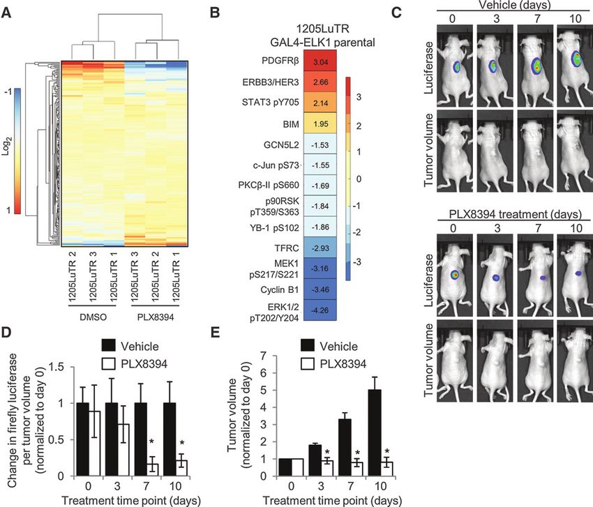
Published OnlineFirst November 13, 2017; DOI: 10.1158/1535-7163.MCT-17-0705

                                                                                         Mutant-Selective, Paradox-Breaking BRAF Inhibitor

previous generations of mutant-selective BRAF inhibitors and the        IHC
combination of BRAF plus MEK inhibitors. Targeted inhibitors              Tissue was fixed in formalin and paraffin embedded. Sections
produce heterogeneous effects in mutant BRAF patients due to            were stained for ERK1/2 phosphorylation (Thr202/Tyr204,
intrinsic mechanisms of resistance and adaptive drug responses.         #4370, Cell Signaling Technology). Staining was scored using
There is an important need for targeted agents to be tested in a        the digital Aperio ScanScope GL system in a blinded fashion by a
personalized manner. Patient-derived xenograft models have              pathologist (A. Goldberg).
been developed but typically take several months to be propa-
gated in mice (25–27). Here, we describe the use of a patient-          Colony formation assays
derived melanoma biopsy explant system (PDeX) and in vivo                  Cells (1.4  104) were seeded in individual wells of 6-well
ERK1/2 reporter models to show that PLX8394 is a potent BRAF            plates in regular culture medium (containing 0.5 mmol/L
inhibitor and does not elicit paradoxical activation of ERK1/2          PLX8394 for PBRTs). The next day, plates were washed and
in vivo and ex vivo.                                                    medium was replaced with medium supplemented with drugs
                                                                        of interest. Medium and drugs were changed every 2 days. After 9
                                                                        days, cells were fixed in buffered formalin with 0.2% crystal violet.
Materials and Methods                                                   Plates were then scanned for quantitation via ImageJ.
Ion Torrent sequencing
   Vehicle-treated and PLX8394-resistant tumors were harvested          Viability assays
for genomic DNA with Wizard Genomic DNA Purification Kit                    Cells (2  103) were seeded in triplicate in wells of a 96-well
(Promega). Samples were barcoded and sequenced using the Ion            plate in regular culture medium (containing 0.5 mmol/L PLX8394
PGM 200 Sequencing Kit (Life Technologies). For full details, see       for PBRTs). On the next day, cells were washed twice with PBS, and
Supplementary Data.                                                     drug-laced media were added. After 4 days (including one medium
                                                                        change), 3-(4,5-dimethylthiazol-2-yl)-2,5-diphenyltetrazolium
Western blot analysis                                                   bromide (MTT) reagent (Sigma-Aldrich Co.) was added for 3
  Western blot analysis was performed as in ref. 28 with volu-          hours. Solubilized formazan was analyzed at 450 nmol/L in a
metric analysis in Quantity One (Bio-Rad). Antibodies were              Multiskan Spectrum spectrophotometer (Thermo Fisher Scien-
purchased from Cell Signaling Technology, Santa Cruz Biotech-           tific). Results are normalized to DMSO conditions and are a
nology Inc., Biosciences Inc., Enzo, and Sigma-Aldrich Co. For full     composite of three independent experiments.
details, see Supplementary Data.
                                                                        Statistical analysis
                                                                           Unless noted otherwise, significant values (indicated by an
Inhibitors
                                                                        asterisk) were considered to have P 0.05 as determined by a
  Vemurafenib, dabrafenib, and trametinib (GSK'212) were pur-
                                                                        two-tailed Student t test assuming unequal variance and error
chased from Selleck Chemicals LLC. PLX8394 was provided by Dr.
                                                                        bars are  SEM. The effects of drug treatment on BRAF homo-
Gideon Bollag (Plexxikon Inc.). PLX8394 for in vivo experiments
                                                                        dimers was modeled by considering the treatment and exper-
was sent to Research Diets Inc. for the production of chow.
                                                                        imental replicate (n ¼ 4) as predictors of log(Myc/FLAG).
                                                                        ANOVA analysis was then performed with these considerations.
Cell culture                                                            IC50 calculations for ERK1/2 phosphorylation were performed
   1205LuTR GAL4-ELK1 reporter cells [modified cell line – the           using GraphPad Prism.
parental was a gift from Dr. Meenhard Herlyn (2005, Wistar
Institute, Philadelphia, PA)], PRT #3 (26), Paradox breaker resis-      S-phase entry analysis
tant tumor (PBRT) #15, and #16 cells [in vivo derived resistant cells     Cells (2.0  105) were seeded in 6-well plates. Cells were treated
of 1205LuTR GAL4-ELK1 (2013)] were grown in MCDB 153                    with drug of interest for 48 hours. The thymidine analogue, EdU,
medium containing 20% Leibovitz-L15 medium, 2% FBS,                     was added at a final concentration of 10 mmol/L for the final 16
0.2% sodium bicarbonate, and 5 mg/mL insulin. In addition, PRT          hours. EdU incorporation was measured using the Click-it EdU
#3 cells were cultured in 1 mmol/L PLX4720, and PBRT #15 and            Alexa Flour 647 Flow Cytometry Assay Kit and was utilized as per
#16 cells were cultured in 0.5 mmol/L PLX8394. BOWES cells              the manufacturer's instructions (Molecular Probes). EdU staining
(2013) were grown in MEM containing 10% FBS, 1% nonessen-               was quantified on BD FACSCalibur, and data were analyzed with
tial amino acids, 1% sodium pyruvate, and 1% HEPES buffer. B6,          FlowJo software. Data points are shown as averages of three
MeWo, [gifts from Dr. Barbara Bedogni (2013, Case Western               experimental replicates.
Reserve University, Cleveland, OH)], and CHL-1 cells (purchased
from ATCC in 2013) were cultured in DMEM with 10% FBS.                  Ex vivo explant system
Penicillin/streptomycin (1%) was added to all media. All cells             Tumors were collected following informed patient consent at
were grown at 37 C in a humidified incubator supplemented               Thomas Jefferson University Hospital (Philadelphia, PA) under
with 5% CO2. Cells are routinely assayed for mycoplasma                 an IRB-approved protocol (#10D.341). Less than 16 hours post-
contamination with MycoScope Kit (Genlantis). Cells were                surgery, excess adipose and stromal tissue was removed and
assayed in April, May, and September 2016. Cell line authen-            tumors were cut into 1-mm3 pieces. Vetspon absorbable hemo-
tication via STR analysis was completed in April 2015 for               static gelatin 1-cm3 sponges (Novartis) were presoaked in 12-well
BOWES, MeWo, B6, and CHL-1, and in February 2017 for                    plates for 15 minutes at 37 C in 500 mL of DMEM/10% FBS
1205LuTR GAL4-ELK1 reporter cells and PBRTs. B6 cells pro-              containing drugs or DMSO as a vehicle control. To avoid concerns
duced a unique profile, whereas all other cells matched to               of intratumoral heterogeneity, up to three 1-mm3 pieces from
known profiles.                                                          different locations of the original tumor were placed per sponge

www.aacrjournals.org                                                                                    Mol Cancer Ther; 17(1) January 2018    85

   Downloaded from mct.aacrjournals.org on February 11, 2021. © 2018 American Association for Cancer Research.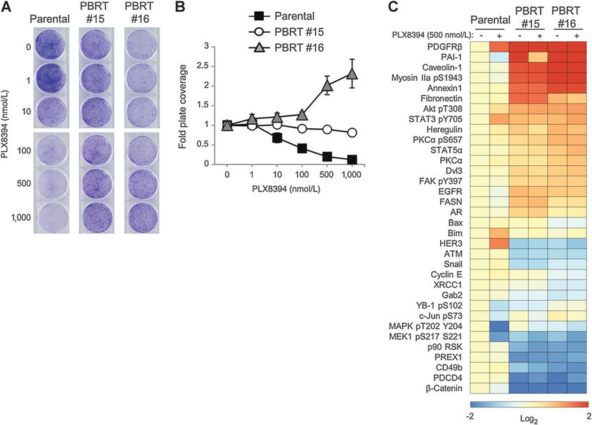
Published OnlineFirst November 13, 2017; DOI: 10.1158/1535-7163.MCT-17-0705
Hartsough et al.
per treatment condition. Similarly, xenograft tumors were dis- (RPPA) analysis on 1205LuTR GAL4-ELK1 reporter cells (Fig. 1A;
sected into 1-mm3 pieces and placed on medium/drug-soaked ref. 28). RPPA allows for quantitative assessment of >200 targets
sponges. Medium was replaced every 24 hours. Tumor pieces for involved in growth factor signaling, cell-cycle progression, apo-
Western blotting were homogenized in modified RPPA lysis ptosis, and histone modification (29). To allow for cell-cycle and
buffer (29) with phosphatase and inhibitors (PhosSTOP and apoptotic changes to take place, we assayed samples at a 24-hour
cOmplete tablets Roche). Laemmli sample buffer was added, and time point compared with an acute time point, which would most
samples were heated at 95 C for 5 minutes. For IHC analysis, likely only affect signaling. PLX8394 treatment significantly (P <
tumor pieces were fixed in formalin for 24 hours. Two of the 0.05 and fold change 1.5) altered 13 targets, including down-
samples (TJU-MEL-27A and TJU-MEL-27B) were different lesions regulation of phosphorylated MEK and ERK1/2, upregulation of
from the same patient, and combination treatment was not the proapoptotic protein BIM, and downregulation of cyclin B1
assayed for TJU-MEL-30. (Fig. 1B). We also observed upregulation of the growth factor
receptor, ERBB3, consistent with previous findings with vemur-
In vivo experiments afenib (30, 31). Phosphorylation of Raf-1/CRAF and Src, which
Seven-week-old female nude mice (The Jackson Laboratory, are implicated in paradoxical ERK1/2 signaling and are sup-
stock# 007850) were injected with 1 106 1205LuTR GAL4- pressed by pan-RAF inhibitors, remained unaffected by PLX8394
ELK1 reporter cells. Tumors were allowed to form to approx- treatment (Supplementary Fig. S1). To quantitatively measure
imately 100 mm3, at which point the mice were randomly the effects of PLX8394 in vivo, we utilized xenografts from BRAF
divided into 2 cohorts and fed either vehicle or PLX8394-laced V600E melanoma cells expressing an ERK1/2 luciferase-based
chow. Tumor volumes and ERK1/2 reporter activity via firefly reporter. This model permits quantitative and temporal anal-
luciferase measurements were recorded every 3 to 4 days. ysis in a noninvasive manner (28). ERK1/2 reporter luciferase
All mouse experiments were performed at Thomas Jefferson levels (adjusted for tumor volume) were significantly reduced
University (Association for Assessment and Accreditation of within 7 days of PLX8394 treatment compared with vehicle
Laboratory Animal Care-accredited) and approved by the Insti- controls (Fig. 1C and D). PLX8394 also significantly reduced
tutional Animal Care and Use Committee. For full details, see tumor growth compared with vehicle-treated mice (Fig. 1E).
Supplementary Data. Together, these results show that PLX8394 inhibits ERK1/2
signaling in vitro, in vivo, and reduces tumor growth in mutant
BRAF melanoma xenografts.
Reverse-phase protein array analysis
1205LuTR GAL4-ELK1 parental reporter cells and PB-resistant PLX8394 suppresses phospho-ERK1/2 and elicits apoptotic
tumor (PBRT) #15 and #16 cells (2.5 105/per condition) were markers in patient samples as efficiently as combination
seeded in 6-well plates in normal growth media (containing 500 treatment
nmol/L PLX8394 for PBRTs). The next day, cells were treated with An ex vivo explant model has been previously utilized in
either DMSO or 0.5 mmol/L PLX8394 for 24 hours. Lysates from prostate cancer (32, 33). These systems are advantageous for
three independent experiments were processed and analyzed as preclinical testing as they contain a stromal component and,
described previously (29), producing triplicates for each. For thus, more closely mimic the tumor microenvironment. We
details of the analysis, see Supplementary Data section. established and validated this model in melanoma, using
explants derived from xenograft tumors of 1205LuTR cells
Immunoprecipitation assays (partially sensitive to PLX4720, the tool compound for vemur-
1205LuTR cells expressing both Myc and FLAG-tagged BRAF afenib) and 1205LuTR-PRT #3 cells, which express a BRAF
V600E DEx 2–8 were seeded (1.0 106) on 10-cm plates V600E splice variant and are resistant to PLX4720 (28, 34).
overnight. Cells were then dosed with 100 ng/mL doxycycline Tumor tissue was treated ex vivo with vemurafenib at 1 mmol/L,
to induce both splice variant expression for 48 hours. Plates a standard concentration for in vitro experiments (6, 7, 34) and
were treated with DMSO, PLX4720, or PLX8394 for an addi- 3D melanoma systems (35) or with 1 mmol/L dabrafenib/16
tional 4 hours. Cells were PBS washed and lysed in an NP40- nmol/L trametinib combination (Supplementary Fig. S2A–
based lysis buffer. Twenty microliters of prewashed anti-FLAG S2D). The dabrafenib and trametinib combination at the given
Affinity Gel (#A2220 Sigma-Aldrich) was used to immunopre- concentration is a clinically relevant molar ratio of the two
cipitate FLAG-tagged target during an overnight incubation at drugs that was found to have significant effect on downstream
4 C. The affinity gel was then washed 3 times with cold TBS, signaling (Supplementary Fig. S2F); this treatment served as a
resuspended in Laemmli sample buffer, and boiled for 5 positive control for ERK1/2 pathway suppression to demon-
minutes. Equal volume was loaded on acrylamide gels for strate the range of response in the ex vivo explant system. These
Western blot analysis. results provided proof of concept for the patient-derived
explant (PDeX) system. Next, we extended PDeX analysis to
fresh human melanomas. Sequence-validated, mutant BRAF
Results V600E melanoma biopsy explants (Supplementary Fig. S3A;
PLX8394 suppresses ERK1/2 signaling and tumor growth Table S1) were treated either with vemurafenib, dabrafenib/
in vivo trametinib combination, or PLX8394 for 48 to 72 hours. By
PLX8394 is a mutant BRAF-selective inhibitor that potently IHC staining of paraffin-embedded tumor sections and quan-
blocks ERK1/2 signaling in BRAF V600E/D–harboring melanoma titative analyses, vemurafenib inhibited ERK1/2 phosphoryla-
cells in vitro (18, 19, 24). The structure of PLX8394 has been tion, but inhibition was partial and variable (Fig. 2A and B).
published previously (18). As an initial assessment of the cellular This observation is consistent with others who report a modest
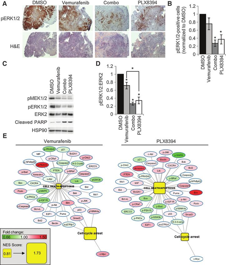
response to PLX8394, we performed reverse-phase protein array response to vemurafenib in 3D tumor systems (35), and in
86 Mol Cancer Ther; 17(1) January 2018 Molecular Cancer Therapeutics
Downloaded from mct.aacrjournals.org on February 11, 2021. © 2018 American Association for Cancer Research.Published OnlineFirst November 13, 2017; DOI: 10.1158/1535-7163.MCT-17-0705
Mutant-Selective, Paradox-Breaking BRAF Inhibitor
Figure 1.
PLX8394 effectively reduces ERK1/2 signaling and tumor volume in vivo. A, 1205LuTR GAL4-ELK1 cells were treated for 24 hours with either DMSO or
0.5 mmol/L PLX8394. Lysates were obtained from three independent experiments and processed for RPPA analysis. A heatmap was generated using median-
centered data across each protein measurement for each sample. B, Proteins with P 0.01 and a fold change of 1.5 were significantly altered following
PLX8394 treatment. C, Mice bearing 1205LuTR GAL4-ELK1 xenografts were fed either vehicle chow or PLX8394 laced chow. Representative images of a vehicle
and PLX8394-treated mouse with overlaid luciferase output across 10 days of treatment are shown. D, Quantification of firefly luciferase. Graph depicts fold
change in luciferase output per tumor volume compared with vehicle for each day of treatment. E, Average fold change in tumor volume between mice fed
vehicle chow and PLX8394-laced chow.
stroma/melanoma coculture settings (36–38). In contrast, both on BRAF V600E human melanoma PDeX treated with targeted
combination and PLX8394 treatment consistently and signif- inhibitors. Pathway analysis of the programmed cell death/
icantly inhibited ERK1/2 phosphorylation in PDeX (Fig. 2A and apoptosis and cell-cycle arrest Gene Ontology pathways by
B). By Western blot analysis, vemurafenib inhibition of ERK1/2 gene set enrichment analysis showed that vemurafenib pre-
phosphorylation was again variable, but statistically significant dominantly enriched a cell-cycle arrest response, whereas
compared with vehicle treatment (Fig. 2C and D; Supplemen- PLX8394 and combination treatment induced an apoptotic/
tary Fig. S3C). Importantly, both PLX8394 and the dabrafenib/ cell death response (Fig. 2E; Supplementary Fig. S3F). In a
trametinib combination inhibited ERK1/2 phosphorylation by tumor with sufficient sample to assay the effects of a dose
>60% (Fig. 2B and D). Furthermore, PARP cleavage is associ- response to PLX8394, we observed a dose-dependent increase
ated with ERK1/2 inhibition following combination and of PARP cleavage, as well as suppression of ERK1/2 phosphor-
PLX8394 treatments (Fig. 2C; Supplementary Fig. S3). To better ylation. RPPA analysis of this sample demonstrated a dose-
understand pathway alterations, RPPA analysis was performed dependent decrease of ERK1/2 pathway targets, and increase in
www.aacrjournals.org Mol Cancer Ther; 17(1) January 2018 87
Downloaded from mct.aacrjournals.org on February 11, 2021. © 2018 American Association for Cancer Research.Published OnlineFirst November 13, 2017; DOI: 10.1158/1535-7163.MCT-17-0705
Hartsough et al.
Figure 2.
PLX8394 effectively inhibits ERK1/2 signaling in patient tumors comparable with dabrafenib/trametinib treatment. A, H&E and IHC analysis of pERK1/2
staining from a representative mutant BRAF patient sample (TJU-MEL-27A) treated with either DMSO, vemurafenib (1 mmol/L), combination (1 mmol/L
dabrafenib/16 nmol/L trametinib), or PLX8394 (0.5 mmol/L). B, Quantitation of A across a panel of 6 different mutant BRAF melanoma patient samples. C, Western
blot analysis of ERK1/2 signaling and PARP cleavage from a representative patient sample (TJU-MEL-27A). D, Western blot quantitation of the normalized
pERK1/2 to ERK2 signal from 5 patient samples. E, RPPA data from mutant BRAF patient samples were analyzed via GSEA. Patient explants treated with
vemurafenib (left) and PLX8394 (right) were grouped and compared with DMSO-treated samples. Enrichment of the programmed cell death/apoptosis and
cell-cycle arrest GO pathways and corresponding changes in RPPA-determined protein levels compared with DMSO are shown. Pathway nodes and protein
levels for all treatments are on the same scale (bottom left).
the proapoptotic protein BIM (Supplementary Fig. S3D and V600E melanomas and elicits effects comparable with the
S3E). Taken together, these data suggest that PLX8394 is a current FDA-approved dabrafenib/trametinib combination in
potent inhibitor of ERK1/2 phosphorylation in human BRAF an explant model.
88 Mol Cancer Ther; 17(1) January 2018 Molecular Cancer Therapeutics
Downloaded from mct.aacrjournals.org on February 11, 2021. © 2018 American Association for Cancer Research.Published OnlineFirst November 13, 2017; DOI: 10.1158/1535-7163.MCT-17-0705
Mutant-Selective, Paradox-Breaking BRAF Inhibitor
PLX8394 is more potent in suppressing ERK1/2 with DMSO control (Fig. 4A and B). In contrast, PLX8394 treat-
phosphorylation than vemurafenib and is efficacious against ment did not produce a statistically significant increase in para-
constitutively dimerized BRAF splice variants doxical activation, and treatment with trametinib strongly
To determine the efficacy of PLX8394 compared with vemur- reduced ERK1/2 phosphorylation. Extending these studies into
afenib in the explant system, xenografts were generated with explants derived from xenografts from the WT/WT BOWES and B6
1205LuTR GAL4-ELK1 parental cells and two different in vivo cells, vemurafenib enhanced ERK1/2 activation in WT/WT xeno-
derived RAF inhibitor resistant lines, PRT #3 and PRT #4 (28). graft explants similar to experiments in 2D (Fig. 4C). In compar-
Xenograft tumors were excised and used in the explant system ison with vemurafenib, PLX8394 did not induce strong paradox-
to assay dose responses. After 48 hours of treatment, Western ical ERK1/2 activation in these samples. Furthermore, we tested
blotting was used to measure ERK1/2 phosphorylation and PARP paradoxical activation by RAF inhibitors using the PDeX system in
cleavage. We found that PLX8394 more efficiently suppressed a WT/WT patient sample (Supplementary Fig. S3B). Western blot
ERK1/2 phosphorylation (IC50 0.01 mmol/L vs. 1.39 mmol/L) and analysis demonstrated a strong paradoxical phosphorylation of
elicited PARP cleavage compared with vemurafenib in parental ERK1/2 induced by vemurafenib, but comparatively weak ERK1/2
1205LuTR (Fig. 3A; Supplementary Fig. S4A). Importantly, phosphorylation in response to PLX8394 (Fig. 4D). As expected,
although vemurafenib treatment was largely ineffective (ERK1/2 the MEK inhibitor trametinib suppressed ERK1/2 signaling (Fig.
phosphorylation IC50 is undefined for PRT #3 and 4.05 mmol/L 4D). Taken together, these data show that when using doses
for PRT #4), PLX8394 inhibited ERK1/2 phosphorylation (0.97 effective in suppressing ERK1/2 signaling in mutant BRAF tumor,
mmol/L and 0.096 mmol/L for PRT #3 and #4, respectively) and PLX8394 does not elicit strong paradoxical signaling in WT BRAF
induced PARP cleavage in BRAF splice variant–expressing tumors, tissue, representing an improvement over the previous generation
PRT #3 and PRT #4 (Fig. 3B and C; Supplementary Fig. S4B and of BRAF inhibitors.
S4C). It is noteworthy that although the PRT tumors were sensitive
to PLX8394, both required a higher dose of PLX8394 than Acquired resistance to PLX8394 is associated with ERK1/2
parental cells to suppress ERK1/2 phosphorylation. This result is reactivation and deregulation of ERK1/2-independent
consistent with other RAF inhibitor–resistant cells treated with pathways
potential second-line RAS–RAF–MEK–ERK pathway targeting Treatment with targeted therapies is invariably associated with
agents (34, 39). As constitutive BRAF splice variant homodimer- acquired resistance; therefore, we investigated the duration of
ization has been linked to vemurafenib resistance (39), we inves- PLX8394 effects on BRAF V600E melanomas in vivo. Mice bearing
tigated whether PLX8394 affects homodimerization of BRAF mutant BRAF xenografts were continued on PLX8394 treatment
splice variants. 1205Lu cell lines were created to inducibly coex- until progression (1,000 mm3 tumor size or displayed signs of
press both Myc-tagged and FLAG-tagged versions of BRAF splice ulceration). Progressing tumors were excised and two PBRT cell
variants lacking exons two through eight (1205LuTR FLAG/Myc lines, #15 and #16, were propagated. PBRT #15 and PBRT #16
BRAF DEx 2–8). BRAF DEx 2–8 is equivalent to the BRAF splice were isolated at days 45 and 35 after drug treatment, respectively.
variant expressed in PRT #4. Similar to the PRT tumors, this cell In 2D colony formation assays, PLX8394 suppressed the growth
line demonstrated a dose-dependent reduction of ERK1/2 path- of 1205LuTR parental cells in a dose-dependent manner (Fig. 5A
way signaling from PLX8394 but not PLX4720 treatment (Fig. and B). Conversely, PBRT #15 maintained growth in PLX8394
3D). Parallel lysates were then used to query homodimerization and PBRT #16 exhibited addiction to PLX8394, similar to a
by immunoprecipitation of the FLAG-tagged BRAF splice variant phenomenon observed in vemurafenib-resistant cells (34). In
and probing for the association of its Myc-tagged binding partner. MTT assays, PLX8394 potently inhibited the viability of parental
Although both drugs impaired homodimerization, PLX8394 cells, but both PBRT #15 and #16 cell lines were highly resistant to
treatment elicited a more profound reduction than PLX4720 (Fig. the inhibitor (Supplementary Fig. S5A).
3E and F). Interestingly, PLX8394 dosing had little effect on To understand pathway alterations associated with resistance to
homodimerization status (Supplementary Fig. S4D), yet higher PLX8394, we performed RPPA analysis on PBRT #15 and #16
doses of PLX8394 were able to inhibit MEK phosphorylation. compared with parental 1205LuTR cells. Compared with parental
These observations suggest that although PLX8394 blocks dimer- 1205LuTR cells, the levels of 34 proteins significantly changed
ization and ERK1/2 pathway more efficiently than PLX4720, the (1.5 fold; P 0.01) in either the DMSO control or PLX8394
extent of BRAF splice variant homodimerization itself may not be conditions (Fig. 5C). Western blotting confirmed the RPPA results
wholly responsible for vemurafenib resistance. Together, these showing maintenance of ERK1/2 phosphorylation in PBRT #15
data demonstrate superior efficacy of PLX8394 as a single-agent and #16 cells treated with PLX8394 (Supplementary Fig. S5B and
RAF inhibitor in comparison with vemurafenib and that PLX8394 S5C). In addition, PBRT cells treated with PLX8394 exhibited
can overcome mutant BRAF splice variants, a common mecha- significantly higher levels of Rb phosphorylation than PLX8394-
nism of BRAF inhibitor resistance. treated parental cells (Supplementary Fig. S5D), reflecting the
ability of PBRT cells to overcome PLX8394-mediated cell-cycle
PLX8394 attenuates the paradoxical ERK1/2 activation in WT/ inhibition. Interestingly, both PBRT cell lines displayed increased
WT melanoma tissue AKT phosphorylation, enhanced PDGFR levels, and reduced
An important goal in the design of PLX8394 is to reduce b-catenin expression compared with 1205LuTR parental cells
hyperactivation of ERK1/2 in WT BRAF-containing tissues. To (Supplementary Fig. S5B and S5E–S5G). These alterations have
demonstrate the "paradox-breaking" ability of PLX8394, we trea- been previously implicated in resistance to vemurafenib (40–44),
ted WT BRAF/WT NRAS (WT/WT) melanoma cell lines with but did not appear to be primary drivers of resistance in the PBRTs
vemurafenib, PLX8394, and trametinib in 2D culture conditions. (Supplementary Fig. S6). Furthermore, BRAF V600E splice var-
Vemurafenib significantly increased ERK1/2 signaling in CHL-1, iants, which drive resistance to vemurafenib, were not detected in
BOWES, MeWo, and B6 WT/WT melanoma cell lines compared PBRT cell lines (Supplementary Fig. S7A). Thus, as with other RAF
www.aacrjournals.org Mol Cancer Ther; 17(1) January 2018 89
Downloaded from mct.aacrjournals.org on February 11, 2021. © 2018 American Association for Cancer Research.Published OnlineFirst November 13, 2017; DOI: 10.1158/1535-7163.MCT-17-0705
Hartsough et al.
Figure 3.
PLX8394 suppresses MEK/ERK signaling in mutant BRAF splice variant–expressing cells and is associated with reduction of splice variant homodimerization.
A, 1205LuTR GAL4-ELK1 cells were used to generate xenograft tumors that were harvested and dissected into approximately 1-mm3 pieces for use in
explant system. After 48 hours of treatment, lysates were collected and analyzed by Western blotting. Using densitometry, the normalized ratio of
phospho-ERK1/2 to ERK2 levels and cleaved PARP to HSP90 for each cohort was quantified and graphed. Data were analyzed with a two-way ANOVA
corrected for multiple comparisons with Tukey analysis. Error bars, SEM; , P 0.05; , P 0.01; , P 0.001; , P 0.0001. B, Similar to A except
PRT#3 cell line was used to generate xenograft tumors. C, Similar to A but PRT#4 xenograft tumors. D, Western blots of whole-cell lysates from
1205LuTR FLAG/Myc BRAF DEx 2–8 cells after 48 hours of doxycycline-induced splice variant expression and an additional 4 hours of PLX4720 or PLX8394
treatment at the indicated concentration. E, Parallel lysates from D were used to immunoprecipitate the FLAG-tagged mutant BRAF splice variant, and
Western blots reveal its associated Myc-tagged binding partner. F, Quantification of splice variant homodimerization after treatment with 1 mmol/L
PLX4720 and 0.5 mmol/L PLX8394 (n ¼ 4).
90 Mol Cancer Ther; 17(1) January 2018 Molecular Cancer Therapeutics
Downloaded from mct.aacrjournals.org on February 11, 2021. © 2018 American Association for Cancer Research.Published OnlineFirst November 13, 2017; DOI: 10.1158/1535-7163.MCT-17-0705
Mutant-Selective, Paradox-Breaking BRAF Inhibitor
Figure 4.
PLX8394 prevents significant
paradoxical ERK1/2 activation in WT
BRAF melanomas. A, Western blot
analysis of WT/WT melanoma cell
lines after treatment with DMSO,
vemurafenib (1 mmol/L), PLX8394
(0.5 mmol/L), or trametinib
(50 nmol/L) for 48 hours. B,
Quantitation of the normalized
pERK1/2 signal from A. C, As in Fig. 2B,
except that two different WT/WT
melanoma cells were used to form
xenografts and were processed/
treated in the ex vivo explant system.
Western blot analysis of lysates
prepared from explants treated with
DMSO, vemurafenib, or PLX8394 after
48 hours. Densitometry results of
pERK1/2 to ERK2 are indicated.
D, WT/WT patient sample explants
(TJU-MEL-29) were treated as in A for
48 hours. Densitometry values are
shown.
inhibitors and MEK–ERK1/2 regimens, prolonged exposure to main (BRD) inhibitor, JQ1. Western blot analysis demonstrated
PLX8394 results in acquired resistance associated with ERK1/2 that JQ1 treatment increased levels of the cyclin-dependent kinase
pathway reactivation and compensatory pathway alterations. inhibitors, p21 and p27, in PBRT #16 (Fig. 6D). This correlated
with increased sensitivity of PBRT #16 to BET/BRD inhibitors in
PBRT cell lines have differential sensitivities to ERK1/2 pathway crystal violet growth assays and EdU incorporation (Fig. 6E and F;
inhibition Supplementary Fig. S7D). Although PBRT #16 was more sensitive
We tested whether vertical targeting of the ERK1/2 pathway to BET/BRD inhibitor treatment, these agents also suppressed
would overcome the acquired resistance to PLX8394, as it does in growth of PBRT #15, suggesting a potential universal second-line
other resistant melanoma models (27, 39). Individual treatments therapy option (Fig. 6E; Supplementary Fig. S7D).
of PLX8394, vemurafenib, trametinib, and the ERK1/2 inhibitor
SCH772984 (SCH772) suppressed ERK1/2 signaling and reduced
Rb phosphorylation in parental cells (Fig. 6A). Although trame- Discussion
tinib reduced phospho-ERK1/2 in both PBRT #15 and #16, Rb FDA-approved mutant BRAF-selective inhibitors have marked-
phosphorylation was only affected in PBRT #15 (Fig. 6A). Sim- ly improved the treatment options and outcomes for BRAF
ilarly, SCH772 treatment reduced Rb phosphorylation in PBRT V600E/K melanoma patients but are limited by the occurrence
#15 but not PBRT #16 (Fig. 6A). Dose escalation of SCH772 was of adverse events associated with paradoxical ERK1/2 activation.
associated with an increase in PARP cleavage and BIM levels, as In this study, we show the efficacy of the next-generation "par-
well as a reduction of both Rb phosphorylation and cyclin A adox-breaking" BRAF inhibitor, PLX8394. Overall, our data sup-
expression in PBRT #15 (Supplementary Fig. S7B). However, port that PLX8394 is a viable treatment option to efficiently
these changes were not observed in PBRT #16 (Supplementary inhibit ERK1/2 signaling while limiting paradoxical effects in WT
Fig. S7B). Similarly, crystal violet growth and EdU incorporation BRAF cells and highlight the use of tumor explants to rapidly
assays demonstrated that both parental and PBRT #15 cell lines assess the utility of targeted agents.
were more sensitive to ERK1/2 inhibitor treatment than PBRT #16 Using an in vivo GAL4-ELK1 reporter system to quantitatively
(Fig. 6B and C; Supplementary Fig. S7C). and temporally measure ERK1/2 signaling in melanoma xeno-
Ion Torrent sequencing results of PBRT #15 and PBRT #16 did grafts (28), we show that PLX8394 effectively inhibits ERK1/2
not yield any missense mutations that would be indicative of signaling and reduces mutant BRAF melanoma growth. This
ERK1/2 inhibitor resistance (Supplementary Tables S2 and S3). approach is complemented by an ex vivo explant system that
Consequently, we postulated that transcriptional alterations may demonstrates PLX8394 effectively inhibits ERK1/2 phosphoryla-
contribute to resistance. Therefore, we utilized epigenetic agents, tion in patient tumors. Vemurafenib treatment only elicited an
the bromodomain and extraterminal domain (BET) bromodo- approximately 20% reduction in ERK1/2 phosphorylation in
www.aacrjournals.org Mol Cancer Ther; 17(1) January 2018 91
Downloaded from mct.aacrjournals.org on February 11, 2021. © 2018 American Association for Cancer Research.Published OnlineFirst November 13, 2017; DOI: 10.1158/1535-7163.MCT-17-0705
Hartsough et al.
Figure 5.
In vivo acquired resistance of 1205LuTR GAL4-ELK1 xenografts to
PLX8394. A, Progressing xenografts were harvested and used to
generate PLX8394-resistant cell lines (PBRTs). Crystal violet staining of
PBRT #15 and #16 cells treated with increasing doses of PLX8394
compared with parental cells is shown. B, Quantified results of three
independent experiments as in B. C, RPPA analysis of PBRT #15
and PBRT #16 cells compared with parental cells treated with either
DMSO or PLX8394 (0.5 mmol/L) for 24 hours. Heatmap showing proteins
with P 0.01 and a fold change of 1.5 found to be significantly altered
in PBRT #15 or #16 cells when compared with parental cells.
explants consistent with others' observations of minimal vemur- treatment in the explant system, compared with 2D culture, may
afenib efficacy in 3D tumor mimics (35) and stroma/melanoma reflect ERK1/2 signaling present in stromal cells that should not be
coculture systems (36–38). Variability in the response of patient inhibited or paradoxically activated.
tumors to vemurafenib may also be due to different treatment PLX8394 was effective in suppressing ERK1/2 signaling and
histories of the patients (Supplementary Table S1) and/or a high eliciting PARP cleavage in BRAF splice variant–expressing, vemur-
stromal component, which displays paradoxical ERK1/2 acti- afenib-resistant samples. This may be in part due to PLX8394's
vation (35). In contrast, PLX8394 consistently inhibited ERK1/2 ability to better suppress splice variant homodimerization, there-
signaling in all patient biopsies independent of treatment by facilitating efficient inhibition of monomeric mutant BRAF
history and was comparable with dabrafenib/trametinib com- kinase. Homodimerization of mutant BRAF splice variant–expres-
bination therapy. sing cells has been linked to RAF inhibitor resistance (28, 39);
PLX8394 demonstrated enhanced efficacy when directly com- however, the dimerization status of these splice variants in the
pared with vemurafenib in parallel ex vivo dosing of xenograft presence of RAF inhibitors has not been tested. Vemurafenib and
tissue. This may not be surprising as the PLX8394 IC50 for ERK1/2 PLX4720 have previously been shown to destabilize homodimer-
phosphorylation is approximately 10-fold lower than vemurafe- ization of full-length mutant BRAF (45) and heterodimerization
nib (18). In contrast, in the explant system, the dose required to of WT BRAF kinase domain with CRAF (46). On the other hand,
reach ERK1/2 phosphorylation IC50 of parental mutant BRAF reports indicate that these drugs may enhance BRAF/CRAF het-
tumor tissue treated with vemurafenib was much higher (44 erodimers (23). In the current study, we utilize differentially
IC50) compared with PLX8394 (3 IC50; Fig. 3A). One expla- tagged V600E BRAF splice variants to measure homodimerization
nation for the larger difference of vemurafenib effectiveness is the status during drug treatment to model the setting in which splice
potential paradoxical activation of ERK1/2 signaling in the stro- variants are expressed. We found that although both PLX4720 and
mal component present in the explant tissue, which is not present PLX8394 reduced homodimerization of V600E BRAF splice var-
in 2D culture systems. Vemurafenib has been shown to paradox- iants, PLX8394 was significantly more effective. It is possible that
ically elicit tumor-protective responses from stromal components the residual dimerized splice variant in PLX4720-treated cells may
in ex vivo systems, as well as stimulate production of mitogenic be adequate for drug resistance as further blocking of homodi-
growth factors from WT BRAF tumors in vivo (18, 35), a phenom- merization by PLX8394 is associated with ERK1/2 pathway inhi-
enon that should be attenuated in PLX8394 treatment. The bition. Alternatively, these data may indicate that although splice
moderate increase in ERK1/2 phosphorylation IC50 for PLX8394 variant homodimerization contributes to signaling in the
92 Mol Cancer Ther; 17(1) January 2018 Molecular Cancer Therapeutics
Downloaded from mct.aacrjournals.org on February 11, 2021. © 2018 American Association for Cancer Research.Published OnlineFirst November 13, 2017; DOI: 10.1158/1535-7163.MCT-17-0705
Mutant-Selective, Paradox-Breaking BRAF Inhibitor
Figure 6.
PBRTs have differential responses to second-line therapies. A, 1205LuTR GAL4-ELK1, PBRT #15, and PBRT #16 were seeded in 6-well plates overnight, and then,
cells were washed and media were replaced and supplemented with DMSO, 0.5 mmol/L PLX8394, 1 mmol/L vemurafenib, 50 nmol/L trametinib, or 1 mmol/L
SCH772. After 24 hours, lysates were harvested and samples were analyzed by Western blotting. B, Quantification of crystal violet 2D growth assays for
1205LuTR GAL4-ELK1, PBRT #15, and PBRT #16 in the presence of increasing SCH772. Data points represent the average percent plate coverage of at least three
independent experiments. Error bars, SEM. C, S-phase entry of 1205LuTR GAL4-ELK1, PBRT #15, and PBRT #16 cells was assayed by EdU incorporation.
Graph is the average EdU positivity from at least three experimental replicates. Error bars, SEM; , P < 0.05 compared with each cell line's DMSO condition using
a two-way Student t test assuming unequal variance. D, Western blot analysis of 1205LuTR GAL4-ELK1, PBRT #15, and PBRT #16 cells after 24-hour drug
treatment of DMSO, 0.5 mmol/L PLX8394, or 1 mmol/L JQ1. E and F, Similar to B and C except cells were treated with JQ1.
presence of vemurafenib, it may not be wholly responsible for may be an appropriate partner with immune-based therapies,
RAF inhibitor resistance. such as CTLA-4 and PD-1/PD-L1 inhibitors (52).
The explant system was also used to show that vemurafenib, but With an increasing number of available therapies for the
not PLX8394, induces a strong paradoxical activation of ERK1/2 treatment of mutant BRAF melanomas, identifying the best ther-
in WT/WT melanoma. Other groups have reported on pan-RAF/ apy for an individual patient is increasingly important. Patient-
Src inhibitors that do not elicit paradoxical activation properties derived tumor xenograft models accurately reproduce a patient's
(47); however, the mutant BRAF selectivity of PLX8394 may response to therapy (53); however, these models are associated
afford a higher therapeutic index than agents that broadly inhibit with long generation times and high cost. As an alternative, we
RAF kinases. Furthermore, the mutant BRAF-specific targeting describe a patient-derived explant system, PDeX, to test multiple
properties of PLX8394 may enable its use in combinatorial regi- treatment strategies using a single patient biopsy that accounts for
mens with immune therapies. As suppression of ERK1/2 signaling intratumoral heterogeneity by assaying multiple sample pieces
is associated with increased melanoma antigen presentation (48), from different parts of the lesion. The ability of PDeX to test the
it is advantageous to use targeted inhibitors as an adjuvant to efficacy of small-molecule inhibitors and mAbs in a short time
improve immunotherapy efficacy. However, there are conflicting period offers an inexpensive and rapid assay that is individualized
reports of how systemic pathway inhibition (i.e., MEK inhibitor and may inform patient treatment options.
treatment) affects the antitumor immune response (49–51). Our As with other targeted therapies, acquired resistance to
results indicate that at doses that would inhibit mutant BRAF PLX8394 eventually occurs in our preclinical studies. Phospho-
tumors, PLX8394 minimally affects ERK1/2 status in WT/WT cells, proteomic analysis implicated well-known BRAF inhibitor resis-
suggesting it will not alter normal T-cell activation. Thus, PLX8394 tance markers in the PLX8394-resistant cell lines (40, 43, 54).
www.aacrjournals.org Mol Cancer Ther; 17(1) January 2018 93
Downloaded from mct.aacrjournals.org on February 11, 2021. © 2018 American Association for Cancer Research.Published OnlineFirst November 13, 2017; DOI: 10.1158/1535-7163.MCT-17-0705
Hartsough et al.
However, initial experiments suggest that enhanced AKT activity Authors' Contributions
and upregulation of PDGFR are not sole drivers of resistance in Conception and design: E.J. Hartsough, C.H. Kugel III, M.J. Vido, A.E. Aplin
these cells (Supplementary Fig. S6). Rather, it is likely that they Development of methodology: E.J. Hartsough, M.J. Schiewer, A.E. Aplin
work in coordination with reactivation of the ERK1/2 pathway. It Acquisition of data (provided animals, acquired and managed patients,
provided facilities, etc.): E.J. Hartsough, C.H. Kugel III, M.J. Vido, A.C. Berger,
is possible that resistance mechanisms to PLX8394 will be unique A.E. Aplin
from vemurafenib and dabrafenib and is underscored by the Analysis and interpretation of data (e.g., statistical analysis, biostatistics,
finding that PBRT #16 is resistant to vertical targeting of the computational analysis): E.J. Hartsough, C.H. Kugel III, M.J. Vido, A.C. Berger,
ERK1/2 signaling pathway but is sensitive to BET/BRD inhibitor T.J. Purwin, A. Goldberg, M.A. Davies, K.E. Knudsen, A.E. Aplin
treatment. As enrollment for PLX8394 phase I/IIa study (Clin- Writing, review, and/or revision of the manuscript: E.J. Hartsough, C.H. Kugel
III, M.J. Vido, A.C. Berger, A. Goldberg, M.A. Davies, A.E. Aplin
icalTrials.gov #NCT02428712) has only recently started, resis-
Administrative, technical, or material support (i.e., reporting or organizing
tance mechanisms in patients remain unknown. data, constructing databases): E.J. Hartsough, A.C. Berger, G. Bollag, A.E. Aplin
In summary, PLX8394 is a promising next-generation mutant Study supervision: A.E. Aplin
BRAF-selective inhibitor that does not elicit strong paradoxical
ERK1/2 activation in nonmutant BRAF cells. PLX8394 monother-
apy is entering clinical trials with hope that it will prevent side
Acknowledgments
effects associated with paradoxical ERK1/2 activation while simul-
We are grateful to Sheera Rosenbaum for generating the WT/WT melanoma
taneously reducing grade 4 toxicities and permanent discontinua- xenografts, Dr. Kevin Basile for help with mutant BRAF xenografts, Dr. Meen-
tions associated with dual inhibitor therapies (14). Our parallel hard Herlyn (Wistar Institute, Philadelphia, PA) for the WM melanoma cell
dosing experiments of tumor tissue suggest that PLX8394 is more lines, and Dr. Barbara Bedogni (Case Western Reserve University, Cleveland,
effective than vemurafenib at suppressing ERK1/2 signaling even OH) for the B6 and MeWo cells.
when considering vemurafenib's lower biochemical potency (18). This work was supported by the National Cancer Center and National
Institutes of Health K99 grant CA207855 (E.J. Hartsough) and F30 fellowship
In addition, the PDeX system utilized in this study provides a
CA203314 (M.J. Vido), National Institutes of Health R01 grants, CA182635 and
rapid and quantitative method to determine the efficacy of CA196278 (A.E. Aplin), and grants from the Dr. Miriam and Sheldon G.
PLX8394 (and other targeted therapies) in patient tissues. As a Adelson Medical Research Foundation (A.E. Aplin and M.A. Davies). The Sidney
result, our data suggest that PLX8394 is a promising new therapy Kimmel Cancer Center core facilities at Thomas Jefferson University are funded
for the treatment of mutant BRAF melanomas refractive to vemur- by National Cancer Institute Support Grant P30 CA56036. The Reverse Phase
afenib and that the PDeX system can potentially be used to guide Protein Array Core Facility at M.D Anderson Cancer Center is supported by the
NCI Cancer Center Support Grant, CA16672.
the treatment of patients in a personalized manner.
Disclosure of Potential Conflicts of Interest The costs of publication of this article were defrayed in part by the
M.A. Davies reports receiving commercial research grants from AstraZeneca, payment of page charges. This article must therefore be hereby marked
BMS, GSK, Oncothyreon, Roche/Genentech, and Sanofi Aventis and is a advertisement in accordance with 18 U.S.C. Section 1734 solely to indicate
consultant/advisory board member for BMS, Novartis, Roche/Genentech, this fact.
Sanofi Aventis, and Vaccinex. A.E. Aplin reports receiving a commercial research
grant from Pfizer. No potential conflicts of interest were disclosed by the other Received July 24, 2017; revised September 8, 2017; accepted October 19,
authors. 2017; published OnlineFirst November 13, 2017.
References
1. Davies H, Bignell GR, Cox C, Stephens P, Edkins S, Clegg S, et al. Mutations 10. Callahan MK, Rampal R, Harding JJ, Klimek VM, Chung YR, Merghoub T,
of the BRAF gene in human cancer. Nature 2002;417:949–54. et al. Progression of RAS-mutant leukemia during RAF inhibitor treatment.
2. Chapman PB, Hauschild A, Robert C, Haanen JB, Ascierto P, Larkin J, et al. N Engl J Med 2012;367:2316–21.
Improved survival with vemurafenib in melanoma with BRAF V600E 11. Carlino MS, Kwan V, Miller DK, Saunders CA, Yip D, Nagrial AM, et al. New
mutation. N Engl J Med 2011;364:2507–16. RAS-mutant pancreatic adenocarcinoma with combined BRAF and MEK
3. Hauschild A, Grob JJ, Demidov LV, Jouary T, Gutzmer R, Millward M, et al. inhibition for metastatic melanoma. J Clin Oncol 2015;33:e52–6.
Dabrafenib in BRAF-mutated metastatic melanoma: a multicentre, open- 12. Robert C, Karaszewska B, Schachter J, Rutkowski P, Mackiewicz A, Stroia-
label, phase 3 randomised controlled trial. Lancet 2012;380:358–65. kovski D, et al. Improved overall survival in melanoma with combined
4. Belum VR, Rosen AC, Jaimes N, Dranitsaris G, Pulitzer MP, Busam KJ, et al. dabrafenib and trametinib. N Engl J Med 2015;372:30–9.
Clinico-morphological features of BRAF inhibition-induced proliferative 13. Flaherty KT, Infante JR, Daud A, Gonzalez R, Kefford RF, Sosman J, et al.
skin lesions in cancer patients. Cancer 2015;121:60–8. Combined BRAF and MEK inhibition in melanoma with BRAF V600
5. Su F, Viros A, Milagre C, Trunzer K, Bollag G, Spleiss O, et al. RAS mutations mutations. N Engl J Med 2012;367:1694–703.
in cutaneous squamous-cell carcinomas in patients treated with BRAF 14. Larkin J, Ascierto PA, Dreno B, Atkinson V, Liszkay G, Maio M, et al.
inhibitors. N Engl J Med 2012;366:207–15. Combined vemurafenib and cobimetinib in BRAF-mutated melanoma.
6. Lee JT, Li L, Brafford PA, van den Eijnden M, Halloran MB, Sproesser K, et al. N Engl J Med 2014;371:1867–76.
PLX4032, a potent inhibitor of the B-Raf V600E oncogene, selectively 15. Long GV, Stroyakovskiy D, Gogas H, Levchenko E, de Braud F, Larkin J, et al.
inhibits V600E-positive melanomas. Pigment Cell Melanoma Res 2010; Combined BRAF and MEK inhibition versus BRAF inhibition alone in
23:820–7. melanoma. N Engl J Med 2014;371:1877–88.
7. Poulikakos PI, Zhang C, Bollag G, Shokat KM, Rosen N. RAF inhibitors 16. Luke JJ, Flaherty KT, Ribas A, Long GV. Targeted agents and immunothera-
transactivate RAF dimers and ERK signalling in cells with wild-type BRAF. pies: optimizing outcomes in melanoma. Nat Rev Clin Oncol 2017;
Nature 2010;464:427–30. 14:463–82.
8. Heidorn SJ, Milagre C, Whittaker S, Nourry A, Niculescu-Duvas I, Dhomen 17. Ribas A, Hodi FS, Callahan M, Konto C, Wolchok J. Hepatotoxicity with
N, et al. Kinase-dead BRAF and oncogenic RAS cooperate to drive tumor combination of vemurafenib and ipilimumab. N Engl J Med 2013;368:
progression through CRAF. Cell 2010;140:209–21. 1365–6.
9. Yaktapour N, Meiss F, Mastroianni J, Zenz T, Andrlova H, Mathew NR, et al. 18. Zhang C, Spevak W, Zhang Y, Burton EA, Ma Y, Habets G, et al. RAF
BRAF inhibitor-associated ERK activation drives development of chronic inhibitors that evade paradoxical MAPK pathway activation. Nature
lymphocytic leukemia. J Clin Invest 2014;124:5074–84. 2015;526:583–6.
94 Mol Cancer Ther; 17(1) January 2018 Molecular Cancer Therapeutics
Downloaded from mct.aacrjournals.org on February 11, 2021. © 2018 American Association for Cancer Research.Published OnlineFirst November 13, 2017; DOI: 10.1158/1535-7163.MCT-17-0705
Mutant-Selective, Paradox-Breaking BRAF Inhibitor
19. Le K, Blomain ES, Rodeck U, Aplin AE. Selective RAF inhibitor impairs 36. Straussman R, Morikawa T, Shee K, Barzily-Rokni M, Qian ZR, Du J, et al.
ERK1/2 phosphorylation and growth in mutant NRAS, vemurafenib- Tumour micro-environment elicits innate resistance to RAF inhibitors
resistant melanoma cells. Pigment Cell Melanoma Res 2013;26:509–17. through HGF secretion. Nature 2012;487:500–4.
20. Choi J, Landrette SF, Wang T, Evans P, Bacchiocchi A, Bjornson R, 37. Fedorenko IV, Wargo JA, Flaherty KT, Messina JL, Smalley KS. BRAF
et al. Identification of PLX4032-resistance mechanisms and implica- inhibition generates a host-tumor niche that mediates therapeutic escape.
tions for novel RAF inhibitors. Pigment Cell Melanoma Res 2014;27: J Invest Dermatol 2015;135:3115–24.
253–62. 38. Capparelli C, Rosenbaum S, Berger AC, Aplin AE. Fibroblast-derived
21. Sievert AJ, Lang SS, Boucher KL, Madsen PJ, Slaunwhite E, Choudhari N, neuregulin 1 promotes compensatory ErbB3 receptor signaling in mutant
et al. Paradoxical activation and RAF inhibitor resistance of BRAF protein BRAF melanoma. J Biol Chem 2015;290:24267–77.
kinase fusions characterizing pediatric astrocytomas. Proc Natl Acad Sci 39. Poulikakos PI, Persaud Y, Janakiraman M, Kong X, Ng C, Moriceau G, et al.
U S A 2013;110:5957–62. RAF inhibitor resistance is mediated by dimerization of aberrantly spliced
22. Bollag G, Hirth P, Tsai J, Zhang J, Ibrahim PN, Cho H, et al. Clinical efficacy BRAF(V600E). Nature 2011;480:387–90.
of a RAF inhibitor needs broad target blockade in BRAF-mutant melanoma. 40. Shao Y, Aplin AE. Akt3-mediated resistance to apoptosis in B-RAF-targeted
Nature 2010;467:596–9. melanoma cells. Cancer Res 2010;70:6670–81.
23. Karoulia Z, Wu Y, Ahmed TA, Xin Q, Bollard J, Krepler C, et al. An integrated 41. Paraiso KH, Xiang Y, Rebecca VW, Abel EV, Chen YA, Munko AC, et al.
model of RAF inhibitor action predicts inhibitor activity against oncogenic PTEN loss confers BRAF inhibitor resistance to melanoma cells through the
BRAF signaling. Cancer Cell 2016;30:485–98. suppression of BIM expression. Cancer Res 2011;71:2750–60.
24. Basile KJ, Le K, Hartsough EJ, Aplin AE. Inhibition of mutant BRAF splice 42. Nazarian R, Shi H, Wang Q, Kong X, Koya RC, Lee H, et al. Melanomas
variant signaling by next-generation, selective RAF inhibitors. Pigment Cell acquire resistance to B-RAF(V600E) inhibition by RTK or N-RAS upregula-
Melanoma Res 2014;27:479–84. tion. Nature 2010;468:973–7.
25. Girotti MR, Gremel G, Lee R, Galvani E, Rothwell D, Viros A, et al. 43. Biechele TL, Kulikauskas RM, Toroni RA, Lucero OM, Swift RD, James RG,
Application of sequencing, liquid biopsies, and patient-derived xeno- et al. Wnt/beta-catenin signaling and AXIN1 regulate apoptosis triggered by
grafts for personalized medicine in melanoma. Cancer Discov 2016;6: inhibition of the mutant kinase BRAFV600E in human melanoma. Sci
286–99. Signal 2012;5:ra3.
26. Kemper K, Krijgsman O, Kong X, Cornelissen-Steijger P, Shahrabi A, Weeber 44. Chien AJ, Haydu LE, Biechele TL, Kulikauskas RM, Rizos H, Kefford RF,
F, et al. BRAF(V600E) kinase domain duplication identified in therapy- et al. Targeted BRAF inhibition impacts survival in melanoma patients with
refractory melanoma patient-derived xenografts. Cell Rep 2016;16:263–77. high levels of Wnt/beta-catenin signaling. PLoS One 2014;9:e94748.
27. Krepler C, Xiao M, Sproesser K, Brafford PA, Shannan B, Beqiri M, et al. 45. Thevakumaran N, Lavoie H, Critton DA, Tebben A, Marinier A, Sicheri F,
Personalized preclinical trials in BRAF inhibitor-resistant patient-derived et al. Crystal structure of a BRAF kinase domain monomer explains basis for
xenograft models identify second-line combination therapies. Clin Cancer allosteric regulation. Nat Struct Mol Biol 2015;22:37–43.
Res 2016;22:1592–602. 46. Hatzivassiliou G, Song K, Yen I, Brandhuber BJ, Anderson DJ, Alvarado R,
28. Basile KJ, Abel EV, Dadpey N, Hartsough EJ, Fortina P, Aplin AE. In vivo et al. RAF inhibitors prime wild-type RAF to activate the MAPK pathway
MAPK reporting reveals the heterogeneity in tumoral selection of resistance and enhance growth. Nature 2010;464:431–5.
to RAF inhibitors. Cancer Res 2013;73:7101–10. 47. Girotti MR, Lopes F, Preece N, Niculescu-Duvaz D, Zambon A, Davies L,
29. Tibes R, Qiu Y, Lu Y, Hennessy B, Andreeff M, Mills GB, et al. Reverse phase et al. Paradox-breaking RAF inhibitors that also target SRC are effective in
protein array: validation of a novel proteomic technology and utility for drug-resistant BRAF mutant melanoma. Cancer Cell 2015;27:85–96.
analysis of primary leukemia specimens and hematopoietic stem cells. Mol 48. Kono M, Dunn IS, Durda PJ, Butera D, Rose LB, Haggerty TJ, et al. Role of
Cancer Ther 2006;5:2512–21. the mitogen-activated protein kinase signaling pathway in the regulation of
30. Abel EV, Basile KJ, Kugel CH III, Witkiewicz AK, Le K, Amaravadi RK, et al. human melanocytic antigen expression. Mol Cancer Res 2006;4:779–92.
Melanoma adapts to RAF/MEK inhibitors through FOXD3-mediated upre- 49. Ebert PJ, Cheung J, Yang Y, McNamara E, Hong R, Moskalenko M, et al.
gulation of ERBB3. J Clin Invest 2013;123:2155–68. MAP kinase inhibition promotes T cell and anti-tumor activity in combi-
31. Kugel CH III, Hartsough EJ, Davies MA, Setiady YY, Aplin AE. Function- nation with PD-L1 checkpoint blockade. Immunity 2016;44:609–21.
blocking ERBB3 antibody inhibits the adaptive response to RAF inhibitor. 50. Boni A, Cogdill AP, Dang P, Udayakumar D, Njauw CN, Sloss CM, et al.
Cancer Res 2014;74:4122–32. Selective BRAFV600E inhibition enhances T-cell recognition of melanoma
32. Centenera MM, Gillis JL, Hanson AR, Jindal S, Taylor RA, Risbridger without affecting lymphocyte function. Cancer Res 2010;70:5213–9.
GP, et al. Evidence for efficacy of new Hsp90 inhibitors revealed by ex 51. Allegrezza MJ, Rutkowski MR, Stephen TL, Svoronos N, Tesone AJ, Perales-
vivo culture of human prostate tumors. Clin Cancer Res 2012;18: Puchalt A, et al. IL15 agonists overcome the immunosuppressive effects of
3562–70. MEK inhibitors. Cancer Res 2016;76:2561–72.
33. Schiewer MJ, Goodwin JF, Han S, Brenner JC, Augello MA, Dean JL, et al. 52. Hu-Lieskovan S, Robert L, Homet Moreno B, Ribas A. Combining targeted
Dual roles of PARP-1 promote cancer growth and progression. Cancer therapy with immunotherapy in BRAF-mutant melanoma: promise and
Discov 2012;2:1134–49. challenges. J Clin Oncol 2014;32:2248–54.
34. Hartsough EJ, Basile KJ, Aplin AE. Beneficial effects of RAF inhibitor in 53. Hidalgo M, Amant F, Biankin AV, Budinska E, Byrne AT, Caldas C, et al.
mutant BRAF splice variant-expressing melanoma. Mol Cancer Res Patient-derived xenograft models: an emerging platform for translational
2014;12:795–802. cancer research. Cancer Discov 2014;4:998–1013.
35. Hirata E, Girotti MR, Viros A, Hooper S, Spencer-Dene B, Matsuda M, et al. 54. Sabbatino F, Wang Y, Wang X, Flaherty KT, Yu L, Pepin D, et al. PDGFRal-
Intravital imaging reveals how BRAF inhibition generates drug-tolerant pha up-regulation mediated by sonic hedgehog pathway activation leads to
microenvironments with high integrin beta1/FAK signaling. Cancer Cell BRAF inhibitor resistance in melanoma cells with BRAF mutation. Onco-
2015;27:574–88. target 2014;5:1926–41.
www.aacrjournals.org Mol Cancer Ther; 17(1) January 2018 95
Downloaded from mct.aacrjournals.org on February 11, 2021. © 2018 American Association for Cancer Research.You can also read