An effective strategy for development of docetaxel encapsulated gold nanoformulations for treatment of prostate cancer - Nature
←
→
Page content transcription
If your browser does not render page correctly, please read the page content below
www.nature.com/scientificreports
OPEN An effective strategy
for development of docetaxel
encapsulated gold
nanoformulations for treatment
of prostate cancer
S. Thambiraj1, R. Vijayalakshmi2 & D. Ravi Shankaran1*
Nanoformulation based drug delivery is one of the most important research areas in the field
of nanomedicine, which provides promising alternatives to the limitations of conventional
chemotherapy. Nano drug delivery enables improved pharmacokinetic profile, bioavailability and
therapeutic efficiency compared to the regular chemotherapeutic drugs. Herein, we have established
a simple method for the synthesis of docetaxel (Dtx) encapsulated poly (ethylene glycol) (PEG)
functionalized gold nanoparticles (AuNPs) for targeted drug delivery to prostate cancer. AuNPs were
synthesized by the citrate ion reduction method followed by functionalization with thiol-PEG-amine
(SH-PEG-NH2). SH-PEG-NH2 functionalized AuNPs were conjugated with the targeting vehicle, folic
acid (FA). The anticancer drug, Dtx was encapsulated within AuNPs by the non-covalent linkage
method. The physicochemical characteristics of the synthesized nanoformulations were extensively
characterized by various spectral and microscopic studies. HR-TEM indicates the average size of the
AuNPs is 16 nm and the nanoformulations is 18 nm. The encapsulation efficiency of the Dtx is ~ 96%
which is confirmed by the elemental mapping analysis. The in vitro drug release profile of Dtx and
AuNPs nanoformulations were studied by the dialysis membrane method. The anticancer activity
of docetaxel encapsulated AuNPs were evaluated with prostate cancer cell lines (PC3). The drug
encapsulated nanoformulations reduced the cell viability to about 40% (40 µM concentration at 24,
48 and 72 h of treatment). The optical microscopy observation reveals that the damage of prostate
cancer cells after exposure to Dtx encapsulated AuNPs. The good cytotoxic activity of the present
nanoformulation against prostate cancer cell lines enables its application for targeted drug delivery to
prostate cancer.
Nanomedicine is an important research area which plays a significant role in the health and wealth of current
and future generation. Nanomedicine involves the design and fabrication of highly desirable and tailor-made
nanoformulation for efficient use in diagnosis and drug delivery. The nanoformulations have unique salient
features to transport and deliver the drug to the various biological entities for diagnosis, imaging and treatment
of various d iseases1–5. In the biological transport process, the actions of nanoformulations are challenged by the
internal and external barriers (skin, mucosa, blood, extracellular matrix and cellular membranes) and various
physical characteristics such as size, shape, surface charges and intrinsic chemical properties6–10. Due to their
unique structural properties including the large surface area and long blood circulation time, nanoparticles are
promising candidate for optimizing therapy to any d isease11,12. In fact, the size, shape and structural character-
istics of nanoparticles play a vital role in the biodistribution of in vivo analysis13.
Over the years, the advancements in nanotechnology, materials science and biochemical processes resulted
in the development of advanced nanoformulations with desired functionalities for specific applications. The
biocompatible nanocarriers like liposome, dendrimers, polymeric nanoparticles, and inorganic nanoparticles
1
Nano‑Bio Materials and Sensors Laboratory, National Centre for Nanoscience and Nanotechnology, University
of Madras, Guindy Campus, Chennai, Tamil Nadu 600 025, India. 2Department of Preventive Oncology, Cancer
Institute (WIA), Adyar, Chennai 600 020, India. *email: dravishankaran@hotmail.com
Scientific Reports | (2021) 11:2808 | https://doi.org/10.1038/s41598-020-80529-1 1
Vol.:(0123456789)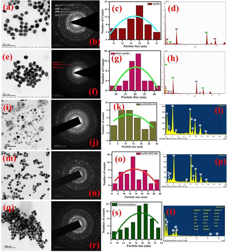
www.nature.com/scientificreports/
have been used for more efficient and safer delivery of a myriad of drugs exploring the advantages of long blood
circulation time, improved pharmacokinetics and reduced the side e ffects2,14–16.
Docetaxel (Dtx) is a semisynthetic anticancer mitotic ("antineoplastic" or "cytotoxic") chemotherapy drug in
the taxoid family. It is derived from the European yew tree (Texus baccata). The Dtx is recommended for optional
treatment in cancer patients with hormone-refractory metastatic prostate c ancer17–21. Dtx is a chemotherapeutic
medication (antitumor activity) against a wide range of solid tumors, including breast, lung, head, prostate, neck,
non-small cell lung and ovarian cancers19–26. Many researchers have been established that Dtx bind to β-tubulin,
which interferes with the normal function of the microtubules polymerization dynamics, dividing cell mitosis,
interface microtubule function and triggering apoptosis27,28. Dtx has the limitations of low water solubility, severe
allergic reactions and systemic toxicity29. To overcome these drawbacks of Dtx in clinical use, nanoformulations
based drug delivery systems like liposome30, inorganic n anoparticles25, magnetic n anoparticles31, polymeric
nanoconjugates32 and nanotubes33 that have been formulated for Dtx delivery.
Gold based nanoformulations are promising vehicles for Dtx administration due to fascinating properties
including tunable size and shape, biocompatibility and facile conjugation to b iomolecules34–37. Moreover, the
unique optical, electrical, physical and chemical properties (size and shape) of gold nanoparticles (AuNPs) make
them an excellent candidate for biomedical applications38, including targeted drug delivery39, photothermal
therapy40, cancer diagnosis41 and tumor i maging42. The surface plasmon resonance (SPR) properties of the AuNPs
play a major role in the biological system to allow their characterization and detection properties43.
Various synthesis methods have been used for the synthesis of AuNPs and nanoformulations. Surface func-
tionalization is one of the most important option for the development of nanoformulations with enhanced
recognition and biocompatibility for biomedical applications44. It has been reported that functionalization of
nanoformulations with folic acid (FA) enables targeted delivery of drugs due to its interaction with folate recep-
tors surrounding the cancer c ells45. Ngernyuang et al. reported the conjugation of FA onto the surface of PEG
functionalized AuNPs by simple physical agitation and mixing m ethod46. Neshastehriz et al. demonstrated
FA conjugation with AuNPs by mixing of FA onto the citrate-stabilized AuNPs by ultra-probe-sonication47.
However, the stability of the FA conjugated AuNPs has to be increased for its prolonged and safe action under
physiological conditions (pH and temperature). Hence, interest has been shown to formulate the covalent attach-
ment of FA onto the surfaces of the AuNPs. Owing to the high affinity of the thiol (SH) groups toward AuNPs,
bi-functional (SH-PEG-NH2) molecules can be used for functionalization enabling the formation of the gold-
sulfur bond (Au-S-PEG-NH2) and free functional groups of alkyne, carboxylate, and amine groups may be used
for covalent coupling with FA. This conjugation process has to be carefully optimized for better performance
of the nanocarrier48–51.
Cancer (an abnormal and uncontrolled proliferation of cells) is a serious disease leading to increasing mortal-
ity throughout the world. According to the National Cancer Institute (NCI), around 14 million new cases were
admitted and 26 million cancer-related deaths from 1991 to 2016. About 40% of cancer patients may be treated
and diagnosed during their lifetime. Currently, treatment and diagnosis of cancer are a high demand that remains
an ongoing challenge in oncology (pathophysiological and heterogeneous disease)52,53. Prostate cancer (PCa) is
the second most common disease in men and which leading causes of death in m en54. In a 2019 survey of PCa,
around 1,64,690 new cases and 29,430 deaths caused in the United States. However, PCa was typically treated by
radical prostatectomy (surgery), hormonal therapy, chemotherapy and radiation therapy. While the treatment,
cancer patients affect some side effects in different ways such as infertility, incontinence urinary, reduced sexual
desire and hormone-based side e ffects53,54. In these aspects, various research groups are focused on developing
effective imaging and drug delivery methods for prostate cancer. In general, cancer chemotherapy causes severe
toxicity due to the indiscriminate distribution of anticancer drugs upon systemic administration54. Therefore,
selective delivery of these therapeutic drugs to a target tumor cell is desirable. Different cell-specific markers/
receptors are overexpressed in various cancer cells which can be exploited for enhanced cancer selectivity and
endocytosis. The overexpressed FA binding receptors on prostate cancer will be targeted here by functionalizing
the nanocarrier with respective l igands55. The current treatment options available for cancer therapies are inad-
equate and spur demand for improved technologies. Rapid growth in nanotechnology towards the development
of nanomedicine provides a wide range of new materials, tools and possibilities, from earlier diagnostics and
improved imaging and therapies.
In the present work, we have established the sequential process for the synthesis of gold nanoformulations
(AuNPs-PEG-FA-Dtx): (i) AuNPs was synthesized by citrate ion (reducing and stabilizing agent) reduction
method, (ii) AuNPs were functionalized with SH-PEG-NH2 via the formation of an Au−S bond by the addition
method, (iii) the targeting ligand of FA was conjugated with the PEG functionalized AuNPs via N-ethyl-N-(3-
dimethylamino propyl) carbodiimide/N-hydroxy succinamide (EDC/NHS) by the coupling method followed
by covalent linkage method and (iv) the anti-cancer drug, Dtx were encapsulated with AuNPs-PEG-FA by the
non-covalent linkage method. The spectral, diffraction features and microscopic characteristics of the synthesized
AuNPs-PEG-FA-Dtx were examined by various analytical techniques. The encapsulation efficiency and drug
release profile of the nanoformulations were examined. The anticancer activity and drug delivery efficacies of
AuNPs nanoformulations, against prostate cancer cell line (PC3) was investigated in both free drug and drug
encapsulated nanoformulations.
Experimental section
Materials. Gold (III) chloride trihydrate (HAuCl4.3H2O, M.W. 393.83 g/mol), docetaxel (Dtx) (C43H53NO14,
M.W. 807.879 g/mol), folic acid (FA) (C19H19N7O6, MW. 441.40 g/mol), N-ethyl-N-(3-dimethyl aminopropyl)
carbodiimide (EDC) (C8H17N3, MW.155.245 g/mol), N-hydroxy succinimide (NHS) ( C6H5NO3, MW.115.09 g/
mol), Trypsin EDTA, Ham’s F12 (F-12 nutrient medium), l-Glutamine (C5H10N2O3, MW.146.146 g/mol),
Scientific Reports | (2021) 11:2808 | https://doi.org/10.1038/s41598-020-80529-1 2
Vol:.(1234567890)
www.nature.com/scientificreports/
sodium bicarbonate (NaHCO3), non-essential amino acids and fetal bovine serum (FBS) were procured from
Sigma Aldrich Chemicals, USA. Trisodium citrate (Na3C6H5O7, MW. 258.06 g/mol), N,N-dimethyl sulfoxide
(DMSO) (C2H6OS), disodium hydrogen phosphate (Na2HPO4, MW.141.96 g/mol) and orthophosphoric acid
(H3PO4, MW.97.994 g/mol) were purchased from Merck Chemicals, Mumbai. Ethanol (99.9%) was received
from Changshu Hongsheng Fine Chemical Co., Ltd, China. Premix WST-1cell proliferation assay kit was pro-
cured from Takara scientific company (USA). Human prostate cancer cell lines of PC3 were received from
National Centre for Cell Science (NCCS), Pune, India. PC3 cells were maintained with Ham’s F12 (F-12 Nutri-
ent Medium) and 10% FBS (maintenance medium). All the chemicals were used without any purification and
Millipore water was used throughout the experiment.
Methods
Synthesis of gold nanoparticles. The modified procedure has been used for the synthesis of gold nano-
particles from gold (III) chloride trihydrate by chemical reduction method using trisodium citrate as a reducing
agent25,56–58.
Briefly, 5 mL of 1 wt% of trisodium citrate in aqueous medium was added by dropwise to 200 mL aliquot of
1 mM HAuCl4·3H2O boiling with stirring the solution under reflux condition. After adding the reducing agent,
the reaction mixture color was changed from golden yellow to colorless. This reaction was continued until the
solution turned to wine red. This color change appeared within 3 min due to the reduction of Au (III) to Au0
and the reaction was completed within 5 min. The obtained colloidal suspension was allowed to cool at room
temperature and the colloidal suspension was sonicated for 15 min at 37 kHz. Followed by, the loosely bounded
citrate ions were removed by centrifugation for 30 min at 0 °C (10,000 rpm). The resulted AuNPs solution was
stored at 4 °C and used further characterization. Figure 1 illustrates the mechanism for the synthesis of gold
nanoformulations.
Functionalization of gold nanoparticles. Synthesized citrate capped AuNPs were functionalized with
SH-PEG-NH2. 2 mg of SH-PEG-NH2 was dissolved in 5 mL Millipore water, then added drop-wise into 100 mL
of 1 mM concentration of AuNPs solution and stirred for 12 h at room temperature. After functionalization, the
solution was maintained the pH range of 6.5 to prolong the stability of the nanoparticles. Further, the solution
was sonicated for 30 min and centrifuged against Millipore water at 10,000 rpm for 30 min at 0 °C. The stable
SH-PEG-NH2 functionalized AuNPs solution was stored at 4 °C and used further characterization.
Fabrication of FA conjugated AuNPs. In order to fabricate FA conjugated AuNPs, 1 mM of FA was dis-
solved in 10 mL of DMSO and sonicated for 15 min. The sonicated solution was centrifuged at 10,000 rpm for
20 min at 0 °C. The prepared FA solution was mixed with N-(3-dimethyl aminopropyl)-N′-ethylcarbodiimide
hydrochloride (EDC) and N-hydroxy succinamide (NHS) and the molar ratio of the FA: EDC: NHS was of
10:1:1 and the reaction solution was maintained the pH at 6.5. The reaction mixture was continuously stirring
for 12 h followed by sonication and centrifuged at 10,000 rpm for 30 min. The filtered solution was mixed with
SH-PEG-NH2 functionalized AuNPs and stirred continuously for 6 h. The resulted solution was sonicated and
centrifuged at 10,000 rpm for 10 min at 0 °C for three times. The fabricated AuNPs/PEG/FA solution was stored
at 4 °C and used further characterization.
Fabrication of docetaxel encapsulated AuNPs. 1 mM of Dtx was dissolved in 5 mL ethanol, water and
tween 80 (1:1:0.25 ratio) solution and sonicated for 15 min. The sonicated solution was centrifuged at 10,000 rpm
for 20 min at 0 °C. This solution was added into the SH-PEG-NH2 functionalized AuNPs in 50 mL beaker using
the non-covalent binding method with continuously stirred for 4 h. The prepared AuNPs/PEG/Dtx/FA solution
was sonicated in an ice bath for 30 min, followed by centrifuging at 10,000 rpm for 20 min at 0 °C. The obtained
solution was stored at 4 °C and used for further characterization.
Characterization of Dtx encapsulated gold nanoformulations. The physicochemical properties
and step-by-step conjugation of the synthesized AuNPs nanoformulations were extensively characterized by var-
ious analytical techniques. UV–Vis absorption spectra were carried out by using Agilent diodaris spectropho-
tometer and Cary-8453, USA. The surface functional groups of the nanoformulations were determined by FT-IR
spectrophotometer (Bruker, Vertex, 80 V, USA). The crystallinity and phase purity of the nanoformulations
were studied by XRD analysis (Bruker D8 advance PXRD and Rigaku X-ray diffractometer, Smart lab, UK). The
chemical composition and oxidation state were evaluated by X-ray photoelectron spectroscopy (XPS, Omicron
Nanotechnology, ESCA-14 (Germany). The surface morphology and elemental composition of the nanoformu-
lations were examined by a high-resolution transmission electron microscope (HR-TEM) ((JEOL-2100-JEM)
and (Bruker)) and field emission scanning electron microscope (FE-SEM) (Hitachi, SU-6600—Japan) instru-
ment with energy dispersive X-ray spectroscopy (EDS). The chemical bond and surface modification of the
nanoformulations were determined by Raman spectrophotometer (Xplora Plus, Raman spectrometer, Horiba,
Japan with laser excitation of 785 nm). The anticancer activity of the AuNPs nanoformulations was analyzed by
ELIZA Reader (Enzyme-Linked Immunosorbent Assay) microplate reader (Robonik, Mumbai, India) with the
excitation wavelength of 460 nm.
Encapsulation efficiency of gold nanoformulations. The percentage of drug encapsulated in gold
nanoformulations was determined by separating the un-entrapped drug from nanoformulations by centrifuga-
tion at 10,000 rpm for 30 min using cooling centrifuge (CPR-Plus 24, Remi Instruments, Mumbai, India). The
Scientific Reports | (2021) 11:2808 | https://doi.org/10.1038/s41598-020-80529-1 3
Vol.:(0123456789)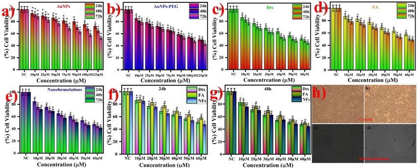
www.nature.com/scientificreports/

                                            Figure 1.  Mechanism for the synthesis of gold nanoformulations.

                                            clear supernatant was analyzed for the contents of Dtx by measuring absorbance in a UV–Visible spectropho-
                                            tometer at λmax = 230 nm59. The percentage encapsulation efficiency and loading capacity were calculated by
                                            the following equation,

          Scientific Reports |   (2021) 11:2808 |               https://doi.org/10.1038/s41598-020-80529-1                                           4

Vol:.(1234567890)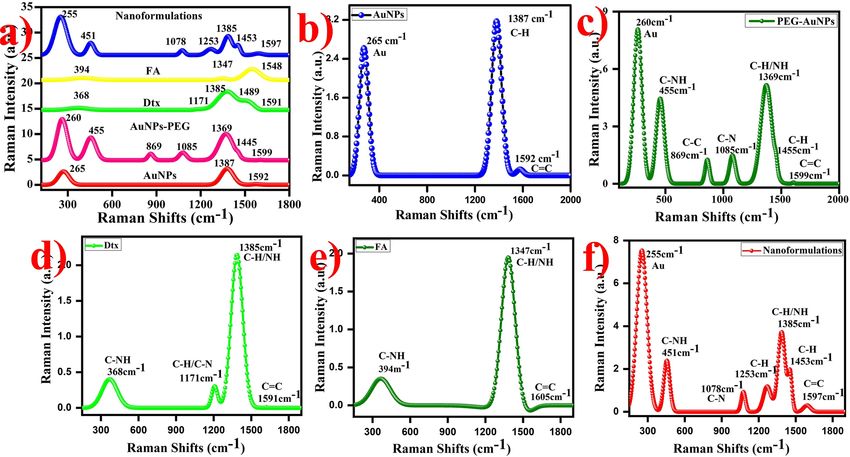
www.nature.com/scientificreports/
Encapsulation efficiency = Amount of the drug in formulation/Total amount of drug ×100
where Dtxt is the total amount of Dtx used in the preparation of nanoformulations and D
txf is the unentrapped
Dtx present in the supernatant.
In vitro drug release. In vitro drug release kinetics of optimized Dtx encapsulated AuNPs nanoformula-
tions and the free drug was investigated by the dialysis membrane method. Briefly, 75 mL of phosphate-buffered
solution (pH 7.4) and 5 mL of 0.1% tween 80 was taken as release media in 150 mL borosil glass beaker. After
that, 5 mL of nanoformulations were poured into the dialysis membrane (molecular weight cut-off 12–14 kDa)
and immersed in an 80 mL PBS solution with the help of a glass rod. The bag was immersed centrally into the
release media using a burette stand. The beaker was placed on the magnetic stirrer with maintained a constant
rotation of 300 rpm with a magnetic pellet. The release kinetics was predetermined at different time intervals
like 1–72 h. At a notable time interval, 3 mL of drug release medium was aspirated and replaced 3 mL fresh drug
release media and also maintained the constant bath volume. The absorbance of the samples was recorded in a
UV–Vis spectrophotometer at λmax of Dtx at 230 nm, FA at 280 and 344 nm, and AuNPs at 528 nm. The calibra-
tion curve was calculated with different concentrations of Dtx in the PBS solution.
The in vitro drug release kinetics were analyzed by different release kinetic models like zero-order, first-order,
Higuchi kinetics, Korsmeyer–Peppas model, and Hixon–Crowell model which enables the identification of the
R2 value (correlation coefficient)60.
Cytotoxicity analysis of gold nanoformulations. In vitro cytotoxicity analysis used to investigate the
cell viability or metabolic activity of prostate cancer cell line (PC3) against gold nanoformulations. This colori-
metric assay is highly sensitive and reliable to the transformation of the metabolic activity of the cells. Briefly,
100 µL (2500 cells per well) of PC3 cells were harvested, counted, and seeded in flat-bottom 96 well plate (Corn-
ing USA) and then incubated for 24 h at 37 °C under 5% C O2 atmospheric conditions which used to adhere the
cells. After 24 h, the cells were washed with PBS (Phosphate Buffer Saline solution) solution twice. Subsequently,
the cells were treated with different concentration of synthesized AuNPs nanoformulations (10 to 60 µM)
includes free AuNPs (10 to 125 µM), PEG functionalized AuNPs (10 to 125 µM), FA (10 to 60 µM), and Dtx (10
to 60 µM) and incubated for different time intervals like 24 h, 48 h, and 72 h. The AuNPs nanoformulations free
cells are considered as control and the experiments were carried out in triplicate. The selected concentration of
the nanoformulations which enables the drug particles were reached in the cancerous cells at the plasma level.
After the incubation time interval, the HAMS F12 medium was aspirated and washed with PBS two times. Then,
3 µL of Premix WST assay solution was added in each well and the cells incubated for 4 h in dark condition. In
this period, the metabolically active cells to transform WST into the formation of insoluble Formazan crystals.
Followed by, the optical absorbance of cells was measure immediately at 460 nm using ELIZA (Enzyme-Linked
Immunosorbent assay) microplate reader (Robonik, Mumbai, India). All the experiments were carried out in
triplicate. The cell viability was calculated by the following equations.
Cell viability (%) = Abssample /Abscontrol × 100
Statistical analysis. The statistical analysis was expressed as a mean ± standard deviation by SPSS 16.0
software (Name: IBM SPSS Software, Version Number: SPSS16.0, and URL Link: https://www.ibm.com/in-en/
products/spss-statistics.Chicago, IL,USA).The statistical comparison was carried out all the AuNPs nanoformu-
lations by one-way analysis of variance (ANOVA) by post hoc hypothesis testing (Tukey test). The p-value is less
than 0.05 (p < 0.05) was considered to be a significance level.
Results and discussion
Synthesis of Dtx encapsulated AuNPs nanoformulations. The stability of the colloidal AuNPs is
a very important feature for surface functionalization. The colloidal suspension of AuNPs was synthesized by
chemical reduction method (citrate ion acts as a reducing agent and stabilizing agent). UV–Vis absorption spec-
tra of the citrate capped AuNPs exhibited the Surface Plasmon Resonance (SPR) band at 527 nm as shown in
Fig. 2a. The magnitude of the SPR peak depends on the concentration and the morphology of the synthesized
AuNPs. This absorption spectrum indicates that the increased concentration of the AuNPs and the SPR peak
height also increased to some extent the absorption peak gets broaden. Followed by, AuNPs were functionalized
with thiolated PEG amine (SH-PEG-NH2). During the thiolation of AuNPs, AuNPs appeared rapid agglomera-
tion which exhibited the SPR signal of PEG functionalized AuNPs observed at 528 nm (Fig. 2b). To overcome
this agglomeration of AuNPs, during the thiolation process the AuNPs solution pH was adjusted at 6.561. This
pH condition, PEG functionalized AuNPs are highly stable to prevent the agglomeration62. The stable solution
of PEG functionalized AuNPs were conjugated with Au–S bond and amine (–NH2) groups. Thereafter, PEG
functionalized AuNPs were conjugated with FA by using EDC/NHS coupling method (the free amine (–NH2)
groups were bonded with the carboxylic group (–COOH) and formed a strong bond between PEG-AuNPs and
FA). UV–Vis absorption spectra of FA were observed in the two bands at 277 and 367 nm (Fig. 2d). The FA
conjugated PEG functionalized AuNPs, it was observed that the one molecule of FA was attached per six mol-
ecules of PEG functionalized AuNPs. This result indicates that the FA was partially attached over the surface of
PEG functionalized AuNPs, which exhibited the steric hindrance of the bulky structure of FA. The SPR signal of
the FA conjugated PEG functionalized AuNPs indicated that the 528 nm, 344 nm, and 277 nm (Fig. 2e). These
results suggest that the FA were strongly attached over the surfaces of PEG functionalized AuNPs. Subsequently,
Scientific Reports | (2021) 11:2808 | https://doi.org/10.1038/s41598-020-80529-1 5
Vol.:(0123456789)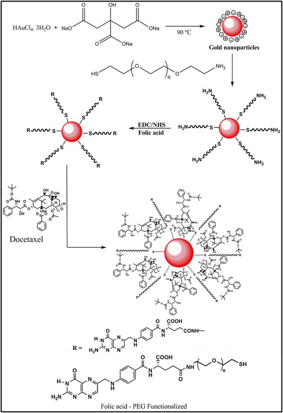
www.nature.com/scientificreports/
Figure 2. UV–Vis absorption spectra of the synthesized gold nanoformulations (a) AuNPs (λmax = 527 nm),
(b) AuNPs-SH-PEG-NH2 (λmax = 528 nm), (c) Dtx (λmax = 231 nm), (d) FA (λmax = 277 and 344 nm), (e)
AuNPs-PEG-FA (λmax = 277, 344 and 528 nm) and (f) AuNPs nanoformulations (λmax = 228, 277, 344 and
528 nm) and inset photographic image of gold nanoformulations at pH = 6.5.
the anticancer drug of Dtx was conjugated by the non-covalent linkages method, which enables the physical
adsorption of Dtx with FA conjugated PEG functionalized AuNPs. UV–Vis spectra of the Dtx were observed at
231 nm ascribed to π–π* transition of carbonyl and hydroxyl groups were present in the sample (Fig. 2c). During
the synthesis of AuNPs nanoformulations (PEG-AuNPs-FA-Dtx), various reaction parameters were optimized
such as the concentration of the reactant, concentration of reducing agent, temperature, reaction time, and pH,
which enables the formation of highly stable nanoformulations. The resulted AuNPs nanoformulations were
examined by UV–Vis absorption spectroscopy. AuNPs nanoformulations (AuNPs-PEG-FA-Dtx) were observed
at the peak at 228, 279, 355, and 528 nm (Fig. 2f). These results indicate the formation of AuNPs nanoformula-
tions with step-by-step conjugation of FA and Dtx.
Characterization studies of nanoformulations. Surface structural analysis. The surface functional
groups and chemical bonding of the synthesized nanoformulations were examined by FTIR spectra shown in
Fig. 3a. The FTIR spectrum of citrate capped AuNPs exhibits shoulder band at 3301 cm−1 and 1636 cm−1 ascribed
to the O–H stretching vibration and C=C stretching vibration of carbonyl groups is present in the AuNPs, re-
spectively (Fig. 3b). The broad band at 2111 cm−1 corresponds to the CO2 (atmosphere)/CΞC week stretching
vibration of alkyne. The doublet peak appeared at 2907 cm−1 and 2849 cm−1 attributed to the C–H stretching
vibration of carboxyl groups also present in the AuNPs. PEG functionalized AuNPs exhibited the major signals
at 3305 cm−1, 2906 cm−1 and 2841 cm−1 (O–H and C–H stretching vibration of carbonyl groups), 2118 cm−1
(atmospheric CO2/CΞC week stretching vibration of alkyne), 1765 cm−1 (C=O strong stretching vibration),
1636 cm−1 (C=C stretching vibration of carboxylic acid) 1454 cm−1 (S=O stretching vibration of sulfonate) and
1074 cm−1 (C–N stretching vibration of amine) present in the sample (Fig. 3c)62. FTIR spectrum of the Dtx indi-
cates the sharper band at 3316 cm−1, 2973 cm−1 and 2885 cm−1 attributed to the N–H and C–H stretching vibra-
tion of secondary amine and alkane, respectively (Fig. 3d)64. A small intense peak at 1636 cm−1 corresponds to
the N–H bending vibration, 1447 cm−1 ascribed to the C–H bending vibration of C H2 and C
H3 and a week band
at 1359 m−1 corresponds to the O–H bending vibration of carboxylic group. A band at 1104 cm−1, 1057 cm−1 and
885 cm−1 corresponds to the C=O, C–N and C=C bending vibration of carbonyl, amide and alkane groups are
appeared in the Dtx, respectively. The appearance of the FTIR spectrum of FA exhibited the characteristic peaks
at 3454 cm−1 (primary amine), 2998 cm−1 and 2898 cm−1 (C–H stretching vibration of alkane), and 1659 cm−1
(C=N amine, conjugated double bond). The doublet band at 1447 cm−1 and 1301 cm−1 representing C–H bend-
ing vibration and bending of alkene are present in the FA, respectively63. The peaks at 1031 cm−1 and 943 cm−1
(S=O/C–N and C=C stretching vibration of sulfoxide/amide and alkane) present in the FA (Fig. 3e). It is evident
that the peak at 3454 cm−1, 1659 cm−1 and 1031 cm−1 corresponds to the formation of primary amine and sulfox-
ide groups are conjugated over folic acid using EDC/NHS coupling method.
FTIR spectra of AuNPs nanoformulations exhibits the major signals at 3308 cm−1 corresponds to the OH
stretching vibration of carboxylic groups (Fig. 3f). The band at 2907 cm−1 corresponds to the CH stretching
vibration of alkanes. The band at 2118 cm−1 corresponds to the atmospheric C O2/CΞC week stretching vibra-
tion of alkyne. The strongest peak at 1630 cm−1 attributed to the C=C stretching vibration of alkane. The intense
band at 1439 cm−1 and 1316 cm−1 ascribed to the C–H and O–H plane bending vibration of alkanes. The weak
band observed at 1009 cm−1 is attributed to the C–N/S=O stretching vibration of amine and sulfonate are present
anoformulations65. A small band at 905 corresponds to the C=C bending vibration of alkane is present
in the n
in the nanoformulations. Evidently, the FTIR spectra exhibited the FA conjugation with PEG functionalized
Scientific Reports | (2021) 11:2808 | https://doi.org/10.1038/s41598-020-80529-1 6
Vol:.(1234567890)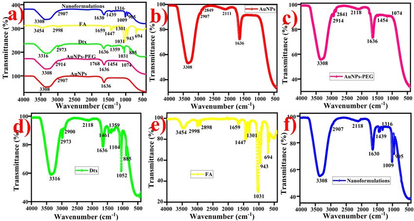
www.nature.com/scientificreports/
Figure 3. FTIR spectra of gold nanoformulations, (a) combined spectra of nanoformulations, (b) AuNPs, (c)
AuNPs-PEG (d) Dtx, (e) FA and (f) AuNPs nanoformulations (AuNPs-PEG-FA-Dtx).
Figure 4. XRD pattern of the gold nanoformulations (a) Combined spectra of AuNPs nanoformulations, (b)
AuNPs (c) AuNPs-PEG, (d) Dtx (e) FA and (f) gold nanoformulations were observed the FCC crystal structure.
AuNPs via amide bond as well as the attachment of Dtx onto the AuNPs-PEG-FA-Dtx nanoformulations via
hydrogen bonding.
The crystalline structure of gold nanoformulations. The crystallinity, phase purity, stability and chemical bond-
ing of the AuNPs nanoformulations were determined by XRD analysis as shown in Fig. 4a. XRD pattern exhibits
the diffraction peaks at 2θ = 36.45°, 38.21°, 44.39°, 64.76° and 77.76° corresponds to the Bragg reflection signals
at (101), (111), (200), (220) and (311) indicates the standard metallic gold ( Au0) [(Joint Committee on Powder
Diffraction Standards, USA-JCPS ID # 00-002-1095] with lattice parameters of a = 4.065 Å and the space group
of Fm-3 m. This Bragg reflection signals exhibited the face centered cubic (FCC) crystal structure and the shoul-
der peak was observed around 30–40° which attributed to the highly crystalline nature of AuNPs (Fig. 4b)66,67.
Scientific Reports | (2021) 11:2808 | https://doi.org/10.1038/s41598-020-80529-1 7
Vol.:(0123456789)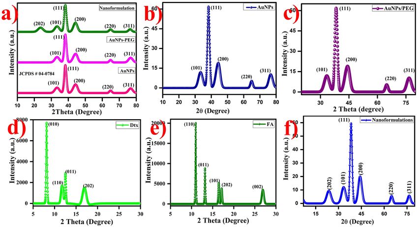
www.nature.com/scientificreports/
The size of the nanoparticles was measured indirectly using a broad bottom width of (111) reflection which
indicates the smaller size of the nanoparticles. Debye–Scherrer’s equation was used to determine the size of
the nanoparticles by (111) width of the Bragg reflection signal. The size of the AuNPs was found to be ~ 18 nm.
The (111) reflection peak exhibited the strong shoulder peak rest of the peaks are weak peaks which indicate
the predominant orientation. There is no small impurities peaks were observed which shows the synthesized
AuNPs are highly pure. Interestingly, the XRD pattern of the SH-PEG-NH2 functionalized AuNPs exhibited
diffraction band is similar to AuNPs (Fig. 4c), whereas, the XRD pattern of the characteristics diffraction peak
of FA at 10.9°, 13.0°, 16.2°, 16.90°, and 27.2° corresponds to the (110), (011), (101), (220) and (002), respectively
(Fig. 4d)68. The XRD pattern of the pure Dtx exhibits the signals at 2θ values of 8.2°,12.18°, 14.12°, 16.89° cor-
responds to the (010), (110), (011) and (202) crystal planes, respectively (Fig. 4e)69.
Finally, the XRD pattern of the synthesized AuNPs-PEG-FA-Dtx exhibits the diffraction signals at 2θ values
of 22.39°, 36.45°, 38.21°, 44.39°, 64.76° and 77.76° corresponds to the (202), (101), (111), (200), (220) and (311)
crystal planes, respectively (Fig. 4f). On comparison with FA, Dtx and AuNPs, the diffraction peaks of the FA and
Dtx signals are slightly suppressed due to the strong attachment between FA-AuNPs-PEG-Dtx. Thus, the XRD
analysis confirmed the obtained AuNPs were FCC crystal structure and the step-by-step functionalization of SH-
PEG-NH2 and FA over AuNPs and Dtx encapsulated over the surface of AuNPs-PEG-FA. The synthesized AuNPs
nanoformulations were highly stable in the aqueous medium over the period of one year at room temperature.
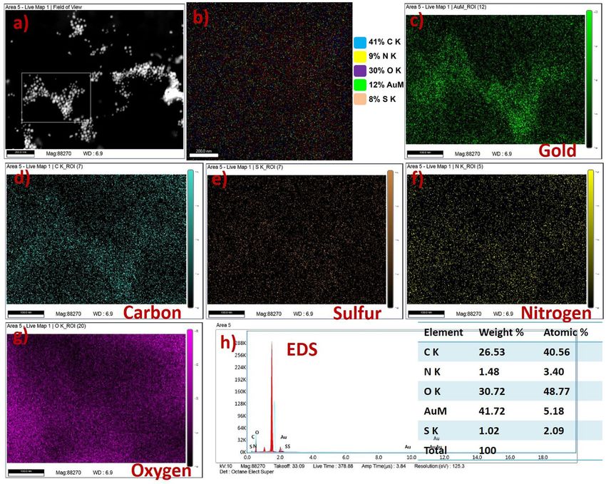
Morphological and elemental mapping analysis of AuNPs nanoformulations. The surface morphology and
microstructure of the synthesized gold nanoformulations were determined by HR-TEM analysis. Typical TEM
micrographs of the citrate capped AuNPs indicates that the particles are spherical in shape and uniformly dis-
tributed over the copper substrate with an average dimension of 16 nm (Fig. 5a–d). AuNPs were uniformly
attached over the SH-PEG-NH2 (Fig. 5e–h). The average diameter is 24 nm, the slight increase in the dimension
of AuNPs is due to the strong interaction between (Au–S) AuNPs-PEG. Figure S1a–d exhibits the FA particles
are spherical in shape and the particles are observed the average dimension of 90 nm. After, FA conjugate over
the surface of AuNPs-PEG exhibited particles are spherical in shape with an average dimension of 16 nm with
slight changes in their shape (Fig. 5i–l). This result confirms that the FA conjugated over the AuNPs-PEG and the
interaction between FA and AuNPs through hydrogen bonding. Figure S1e–h depicts the Dtx particles observed
the core–shell morphology and uniformly distributed over the substrate with an average dimension of 290 nm.
Figure 5m–p exhibits the Dtx encapsulated PEG functionalized AuNPs observed the particles are spherical in
shape with slight changes in the size of the particles. Finally, the gold nanoformulations (AuNPs/PEG/Dtx/FA)
exhibits that the particles are spherical in shape with uniformly distributed over the substrate with an average
dimension of 18 nm, without any aggregation (Fig. 5q–t). Figure 5d shows the SAED pattern of the AuNPs and
the particles are highly crystalline in nature. The crystal lattice spacing distances of AuNPs is about 4.0786 Å.
These results are in accordance with the (111), (200), (220) and (311) diffraction planes of gold (JCPDS ID #
00-002-1095). Thus, the results indicate that the gold particles are arranged in a face centered cubic crystals
system with λ = 1.5406 Å. These results were compared with the XRD pattern. The SAED pattern of the gold
nanoformulations was observed the particles are highly crystalline. Hence, this result confirms the step-by-step
conjugation of Dtx encapsulated gold nanoformulations are highly crystalline nature without changing the crys-
talline morphology.
The elemental compositions of the synthesized gold nanoformulations were analyzed by EDS analysis (Fig. 5t).
The results indicate the carbon of 34.09 wt%, nitrogen of 10.06 wt%, oxygen of 43.22 wt%, Sulfur of 0.22 wt% and
gold of 12.40 wt% elements are present in the sample. These results suggest that the synthesized AuNPs nanofor-
mulations are highly pure with good crystallinity. Hence, TEM and EDS analyses confirmed the microstructure,
size, morphology and elemental composition of the AuNPs nanoformulations during the stepwise conjugation
and drug formulation. The synthesized nanoformulations were highly stable in aqueous medium over the period
of one year at room temperature.
Field emission scanning electron microscope. Elemental mapping analysis of the synthesized nano-
formulations was evaluated by FESEM analysis. Figure 6a shows the AuNPs nanoformulations exhibited low and
high magnification. The high magnification image of nanoformulations exhibited the particles are uniformly
arranged over the substrate which is used for elemental mapping analysis. These images could be used as a
selected area for the mapping analysis. Elemental mapping analysis of the nanoformulations was clearly indicat-
ing the Dtx and FA particles embedded with PEG functionalized AuNPs (Fig. 6b). The mapping images of nano-
formulations showed the embedded elements of CK = 41%, NK = 9%, OK = 30%, metallic gold (AuM = 12%),
and Sk = 8%. Figure 6c depicts the mapping image of gold particles in the metallic form which indicates the gold
particles are present in the sample. The mapping images of carbon, sulfur, nitrogen, and oxygen were exhibited
in Fig. 6d–g, respectively. This image represents the synthesized gold nanoformulations are highly pure without
any impurities.
The elemental composition of the gold nanoformulations was evaluated by EDS spectroscopy as shown in
Fig. 6h. The Dtx encapsulated FA conjugates PEG functionalized gold nanoformulations exhibited the elements of
carbon (CK = 26.53%), sulfur (SK = 1.02), nitrogen (NK = 1.48%), oxygen (OK = 30.72), and gold (AuM = 41.72).
This result represents the synthesized nanoformulations were systematically conjugated with AuNPs which exhib-
ited the nanoformulations are highly pure. The detail FE-SEM images of AuNPs, PEG functionalized AuNPs,
FA, AuNPs-PEG-FA, Dtx and gold nanoformulations were provided in supplementary section (Fig. S2a–i).
XPS analysis. The XPS analysis was used to determine the oxidation state and chemical composition of the
nanoformulations. Figure 7a exhibits the survey spectrum of the AuNPs, AuNPs-PEG, FA, FA-AuNPs-PEG,
Scientific Reports | (2021) 11:2808 | https://doi.org/10.1038/s41598-020-80529-1 8
Vol:.(1234567890)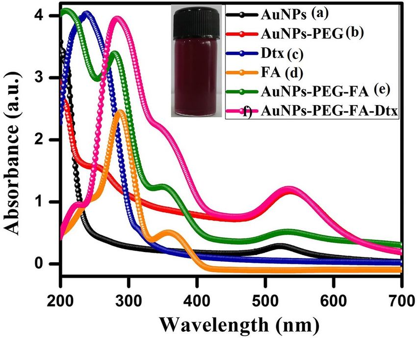
www.nature.com/scientificreports/
Figure 5. HR-TEM images of the gold nanoformulations (a–d) AuNPs TEM image, SAED, particle size
distribution and EDS spectrum followed by the similar characteristics were carried for remaining particle, (e–h)
PEG functionalized AuNPs, (i–l) FA conjugated AuNPs-PEG, (m–p) Dtx loaded AuNPs-PEG and (q–t) drug
encapsulated gold nanoformulations.
Dtx, AuNPs-PEG-Dtx, and AuNPs-PEG-FA-Dtx, which observed the strongest band of gold, sulfur, carbon,
nitrogen, and oxygen (Au4f, S2p, C1s, N1s, and O1s), respectively. Figure 7b depicts the survey spectrum of the
AuNPs. The valence band and binding energy of 4f core level spectrum of AuNPs have observed the peaks at
85.3 eV and 88.9 eV corresponds to the Au 4f7/2 and Au 4f5/2 spin orbitals, respectively (Fig. 7c)25. These bind-
ing energies were compared to the respective core levels of bulk Au crystals. Evidently, this result exhibited the
metallic types of gold (Au0) were present in the sample. Moreover, the resulted value of Au 4f7/2 and Au 4f5/2
exhibits the narrow width revealed that only a single element of gold was present in the system which indi-
cates the synthesized AuNPs are highly stable. The Au4f core level spectrum of the SH-PEG-NH2 functionalized
AuNPs exhibited the two components at 85.22 eV and 89.01 eV corresponds to the Au 4f7/2 and Au 4f5/2 of spin
Scientific Reports | (2021) 11:2808 | https://doi.org/10.1038/s41598-020-80529-1 9
Vol.:(0123456789)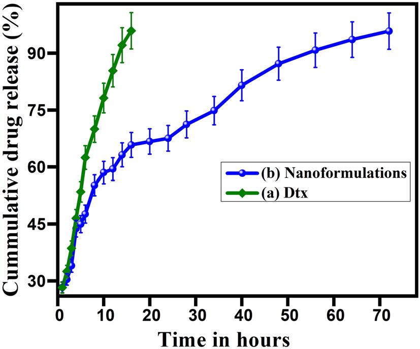
www.nature.com/scientificreports/

                                            Figure 6.  FESEM image and Elemental mapping with EDS spectrum of gold nanoformulations, (a) high
                                            magnification image gold nanoformulations with selected area for mapping analysis, (b) Elemental mapping
                                            images of gold nanoformulations with corresponding elements (C K = 41%, N K = 9%, O K = 30%, AuM = 12%
                                            and S K = 8%), (c) mapping image of metallic gold nanoparticles, (d) carbon, (e) sulfur, (f) nitrogen, (g) oxygen
                                            and (h) EDS spectrum of gold nanoformulations.

                                            orbitals of PEG-AuNPs bonds with alkyl thiol self-assembled AuNPs (Au–S), respectively (Fig. 7f) whereas, the
                                            FA-PEG-AuNPs exhibited the two components at 83.55 eV and 86.88 eV corresponds to the core level of Au
                                            4f7/2 and Au 4f5/2 spin-orbital, respectively and this value confirms the XPS traces of alkyl thiol self-assembled
                                            bonded over AuNPs (Fig. S3h). Figure S3a depicts the XPS survey spectrum of the free docetaxel which observed
                                            the traces of C1s, N1s, and O1s, whereas, Fig. 7k exhibits the XPS survey spectrum of gold nanoformulations.
                                            The Au4f core level XPS spectrum of the AuNPs-PEG-FA-Dtx exhibited the two components at 83.21 eV and
                                            87.56 eV corresponds to the Au 4f7/2 and Au 4f5/2 spin-orbital, respectively (Fig. 7l). Hence, these XPS results
                                            confirm the functionalization of AuNPs nanoformulations.
                                                The C1s core level spectra of the AuNPs exhibited the two XPS traces at 286.93 eV and 290.92 eV attributed to
                                            the C–O–H, C=C/C–O–C groups of the carbon atom, respectively (Fig. 7d), whereas, PEG functionalized AuNPs
                                            exhibited three XPS signals at 286.76 eV, 287.74 eV and 290.68 eV correspond to the C−O or C−S, C=O(amide)
                                            and O–C=O group of the carbon atom, respectively (Fig. 7h)70–72. The appearance of the C1s XPS spectra of
                                            FA-AuNPs-PEG exhibited the signals at 284.02 eV, 285.69 eV ascribed to the C=C/C=N and C−C/C−H groups
                                            confirmed the attachment of FA, respectively (Fig. S3j). The Shifting of carbonyl carbon bond towards the lower
                                            binding energy level compared to PEG-AuNPs indicated the conversion of acid carbonyl group into amide car-
                                            bonyl group and hence, the FA conjugation over the AuNPs-PEG via the formation of the amide bond. The C1s
                                            spectrum of FA exhibited the three XPS traces at 286.63 eV, 288.03 eV and 290.52 eV correspond to the C–N,
                                            C=O, and O–C=O, respectively (Fig. S3e), whereas, the Dtx spectrum exhibits the two signals at 297.96 eV and
                                            301.02 eV corresponds to the O–C=O and C–O–C groups of carbon atoms (Fig. S3b). The Dtx encapsulated
                                            with FA conjugated PEG functionalized AuNPs exhibited the two components corresponding to the 284.05 eV
                                            and 286.69 eV corresponds to the C=C/C=N and C–C/C–H groups of the carbon atom, respectively (Fig. 7p).

          Scientific Reports |   (2021) 11:2808 |                https://doi.org/10.1038/s41598-020-80529-1                                                 10

Vol:.(1234567890)
www.nature.com/scientificreports/
Figure 7. XPS spectra of gold nanoformulations, (a) overall survey spectra, (b–d) Survey spectrum of AuNPs
and corresponding high resolution spectra as follows (c) Au4f5/2 and Au4f7/2, (d) C1s, (e) O1s, (f–j) high
resolution spectra of PEG functionalized AuNPs (f) Au4f, (g) S2p, (h) C1s, (i) N1s and (j) O1s and (k–p) Survey
spectrum of gold based nanoformulations and corresponding high resolution spectra (k) survey spectrum, (l)
Au4f, (m) S2p, (n) C1s, (o) N1s and (p) O1s.
The O1s XPS spectra of the AuNPs exhibited a band at 534.37 eV attributed to C–O/C=O groups of the oxygen
atom (Fig. 7e)25,58,73. PEG functionalized AuNPs exhibited the two XPS traces at 534.19 eV and 535.53 eV cor-
responds to the C=O and C–O–H groups of the oxygen atom, respectively (Fig. 7j). The appearance of O1s XPS
spectra of the AuNPs-PEG-FA exhibited the signals at 532.65 eV corresponds to the carbonyl group of FA and
the minor component at 533.78 eV attributed to the C–O–C group of the oxygen atom (Fig. S3l). The O1s traces
of FA exhibited the components at 533.83 eV and 534.18 eV corresponds to the C–O–C and C–O–H groups of
the oxygen atom, respectively (Fig. S3g). The XPS spectra of Dtx exhibited the signals at 544.9 eV and 545.66 eV
corresponds to the C-O and C=O groups of the oxygen atom present in the samples, respectively (Fig. S3d). The
gold nanoformulations exhibit the O1s traces at 533.09 eV and 534.26 eV correspond to the free carboxylic acid
and C–O–H group of the oxygen atom (Fig. 7o). This result showed that the O1s trace of the amide carbonyl
groups of Dtx was encapsulated over AuNPs-PEG-FA via amide carbonyl groups at 533.09 eV, which confirms
the step-by-step conjugation of Dtx encapsulated AuNPs-PEG-FA nanoformulations.
The S2p XPS spectra of the PEG functionalized AuNPs exhibited the two signals at 161.18 eV and 162.38 eV
along with a 1.2 eV gap corresponds to the S2p3/2 and S2p1/2, (Fig. 7g)74 of the SH-PEG-NH2 functionalized
AuNPs, respectively. Spampinato et al. reported that the unbounded sulfur observed at 164 eV corresponds to
S2p3/2, while, the sulfur traces bound over the surface of AuNPs appeared at 162 eV. The appearance of the FA
conjugated PEG functionalized AuNPs exhibited the S2p3/2 and S2p1/2 XPS signals at 162.02 eV, 163.56 eV and
164 eV correspond to the Au–S, R-SH and S groups of sulfur atoms, respectively (Fig. S3i). The gold nanoformu-
lations exhibit the S2p traces at 162.18 eV and 163.80 eV corresponds to the Au–S and S groups of sulfur atoms
present in the sample, respectively (Fig. 7m)75. Similarly, the entire spectrum exhibited the S2p3/2 XPS traces at
162 eV confirm the conjugation of PEG-FA-Dtx over AuNPs. The AuNPs did not show any N1s of XPS traces,
whereas, N1s spectrum of the PEG-Functionalized AuNPs exhibited the XPS components at 401.27 eV and
Scientific Reports | (2021) 11:2808 | https://doi.org/10.1038/s41598-020-80529-1 11
Vol.:(0123456789)www.nature.com/scientificreports/
Figure 8. Raman spectra of the gold nanoformulations (a) Combined spectra of AuNPs nanoformulations, (b)
AuNPs (c) AuNPs-PEG, (d) Dtx, (e) FA and (f) AuNPs-PEG-FA-Dtx.
402.58 eV corresponds to the C-NH2 and (C=O)N(C=O) groups of nitrogen atoms, respectively (Fig. 7i)25,58,76.
The FA exhibited the N1s signals of 400.01 eV and 401.08 eV corresponds to the C–N/N−H and NH–C–O groups
of nitrogen atoms, respectively (Fig. S3f), whereas, Dtx was observed the N1s energy of 401.2 eV and 402.78 eV
corresponds to the C-NH2 and N–C=O–N groups of nitrogen atoms, respectively (Fig. S3c). The appearance
of the FA conjugated PEG functionalized AuNPs exhibited the N1s XPS signals at 399.93 eV and 400.67 eV
corresponds to the C–N/N−H and NH–C–O groups of the nitrogen atoms, respectively (Fig. S3k). The gold
nanoformulations exhibited the N1s XPS components at 401.07 eV and 403 eV corresponds to the C–NH2 and
N–C=O–N groups of the nitrogen atom present in the nanoformulations, respectively (Fig. 7. (n)). Hence, these
XPS spectra support the stepwise formation of Dtx encapsulated FA conjugated PEG functionalized AuNPs. The
synthesized gold nanoformulations are highly pure without any impurity peaks were appeared.
Raman spectra of gold nanoformulations. Raman spectroscopy is used to identify the structural fin-
gerprints of the particles. Raman spectra of AuNPs exhibited band at 267 cm−1 corresponds to the Au(III) to Au0.
The shoulder band at 1385 cm−1 and 1585 cm−1 is corresponds to the C–H and C=C of carbon (citrate ions),
respectively (Fig. 8b)64, whereas, PEG functionalized AuNPs exhibited the Raman spectra appeared the signals at
260.81 cm-1,455 cm−1 (C–NH amide), 869 cm−1 (C–N in-plane bending), 1085 cm−1(C–C stretching of carbonyl
groups), 1369 cm-1, 1445 cm−1 and 1599 cm−1 corresponds to the Au0, C–O, C–H and C–O–H strongly observed
on the surface, respectively (Fig. 8c). The appearance of the FA exhibited the bands at 394 cm−1, 1347 cm−1
and 1548 cm−1 corresponds to the amide group, carbonyl and carboxylic acid groups were present in the sam-
ple, respectively (Fig. 8d)65. The Dtx exhibited the Raman spectra observed the band at 368 cm−1, 1171 cm−1,
1385 cm−1, 1489 cm−1 and 1591 cm−1 corresponds to the amide group (C–NH), carbonyl (C–H) groups, C–H,
N–H and C=C present in the Dtx surfaces, respectively (Fig. 8e)77.
The Raman spectra of the synthesized gold nanoformulations exhibited the band at 255 cm−1, 451 cm−1,
1078 cm−1, 1253 cm−1, 1385 cm−1, 1453 cm−1 and 1597 cm−1 (Fig. 8f). From the spectra, the shoulder peak
at 260 cm−1 corresponds to the A u0 strongly observed in the surface. The peaks at 451 cm−1, 1078 cm−1 and
1253 cm−1 are attributed to the C–NH, C–N, C–H of carbonyl and amine groups are present in the sample,
respectively. These peaks indicates the conjugation of AuNPs with FA and Dtx. The major peaks at 1385 cm−1,
1455 cm−1 and 1597 cm−1 are ascribed to the C–H/NH, C–H and C=C of bending frequency of amine and car-
bonyl groups due to the formation of FA and Dtx. The combined Raman spectra of gold nanoformulations are
provided due to comparison between the nanoparticles (Fig. 8a). Thus, the results suggest that the synthesized
AuNPs are highly pure.
In vitro analysis of gold nanoformulations. The encapsulation efficiency of gold nanoformulations
was observed at approximately ~ 95%. Here, 1 mM con of Dtx solution was prepared and encapsulated with
PEG functionalized FA conjugated AuNPs by the non-covalent method. The resulted nanoformulations were
sonicated and centrifuged at 10,000 rpm at 0 °C for 15 min. The obtained solution was used for further charac-
terization and drug release studies.
Scientific Reports | (2021) 11:2808 | https://doi.org/10.1038/s41598-020-80529-1 12
Vol:.(1234567890)www.nature.com/scientificreports/
Figure 9. In vitro drug release profile of (a) docetaxel and (b) gold nanoformulations in PBS solution at pH 7.4.
In vitro drug release studies. The controlled drug discharge profile exhibited the drug was smoothly
circulated in blood over the period and which minimizes side effects, reduces drug usages, and maximizes thera-
peutic efficacy. In vitro, drug release pattern of free docetaxel and Dtx encapsulated gold nanoformulations were
evaluated using the dialysis membrane diffusion method. The release pattern was expressed by the percentage of
cumulative drug release against time in hours as shown in Fig. 9. Initially, a very fast and continuous drug (Dtx)
release behavior was observed (i) burst discharge up to 45 to 50% in 1.5 h followed by (ii) the sustained discharge
at the maximum level of 96% in 18 h (Fig. 9a). The cumulative drug discharge behavior of Dtx encapsulated
gold nanoformulations were observed during the prolonged discharge. The Dtx encapsulated AuNPs nanofor-
mulations exhibited the tri-phasic release pattern was observed. At first, burst discharge was observed about
28% of Dtx released from the nanoformulations in 16 h than, sustained discharge up to 35 h and finally slow
with constant discharge up to 72 h at the maximum of the release of 96%. These results clearly indicate that the
maximum Dtx release in gold nanoformulations was observed up to 96% and the discharge behavior (rate and
style) varies which exhibited in Fig. 9b. This drug release system contains a tri-phasic discharge system, initial
burst release due to the release of drugs from the surface of nanoparticles followed by the second phase which
shows a sustained release due to the release of drugs from the matrix which represents the Fickian diffusion and
finally, slow with a constant discharge of drugs from the nanoformulations. This release pattern has followed the
characteristics of Higuchi’s square root kinetics with a correlation coefficient of r = 0.9996. This drug discharge
as follows the diffusion with erosion mechanism.
Cytotoxicity analysis of gold nanoformulations. The anticancer activity of the synthesized gold nano-
formulations includes AuNPs, AuNPs-PEG, FA, Dtx and Dtx encapsulated gold nanoformulations (AuNPs-
PEG-FA-Dtx) were investigated against prostate cancer cell of PC3 cells (Fig. 10a–h). Figure 10a and b clearly
indicates that the cell viability of the AuNPs and PEG-AuNPs was observed the 75% and 60%, respectively at
72 h post treatment, this difference due to the amine and thiol groups are attached over the AuNPs surface. The
above 60% of cell viability was detected at 72 h treatment of FA (Fig. 10d). This result indicates that the AuNPs-
PEG-FA showed less cytotoxicity effect on PC3 cells up to 60 μM of AuNPs and 50 μM of FA over 72 h. The
anticancer drug of docetaxel was detected the 50% cell viability of 40 μM (32.28 μg/mL) at 72 h treatment. I C50
was determined to be 40 µM for free docetaxel at 48 h (Fig. 10c). At the same time point the concentrations for
AuNPs, AuNPs-PEG and FA were also determined at which we have maximum cell death, thus contributing to
an optimum value for the formation of nanoformulations. We have determined that at the maximum concentra-
tion of 60 µM for AuNPs and the cell death has approximately 25% of the control, while FA has almost 80% cell
viability in the 50 µM concentrations. The results provide an idea of the optimal concentrations of the compo-
nents of the nanoformulation with better cytotoxic activity.
Interestingly, AuNPs-PEG-FA-Dtx exhibited only 60–70% of cell viability at the concentration of 10 µM to
60 µM at 24 h treatment, and the PC3 cells proliferation was decreased about 75%, 68%, 56%, 47 and 39% over
73 h (Fig. 10e). The result indicates that the potential cytotoxic effect of AuNPs-PEG-FA-Dtx against PC3 cells
at 10 µM to 60 µM. Moreover, this results compared with Dtx to an increase in the cytotoxicity up to 50% and
the significance (p < 0.05) results also investigated. The comparison between Dtx, FA and nanoformulations
were observed different cytotoxic activity against prostate cancer cell lines as shown in Fig. 10f, g. The conjuga-
tion of FA into AuNPs-PEG-Dtx could increase the targeting efficacy of the obtained nanoformulations due to
the relatively higher occurrence of folate receptors present on the surface of the PC3 cancer cells. However, the
anticancer activity of the gold nanoformulations possesses to their anti-folate activity of folate-Dtx released
from the AuNPs-PEG-FA-Dtx under lysosomal pH condition in PC3 cells. Hence, the anticancer activity of the
AuNPs-PEG-FA-Dtx possesses potential nanoformulations for the promising chemotherapeutic agents for the
treatment of various cancers. The morphological analysis of AuNPs nanoformulations, based on the cytotoxic-
ity effect of the PC3 cell lines treated against nanoformulations has been visualized by optical microscope. The
corresponding untreated cells as well as cells treated images are shown in Fig. 10. Figure 10h depicts the normal
Scientific Reports | (2021) 11:2808 | https://doi.org/10.1038/s41598-020-80529-1 13
Vol.:(0123456789)www.nature.com/scientificreports/
Figure 10. In vitro cytotoxicity analysis of gold nanoformulations against PC3 cell lines at different time
intervals such as 24 h, 48 h, and 72 h using premix WST cell proliferation assay kit. (a) AuNPs, (b) PEG-AuNPs,
(c) Dtx, (d) FA (e) AuNPs nanoformulations and (f,g) Comparison between Dtx, FA and nanoformulations at
24 and 48 h post treatment, and (h) PC3 cell images at control and nanoformulations. [*Corresponds to the
statistical significance values (p < 0.05)].
cells and Dtx encapsulated nanoformulations treated cells with various spots of the 96 well plates. This result
suggests that the cytotoxicity results are in accordance with the MTT assays.
Conclusion
In this work, we have established a simple, stable and highly promising AuNPs nanoformulations for targeted
drug delivery to prostate cancer. The SH-PEG-NH2 was successfully functionalized over citrate capped AuNPs
without any agglomeration at pH 6.5 and FA conjugated onto AuNPs-PEG by EDC/NHS coupling and covalent
linkage method. The anticancer drug, Dtx was encapsulated within AuNPs-PEG-FA by the non-covalent link-
age method. The FTIR, Raman and XPS structural analysis confirmed the step-by-step chemical bonding of FA
over AuNPs-PEG and Dtx over AuNPs-PEG-FA nanoformulations. The XRD and SAED pattern confirms the
AuNPs nanoformulations exhibited the face centered cubic crystal structure of AuNPs. The HR-TEM and FE-
SEM confirm the morphology and microstructure of the gold nanoformulations are spherical in shape with an
average size of 16 nm and 18 nm for AuNPs and nanoformulations, respectively. The elemental mapping analysis
also evident that the encapsulation of Dtx over AuNPs and the encapsulation efficiency of the Dtx was found
to be ~ 96%. The drug release profile of the nanoformulations was found three different kinetics of slow, burst
and sustained release which mainly follows the Higuchi kinetics. The cell viability of the gold nanoformulations
treated against PC3 cells increases with increasing time, indicating that the nanoformulations decelerate cell
proliferation of PC3 cells. Hence, the synthesized AuNPs-PEG-FA-Dtx nanoformulations could be used as an
effective and alternative drug delivery system for prostate cancer treatment to achieve improved therapeutic
efficacy with decreased drug dosage.
Received: 28 January 2020; Accepted: 3 December 2020
References
1. Kim, B. Y., Rutka, J. T. & Chan, W. C. Nanomedicine. N. Engl. J. Med. 363(25), 2434–2443 (2010).
2. Peer, D. et al. Nanocarriers as an emerging platform for cancer therapy. Nat. Nanotechnol. 2(12), 751–760 (2007).
3. Zhang, L. et al. Nanoparticles in medicine: Therapeutic applications and developments. Clin. Pharmacol. Ther. 83(5), 761–769
(2008).
4. Riehemann, K. et al. Nanomedicine: Challenge and perspectives. Angew. Chem. Int. Ed. Engl. 48(5), 872–897 (2009).
5. Doane, T. L. & Burda, C. The unique role of nanoparticles in nanomedicine: Imaging, drug delivery and therapy. Chem. Soc. Rev.
41(7), 2885–2911 (2012).
6. Ferrari, M. Cancer nanotechnology: Opportunities and challenges. Nat. Rev. Cancer. 5(3), 161–171 (2005).
7. Davis, M. E., Chen, Z. G. & Shin, D. M. Nanoparticle therapeutics: An emerging treatment modality for cancer. Nat. Rev. Drug.
Discov. 7(9), 771–782 (2008).
8. Farokhzad, O. C. & Langer, R. Impact of nanotechnology on drug delivery. ACS Nano 3(1), 16–20 (2009).
9. Petros, R. A. & DeSimone, J. M. Strategies in the design of nanoparticles for therapeutic applications. Nat. Rev. Drug. Discov. 9(8),
615–627 (2010).
10. Lammers, T., Kiessling, F., Hennink, W. E. & Storm, G. Drug targeting to tumors: Principles, pitfalls and (pre-) clinical progress.
J. Control. Release. 161(2), 175–187 (2012).
11. Jain, K. K. Role of nanobiotechnology in the development of personalized medicine. Nanomedicine 4(3), 249–252 (2009).
Scientific Reports | (2021) 11:2808 | https://doi.org/10.1038/s41598-020-80529-1 14
Vol:.(1234567890)You can also read