Molecular basis of host-adaptation interactions between influenza virus polymerase PB2 subunit and ANP32A - Nature
←
→
Page content transcription
If your browser does not render page correctly, please read the page content below
ARTICLE
                  https://doi.org/10.1038/s41467-020-17407-x                OPEN

                  Molecular basis of host-adaptation interactions
                  between influenza virus polymerase PB2 subunit
                  and ANP32A
                  Aldo R. Camacho-Zarco 1, Sissy Kalayil2, Damien Maurin1, Nicola Salvi 1, Elise Delaforge1, Sigrid Milles1,
                  Malene Ringkjøbing Jensen1, Darren J. Hart1, Stephen Cusack 2 & Martin Blackledge 1 ✉
1234567890():,;

                  Avian influenza polymerase undergoes host adaptation in order to efficiently replicate in
                  human cells. Adaptive mutants are localised on the C-terminal (627-NLS) domains of the
                  PB2 subunit. In particular, mutation of PB2 residue 627 from E to K rescues polymerase
                  activity in mammalian cells. A host transcription regulator ANP32A, comprising a long C-
                  terminal intrinsically disordered domain (IDD), is responsible for this adaptation. Human
                  ANP32A IDD lacks a 33 residue insertion compared to avian ANP32A, and this deletion
                  restricts avian influenza polymerase activity. We used NMR to determine conformational
                  ensembles of E627 and K627 forms of 627-NLS of PB2 in complex with avian and human
                  ANP32A. Human ANP32A IDD transiently binds to the 627 domain, exploiting multivalency
                  to maximise affinity. E627 interrupts the polyvalency of the interaction, an effect compen-
                  sated by an avian-unique motif in the IDD. The observed binding mode is maintained in the
                  context of heterotrimeric influenza polymerase, placing ANP32A in the immediate vicinity of
                  known host-adaptive PB2 mutants.

                  1 Univ. Grenoble Alpes, CNRS, CEA, IBS, F-38000 Grenoble, France. 2 European Molecular Biology Laboratory - EMBL, 71, Avenue des Martyrs,

                  Grenoble, France. ✉email: martin.blackledge@ibs.fr

                  NATURE COMMUNICATIONS | (2020)11:3656 | https://doi.org/10.1038/s41467-020-17407-x | www.nature.com/naturecommunications                    1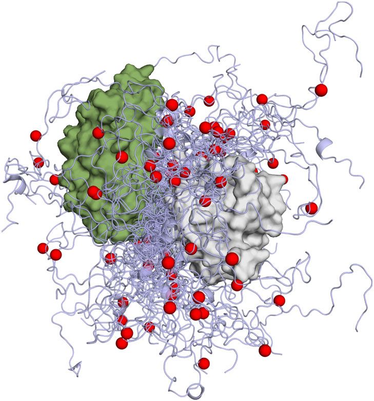
ARTICLE                                                                 NATURE COMMUNICATIONS | https://doi.org/10.1038/s41467-020-17407-x

I
    nfluenza A virus (IAV) is responsible for 3–5 million severe              of the IDD in the function of putative ANP32A:polymerase
    cases every year, resulting in 250–500,000 deaths1. Most                 complexes.
    influenza strains evolve exclusively in the large reservoir of
water birds, but some highly pathogenic avian strains (e.g., H5N1,
H5N8 and H7N9) can infect humans with lethal consequences                    Results
(up to 60% mortality) and are potential pandemic threats for                 IDDs of h and avANP32A and their interactions with 627-NLS.
humanity if they develop human-to-human transmissability2.                   We have compared the two complexes using solution state NMR,
However, for these avian (av) viruses to efficiently replicate in             initially from the side of ANP32A. The IDDs of h and avANP32A
mammalian cells, host adaptation of the viral polymerase is                  comprise 63/96 and 79/129 Asp or Glu residues, respectively,
necessary. Replication of IAV is carried out by the RNA-                     leading to extensive spectral overlap. h and avANP32A IDDs
dependent RNA viral polymerase that functions as a hetero-                   differ principally due to a 33 amino acid insert in avANP32A
trimeric complex, formed from separate components PA, PB1                    (176–209) comprising an av-unique hexapeptide, 176VLSLVK181,
and PB2. Few mutations are required for avian to human adap-                 followed by a duplication of 27 amino acids also present in
tation3–6, and a number of these cluster on the surface of the C-            hANP32A IDD. Backbone resonance assignment was completed
terminal 221 amino acid section of PB2, comprising separate ‘627’            to 78% and 58%, respectively, revealing that h and avANP32A
and ‘NLS’ domains4,7. In particular mutation of residue 627 from             IDDs are indeed both intrinsically disordered (Supplementary
E to K in avPB2 rescues polymerase activity and viral replication            Figs. 1 and 2), with a slight tendency (20%) towards helical
in mammalian cells8–11. Members of the host transcription reg-               conformation for the hexapeptide, but negligible secondary
ulator family ANP3212, comprising a long low-complexity acidic               structural tendency elsewhere.
intrinsically disordered domain (IDD; sometimes known as                        Upon addition of the 627(K) domain, 1H and 15N chemical
LCAR) at the C-terminus, have been shown to be responsible for               shift perturbations (CSPs) are seen for a large number of
this viral adaptation13. Human ANP32A (hANP32A) lacks an                     resonances in the IDD of hANP32A (Fig. 1b, Supplementary
insertion of 33 disordered residues compared to avANP32A,                    Fig. 1d). NMR relaxation rates measured at increasing titration
restricting avH5N1 polymerase activity in mammalian cells. This              admixtures (Fig. 1c) show maximal effects for the acidic strand
restriction is lifted by E627K mutation, suggesting an essential             180DEDA183, while the largest CSPs are seen for the adjacent
role for ANP32A through interaction with PB214–24, although                  hydrophobic residues 184QVV186 (Fig. 1b, Supplementary
there are currently no molecular descriptions of these interac-              Fig. 1d). Additional interactions are seen throughout the chain,
tions. The interaction between members of the ANP32 family and               in particular at 164VE165 and 214YND216. Comparison of
influenza polymerase is critical in supporting IAV replication,               backbone 13C shifts in the free and fully bound states reveals
and is attracting increasingly intense interest16,18,19. Recent stu-         that only 179YDED182 shows any evidence of folding upon
dies also demonstrate that the interaction occurs in the nucleus24,          binding (Supplementary Fig. 1d), in this case into an extended β-
and further studies point to the importance of related members of            sheet conformation, while the remainder of the chain remains
the ANP32 family, in particular ANP32B20–22, as well as the role             highly flexible in the complex, retaining its random coil nature
of surface residues in the folded leucine-rich region (LRR) of               (Supplementary Fig. 1b). Similar evidence of multiple interaction
ANP32A23.                                                                    sites is seen for the IDD of avANP32A in complex with the 627
   Conformationally, the 627-NLS region of PB2 exhibits                      (E) domain (Supplementary Fig. 1d, Fig. 1d). Relaxation proper-
intriguing behaviour7,25. X-ray crystallographic structures of               ties (Fig. 1c, h) and more specifically CSPs (Fig. 1e) of the
both h and av-adapted 627-NLS revealed a compact two-                        hexapeptide are strongly influenced by the presence of both
domain structure4,7, a conformation also found in full-length                domains of 627-NLS, compared to constructs each comprising
transcriptionally active polymerase26. Solution NMR, however,                only the 627 or NLS domains and the linker. This implies that the
revealed the coexistence of two forms of 627-NLS, corre-                     specific conformational behaviour of the linker in integral 627-
sponding to ‘open’ and ‘closed’ states that interchange in a                 NLS, or the relative position of the two domains, are essential for
highly dynamic equilibrium27, while crystallographic investi-                the interaction with the hexapeptide of avANP32A IDD.
gation of the transcriptionally inactive polymerase complex                     Interaction of 627-NLS and ANP32A reveals similar profiles
suggested a role for this open form in viral replication or                  to those measured for the IDDs alone, with additional
polymerase assembly28,29.                                                    interactions involving the LRR for both h and avANP32A
   Here, we combine NMR and quantitative ensemble analysis to                (Fig. 1f–i). In both h and avANP32A, the largest CSPs in the
describe and compare the complexes formed between avANP32A                   LRR are centred on 120LFN122, with additional shifts induced
and av-adapted 627-NLS (627-NLS(E)), and between hANP32A                     following the spine of the beta-helix (Fig. 1g). Notably, the
and h-adapted 627-NLS (627-NLS(K)). Although the complexes                   cross-interaction, between hANP32A and 627-NLS(E) shows
are both found to be highly dynamic they exhibit significant                  much smaller shifts than hANP32A:627-NLS(K), while
differences. Polyvalent combinations of transient interactions               comparison with avANP32A:627-NLS(E) clearly identifies
between the acidic IDD and the positively charged 627(K)                     increased shifts centred on the hexapeptide (Fig. 1i).
domain stabilise the hANP32A:627-NLS(K) complex. This poly-                     Although most of the IDD is involved in the interaction, the
valency is less efficient for avANP32A:627-NLS(E), due to the                 strongest binding or highest populations of binding interactions,
interruption of an exposed basic surface on the 627 domain by                occur at a distance of 25–35 amino acids from the LRR. In the
the presence of E627. The weaker interaction is however com-                 case of avANP32A, this concerns the avian-unique hydrophobic
pensated by the recruitment of additional sequences on the longer            hexapeptide, and in hANP32A the sequence 180DEDAQVV186.
avIDD, in particular an avian-specific hexapeptide motif that                 Further interactions are observed downstream of these interac-
interacts with the linker between 627 and NLS. Notably the cross-            tion sites until the nuclear localisation sequence (KRKR), situated
interaction between hANP32A and 627-NLS(E) exhibits neither                  ~15 amino acids from the end of the chain, beyond that point no
of these possible stabilisation mechanisms, which may be related             significant interactions are observed.
to the inability of IAV polymerase to function in human cells                   Despite the evidence of clear interaction between ANP32A and
without the E627K mutation. Importantly, we show that the                    627-NLS, the complex is polyvalent, and in all cases weak
interaction exhibits the same properties in the presence of het-             (Supplementary Table 1, Supplementary Fig. 3), with none of the
erotrimeric influenza polymerase, providing insight into the role             individual interactions exhibiting a stronger affinity than 800 μM

2                NATURE COMMUNICATIONS | (2020)11:3656 | https://doi.org/10.1038/s41467-020-17407-x | www.nature.com/naturecommunications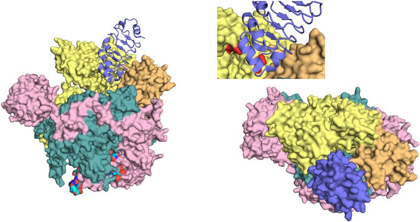
NATURE COMMUNICATIONS | https://doi.org/10.1038/s41467-020-17407-x ARTICLE
a
149 H 281
av
LRR IDD 149 249
h
LRR IDD
149 249/281
b f 70
60
184Q
50
204S 161E
120
R1ρ (s-1)
40
30
(ppm)
20
10
ω 1 - 15N
g 0
0.7
CSP (ppm)
123 185V 0.6
183A 0.5
0.4
0.3
0.2
164V
0.1
186V 0
126 187E 0 50 100 150 200 250
8.6 8.4 8.2 8.0
1H
h 60
(ppm)
50
c H
40
R1ρ (s-1)
16
R1ρ (s )
-1
12 30
8
20
4
10
d 0
12
i 0
R1ρ (s )
-1
8
0.5
4
CSP (ppm)
0.4
e 0
0.3
0.8 H
CSP (ppm)
0.6 0.2
0.4 0.1
0.2
0 0 50 100 150 200 250
140 160 180 200 220 240 260 280
Sequence
Sequence
Fig. 1 NMR of ANP32A with 627-NLS reveals a highly dynamic polyvalent complex. a Representation of the domains of ANP32A. Blue surface shows the
LRR domain, pale blue ribbon-disordered IDD (100 or 132 amino acids). Sequence distribution of IDDs shown on right. Red = acidic, blue = basic and
beige = hydrophobic or polar. AvIDD contains a 33 amino acids insert (horizontal line) comprising an avian-unique hexapeptide (H). b Chemical shift
titration of 627(K) into 15N-labelled IDD of hANP32A. hANP32A was 25 μM throughout. 627(K) concentrations were 0 (blue), 25 (green), 50 (grey-blue),
100 (orange) and 200 (red) μM. Measurement at 850 MHz, 293 K (see ‘Methods’ section). c 15N rotating frame relaxation (R1ρ) of free (blue) 15N-labelled
IDD of hANP32A at 300 μM, and upon mixing with 627(K) at 1:1 (orange) and 1:4 (red) ratio. Measurement at 850 MHz, 293 K. d 15N rotating frame
relaxation (R1ρ) of free (blue) 15N-labelled IDD of avANP32A at 300 μM, and upon mixing with 627(E) at 1:1 (orange) and 1:4 (red) ratio. Maximum effect is
seen at the equivalent motif, 33 amino acids downstream, from hANP32A. e Chemical shift perturbation (CSP) of full-length avANP32A upon addition of
627(E) (orange), NLS (red) and 627-NLS(E) (blue). Concentration of avANP32A was 25 μM and the partner 50 μM throughout. Resonances from the LRR
were not observable at this low concentration. f 15N rotating frame relaxation (R1ρ) of free (red) 15N-labelled full-length hANP32A at 300 μM, and upon
mixing with 627-NLS(K) at 1:1 ratio (blue). Measurement at 850 MHz, 293 K. g CSP of full-length hANP32A upon addition of 627-NLS(K) (blue).
Concentration of hANP32A was 300 μM and 627-NLS(K) 600 μM. The cross-interaction between hANP32A and 627-NLS(E) is shown for comparison at
the same stoichiometric ratio (orange). h 15N rotating frame relaxation (R1ρ) of free (red) 15N-labelled full-length avANP32A at 300 μM, and upon mixing
with 627-NLS(E) at 1:1 ratio (blue). Measurement at 850 MHz, 293 K. i CSP of full-length avANP32A upon addition of 627-NLS(E) (blue). Concentration of
avANP32A was 300 μM and 627-NLS(E) 600 μM. The cross-interaction between hANP32A and 627-NLS(E) is shown for comparison at the same
stoichiometric ratio (orange). The avian-specific insert is highlighted by shifting data from hANP32A from residues following the beginning of the insert by
33 amino acids.
NATURE COMMUNICATIONS | (2020)11:3656 | https://doi.org/10.1038/s41467-020-17407-x | www.nature.com/naturecommunications 3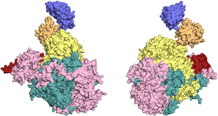
ARTICLE                                                                 NATURE COMMUNICATIONS | https://doi.org/10.1038/s41467-020-17407-x

for the interaction of hANP32A with 627(K) (1700 μM for                      the equilibrium at 1:2 ratio of 627-NLS(E):avANP32A and falling
avANP32A with 627(E)). Notably, the interactions between                     from 65 to 40% at the same stoichiometry in 627(K)-NLS:
hANP32A and 627(E) are also weaker (>1400 μM), suggesting                    hANP32A.
that the single E627K mutation plays an important role.                         The polyvalent nature of the interaction of the IDD with 627-
Interaction with integral 627-NLS is weaker due to the open-                 NLS is further substantiated by the comparison of the CSP-
closed equilibrium reducing the population of available binding              derived profiles measured when individual adjacent peptides from
states. Interaction of the LRR of ANP32A alone with the 627                  huANP32A IDD interact with the open-mutant of 627-NLS(K)
domain reveals weaker affinity (>1500 μM). Although these                     (Supplementary Fig. 6). These results demonstrate that both
interactions are weak, the extended interaction surface involving            peptides interact with residues on the surface of 627-NLS, some
80–100 disordered amino acids in h and avANP32A nevertheless                 that are common to both peptides and to the full-length IDD
results in tighter binding as experienced by 627-NLS.                        (Fig. 2b), and others that are unique to one or other of the
                                                                             peptides, confirming the multivalent, transient and dynamic
                                                                             nature of the interaction of the full-length huANP32A IDD with
Interactions of av and h-adapted 627-NLS with av and                         627-NLS(K). One of these peptides, that comprises 11 consecutive
hANP32A. The interaction was also investigated from the side of              acidic amino acids, also binds much tighter to the open form of
627-NLS domains of PB2. The two domains exhibit an open-                     627-NLS(K) than to the open form of 627-NLS(E) (Supplemen-
closed equilibrium (Fig. 2a) that is populated ~40:60 at 293 K               tary Fig. 7), again supporting the observation that the contribu-
leading to two sets of resonances for the majority of the protein27.         tion of K627 to the positively charged surface of the 627(K)
Addition of hANP32A to 627-NLS(K) resulted in CSPs                           domain is the essential factor for the tighter interaction with the
throughout the protein (Supplementary Fig. 4). The largest shifts            negatively charged regions of huANP32A IDD. These data
are observed in the open form, suggesting that ANP32A interacts              therefore further support our model of differentiation of the
preferentially with this conformation. The closed form of 627-               binding modes of the two complexes.
NLS(K) is stabilised by a tripartite salt bridge, and can be
removed from the equilibrium by mutation of the implicated
amino acids (R650 or D730/E687)27. CSPs induced upon inter-                  Ensemble descriptions of dynamic ANP32A:627-NLS com-
action with the ANP32A IDD are illustrated for clarity using an              plexes. To develop a more detailed description of the dynamic
open-only mutant (Fig. 2b), and the distribution of CSPs as                  interaction between 627-NLS and ANP32A, we have incorporated
measured on the wild-type proteins (Fig. 2c). Figure 2d illustrates          eight cysteine mutants into both 627-NLS(K) and (E), and indi-
the major shifts observed for 627-NLS(K) upon addition of                    vidually labelled the proteins with single TEMPO-based para-
hANP32A IDD. Although basic sidechains are found on both                     magnetic spin-label. This allows the detection of paramagnetic
sides of the domain (Fig. 2e), CSPs are observed mainly on one               relaxation enhancements (PREs), from the perspective of five
face of the 627 domain, again suggesting that the interaction is             positions on the 627 domain and three on the NLS domain
not uniquely driven by electrostatic attraction with the acidic              (Fig. 3a–c). PREs report on weak, or sparsely populated contacts
IDD. Despite strong similarity of the CSP profiles, clear addi-               between the two proteins, providing long-range positional con-
tional shifts are measurable for 627-NLS(K):hANP32A compared                 straints that are complementary to the short-range modulation of
to 627-NLS(E):avANP32A (Fig. 2c, Supplementary Fig. 4), par-                 the electronic and chemical environment probed by CSPs, and
ticularly in the vicinity of R630, L636 and 587–591, forming a               the modulation of the dynamics of each site as measured by spin
continuous interaction surface that is strongly enhanced in 627-             relaxation. These orthogonal experimental probes reveal different
NLS(K) (Fig. 2f). It is interesting to note that residues 590 and            aspects of the same interfaces, while providing a high level of
591 provide an alternative pathway to host adaptation, as evi-               confirmatory experimental validation (vide infra).
denced in the 2009 pandemic strain5,30. The long extended loop                  The experimental results show strong PREs, reporting on
comprising 627E/K, that is highly flexible in both 627E and 627 K             tighter or more populated interactions, distributed over long
(Supplementary Fig. 5), wraps around the 588–605 helix and                   stretches of the IDD for certain spin-label positions, and little
interaction with hANP32A appears to enhance the accessibility of             broadening for other positions. Such profiles again suggest
the N-terminus of this helix to the ANP32A IDD uniquely when                 a polyvalent interaction, whereby distinct sites dispersed along
627 K is present.                                                            the IDD visit the same sites on 627-NLS. Interestingly, some
   The affinity measured when observing resonances from 627-                  labels (in particular 587 and 631) induce a well-defined pattern
NLS(K) is considerably tighter than from the side of hANP32A,                over the long β-sheet on the concave face of the LRR of ANP32A,
with values of 20 μM for 627(K) and 50 μM for 627-NLS(K). This               allowing the determination of its orientation with respect to the
increased affinity apparently occurs due to avidity with the                  627 domain (Fig. 3d, see ‘Methods’ section), despite the weak
extensive interaction surface presented by hANP32A. Although                 interaction between the two domains (Supplementary Table 1).
the stoichiometry cannot be determined accurately, titration                 This orientation is in full agreement with the observed CSP on the
curves imply that it is significantly different to 1:1 from the side of       surface of the LRR of ANP32A (Fig. 1), where the largest
627-NLS (Supplementary Fig. 3), again suggesting that the                    chemical shifts are seen for F121, Y122 and C123. These residues
increased affinity occurs due to polyvalent binding of multiple,              are positioned in closest proximity to the 627 domain in the
weak binding sites on ANP32A to each site on the surface of 627-             ensemble of conformers that best fit the entire set of experimental
NLS(K). Notably, the affinity for avANP32A IDD measured when                  data. The interaction is apparently stabilised by hydrophobic
observing resonances of 627(E) is ~20 times weaker than for the              interactions, involving residues on the surface of 627 and
equivalent human IDD:627(K) complex, with values >600 μM. In                 ANP32A, and an electrostatic interaction between D130
combination with observations measured from the side of                      (ANP32A) and R646 (627) (Fig. 3e). It seems likely that the
ANP32A, it therefore appears that the absence of K627, which                 observed interaction relates to observations that have recently
disrupts the continuity of the positively charged surface on 627,            implicated D130 in host adaptation20,21,23. The optimal poses
strongly abrogates this component of the interaction.                        determined for the 627-NLS(K):hANP32A and 627-NLS(E):
   In addition, the interaction with ANP32A strongly favours the             avANP32A complexes are very similar. The interface between
open form of 627-NLS (Fig. 2g), especially for the avian                     627 and ANP32A is bordered by a nearly continuous ridge of
interaction, with the closed form essentially disappearing from              solvent accessible basic sidechains, including K589, R630, R641

4                NATURE COMMUNICATIONS | (2020)11:3656 | https://doi.org/10.1038/s41467-020-17407-x | www.nature.com/naturecommunications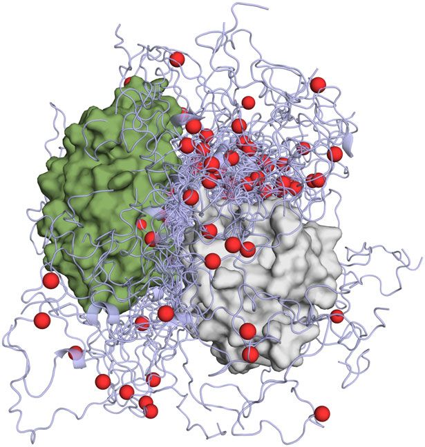
NATURE COMMUNICATIONS | https://doi.org/10.1038/s41467-020-17407-x ARTICLE
a 627 Linker
627 Linker NLS NLSp NLS
538 675 691 734 758
Closed Open
b d D605
I696
698 Q591 K721
105
S688
685 651 A587 R650
682 π
110 R641
(ppm)
115
ω 1 - 15N
650
652
G729
120
e
R630
649
K627
125
717
645
591
697
π
648
130
10 9 8 7
ω 2 - 1H (ppm)
c 1.2
1.0
CSP
0.8
(ppm)
f S629
K627
0.6
Q591
M631
0.4
A587 S635
T637
0.2
0.0
550 575 600 625 650 675 700 725 750
Sequence
g av h av h av h
129.2
119.6 723o 723o 123.6 707o 707o
119.8
129.4 725c 725o 725c 725o
120.0 723c 723c 124.0 707c 707c
8.35 8.25 8.35 8.25 9.15 9.05 9.15 9.05 8.50 8.50
ω 2 - 1H (ppm) ω 2 - 1H (ppm)
and R650, that is completed by the presence of K627 in the case of the spin-label at position 699 in the NLS broadens the IDD
627-NLS(K) (Fig. 3f). Inspection of the PRE data from the IDD maximally at the position of the hexapeptide of avANP32A. This
regions of h and avANP32A reveals weaker effects in the region again confirms that the interaction with the immediately
immediately following the LRR for avANP32A, and more proximal basic face of 627, defined by the differential CSPs
contacts with the dislocated NLS domain (Fig. 3a, b), in particular (Fig. 2f), is weaker in the case of avANP32A.
NATURE COMMUNICATIONS | (2020)11:3656 | https://doi.org/10.1038/s41467-020-17407-x | www.nature.com/naturecommunications 5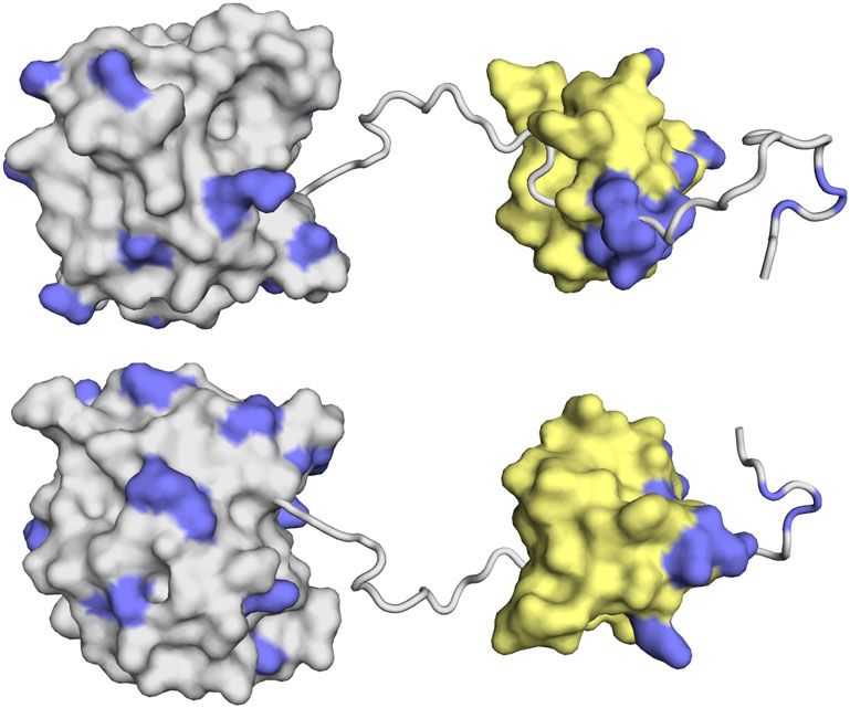
ARTICLE                                                                                   NATURE COMMUNICATIONS | https://doi.org/10.1038/s41467-020-17407-x

Fig. 2 Interaction of 627-NLS and ANP32A observed from the perspective of 627-NLS. a Representation of the domains of 627-NLS. The C-terminal
domains of PB2 comprise two sub-domains (627—orange and NLS—grey) connected by a linker (red) and terminated by an NLS peptide (NLS). The
molecule exists in equilibrium between closed and open forms that exchange at 50 s−1 at room temperature. The position of the E627K adative mutation is
shown in blue. b HSQC of the open-only form of 2D, 13C and 15N-labelled 627-NLS(K) (D730A/E687A; 200 μM; red), indicating chemical shifts of selected
sites upon addition of hANP32A IDD (400 μM blue). c CSP of 627-NLS(K) (300 μM) upon addition of hANP32A IDD at a ratio of 1:4 (red) and 627-NLS(E)
(300 μM) upon addition of avANP32A IDD at a ratio of 1:4 (blue). Spectra recorded on 2D, 13C and 15N-labelled 627-NLS at 293 K and 850 MHz. Only
shifts affecting resonances corresponding to the open form are shown for clarity. d CSP of 627-NLS(K) (red) induced by addition of hANP32A derived from
b above the threshold of 0.25 (dashed line) for 627 (grey) and NLS (yellow) domains. Two orientations of the protein are shown. e Distribution of basic
sidechains (blue) on the surface of the 627 and NLS domains. By comparison with c, it is evident that one of the two faces preferentially interacts with the
IDD. f Represention of the largest differential shifts on the surface of the 627 domain upon addition of ANP32A as shown in c. Amino acids showing the
largest difference in CSP (>0.15) in the 627-NLS(K):hANP32A IDD complex compared to the 627-NLS(E):avANP32A IDD are highlighted in red (position
of 627 shown in blue). g Interaction of full-length 627-NLS with ANP32A induces a change in the open-closed equilibrium. Red—peaks reporting on the
open and closed forms of 627-NLS(E) or 627-NLS(K) before the addition (blue) of h or avANP32A, respectively. The interaction with ANP32A potentiates
the equilibrium in both cases, fully removing the closed form in the case of the avian pair. Residues distal from the main interaction site were chosen to
reduce the risk of peak disappearance due to direct interaction.

    a      1.2                                                                                b      1.2
             1                                                                                         1
           0.8                                                                                       0.8
           0.6                                                                                       0.6
    I /I 0 0.4                                                                                I /I 0 0.4
           0.2       539                                          643                                0.2       539                                      643
             0                                                                                         0
           1.2                                                                                       1.2
             1                                                                                         1
           0.8                                                                                       0.8
           0.6                                                                                       0.6
           0.4                                                                                       0.4
           0.2       587                                          699                                0.2       587                                      699
             0                                                                                         0
           1.2                                                                                       1.2
             1                                                                                         1
           0.8                                                                                       0.8
           0.6                                                                                       0.6
           0.4                                                                                       0.4
           0.2       605                                          707                                0.2       605                                      707
             0                                                                                         0
           1.2                                                                                       1.2
             1                                                                                         1
           0.8                                                                                       0.8
           0.6                                                                                       0.6
           0.4                                                                                       0.4
           0.2       631                                          717                                0.2       631                                      717
                 0   50      100     150         200       0       50   100   150   200                    0   50    100   150   200    250   0         50    100   150     200    250
                                                       Sequence                                                                           Sequence

    c                                                  d                                  e                                f           LRR
                      643      631
                                       707                                                         F121/Y144                                      π/2                      586
     539                                                                                                   F633/Y592                                                         589
                                                                                                                                                                                   627
                                           699

                                           717
                                                                                                                                                                    630
                                     587                                                                                                                             650
                            605                                                               D130 R646                                627

Fig. 3 Experimental characterisation of the dynamic 627-NLS:ANP32A interaction complexes. a Experimental (orange) and calculated (blue)
paramagnetic relaxation enhancements measured on hANP32A in the presence of paramagnetically labelled 627-NLS. Intensity ratios compare spectra
recorded in the presence of oxidised and reduced forms of TEMPO-maleimide for complex admixtures of hANP32A (300 μM) and 627-NLS(K) (150 μM).
Calculated values result from representative ensembles selected using the ASTEROIDS approach. b Experimental paramagnetic relaxation enhancements
measured on avANP32A in the presence of paramagnetically labelled 627-NLS. Intensity ratios compare spectra recorded in the presence of oxidised and
reduced forms of TEMPO-maleimide for complex admixtures of avANP32A (220 μM) and av627-NLS (400 μM). This admixture was chosen to replicate
the population of bound state estimated in the case of 627-NLS(K):hANP32A. c Position of the eight cysteine mutations used to label 627-NLS. Five
mutations were selected over the surface of the 627 domain and three on NLS. One mutant protein was expressed and purified for each site on both 627-
NLS(K) and 627-NLS(E), and labelled with TEMPO-maleimide (see ‘Methods’ section). d Optimisation of the position of the folded domains of hANP32A
and 627-NLS(K). Two thousand starting poses generated by the programme Haddock were optimised with respect to experimental PREs measured on the
folded domain. The ten best fitting poses are shown. The same procedure was used for PREs measured on the avANP32A: 627-NLS(E) complex, resulting
in similar best fitting structures. e Localisation of intermolecular interactions possibly stabilising the interface between the two proteins, involving
hydrophobic (F121 and Y144 in ANP32A, and Y592 and F633 in 627-NLS) and electrostatic interactions (D130 in ANP32A and R646 in 627-NLS).
f Position of positively charged ridge of solvent-exposed basic sidechains in the vicinity of the interface between the folded domains.

  Ensemble analysis of the PRE data using the ASTEROIDS                                        sampling of conformational space of the IDD and NLS domains
approach31, accounting for the flexibility in the 627-NLS linker                                relative to the position of 627 and the LRR of ANP32A.
and IDD domains of ANP32A (Supplementary Fig. 8), allows us                                    Comparison of the position of the IDDs over the ensembles
to propose a molecular description of the conformational                                       (Fig. 4d, e) reveals more restricted sampling for hANP32A,
sampling of the entire complex. Representative ensembles of                                    localising residues 175–200 in the vicinity of the basic ridge on
conformations of the multi-domain complex (Fig. 4) reveal the                                  the surface of 627. Sampling of avANP32A is more dispersed

6                           NATURE COMMUNICATIONS | (2020)11:3656 | https://doi.org/10.1038/s41467-020-17407-x | www.nature.com/naturecommunications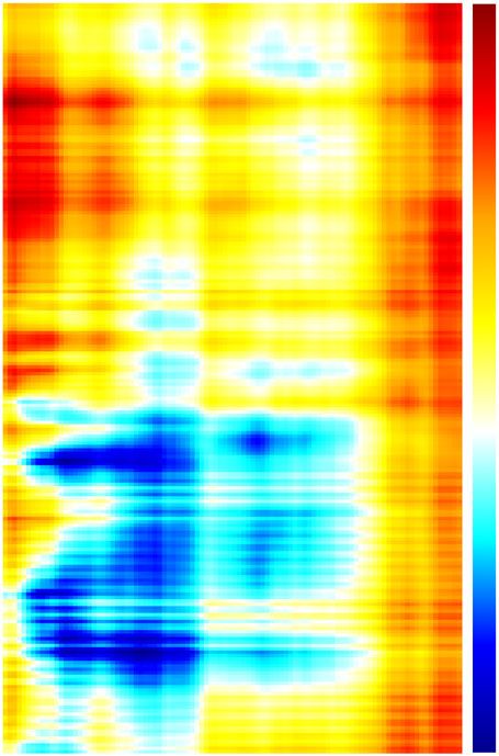
NATURE COMMUNICATIONS | https://doi.org/10.1038/s41467-020-17407-x                                                                           ARTICLE

      a                                            10      b                                                c
               750
                                                             LRR                                                                          LRR
      NLS

                                                   5
               700
      Linker

                                                                                                                                                av IDD
                                                                                            hIDD
               650                                 0

                                                                                                                                                     NLS
                                                                                      627
      627

                                                                                                   NLS
               600                                 –5                                                                            Linker
                                                                             Linker

               550
                                                   –10                                                                               hexapeptide
                     160 180 200 220 240
                      Sequence (ANP32A IDD)

                     d                                                         e

                                                   hIDD                                      LRR

                     LRR

                                                            627

                                                                                                                           627

                                                                                   avIDD

Fig. 4 Comparison of 627-NLS(K):hANP32A and 627-NLS(E):avANP32A interaction complexes. a Average distance difference matrix, showing the
average difference (dhh − davav) in distance between amino acid positions in the IDD of ANP32A (x-axis) and the 627-NLS domains (y-axis) over the two
ensembles. The colour code shown on the right is measured in Å. b Representation of the key interactions between hANP32A and 627-NLS(K). Polyvalent
interactions between 627 and the IDD localise the disordered domain in the vicinity of the basic patch on the surface of 627 (see Fig. 3). The different IDD
chains are shown to represent different binding modes and are truncated at residue 200 for clarity. Red positions in the IDD indicate the acidic sidechains.
Note that this representation illustrates the tendency over the entire ensemble that is highly disperse (see d and e). c Representation of the key
interactions between avANP32A and 627-NLS(E). In this case, the average distance between the IDD and the surface of 627 is larger, but in general closer
to the NLS domain, in particular the hexapaptide of ANP32A, and the linker between 627 and NLS. The two IDD chains represent reduced polyvalency
compared to hANP32A and 627-NLS(K), and are again truncated at residue 200 for clarity. d Representative ensemble of conformations describing the
conformational space sampled by the hANP32A:627-NLS(K) complex. The NLS domain has been removed for clarity, and the IDD truncated at residue 210.
The position of residue 190 is indicated as a red sphere. e Representative ensemble of conformations describing the conformational space sampled by the
avANP32A:627-NLS(E) complex. Representation as in d.

(Fig. 4b–e), as shown quantitatively in the average distance map                abrogates the extensive interaction surface and the linker-specific
(Fig. 4a), that identifies closer contacts in the 627-NLS(K):                    contact present in avANP32A:627-NLS(E) (compare profiles
hANP32A complex for residues 160, 180 and 205 with 570, 586                     induced by probe attached at 699). The required components for
and 627, and closer contacts between the linker and NLS regions,                interaction modes specific to either 627-NLS(K):hANP32A or
and avANP32A region 150–200 (Fig. 4a).                                          627-NLS(E):avANP32A are therefore both weakened when avian
   Importantly, comparison of PRE profiles of hANP32A:627-                       polymerase interacts with hANP32A.
NLS(E) demonstrates that the cross-interaction, that represents
the case encountered when a non-adapted avian IAV infects
human cells, shows less extensive and in general weaker contacts                hANP32A:627-NLS in the context of integral FluB polymerase.
compared to hANP32A:627-NLS(K) and avANP32A:627-NLS(E)                          To determine whether the interactions characterised here are
(Fig. 5). The absence of K627 is again seen to diminish the                     relevant in the context of a human-adapted full-length influenza
numerous polyvalent contacts present in hANP32A:627-NLS(K)                      polymerase, we initially repeated the chemical shift titrations
as illustrated by the profiles induced by probes attached to 587                 using the heterotrimeric influenza B (FluB) polymerase (bound to
and 631, while the shorter IDD and lack of the hexapeptide                      vRNA), with the IDD of hANP32A (Fig. 6a). The interaction sites

NATURE COMMUNICATIONS | (2020)11:3656 | https://doi.org/10.1038/s41467-020-17407-x | www.nature.com/naturecommunications                                   7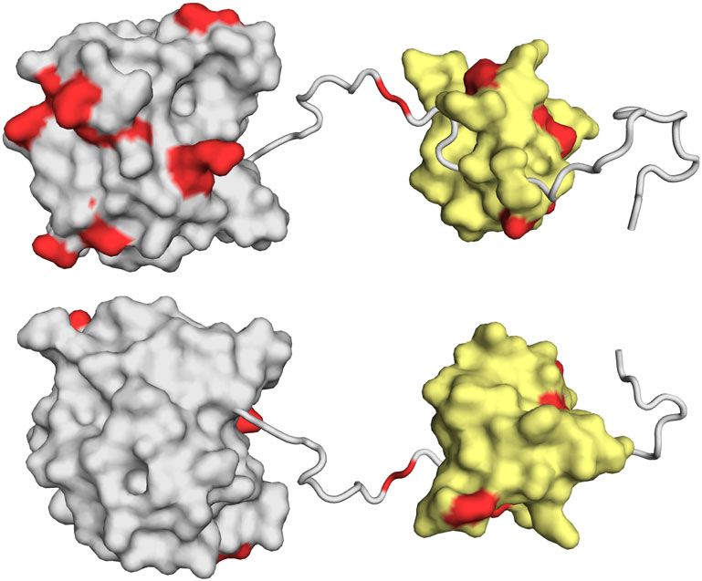
ARTICLE NATURE COMMUNICATIONS | https://doi.org/10.1038/s41467-020-17407-x
a Fig. 5 Adapted interaction modes are significantly weakened in the
1.2
1
cross-interaction hANP32A:627-NLS(E). Comparison of intermolecular
0.8 contacts in hANP32A:627-NLS(K) (light orange), avANP32A:627-NLS(E)
I /I 0 0.6 (dark orange) and hANP32A:627-NLS(E) (green). Three PREs are
0.4 compared, a 631, b 587 and c 699. The comparison of PRE profiles due to
0.2 the presence of a spin probe on residue 631 illustrates the reduced number
0 and strength of contacts of the IDD, with the 627 domain in the
1 631 hANP32A:627-NLS(E) complex compared to hANP32A:627-NLS(K).
0.8 Residue 587 shows a similar effect. Comparison of PRE profiles due to the
0.6
presence of a spin probe on residue 699 illustrates the lack of interaction
0.4
with the linker-NLS region in the hANP32A:627-NLS(E) compared to
0.2
avANP32A:627-NLS(E). The position of the hexapeptide region is identified
0
1 with dotted vertical lines in c.
0.8
0.6
0.4
0.2 with IAV 627-NLS and full-length FluB polymerase, it is inter-
0 esting to speculate whether the interaction described in detail for
0 50 100 150 200 250
IAV 627-NLS can be accommodated within known structures of
Sequence
the polymerase.
b 1.2
Superposition of the 627 domains of the ensembles describing
1 the complex onto the 627 domain in the transcription-active
0.8 conformation of FluB polymerase (the form used in the
I /I 0
0.6 interaction study described above)26,32 reveals that the LRR of
0.4 ANP32A can indeed be inserted into a broad cylindrical pocket
0.2 formed by 627, the mid and cap-binding domains of PB2, also
0 bordered by the C-terminal domain (CTD) of PB1, allowing
1 587 ANP32A to adopt the pose determined in solution (Fig. 6d).
0.8
Interestingly, the site of recently described adaptive mutations of
0.6
0.4
PB2 at 521 and 35533 lie in the immediate vicinity of the surface
0.2
of ANP32A in this pose (inset Fig. 6d), suggesting that the
0 importance of these mutations involves interaction with
1
ANP32A. Figure 6e, f illustrates the expected conformational
0.8
space sampled by the IDDs of av and hANP32A within the 627-
0.6 NLS:ANP32A complexes, indicating a broader capture radius for
0.4 the avian complex. A similar procedure was applied to the
0.2 conformationally very distinct transcriptionally inactive IAV
0
0 50 100 150 200 250
polymerase bound to the cRNA 5′ terminus29, where the 627
Sequence
domain sits on the surface, and is displaced and rotated relative to
the polymerase core by ~70°34. In this case, the 627-NLS:ANP32A
c 1.2 complex can be easily accommodated (Fig. 6g, h). The position of
1 NLS on the surface of the polymerase, and its observed positional
I /I 0 0.8 variability in existing structures, suggests that the open form of
0.6 627-NLS is sampled in both transcriptionally active and inactive
0.4 polymerases. Finally, the recently determined dimeric structure of
0.2
apo IAV polymerase accommodates the ensemble equally well
0
699
(Supplementary Fig. 9)35.
1
0.8
0.6 Discussion
0.4 In this study, we describe and compare the molecular complexes
0.2 formed by the human-adapted or avian-adapted 627-NLS
0 domains with the respective ANP32A host proteins, in order to
1
understand the nature and specificity of these interactions. All of
0.8
0.6
the implicated proteins exhibit extensive intrinsic disorder. The
0.4
elaboration of atomic resolution descriptions of such highly dis-
0.2 ordered complexes requires methodologies that can account for
the ensemble of structures sampled by the two proteins in
0 50 100 150 200 solution36,37. Investigation of the hANP32A:627-NLS(K) and
Sequence
hANP32A:627-NLS(E) complexes using complementary struc-
tural data from NMR chemical shifts, spin relaxation and para-
are found to be the same as for 627-NLS from IAV (Fig. 6b). In magnetic relaxation combined with quantitative ensemble
the case of full-length hANP32A (Fig. 6c), the first 50 amino acids modelling reveals the existence of highly dynamic molecular
of the IDD disappear from the spectrum, likely because the large assemblies that exhibit very different interaction modes.
particle results in extreme line broadening for the residues that The LRR of ANP32A interfaces with the 627 domain of PB2 at
are closest to the LRR domain due to slow tumbling in solution. the C-terminal end of its concave surface, apparently stabilised
Given the spectroscopic similarities of the interaction of ANP32A via hydrophobic and electrostatic interactions. This interaction is
8 NATURE COMMUNICATIONS | (2020)11:3656 | https://doi.org/10.1038/s41467-020-17407-x | www.nature.com/naturecommunications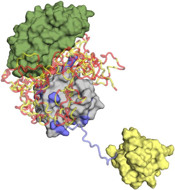
NATURE COMMUNICATIONS | https://doi.org/10.1038/s41467-020-17407-x ARTICLE
a d
ANP32A D521
184Q
120 204S
K355
PB2 Cap
(ppm)
627
PA Endo
ω 1 - 15N
185V
π/2
125 164V
187E 186V PB1
PA Endo
627
8.8 8.6 8.4 8.2 8.0
ω2 - 1H (ppm)
ANP32a
b e f
0.15
0.10
CSP
0.05
c
0.8
0.6
I /I0
0.4
0.2
0.0
150 160 170 180 190 200 210 220 230 240
Sequence
g h
ANP32A
π π
627
PB2 Cap
NLS
PA Endo
PB1
Fig. 6 Interaction of hANP32A with full-length FluB polymerase. a Chemical shifts of 15N-labelled IDD of hANP32A (4 μM) upon addition of full-length
FluB polymerase (32 μM) bound to viral promoter RNA (vRNA). Measurement at 850 MHz, 293 K. Red—free protein, blue—in presence of polymerase.
Notably the distribution of chemical shifts is highly similar to that induced in hANP32A upon addition of IAV 627-NLS(K). b CSP associated with
a. c Intensity ratio of 15N-labelled full-length hANP32A (4 μM) upon addition of full-length FluB polymerase (32 μM) bound to vRNA. d Compatibility of
the experimentally observed binding mode in the context of the vRNA-bound conformation of FluB polymerase26. The 627 domain was superimposed on
the 627 domain of PB2 in the full-length polymerase structure (4wsa). In this position, ANP32A LRR can be accommodated in a large pocket formed by
627 (yellow–orange), and cap-binding domains of PB2 (yellow) and bordered by PB1 (dark-cyan). Inset: PB2 adaptive mutants D521 and K355 lie in the
immediate vicinity of ANP32A LRR. e Conformational sampling of the IDD of hANP32A, assuming the position of the LRR of ANP32A shown in d. The
linker and NLS domains are not shown for clarity and are assumed flexible. f Conformational sampling of the IDD of avANP32A (otherwise as in e).
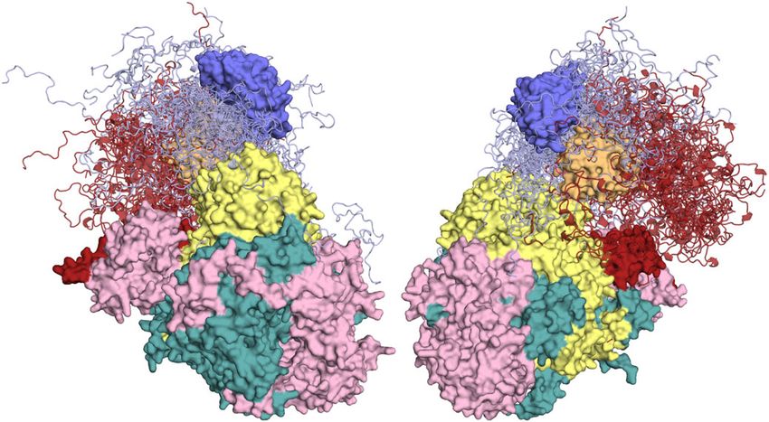
g Compatibility of the experimentally observed binding mode in the context of the cRNA-bound conformation of FluB29. The 627 domain was
superimposed on the 627 domain of PB2 in the full-length polymerase structure (5epi), which is dislocated relative to the vRNA-bound form. ANP32A LRR
can be accommodated easily on the accessible surface of 627. The NLS domain (brick red) is detached from 627 in this structure. h Conformational
sampling of the IDD of hANP32A, assuming the position of the LRR of ANP32A shown in g. Sampling of the IDD (light blue) linker and NLS domains are
shown (brick red).
intrinsically very weak, but experimental NMR, including CSP case of hANP32A:627-NLS(K), involving weak, polyvalent
and PREs, accurately report on the relative position of the two interactions of acidic and hydrophobic residues of the IDD. The
domains. The disordered domain interacts transiently with a presence of multiple low-affinity interactions (in the millimolar
basic patch on the surface of the 627 domain, preferentially in the range individually) distributed throughout 80 amino acids of the
NATURE COMMUNICATIONS | (2020)11:3656 | https://doi.org/10.1038/s41467-020-17407-x | www.nature.com/naturecommunications 9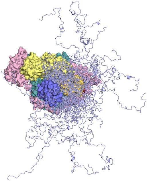
ARTICLE                                                                  NATURE COMMUNICATIONS | https://doi.org/10.1038/s41467-020-17407-x

IDD of hANP32A results in an effective increase in affinity to                 solution. It is also possible that the highly dynamic and transient
~50 μM for 627-NLS(K), an effect of avidity that has been                     nature of the interaction with ANP32A enables or enhances
observed in a number of systems exhibiting extensive intrinsic                exchange between these forms during the viral cycle. It is there-
disorder38,39. The critical E627K mutation completes a con-                   fore of interest to investigate the possible impact of our observed
tinuous ridge of solvent-exposed positively charged residues4 that            binding mode on other known states of influenza polymerase.
are available for interaction with the highly dynamic acidic IDD.             The proximity of ANP32A IDD to the interface between the 627
This ridge colocalizes interacting residues of the IDD in the                 and mid-domains of PB2, both of which undergo large-scale
vicinity of the surface, that differentially implicates residues 589          reorientations and dislocation between apo-, cRNA-bound and
and 591, 629, 631, 635 and 637 when the E627K mutation is                     vRNA-bound polymerase, also raises the possibility that ANP32A
present.                                                                      interaction is associated with these conformational changes. In
   By contrast the IDD of avANP32A interacting with 627-NLS(E)                the transcriptionally inactive structure, the 627 domain appears
populates fewer conformations in the vicinity of the surface of 627.          almost dislocated from the core, a conformation that can easily
This lack of interaction, due to the interruption of the positively           accommodate the binding pose of ANP32A determined for the
charged surface by the presence of E627, is compensated by an                 minimal complex.
even broader conformational sampling of the IDD that exploits a                  The information contained in our ensemble descriptions of the
more extensive interaction surface, implicating the hexapeptide               ANP32A:627-NLS interactions therefore allow us to speculate fur-
motif specific to avANP32A and the NLS domain, and in parti-                   ther on its role in viral function. Recent observations have estab-
cular the linker region of 627-NLS. It is again interesting to note           lished that dimerisation of influenza polymerase is essential for the
that two adaptative mutations (V683T and A684S)40 have been                   initiation of vRNA synthesis during replication35. The apo-
identified in this linker region. By binding predominantly to the              polymerase structure of IAV polymerase that was recently solved
open form of 627-NLS, ANP32A potentiates the equilibrium of                   in its dimeric form is also found to be compatible with the binding
open and closed forms of 627-NLS, an effect that is more efficient             pose determined here (Supplementary Fig. 9), again with the 627
in the case of the 627-NLS(E):avANP32A interaction, likely as a               domain dislocated and sitting on the surface of the catalytic core
result of the interaction of the hexapeptide. Investigating the               domains. It has been suggested that ANP32A plays a role in
interaction of individual peptides isolated from the IDD, we are              assembly or regulation of this dimerisation process, for example, by
able to confirm the multivalent nature of the interaction of the               recruitment of a second apo ‘packaging’ polymerase to a replicating
huIDD with 627-NLS, and to strongly substantiate the differential             polymerase to initiate formation of the progeny viral RNP43. In this
binding modes of the two complexes.                                           context, the polyvalent nature of the interaction between ANP32A
   Notably, the ‘cross-interaction’ between hANP32A and 627-NLS               and 627-NLS may be of functional relevance, allowing for more
(E) exhibits neither the stabilisation properties mediated by the             than one polymerase to simultaneously bind to ANP32A thereby
avIDD in the avANP32A:627-NLS(E) complex, nor the polyvalent                  colocalizing two polymerases to facilitate viral replication. In all of
binding specific to 627(K) as observed in the hANP32A:627-NLS                  these aspects, the more extensive effective capture radius of the IDD
(K) interaction. This lack of adapted molecular mechanisms results            of avANP32A may be important.
in fewer and weaker contacts between human ANP32A and avian-                     It is known that the intrinsically disordered, phosphorylated
adapted 627-NLS. In combination, these effects may explain the                CTD of host RNA polymerase II (Pol II) binds to the surface of
inefficiency of avian polymerase in human cells in the absence of              PA of influenza polymerase to facilitate the cap-snatching
avANP32A or 627-NLS(K).                                                       mechanism44,45. Given the highly negatively charged nature of
   It is interesting to compare the interaction results measured by           the IDD of ANP32A it seems possible that it may play a reg-
different NMR-based techniques with existing studies, where results           ulatory role in this interaction, for example, by competing with
measured using different techniques seem to paint a slightly dif-             the phosphorylated CTD to inhibit the interaction with Pol II.
ferent picture. While the individual interaction sites reported in our        Notably, the extremely long IDDs, allied to the fact that ANP32A
study indicate a weaker binding between avANP32A and 627-NLS                  is bound to the 627 domain that exhibits extensive mobility with
(E) as compared to hANP32A and 627-NLS(K), some biochemical                   respect to the rest of the polymerase core, would appear to
and cell biology studies report on a higher affinity of avANP32 to             facilitate the kind of flexible chaperoning action seen in other
avian-adapted polymerase17,24. This apparent contradiction may                highly dynamic viral proteins46.
result from different tagging techniques used in the reported pull-              In summary, the description of these highly dynamic species-
down assays, but we also note that overall affinities between pro-             specific assemblies reveals unique mechanistic insight into the
teins are not necessarily comparable to individual multivalent                role of the ANP32 family in host adaptation of avian influenza
interactions in terms of affinities, and it will be interesting to resolve     polymerase to the human cells, and provides a molecular fra-
the dependencies in the future.                                               mework for understanding the considerable volume of experi-
   Importantly, NMR indicates that the complex characterised for              mental observation measured on this complex system, as well as
the minimal 627-NLS:ANP32A interaction is maintained in the                   informing the identification of novel targets for IAV inhibition.
context of the integral FluB polymerase in its transcriptionally
active form, showing very similar NMR-binding characteristics to
hANP32A. Superposition of the binding pose of ANP32A with                     Methods
respect to 627 onto the 627 domain in the associated tran-                    Constructs. A codon optimised 627-NLS construct from PB2 subunit was syn-
scriptionally active polymerase structure26 would place the LRR               thesised encoding amino acids 538–759 from avian H5N1 A/duck/Shantou/4610/
                                                                              2003 for expression in Escherichia coli (Geneart, Regensburg, Germany)27. In
domain in a similarly dimensioned pocket bounded by PB2                       addition, constructs containing just the 627 domain (amino acids 538–693) or the
domains (Fig. 6d). Intriguingly, recently characterised host-                 NLS domain (amino acids 678–759) were generated. Avian ANP32A (Gallus gallus,
adaptive PB2 mutants33 lie in the immediate vicinity of the sur-              XP_413932.3) was synthesised and codon optimised for expression in E. coli
face of the folded domain of ANP32A in this binding con-                      (GenScript, New Jersey, USA). Plasmids containing just the intrinsically disordered
                                                                              region of avian ANP32A (avIDD, amino acids 149–281) or human ANP32A
formation. Influenza polymerase exhibits extensive plasticity in               (hIDD, amino acids 144–249) were generated. All constructs were cloned into a
solution, as demonstrated by recent electron microscopy and X-                plasmid derived from pET9a with an N-terminal His-tag and a TEV cleavage site
ray crystallographic studies describing multiple states of the                (MGHHHHHHDYDIPTTENLYFQG). pQTEV-ANP32A was a gift from Konrad
polymerase26,28,29,34,35,41,42, and it appears highly likely that             Buessow (Addgene plasmid # 31563; http://n2t.net/addgene:31563; RRID:
many of these states will be in conformational exchange in                    Addgene_31563)47.

10                NATURE COMMUNICATIONS | (2020)11:3656 | https://doi.org/10.1038/s41467-020-17407-x | www.nature.com/naturecommunications
NATURE COMMUNICATIONS | https://doi.org/10.1038/s41467-020-17407-x ARTICLE
Protein expression and purification. Plasmids were transformed into E. coli Ensemble descriptions of ANP32A:627-NLS complexes. Having determined the
Rosetta cells, and the cultures were grown in LB and induced with IPTG for 16 h at relative position of the two domains, the flexible parts were constructed onto this
18 °C. Bacteria were harvested by centrifugation, resuspended in buffer A (50 mM conformation. For both h and av complexes, the statistical coil model flexible mec-
Tris-HCl pH 7.5 and 200 mM NaCl) with protease inhibitors (complete, Roche) cano57 was used to predict 10,000 conformations of the linker region of 627-NLS, the
and bacterial lysis was performed by sonication. All proteins were purified by NLS domain, the NLS peptide that terminates 627-NLS, and the 96 or 128 amino acid
affinity chromatography on Ni-NTA agarose (Qiagen), followed by incubation with IDD of h and avANP32A. Conformers were calculated using amino acid-specific
TEV protease at 4 °C coupled with dialysis into buffer A. A second affinity column potentials that reproduce the experimentally observed behaviour of the IDD domains,
with Ni-NTA agarose was performed and the flow-through was loaded into a and were calculated to avoid steric overlap between any of the domains (Supple-
Superdex 75 column (GE Healthcare) for size-exclusion chromatography in mentary Fig. 8). PREs over the entire ANP32A molecule (folded and unfolded
buffer A. domains) were calculated for each of the conformers calculated for each complex, and
To produce 15N-labelled or 15N, and 13C-labelled proteins for NMR these conformations were used as a basis set from which ensembles were selected using
spectroscopy, bacteria were grown in M9 minimal medium containing MEM the ASTEROIDS approach36,58. Ensemble size was estimated on the basis of direct and
vitamins (Gibco), supplemented with 1 g L−1 of 15NH4Cl and 2 g L−1 of unlabelled cross-validated PRE profiles (60 conformers were used for both h and av complexes).
or 13C-glucose. To produce additionally 2H-labelled proteins, the M9 minimal Distance matrices were calculated by calculating the average distance between
medium was prepared in D2O and 2 g L−1 of deuterated 13C-glucose. Protein Cα atoms between the two proteins in the selected ensembles from the two
purity was checked by SDS–PAGE and mass spectrometry. Single-point mutations complexes, and the distance difference matrix shown in Fig. 5a by subtracting the
of 627-NLS were done using the Quick change method48, using Phusion high- matrix from hANP32A:627-NLS(K) from the avANP32A:627-NLS(E) matrix.
fidelity DNA polymerase and DpnI (Thermo Scientific). Cysteine mutants were
purified as mentioned above for wild-type protein; however, 10 mM of
Reporting summary. Further information on research design is available in the Nature
dithiothreitol (DTT) was added after the second Ni-NTA column to keep proteins
Research Reporting Summary linked to this article.
in a reduced state until labelling. The heterotrimeric human influenza polymerase
from B/Memphis/13/03 (FluB) was expressed as a self-cleaving polyprotein and
purified, using NTA affinity and heparin columns followed by size-exclusion Data availability
chromatography32. ANP32A peptides were purchased from Caslo, Denmark. All of the NMR data presented in the article are available from the authors upon request.
The NMR chemical shift assignments, and PREs of avian and human ANP32A have been
NMR spectroscopy. All samples for NMR were measured in 50 mM Tris-HCl deposited in the Biological Magnetic Resonance Bank (BMRB; bmrb.wisc.edu/) with
buffer pH 6.5, 200 mM NaCl and 10% D2O. The assignment of the intrinsically accession codes 28134 and 28135, respectively.
disordered regions of avian and human ANP32A were obtained using 15N,13C-
labelled samples (700 μM) using BEST-TROSY tridimensional experiments Received: 13 March 2020; Accepted: 29 June 2020;
recorded on a Bruker spectrometer equipped with a cryoprobe operating at 20 °C
and a 1H frequency of 850 MHz. All spectra were processed using NMRPipe49 and
analysed in Sparky50. MARS51 was used for spin system identification, followed by
manual verification. The folded domain of ANP32A has been assigned
previously52,53. 13Cα chemical shifts of the intrinsically disordered regions were
compared to random coil values using the software SSP54.
15N R relaxation rates were measured at 293 K and a 1H frequency of
1ρ References
850 MHz using a spin lock of 1.5 kHz as described55. A typical set of relaxation 1. Lozano, R. et al. Global and regional mortality from 235 causes of death for 20
delays included points measured at 1, 15, 30, 50, 100, 140, 200 and 230 ms, age groups in 1990 and 2010: a systematic analysis for the Global Burden of
including repetition of one delay. Relaxation rates were determined using in-house Disease Study 2010. Lancet 380, 2095–2128 (2012).
software and errors were estimated on the basis of noise-based Monte Carlo 2. Nicholls, H. Pandemic Influenza: the Inside Story. PLoS Biol. 4, e50 (2006).
simulation. Interaction experiments with full-length polymerase were acquired 3. Almond, J. W. A single gene determines the host range of influenza virus.
with 15N-labelled hIDD or full-length 15N-hANP32A at a concentration of 4 μM
Nature 270, 617–618 (1977).
after the addition of 32 μM of human FluB polymerase bound to the 5′ terminal
4. Tarendeau, F. et al. Host determinant residue lysine 627 lies on the surface of a
viral RNA promoter (5′-pAGUAGUAACAAGAG-3′ OH). These experiments were
discrete, folded domain of influenza virus polymerase PB2 subunit. PLoS
recorded at 293 K and a 1H frequency of 850 MHz.
Pathog. 4, e1000136 (2008).
PRE effects used to model the complex formed by ANP32A (15N labelled,
5. Mehle, A. & Doudna, J. A. Adaptive strategies of the influenza virus
human or avian) and 627-NLS (E627 or K627) were measured from the peak
intensity ratios between a 15N-HSQC 2D spectrum recorded on a sample polymerase for replication in humans. Proc. Natl Acad. Sci. USA 106,
containing 627-NLS labelled with TEMPO, and a reference diamagnetic sample 21312–21316 (2009).
that was incubated previously with 5 mM of DTT. For these experiments, single- 6. Gabriel, G., Czudai-Matwich, V. & Klenk, H.-D. Adaptive mutations in the
cysteine mutants at positions 539, 587, 605, 631, 643, 699, 707 and 717 were tagged H5N1 polymerase complex. Virus Res. 178, 53–62 (2013).
using 4-maleimido-TEMPO. Briefly, purified 627-NLS single-cysteine mutants 7. Tarendeau, F. et al. Structure and nuclear import function of the C-terminal
were reduced with 10 mM of DTT at 4 °C for 12 h and then dialysed throughly into domain of influenza virus polymerase PB2 subunit. Nat. Struct. Mol. Biol. 14,
50 mM phosphate buffer pH 7.0 containing 150 mM NaCl without DTT. A fivefold 229–233 (2007).
molar excess of 4-maleimido-TEMPO dissolved in DMSO was added to the 8. Subbarao, E. K., Kawaoka, Y. & Murphy, B. R. Rescue of an influenza A virus
reduced 627-NLS cysteine mutants. The reaction was incubated for 12 h at 4 °C and wild-type PB2 gene and a mutant derivative bearing a site-specific temperature-
then injected into a Superdex S75 column to eliminate the excess of TEMPO sensitive and attenuating mutation. J. Virol. 67, 7223–7228 (1993).
through size-exclusion chromatography. Complete labelling with TEMPO was 9. Massin, P., van der Werf, S. & Naffakh, N. Residue 627 of PB2 is a
verified by mass spectrometry. Measurement of PRE effects was performed in determinant of cold sensitivity in RNA replication of avian influenza viruses. J.
samples containing 200 μM of 15N-labelled hIDD, and 100 or 200 mM of the Virol. 75, 5398–5404 (2001).
respective TEMPO-labelled 627-NLS (K627) mutants. Measurements in full-length 10. de Jong, M. D. et al. Fatal outcome of human influenza A (H5N1) is
hANP32A were performed with 300 μM of 15N-hANP32A and 150 μM of the 627- associated with high viral load and hypercytokinemia. Nat. Med. 12,
NLS (K627) mutants, and measurements on full-length avANP32A were carried 1203–1207 (2006).
out with 220 μM of 15N-labelled protein and 400 μM of the 627-NLS (E627) 11. Kirui, J., Bucci, M. D., Poole, D. S. & Mehle, A. Conserved features of the PB2
mutants. 627 domain impact influenza virus polymerase function and replication. J.
Virol. 88, 5977–5986 (2014).
Determination of the relative position of ANP32A and 627. Experimentally 12. Reilly, P. T., Yu, Y., Hamiche, A. & Wang, L. Cracking the ANP32 whips:
determined PREs measured on h and avANP32A in the presence of different spin- important functions, unequal requirement, and hints at disease implications.
labelled forms of h and av627-NLS, respectively, were used to determine the BioEssays N. Rev. Mol. Cell. Dev. Biol. 36, 1062–1071 (2014).
relative position of the folded domain of ANP32A with respect to the 627 domain. 13. Long, J. S. et al. Species difference in ANP32A underlies influenza A virus
Two thousand different positions of the two domains were generated using the polymerase host restriction. Nature 529, 101–104 (2016).
programme Haddock56, varying over a wide range of distances and orientations. 14. Sugiyama, K., Kawaguchi, A., Okuwaki, M. & Nagata, K. pp32 and APRIL are
Positions of spin-label-bearing sidechains were generated on the basis of rotameric host cell-derived regulators of influenza virus RNA synthesis from cRNA.
libraries (see Supplementary Fig. 8)31,37, and an ensemble of sidechain positions eLife 4, e08939 (2015).
was used to calculate expected PREs on ANP32A for a given position of the 627 15. Lowen, A. C. Virology: Host protein clips bird flu’s wings in mammals. Nature
domain for each label. Admixtures were adjusted to ensure a population of the 529, 30–31 (2016).
bound state of 10% for both complexes. The position of each of the 2000 starting 16. Mehle, A. The avian influenza virus polymerase brings ANP32A home to
conformations were varied over a range of ±10 Å along three orthogonal cartesian roost. Cell Host Microbe 19, 137–138 (2016).
axes at a resolution of 0.1 Å, and the best fitting position retained. The ten best 17. Domingues, P. & Hale, B. G. Functional insights into ANP32A-dependent
fitting structures are shown in Fig. 3d. influenza a virus polymerase host restriction. Cell Rep. 20, 2538–2546 (2017).
NATURE COMMUNICATIONS | (2020)11:3656 | https://doi.org/10.1038/s41467-020-17407-x | www.nature.com/naturecommunications 11You can also read