Comparison of two fluorescent probes in preclinical non invasive imaging and image guided debridement surgery of Staphylococcal biofilm implant ...
←
→
Page content transcription
If your browser does not render page correctly, please read the page content below
www.nature.com/scientificreports
OPEN Comparison of two fluorescent
probes in preclinical non‑invasive
imaging and image‑guided
debridement surgery
of Staphylococcal biofilm implant
infections
Howard Y. Park1, Stephen D. Zoller1, Vishal Hegde1, William Sheppard1, Zachary Burke1,
Gideon Blumstein1, Christopher Hamad1, Marina Sprague1, John Hoang1, Ryan Smith1,
Francisco Romero Pastrana3, Julie Czupryna2, Lloyd S. Miller4,5, Marina López‑Álvarez3,
Mafalda Bispo3, Marleen van Oosten3, Jan Maarten van Dijl3, Kevin P. Francis1,2 &
Nicholas M. Bernthal1*
Implant-associated infections are challenging to diagnose and treat. Fluorescent probes have been
heralded as a technologic advancement that can improve our ability to non-invasively identify
infecting organisms, as well as guide the inexact procedure of surgical debridement. This study’s
purpose was to compare two fluorescent probes for their ability to localize Staphylococcus aureus
biofilm infections on spinal implants utilizing noninvasive optical imaging, then assessing the broader
applicability of the more successful probe in other infection animal models. This was followed by
real-time, fluorescence image-guided surgery to facilitate debridement of infected tissue. The two
probe candidates, a labelled antibiotic that targets peptidoglycan (Vanco-800CW), and the other, a
labelled antibody targeting the immunodominant Staphylococcal antigen A (1D9-680), were injected
into mice with spine implant infections. Mice were then imaged noninvasively with near infrared
fluorescent imaging at wavelengths corresponding to the two probe candidates. Both probes localized
to the infection, with the 1D9-680 probe showing greater fidelity over time. The 1D9-680 probe was
then tested in mouse models of shoulder implant and allograft infection, demonstrating its broader
applicability. Finally, an image-guided surgery system which superimposes fluorescent signals over
analog, real-time, tissue images was employed to facilitate debridement of fluorescent-labelled
bacteria.
Orthopaedic implant infections are clinically disastrous for patients and economically burdensome for the health
system1–4. Once bacteria adhere to avascular implants and form protective biofilms, they become highly recalci-
trant to systemic and local antibiotics. Thus, biofilm-associated implant infections across all medical and surgical
disciplines often require a combination of debridement surgery of wound beds, explantation of implants, and
long-term antibiotic t reatment5–7. Despite these aggressive surgical interventions, treatment failure is common
and highly morbid to the p atient5–7. The current approach to debride infected tissue is imprecise and rudimentary
at best, consisting of excision of visually pathologic tissue at the discretion of the surgeon. Therefore, due to the
1
Department of Orthopaedic Surgery, David Geffen School of Medicine at University of California Los Angeles,
1250 16th Street, Suite 2100, Santa Monica, CA 90404, USA. 2PerkinElmer, 68 Elm Street, Hopkinton, MA 01748,
USA. 3Department of Medical Microbiology, University of Groningen, University Medical Center Groningen,
Hanzeplein 1, Groningen 9700 RB, The Netherlands. 4Department of Dermatology, Johns Hopkins University
School of Medicine, Baltimore, MD, USA. 5Department of Orthopaedic Surgery, Johns Hopkins University School
of Medicine, Baltimore, MD, USA. *email: nbernthal@mednet.ucla.edu
Scientific Reports | (2021) 11:1622 | https://doi.org/10.1038/s41598-020-78362-7 1
Vol.:(0123456789)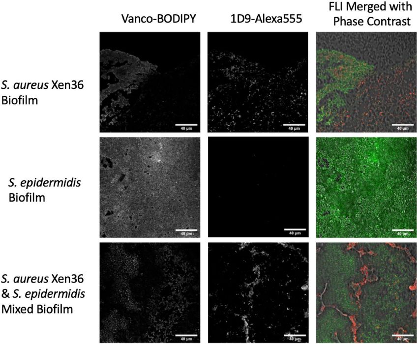
www.nature.com/scientificreports/
inability to discriminate infected from native tissue, reoperations with serial debridements are commonplace,
escalating costs and risks to the patient8–10.
Accurate diagnosis and treatment of biofilm infections remains a challenge due to difficulties with localiza-
tion and identification of bacterial pathogens seated deep within tissues. Many imaging modalities including
X-ray (XR), ultrasound (US), computer tomography (CT), or magnetic resonance imaging (MRI) are limited
in their ability to achieve high resolution adjacent to an implant; whereas nuclear medicine methodologies,
like positron emission tomography (PET), cannot accurately discriminate infection from other entities, such
as sterile inflammation or fluid collections11–15,39. Furthermore, the current standard of infection diagnosis by
culture growth of the pathogen suffers from false-negative and false-positive culture results at alarmingly high
rates, and there exist inherent risks to the patient with invasive procurement of culture samples13,15–17. As a result,
current treatment algorithms of implant infections are often based on clinical judgments that utilize diagnostics
that are known to be unreliable.
The development of fluorescent molecular targeting probes that specifically bind to specific bacterial patho-
gens has been widely heralded as a major advancement to improve diagnosis and treatment of biofilm infections18.
Staphylococcus species are identified as the most common pathogens of bacterial implant infections19–21. Van-
comycin-IRDye800CW (Vanco-800CW) consists of fluorescently-labelled vancomycin, an antibiotic that binds
to peptidoglycan in the cell wall of Gram-positive b acteria22. In contrast, the immunodominant staphylococcal
antigen A (IsaA) antibody labelled with a cyanine 680 dye (1D9-680) is a fluorescent probe that labels ubiq-
uitously expressed proteins involved in cell wall metabolism of S. aureus, but not other types of bacteria23. In
addition, IsaA is bound by the IgG-binding proteins Spa and Sbi of S. aureus23. Both molecular targeting probes
have been shown to accurately label S. aureus bacteria within in vitro and in vivo m odels22,23. Targeted fluores-
cent imaging (TFLI) applied to these novel staphylococcal targeting molecules can, thus, illuminate infections
in vivo with the potential to increase diagnostic sensitivity and specificity. Furthermore, image-guided surgical
debridement, achieved using a specialized fluorescence camera system in the surgical suite, may improve the
accuracy of infection excision, which currently relies solely on the surgeon’s discretion based on imprecise visual
inspection of infected tissue.
The purpose of this study is to: (1) compare the diagnostic accuracy of two candidate probes targeting Staphy-
lococcus aureus, (2) assess the broader applicability of the more specific probe in the spinal implant model to
identify S. aureus infections in other models of implant-associated infection, and (3) test the functionality of the
probe within a real-time, fluorescence image-guided surgery system in a proof-of-concept study evaluating the
feasibility of image-guided infection debridement surgery.
Results
In vitro assessment of staphylococcal biofilm targeting using Vancomycin‑ and 1D9 anti‑
body‑based fluorescent probes. Staphylococcal biofilms composed of either S. aureus, S. epidermidis or
a mixed culture of both strains were established on the surface of 18 mm chemically resistant borosilicate glass
coverslips. These biofilms were then incubated with vancomycin-BODIPY FL (Vanco-BODIPY) and 1D9-Alex-
aFluor555 (1D9-Alexa555) simultaneously. Of note, the latter two probes were implemented for the in vitro
assessment of staphylococcal biofilm targeting, because they are more suitable for the fluorescence microscopic
analysis of biofilms than the equivalent Vanco-800CW and 1D9-680 probes used for subsequent in vivo imaging
of infection. As shown in Fig. 1, the vancomycin probe is capable of binding to both S. aureus and S. epidermidis.
In contrast, the 1D9 probe binds exclusively to S. aureus, because this strain exposes the IsaA epitope on its
surface, whereas the selected S. epidermidis strain is incapable of IsaA expression. Interestingly, Vanco-BODIPY
appears to be more density/biomass-dependent, with the probe seeming to penetrate deeper into the biofilm
matrix compared to 1D9, which appears more surface-bound (top panels of Fig. 1). This may relate to the fact
that the expression of IsaA is downregulated in staphylococal biofilms and so 1D9 may bind preferentially to
more actively growing bacteria on the surface of the biofilm38. The apparently lower biofilm penetration by the
1D9 probe compared to the vancomycin probe may also relate to the fact that the full-size 1D9 IgG1 molecule is
substantially larger in size than vancomycin.
Establishing infected spinal implants within a mouse model. To compare the abilities of the Vanco-
800CW and 1D9-680 probes in diagnosing implant infections, an established mouse model of spine implant
biofilm infection was e mployed24. Briefly, in this infection model a midline skin incision measuring ~ 2 cm cen-
tered over the lumbar spine was made followed by exposure of the spinous processes along the entire length of
the incision. Once the spinous processes were exposed, an L-shaped stainless-steel implant was fixed into the
spinous process and inoculated with a bioluminescent S. aureus (Xen36 strain) (Fig. 2A). 10 mice were subjected
to the surgery as described and 5 mice were subjected to this surgery without inoculation of S. aureus, receiving
an equivalent volume of sterile saline. Non-invasive in vivo bioluminescence imaging (BLI) was performed on
post-operative days (POD) 0, 1, 3, 5, and 7 to assess the bacterial burden, which showed that the mean in vivo
BLI signal of the Xen36 bacteria was significantly higher in infected mice compared to sterile mice at all POD
time-points (P < 0.05) (Fig. 2B). All 10 mice in the infection group were verified to have spinal infections and all
5 control mice had no evidence of infection, as determined by in vivo BLI signal detection on POD 0 (Fig. 2C).
Candidate probes administered to mice and subsequent two‑dimensional imaging results. On
POD 7, which was previously shown to correlate with the peak of infection in our prior work, the 10 infected
and 5 sterile control mice were subjected to injections of both the Vanco-800CW and 1D9-680 fluorescently
labelled probes18,23. Mice were imaged for both in vivo BLI and in vivo fluorescent imaging (FLI) signals, 24-,
48- and 72-h post-injection (PI) of the probes. In vivo FLI was performed at excitation/emission wavelengths
Scientific Reports | (2021) 11:1622 | https://doi.org/10.1038/s41598-020-78362-7 2
Vol:.(1234567890)
www.nature.com/scientificreports/
Figure 1. Imaging of staphylococcal biofilms with vancomycin- and 1D9-based fluorescent probes. Biofilms of
S. aureus Xen36, S. epidermidis ATCC 38984, or a mixture of both Staphylococcus species were grown on cover
slides for confocal microscopy. The established biofilms were simultaneously incubated with the Vanco-BODIPY
and 1D9-Alexa555 probes. Subsequently, images were recorded by confocal fluorescence microscopy, revealing
binding of the vancomycin probe to all biofilms and the 1D9-Alexa555 probe to S. aureus biofilms only. Of note,
we attribute the “patchy” fluorescence pattern of the S. aureus cells in the mixed biofilm, as detected with the
1D9-Alexa555 probe, to the fact that S. epidermidis secretes serine proteases that can inhibit biofilm formation
by S. aureus41. The magnification is indicated by scale bars.
Figure 2. Spinal implant infection model. (A) Post-operative day 7 (POD 7) lateral XR revealing the position
of the pin fixed within the spinous process in the lumbar spine and overlying bioluminescent signal of S. aureus
Xen36. (B) Mean total bioluminescence represented as total flux (photons/sec) at each POD with error bars
derived from Standard Error of the Mean for the Infected (n = 10) and Sterile (n = 5) cohorts. (C) Representative
bioluminescence images of four mice from the Infected and Sterile Cohorts at post-operative day 7.
Scientific Reports | (2021) 11:1622 | https://doi.org/10.1038/s41598-020-78362-7 3
Vol.:(0123456789)
www.nature.com/scientificreports/
Figure 3. Bioluminescence and fluorescence images of mice with spinal implant infection at 24 h and 72 h
post probe injection (PI). Bioluminescence imaging of two representative mice reveals bacterial signal about
the lumbar spine. At 24 h PI, the 1D9-680 probe was imaged with an excitation/emission pair of 675/720 nm
revealing a similar pattern of signal in comparison to bioluminescence. The Vanco-800CW probe was imaged
with an excitation/emission pair of 745/800 nm revealing a relatively non-specific signal about the lumbar spine
as well as caudally to the base of the tail and feet due to the ubiquitous pooling of the probe within the tissues
as a consequence of the pharmacokinetics of the Vanco-800CW probe. Clearance of this pooling effect was
resolved by 72 h PI with remaining probe signal localizing to the site of infection. By 72 h PI, the 1D9-680 and
Vanco-800CW signals both revealed a similar pattern in comparison to bioluminescence.
of 675/720 nm and 745/800 nm, corresponding to the 1D9-680 and Vanco-800CW fluorescent probes, respec-
tively. Importantly, these probes did not exhibit spectral overlap; thus upon co-injection in vivo, an accurate,
head-to-head comparison could be made of their accuracy and specificity of targeting the S. aureus infection.
At 24 h PI of the probes, in-vivo FLI revealed a highly intense signal from both probes (Fig. 3). However, in
the case of the Vanco-800CW probe, this signal still lacked resolution and was found to be equally as strong in
the sterile control animals due to non-specific tissue distribution. By 72 h PI of the probes, the Vanco-800CW
signal appeared to be accumulating at the site of the bacterial infection to a similar degree as the 1D9-680 probe
when the animals were imaged two-dimensionally (2D) in the dorsal plane. In contrast to the apparent slower
clearance of the Vanco-800CW probe, the 1D9-680 probe appeared to specifically target the S. aureus infections
as early as 24 h PI. Moreover, the antibody probe seemed to clear more rapidly from the animal’s tissues, since
none of the sterile control mice showed non-specific background signals, as seen with the Vanco-800CW probe
(Fig. 3). Possibly, this relates to the fact that 1D9-680 is cleared by the liver, while the Vanco-800CW probe is
excreted via the kidney and bladder22,23.
Three‑dimensional computed tomography scans with bioluminescent and fluorescent over‑
lay. In order to assess the distribution of the 1D9-680 and Vanco-800CW probes throughout the mouse,
three-dimensional (3D) computed tomography scans with 3D in vivo BLI and FLI overlay were completed at
24-, 48- and 72-h PI of the probes. The 1D9-680 probe’s fluorescence signal tightly correlated with the in vivo
BLI signals in all planes from 48 h PI onwards (Fig. 4). In contrast, the Vanco-800CW probe’s fluorescence signal
was less discriminatory. Although it clearly accumulated at the site of the bacterial in vivo BLI signals, it was also
detectable in the abdomen and pelvis of the animals. This off-target accumulation of Vanco-800CW in the mice
is possibly due to non-specific retention in tissues or the bladder, non-specific targeting by breakdown products
of the probe, and/or dissemination of bacterial debris towards the murine abdomen. Importantly, this 3D assess-
Scientific Reports | (2021) 11:1622 | https://doi.org/10.1038/s41598-020-78362-7 4
Vol:.(1234567890)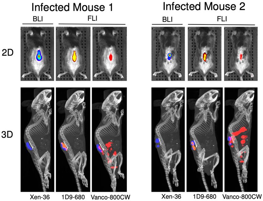
www.nature.com/scientificreports/
Figure 4. Three-dimensional bioluminescence and fluorescence imaging of spinal implant infection. At
72 h following PI, the mice were subjected to 3D-CT with bioluminescence and fluorescence imaging. Two
representative infected mice are shown. The 1D9-680 probe signal colocalizes with the bioluminescent bacterial
signal on the infected implant. The Vanco-800CW probe signal colocalizes with the infected implant, but is also
detected in the pelvic and abdominal cavities.
ment revealed better performance by 1D9-680 in terms of accuracy and specificity of co-localization with living
bacteria within the biofilm on spinal implants.
Ex vivo confirmation. At 72 h PI of the probes, following 3D imaging, necropsy surgery was performed
to determine the ex vivo co-localization of the probes with the normative ex vivo BLI signals. Both 1D9-680
and Vanco-800CW colocalized with the ex vivo BLI signals of the bacteria in the explanted periprosthetic bone
(Fig. 5, Middle Row), however, the Vanco-800CW also localized to an uninfected piece of bone that showed no
presence of living bacteria by ex vivo BLI (Fig. 5, Bottom Row). This again shows the adequate sensitivity of both
probes, but demonstrates a higher specificity of the 1D9-680 probe for living bacteria in the present infection
model.
Assessing broader applicability of the 1D9‑680 probe. Given the off-target binding of Vanco-
800CW demonstrated in Fig. 3 and the greater specificity of 1D9-680 demonstrated in Fig. 4 (Bottom row),
all additional experiments involved assessing only the 1D9-680 probe. Since all prior spinal implant infection
imaging studies had been conducted using both 1D9-680 and Vanco-800 in combination, a second group of 8
infected mice and 4 control mice were intravenously injected with the 1D9-680 probe alone, to allow for an inde-
pendent assessment of this antibody probe (Fig. 6). Based on previous imaging results, all 12 mice were imaged
48 h PI. In Fig. 6, 1D9-680 co-localized consistently with the Xen36 in vivo BLI signals. Moreover, the magnitude
of the in vivo FLI signals were somewhat comparable to the in vivo BLI signals, with only the weakest in vivo
BLI signals showing in vivo FLI signals close to background levels (mouse #7 from the left) and all sterile control
mice demonstrated negligible background in vivo FLI signals.
Next, the 1D9 probe was tested in two additional established models of implant-associated infection: an
allograft (Fig. 7) and a shoulder implant (Fig. 8) model37. First, in 7 mice, a dorsally placed cancellous human
allograft implant was either infected with bioluminescent Xen36 (4 mice) or treated with PBS (3 mice). The 1D9-
680 probe was then intravenously injected in an identical manner to that of the spine model, and all 7 mice were
imaged 72 h PI (Fig. 7). As observed with the spinal model, the in vivo FLI signals corresponded closely with
the in vivo BLI signal, demonstrating the 1D9-680 antibody accumulated at the infection site equally well in this
animal implant model. In this case, even the weakest in vivo BLI signal (mouse #4 from the left) was accurately
detected by this probe. Finally, the 1D9-680 probe was evaluated in a shoulder arthroplasty model (Fig. 8), with
4 mice infected with Xen36 and 3 mice acting as sterile controls37. In contrast to the spine and allograft models,
where even infections with the weakest in vivo BLI signals could be detected by the 1D9-680 probe to give a con-
comitant in vivo FLI signal, only the strongest in vivo BLI signal could be detected above background. Whether
this more limited detection is due to greater depth of tissue or less efficient labelling by the 1D9-680 probe is not
Scientific Reports | (2021) 11:1622 | https://doi.org/10.1038/s41598-020-78362-7 5
Vol.:(0123456789)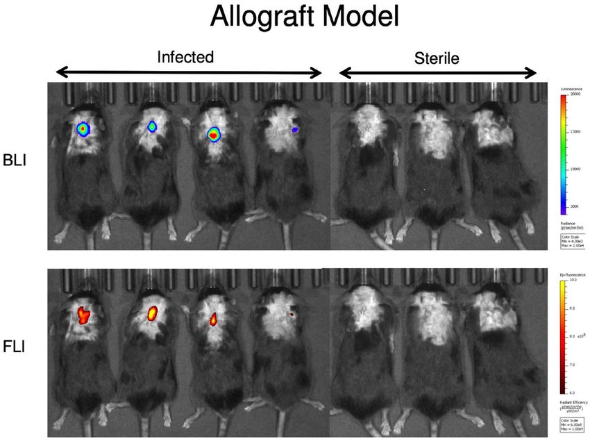
www.nature.com/scientificreports/

                                            Figure 5.  Bioluminescence and fluorescence imaging of infected necropsy specimen from spinal implant
                                            infection. Following necropsy of the infected tissue bed, bioluminescence and fluorescence imaging were
                                            repeated revealing co-localization of the 1D9-680 probe with infected tissue. As shown for a representative
                                            mouse, the Vanco-800CW imaging revealed co-localization with the infected necropsy specimen, but
                                            fluorescence signal was also detected on supplemental tissue without bioluminescence.

                                            Figure 6.  Bioluminescence and fluorescence imaging of mice with a spinal implant infection 72 h post 1D9-
                                            680 probe injection. At 72 h PI of the 1D9-680 probe, co-localization of the probe in 8 mice with infected
                                            implants is evidenced by bioluminescence and fluorescence imaging as described for Fig. 3. As expected, neither
                                            bioluminescence nor fluorescence are detectable in the 4 control mice with sterile spinal implants.

                                            known. However, tissue penetration should not pose a problem in surgical settings, where infectious sites are
                                            exposed for imaged-guided debridement of fluorescently-labelled infected tissue.

                                            Testing 1D9‑680 for its ability to delineate infection using image‑guided surgery.              Image-
                                            guided infection debridement surgery was performed using the Solaris Fluorescence Image-Guide Surgery
                                            System (PerkinElmer, Hopkinton, MA). This system utilizes an overhead camera with fluorescence input to

          Scientific Reports |   (2021) 11:1622 |               https://doi.org/10.1038/s41598-020-78362-7                                                6

Vol:.(1234567890)
www.nature.com/scientificreports/

                                  Figure 7.  Bioluminescence and fluorescence imaging of mice with a spinal allograft infection 72 h post
                                  1D9-680 probe injection. At 72 h PI of the 1D9-680 probe, co-localization of the probe in 4 mice with infected
                                  dorsally placed cancellous human allograft implants is evidenced by bioluminescence and fluorescence imaging
                                  as described for Fig. 3. As expected, neither bioluminescence nor fluorescence are detectable in the 3 control
                                  mice with sterile allograft implants.

                                  Figure 8.  Bioluminescence and fluorescence imaging of mice with a spinal allograft infection 72 h post
                                  1D9-680 probe injection. At 72 h PI of the 1D9-680 probe, co-localization of the probe in 1 out of 4 mice with
                                  infected shoulder arthroplasty is evidenced by bioluminescence and fluorescence imaging as described for
                                  Fig. 3. As expected, neither bioluminescence nor fluorescence are detectable in the 3 control mice with sterile
                                  shoulder arthroplasty.

Scientific Reports |   (2021) 11:1622 |                https://doi.org/10.1038/s41598-020-78362-7                                                   7

                                                                                                                                             Vol.:(0123456789)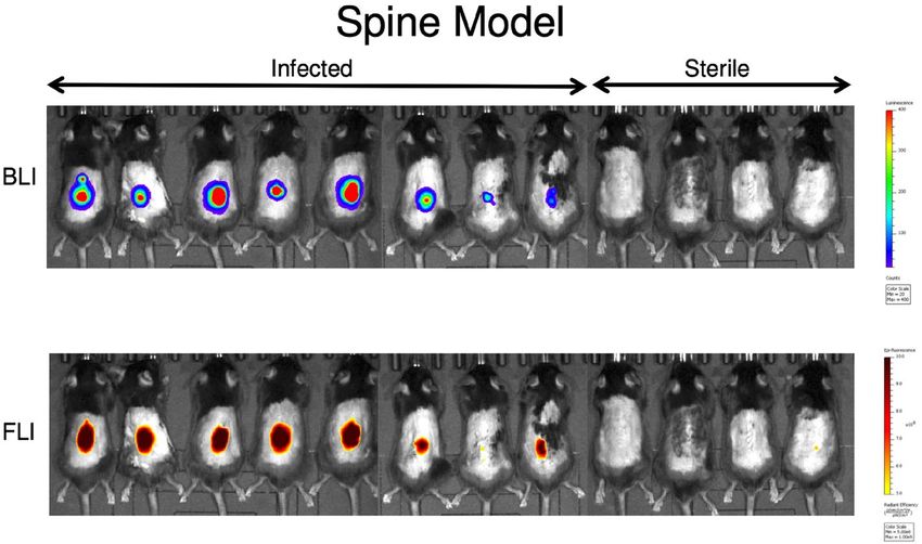
www.nature.com/scientificreports/
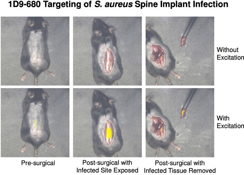
Figure 9. Image-guided surgical debridement of infected tissue in the spinal implant model. Image-guided
surgery was performed utilizing the PerkinElmer Solaris Image-Guided Surgery System. Both the real-time
white light images and fluorescence overlay images of a surgical debridement are shown. Pseudo-color yellow
fluorescence can be seen in the pre-surgical panel, bottom image. Once the skin is opened, the wound bed is
bright yellow in the bottom panel, which is indicative of the stronger probe signal. Following debridement
of tissue, the bottom panel reveals no signal within the wound bed and persistent probe signal in the tissue
specimen held by forceps.
display real-time images of the surgical specimen with fluorescence signal overlay. Based on the tightly cor-
related in vivo FLI signals of the 1D9-680 probe and in vivo BLI signals of the bacteria, image-guided surgery
was performed with an excitation wavelength of 660 nm. Prior to skin incision, the in vivo FLI signals overlying
the spinal surgical site was readily visualized. The skin was incised and the visualized fascia exhibited persistent
in vivo FLI signals. Fluorescent tissue was sharply debrided, and the excised tissue specimen was imaged with
ex vivo BLI and FLI, which yielded closely correlated signals (Fig. 9). The wound was debrided until all remain-
ing fluorescent tissue was resected.
Discussion
The findings presented in this study address the pressing clinical shortcomings in the diagnosis and treatment
of implant infections. In cases that require surgical debridement, the basic tasks of accurate localization and
identification of pathogens remain a challenge, often to the detriment of the patient12,25. Imaging modalities
such as US, XR, CT, PET, and MRI are limited in their ability to discriminate infections from other differential
processes13–17. Successful biopsy or culture of the bacterial foci are vital to successful treatment, yet current tech-
niques suffer from considerable false-negative and false-positive results, which contribute to treatment failures
and patient m orbidity17,18,25. Moreover, surgical debridement is rudimentary at best, depending on the individual
surgeon’s visual assessment of pathologic tissue, such as discoloration or changes in texture; this assessment is
fundamentally subjective and prone to error.
In order to address these shortcomings, the purpose of this study was to compare two novel S. aureus target-
ing fluorescent imaging (TFLI) probes in their ability to identify and delineate this bacterium in implant infec-
tions, and to assess whether the more accurate probe could be utilized in a real-time, fluorescence image-guided
surgical system. The two candidate probes, Vanco-800CW and 1D9-680, were both found to target S. aureus
in vitro and in vivo with high affinity. However, the Vanco-800CW probe had background signals within the
pelvis and abdomen, whereas the 1D9-680 probe specifically localized to the site of the in vivo BLI signals of the
bacteria, which was most apparent upon 3D co-registration of CT and in vivo BLI/FLI analysis. The 1D9 probe
demonstrated excellent specificity for S. aureus in three different mouse models of surgical implant infections
and good sensitivity in all but the low-grade shoulder implant infection. Mice with infected spinal implants
were then subjected to a fluorescence image-guided surgical system, in which the 1D9-680 probe successfully
guided debridement surgery. These data provide a proof-of-concept for the specific, non-invasive diagnosis and
localization of bacterial infection while leveraging TFLI within an image-guided surgery system to aid in the
resection of infected tissue.
The implementation of fluorescent probes for TFLI of bacterial infections is a burgeoning theranostic
approach for clinical infections. Aside from the Vanco-800CW and 1D9-680 utilized in this study, previous work
Scientific Reports | (2021) 11:1622 | https://doi.org/10.1038/s41598-020-78362-7 8
Vol:.(1234567890)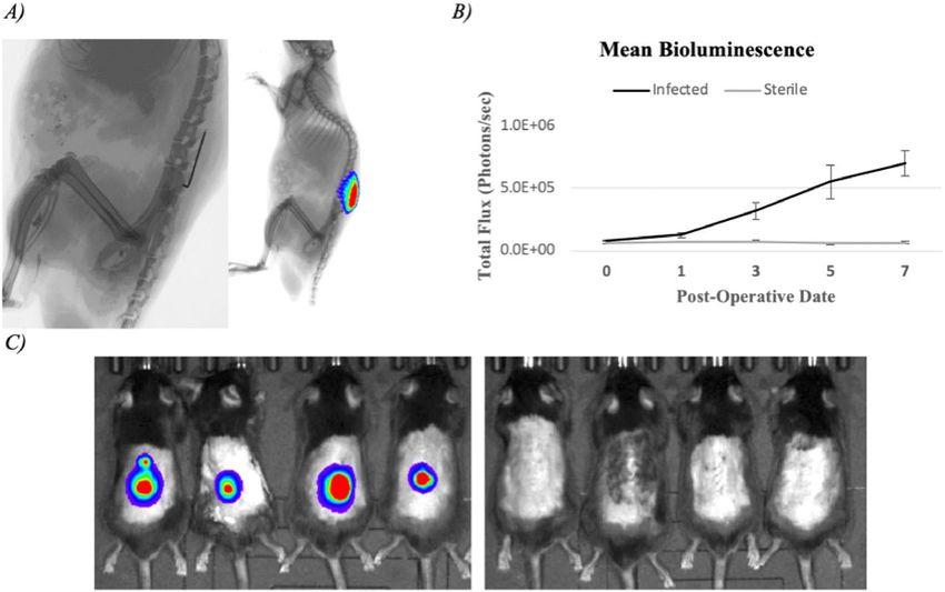
www.nature.com/scientificreports/
has focused on probes that target the cell membrane, membrane transporters, bacterial enzymes, genetic mark-
ers, and specific organelles, in a wide range of bacteria, including Staphylococcus, Enterococcus, Bifidobacterium,
Salmonella, Mycobacterium, Klebsiella, and Pseudomonas species26–32,39. This recent torrent of studies points to the
substantial interest in targeted diagnostic and therapeutic technologies, especially with the widespread emergence
of multi-drug resistant organisms that do not respond to standard a ntibiotics33,34. The development of multiple
bacterial probes with unique optical outputs may enhance the ability to rapidly discriminate between bacterial
pathogens and pave the way for unique therapeutic modalities. TFLI technology allows real-time, image-guided
infection debridement surgery which we believe represents the next translational step of this technology. At pre-
sent, no bacteria-specific molecular probes exist that are licensed for human use, although several candidates are
in preclinical development. It has been postulated that the ideal probe for human use will likely be a fluorescently
labelled antibody, antibiotic derivative, or modified antimicrobial peptide34,39. Both Vanco-800CW and 1D9-680
fit this description. However, the Vanco-800CW probe non-specifically bound to areas within the abdomen and
pelvis. Based on these data, we believe 1D9-680 represents a more specific and stable probe for S. aureus infec-
tions, illuminating infections for at least 72 h following injection. On the other hand, it is important to bear in
mind that Vanco-800CW targets a wide range of Gram-positive bacteria that cannot be detected with the IsaA
target-specific 1D9-680 probe as exemplified in Fig. 1 of this s tudy22,23. This view is underlined by prosthetic joint
infections, which are more frequently caused by Gram-positive bacteria other than S. aureus40. In this respect, the
simultaneous imaging with both the 1D9-680 and Vanco-800CW probes could offer an opportunity for in vivo
diagnostics to distinguish infections caused by S. aureus or other Gram-positive bacteria.
There are several limitations to this animal study. First, the extrapolation of findings from animal models to
human clinical conditions should be interpreted with caution. In particular, the methods implemented in this
study, including the injection of probe at the known peak of infection within the animal model might not repre-
sent the most ideal conditions in which to detect bacterial infection in humans. Second, in vivo FLI is inversely
related to the amount of intervening tissue, which in humans may prove to be a challenge in the diagnosis of
deep-seated infections such as peri-prosthetic total hip arthroplasty infection. This issue may have been high-
lighted by the inability of 1D9-680 to detect a low-grade shoulder girdle infection in the mouse model, where a
low bacterial burden combined with a thicker overlying tissue led to limited sensitivity.
In conclusion, the 1D9-680 probe, but not the Vanco-800CW probe, more specifically co-localized to the S.
aureus infection in a mouse model of spinal implant infection as evidence by 2D and 3D in vivo BLI and FLI.
This specificity was maintained in allograft and shoulder implant infection models. The fluorescence emitted
by this probe aided in the debridement of infected tissue within an open air, fluorescence image-guided surgery
system, which we believe is the next advancement in the utilization of this technology that can be translated to
clinical use. These findings provide proof-of-concept of TFLI technology enhancing the diagnosis and treatment
of bacterial implant infections, which are currently beleaguered by inaccurate diagnosis and empiric or incorrect
treatment to the detriment of countless patients.
Methods
Ethics statement. All animals were maintained and utilized in accordance with good animal practice as
defined in the federal regulations set forth in the Animal Welfare Act (AWA), the 1996 Guide for the Care and
Use of Laboratory Animals, and PHS Policy for the Humane Care and Use of Laboratory Animals. All animal
use was in accordance with institutional animal care and use committee protocols, and was approved by the
UCLA Chancellor’s Animal Research Committee (ARC# 2012-104-03J).
Bioluminescent S. aureus strain. Staphylococcus aureus Xen36 strain (PerkinElmer, Hopkinton, MA) is
a bioluminescent derivative of the clinical isolate ATCC 49525 (Wright)35. S. aureus Xen36 possesses a Gram-
positive optimized luxABCDE operon stably integrated into a large native plasmid35 yielding a maximal emis-
sion wavelength of approximately 490 nm constitutively produced by live, metabolically active bacteria. It has
been previously shown to be the optimal S. aureus strain for use in such experiments due to the strength and
consistency of its bioluminescence signal36. S. aureus Xen36 is kanamycin resistant and is grown with 200 µg/ml
kanamycin (Sigma–Aldrich) to avoid contamination. S. aureus was streaked onto tryptic soy agar plates (tryptic
soy broth [TSB] plus 1.5% bacto agar; BD Biosciences) and grown at 37 °C overnight. Single colonies of S. aureus
were then cultured in TSB and again grown overnight at 37 °C in a shaking incubator (240 rpm) (MaxQ 4,450,
Thermo Fisher Scientific). Mid-logarithmic phase bacteria were obtained after a 2 h subculture of a 1:50 dilution
of the overnight culture. Bacterial cells were pelleted, re-suspended, and washed three times in PBS. Bacterial
inocula of 1 × 10³ colony forming units (CFU) were ascertained by measuring the absorbance at 600 nm (A600,
Biomate 3 [Thermo]) and comparing to a reference standard. This dose was derived from previous described
methods utilizing this model24.
In vitro imaging of staphylococcal biofilms. S. aureus Xen36 and S. epidermidis ATCC 38984 were
grown overnight in tryptic soy broth (TSB) in a shaking incubator at 37 °C. Bacterial biofilms were grown
on chemically resistant borosilicate 18 mm glass coverslips (Paul Marienfeld GmbH, Lauda-Königshofen, Ger-
many) in a 12-well microtiter plate containing TSB supplemented with 5% glucose and 4% NaCl. To this end,
the wells were inoculated from the overnight cultures to a final optical density at 600 nm (OD600) of 0.1, and the
plates were incubated at 37 °C for 48 h. Co-cultures of S. aureus and S. epidermidis were prepared by inoculating
the wells at a 1:1 ratio from the respective overnight cultures. Coverslips with biofilm were incubated with a mix
of 0.2 µM 1D9-Alexa555 and 0.2 µM Vanco-BODIPY FL (Thermo Fisher Scientific) for 30 min in PBS. Subse-
quently, the biofilms were washed 1× with PBS to remove unbound fluorescent probes and, thereafter, they were
fixed in 4% paraformaldehyde. Finally, the coverslips were mounted on microscopy slides. Image acquisition was
Scientific Reports | (2021) 11:1622 | https://doi.org/10.1038/s41598-020-78362-7 9
Vol.:(0123456789)www.nature.com/scientificreports/
performed with a Leica TCS SP8 confocal microscope. The recorded images were processed using ImageJ soft-
ware (National Institutes of Health) and LAS X Life Science. The 1D9-Alexa555 probe used in these experiments
was obtained by crosslinking the Alexa Fluor 555 dye (Thermo Fisher Scientific) to the human monoclonal
antibody 1D9 via activated N-hydroxysuccinimide ester chemistry.
Mouse model of spinal implant infection. The following description is sourced from work previously
published by our g roup24. The mice used within these experiments were twelve-week-old male C57BL/6 wild-
type mice (Jackson Laboratories, Bar Harbor, ME). Mice were kept at 4 per cage in standard cages with a 12 h
light and dark cycle. Free access to water and standard pellet feed were utilized in compliance with the standards
of the UCLA Chancellor’s Animal Research Committee. Veterinary staff ensured the well-being of all animals
throughout the experiment by daily assessment. Mouse spinal implant surgeries were performed as previously
described24. In brief, animals were shaved along the dorsum of their lumbar spine, skin was prepped with alcohol
and betadine, and inhalation isoflurane (2%) was utilized for anesthesia. A dorsal skin incision ~ 2 cm in length
was utilized, centered over the lower lumbar spine. The lumbar spinous processes were exposed by carefully
peeling away the soft tissue from the posterior elements of the spine, unilaterally. The spinous process of L4 was
reamed with a 25-gauge needle, and this reamed cavity received an “L-shaped” surgical-grade 0.1 mm diameter
stainless steel implant (Modern Grinding, Port Washington, WI). The short arm of the “L-shaped” implant was
placed through the reamed cavity of the spinous process, and the long arm extended toward the head of the
mouse. Two 4–0 Vicryl sutures were placed in preparation for closure. An inoculation of 1 × 10³ CFU of biolumi-
nescent Xen36 S. aureus or sterile saline was pipetted onto the bend of the implant, the previously placed sutures
were expeditiously tied to avoid contamination of the wound, and a running 4–0 Vicryl suture was utilized to
close the overlying skin. Buprenorphine (2.5 mg/kg) (ZooPharm, Fort Collins, CO) was administered for anal-
gesia, subcutaneously, every 72 h for the duration of the experiment. Mice were then subjected to high resolution
X ray (Faxitron LX-60 DC-12 imaging system) to confirm placement of the implant.
Mouse model of allograft infection. As previously described, 12-week-old male C57BL/6 wild-type
mice were used (Jackson Laboratories, Bar Harbor, ME) for the allograft infection experiment with a similar care
protocol. Human fibular cortical allograft was obtained from, and sterilized in the standard manner, by Mus-
culoskeletal Transplant Foundation (Edison, NJ). Bulk allograft was shaped into 2.5 mm diameter discs using a
high-speed saw and wire cutter. Scissors were used to trim rough edges, and implants were sterilized in an auto-
clave. Survival surgery was performed in which the allograft was implanted in the subcutaneous space dorsal to
the caudal cervical spine. Mice were anesthetized via inhalation isoflurane (2%). The level of the implantation
was approximated by palpating the maximal point of lordosis with minimum skin tension. A 1 cm midline inci-
sion was then made and carried down to the fascia, exposing subcutaneous muscle. The dissection was directed
bilaterally superficial to the paraspinal musculature, developing a subcutaneous pocket for the allograft implant.
Fine-toothed forceps were used to gently place the sterile implant into the pocket created by dissection. An
inoculation of 1 × 10² CFUs of bioluminescent Xen36 S. aureus in 2 µL phosphate buffered solution or 2 µL sterile
saline (control group) was pipetted onto the allograft. A single 4.0 Vicryl suture was then placed in a running
fashion to approximate the skin. Quick-release buprenorphine (0.3 mg/kg) (Zoo-Pharm, Fort Collins, CO) was
administered subcutaneously every 12 h for 72 h as post-operative analgesic.
Mouse model of shoulder implant infection. As previously described, 12-week-old male C57BL/six
wild-type mice were used (Jackson Laboratories, Bar Harbor, ME) for the shoulder implant infection experiment
with a similar care protocol37. The mice were anesthetized for survival surgery. The shoulder joint was approxi-
mated by palpating superiorly towards the most proximal aspect of the humerus. An incision was made starting
at the sternum and extending laterally across the deltoid, which exposed the deltopectoral groove beneath. Next,
the pectoralis musculature was removed from its insertion on the humerus. Once near the humeral head, the
joint was exposed by applying anterior directed pressure on the posterior aspect of an extended and externally
rotated humerus, which subluxed the joint anteriorly. A 25-gauge needle was used to ream the most proximal
aspect of the humeral head, with the needle aimed towards the palpable deltoid tuberosity. The needle was then
removed, allowing the placement of the implant, with the implant’s most proximal aspect communicating with
the glenoid fossa within the shoulder joint. 5–0 vicryl was then used to approximate the deep fascial layers.
These sutures were placed but not tied to allow for expedient closure after inoculation and to restrict bacteria to
the immediate area of the implant. Next, a 2 µL inoculation of 1 × 10³ (e3 group) or 1 × 10⁴ (e4 group) CFUs of
bioluminescent S. aureus Xen-36 or sterile saline (control group) was pipetted onto the tip of the implant. Deep
sutures were tied and a running 5–0 vicryl was used to approximate the skin. Sustained release buprenorphine
(2.5 mg/kg) (Zoo-Pharm, Fort Collins, CO) was then administered subcutaneously every 72 h as analgesic for
the duration of the experiment.
Description of probes and injection protocol. The 1D9-680 probe is composed of the anti-IsaA IgG1
antibody conjugated to the near infrared fluorophore NIR680 (PerkinElmer, Hopkinton, MA) with peak exci-
tation of 680 nm as previously described18,23. The IsaA epitope is a ubiquitously expressed protein involved
in the cell wall metabolism of S. aureus and a few other staphylococcal s pecies23. Vancomycin-IRDye800CW
(Vanco-800CW) is a vancomycin molecule conjugated to IRDye 800CW, a near-infrared fluorophore with peak
excitation at 778 nm and emission at 794 nm. Vanco-800CW was synthesized as previously d escribed22. Both
1D9-680 and Vanco-800CW were administered to infected and control group mice on POD 7. To this end, the
probes were dissolved in phosphate-buffered saline to deliver 25 nmol of Vanco-800CW and 0.1 μg 1D9-680 in a
100 µL final injection volume. Mice were anesthetized via inhalation isofluorane (2%) and probe solutions were
Scientific Reports | (2021) 11:1622 | https://doi.org/10.1038/s41598-020-78362-7 10
Vol:.(1234567890)www.nature.com/scientificreports/
administered via tail vein injection as previously described22,23. Mice were monitored daily for systemic reaction
and adverse effects.
Bioluminescence and fluorescence imaging. Mice were anesthetized via inhalation isoflurane (2%)
and in vivo bioluminescence and fluorescence imaging was performed using an IVIS Spectrum-CT (Perki-
nElmer, Hopkinton, MA). Anesthesia was maintained at 1.5% during imaging. 2D Bioluminescent imaging
was performed as previously described24. Fluorescent excitation/emission pairs of 675/720 nm and 745/800 nm
were used for 1D9-680 and Vanco-800CW probes, respectively. 2D Images were obtained 24, 48, and 72 h after
probe injection. 2D data are presented via color scale overlaid on a grayscale photograph of mice and quantified
within a circular region of interest (16,103 pixels) as mean maximum flux (photons per second (s) per c m2 per
steradian (sr) [p/s/cm2/sr]) for bioluminescence and as maximum radiant efficiency ([photons/s]/[mW/cm2])
for fluorescent imaging using Living Image 4.2 software (PerkinElmer, Hopkinton, MA).
At the indicated timepoints, Diffuse Light Imaging Tomography (DLIT—3D bioluminescence) images were
acquired with a Spectrum-CT using emission filters that correspond to the spectral profile of lux. Immediately
following DLIT, Fluorescence Light Imaging Tomography (FLIT—3D fluorescence) images were acquired with
a Spectrum-CT using the filter pairs mentioned above over a scan field surrounding the implants. 3D images
were reconstructed via tomographic algorithms and automatically coregistered with a CT scan using the Living
Image software package.
Solaris fluorescence image‑guided surgery system. Image-guided operative debridement of infected
tissue and implant retrieval were performed 72 h following PI utilizing a Solaris Fluorescence Image-Guided
Surgery System (PerkinElmer, Hopkinton, MA) under ambient light conditions. Mice were sacrificed immedi-
ately prior to operative debridement. An excitation filter of 660 nm was utilized corresponding to the excitation
wavelength of the 1D9-680 probe. Still images and videos were periodically saved throughout the procedure
using the Solaris software (PerkinElmer, Hopkinton, MA).
Statistical analysis. Data between two groups were compared by using either one or two-tailed Student’s
t-test and data form three or more groups were compared using a one-way ANOVA. All data are expressed as
mean ± standard error of the mean (SEM). Values of P < 0.05 were considered statistically significant.
Data availability
All data generated or analyzed during this study are included in this published article (and its Supplementary
Information files).
Received: 28 February 2020; Accepted: 17 November 2020
References
1. Kirkland, K. B., Briggs, J. P., Trivette, S. L., Wilkinson, W. E. & Sexton, D. J. The impact of surgical-site infections in the 1990s:
attributable mortality, excess length of hospitalization, and extra costs. Infect. Control Hosp. Epidemiol. 20(11), 725–730 (1999).
2. de Lissovoy, G. et al. Surgical site infection: incidence and impact on hospital utilization and treatment costs. Am. J. Infect. Control
37(5), 387–397 (2009).
3. Emohare, O. et al. Cost savings analysis of intrawound vancomycin powder in posterior spinal surgery. Spine J. 14(11), 2710–2715
(2014).
4. Godil, S. S., Parker, S. L., O’Neill, K. R., Devin, C. J. & McGirt, M. J. Comparative effectiveness and cost-benefit analysis of local
application of vancomycin powder in posterior spinal fusion for spine trauma: clinical article. J. Neurosurg. Spine 19(3), 331–335
(2013).
5. Costerton, J. W., Stewart, P. S. & Greenberg, E. P. Bacterial biofilms: a common cause of persistent infections. Science 284(5418),
1318–1322 (1999).
6. Joost, I. et al. Staphylococcus aureus bloodstream infection in patients with ventricular assist devices-management and outcome
in a prospective bicenter cohort. J. Infect. 77(1), 30–37 (2018).
7. Renz, N., Özdirik, B., Finger, T., Vajkoczy, P. & Trampuz, A. Infections after cranial neurosurgery: prospective cohort of 103 episodes
treated according to a standardized algorithm. World Neurosurg. 116, 491–499 (2018).
8. Rodríguez-Pardo, D. et al. Gram-negative prosthetic joint infection: outcome of a debridement, antibiotics and implant retention
approach. A large multicentre study. Clin. Microbiol. Infect. 20(11), O911–O919 (2014).
9. Kalra, K. P., Lin, K. K., Bozic, K. J. & Ries, M. D. Repeat 2-stage revision for recurrent infection of total hip arthroplasty. J. Arthro-
plasty 25(6), 880–884 (2010).
10. Ho, C., Skaggs, D. L., Weiss, J. M. & Tolo, V. T. Management of infection after instrumented posterior spine fusion in pediatric
scoliosis. Spine 32(24), 2739–2744 (2007).
11. Kwee, T. C., Kwee, R. M. & Alavi, A. FDG-PET for diagnosing prosthetic joint infection: systematic review and metaanalysis. Eur.
J. Nucl. Med .Mol. Imaging 35(11), 2122–2132 (2008).
12. Gratz, S. et al. 18F-FDG hybrid PET in patients with suspected spondylitis. Eur. J. Nucl. Med. Mol. Imaging 29(4), 516–524 (2002).
13. Smids, C. et al. A comparison of the diagnostic value of MRI and 18F-FDG-PET/CT in suspected spondylodiscitis. Infection 45(1),
41–49 (2017).
14. Bassetti, M. et al. 18F-Fluorodeoxyglucose positron emission tomography and infectious diseases: current applications and future
perspectives. Curr. Opin. Infect. Dis. 30(2), 192–200 (2017).
15. Termaat, M. F. et al. The accuracy of diagnostic imaging for the assessment of chronic osteomyelitis: a systematic review and meta-
analysis. J. Bone Jt. Surg. Am. 87(11), 2464–2471 (2005).
16. Neut, D., van Horn, J. R., van Kooten, T. G., van der Mei, H. C. & Busscher, H. J. Detection of biomaterial-associated infections in
orthopaedic joint implants. Clin. Orthop. Relat. Res. 413, 261–268 (2003).
17. Trampuz, A., Osmon, D. R., Hanssen, A. D., Steckelberg, J. M. & Patel, R. Molecular and antibiofilm approaches to prosthetic joint
infection. Clin. Orthop. Relat. Res. 414, 69–88 (2003).
Scientific Reports | (2021) 11:1622 | https://doi.org/10.1038/s41598-020-78362-7 11
Vol.:(0123456789)www.nature.com/scientificreports/
18. Zoller, S. D. et al. Multimodal imaging guides surgical management in a preclinical spinal implant infection model. JCI Insight
4(3), e124813 (2019).
19. Arciola, C. R., Campoccia, D., Ehrlich, G. D. & Montanaro, L. Biofilm-based implant infections in orthopaedics. Adv. Exp. Med.
Biol. 830, 29–46 (2015).
20. Costerton, J. W. et al. New methods for the detection of orthopedic and other biofilm infections. FEMS Immunol. Med. Microbiol.
61(2), 133–140 (2011).
21. Montanaro, L. et al. Scenery of Staphylococcus implant infections in orthopedics. Future Microbiol. 6(11), 1329–1349 (2011).
22. van Oosten, M. et al. Real-time in vivo imaging of invasive- and biomaterial-associated bacterial infections using fluorescently
labelled vancomycin. Nat. Commun. 4, 2584 (2013).
23. Romero Pastrana, F. Noninvasive optical and nuclear imaging of Staphylococcus-specific infection with a human monoclonal
antibody-based probe. Virulence 9(1), 262–272 (2018).
24. Dworsky, E. M. et al. Novel in vivo mouse model of implant related spine infection. J. Orthop. Res. 35(1), 193–199 (2017).
25. Campoccia, D., Montanaro, L. & Arciola, C. R. The significance of infection related to orthopedic devices and issues of antibiotic
resistance. Biomaterials 27(11), 2331–2339 (2006).
26. Bardhan, N. M., Ghosh, D. & Belcher, A. M. Carbon nanotubes as in vivo bacterial probes. Nat. Commun. 5, 4918 (2014).
27. Chen, H. et al. Bacteria-targeting conjugates based on antimicrobial peptide for bacteria diagnosis and therapy. Mol. Pharm. 12(7),
2505–2516 (2015).
28. Hernandez, F. J. et al. Noninvasive imaging of Staphylococcus aureus infections with a nuclease-activated probe. Nat. Med. 20(3),
301–306 (2014).
29. Kong, Y. et al. Imaging tuberculosis with endogenous beta-lactamase reporter enzyme fluorescence in live mice. Proc. Natl. Acad.
Sci. U.S.A. 107(27), 12239–12244 (2010).
30. Ning, X. et al. Maltodextrin-based imaging probes detect bacteria in vivo with high sensitivity and specificity. Nat. Mater. 10(8),
602–607 (2011).
31. Stanton, M., Cronin, M., Lehouritis, P. & Tangney, M. In vivo bacterial imaging without engineering; A novel probe-based strategy
facilitated by endogenous nitroreductase enzymes. Curr. Gene Ther. 15(3), 277–288 (2015).
32. Welling, M. M. et al. Development of a hybrid tracer for SPECT and optical imaging of bacterial infections. Bioconjug. Chem.
26(5), 839–849 (2015).
33. Heuker, M. et al. Preclinical studies and prospective clinical applications for bacteria-targeted imaging: the future is bright. Clin.
Transl. Imaging 4, 253–264 (2016).
34. Mills, B., Bradley, M. & Dhaliwal, K. Optical imaging of bacterial infections. Clin. Transl. Imaging 4, 163–174 (2016).
35. Francis, K. P. et al. Monitoring bioluminescent Staphylococcus aureus infections in living mice using a novel luxABCDE construct.
Infect. Immun. 68(6), 3594–3600 (2000).
36. Pribaz, J. R. et al. Mouse model of chronic post-arthroplasty infection: noninvasive in vivo bioluminescence imaging to monitor
bacterial burden for long-term study. J. Orthop. Res. 30(3), 335–340 (2012).
37. Sheppard, W. L. et al. Novel in vivo mouse model of shoulder implant infection. J. Shoulder Elbow Surg. 29(7), 1412–1424 (2020).
38. Resch, A. et al. Comparative proteome analysis of Staphylococcus aureus biofilm and planktonic cells and correlation with tran-
scriptome profiling. Proteomics 6(6), 1867–1877 (2006).
39. van Oosten, M. et al. Targeted imaging of bacterial infections: advances, hurdles and hopes. FEMS Microbiol. Rev. 39(6), 892–916
(2015).
40. Tande, A. J. & Patel, R. Prosthetic joint infection. Clin. Microbiol. Rev. 27(2), 302–345 (2014).
41. Iwase, T. et al. Staphylococcus epidermidis Esp inhibits Staphylococcus aureus biofilm formation and nasal colonization. Nature
465(7296), 346–349 (2010).
Acknowledgements
Research reported in this publication was supported by the National Institute of Arthritis and Musculoskeletal
and Skin Diseases of the National Institutes of Health under Ruth L. Kirschstein National Research Service Award
T32AR059033 and Award 5K08AR069112-01. The content is solely the responsibility of the authors and does
not necessarily represent the official views of the National Institutes of Health. We thank Gooitzen M. van Dam
for helpful discussions. M.L.Á. and M.B. received funding from CEC MSCI-ITN grant 713660 (Pronkjewail).
Part of this work was performed at the University Medical Center Groningen Imaging and Microscopy Center,
which is sponsored by grants from the Netherlands Organisation for Scientific Research (40-00506-98-9021,
TissueFaxs, and 175.010.2009-023, Zeiss 2p).
Author contributions
H.Y.P., S.D.Z., V.H., K.P.F., L.S.M., and N.M.B. made substantial contributions to the conception of the work,
analysis, interpretation of data, and writing the manuscript. W.S., Z.B., G.B., C.H., M.S., J.H., and R.S. made
substantial contributions in the acquisition of data, interpretation of data, and figure development. F.R.P., J.C.,
M.L.A., M.B., M.V.O., J.M.V.D. all made contributions in the acquisition of data, and writing of the manuscript.
Competing interests
L.S.M. reports that he has grant support from MedImmune, Pfizer, Moderna Therapeutics, Regeneron Phar-
maceuticals, Boehringer Ingelheim and consulting fees from Integrated BioTherapeutics outside this study but
related to Staphylococcus aureus vaccines and therapeutics. M.L.A., M.B. and M.v.O. report that they have no
competing interests relevant to this study. J.M.vD. reports that he has the following competing interests relevant
to this study: he has filed a patent application on the use of 1D9, which is owned by his employer University
Medical Center Groningen. K.P.F. reports that he is an employee of PerkinElmer, Inc., manufacturer of optical and
PET imaging equipment. N.M.B. reports that he has consulting fees from Biomet, Bone Support, Daiichi Sankyo,
and Onkos, and he is a board or committee member of the Musculoskeletal Tumor Society and Orthopaedic
Research and Education Foundation. The remaining authors declare no competing interests.
Additional information
Supplementary Information The online version contains supplementary material available at https://doi.
org/10.1038/s41598-020-78362-7.
Correspondence and requests for materials should be addressed to N.M.B.
Scientific Reports | (2021) 11:1622 | https://doi.org/10.1038/s41598-020-78362-7 12
Vol:.(1234567890)www.nature.com/scientificreports/
Reprints and permissions information is available at www.nature.com/reprints.
Publisher’s note Springer Nature remains neutral with regard to jurisdictional claims in published maps and
institutional affiliations.
Open Access This article is licensed under a Creative Commons Attribution 4.0 International
License, which permits use, sharing, adaptation, distribution and reproduction in any medium or
format, as long as you give appropriate credit to the original author(s) and the source, provide a link to the
Creative Commons licence, and indicate if changes were made. The images or other third party material in this
article are included in the article’s Creative Commons licence, unless indicated otherwise in a credit line to the
material. If material is not included in the article’s Creative Commons licence and your intended use is not
permitted by statutory regulation or exceeds the permitted use, you will need to obtain permission directly from
the copyright holder. To view a copy of this licence, visit http://creativecommons.org/licenses/by/4.0/.
© The Author(s) 2021
Scientific Reports | (2021) 11:1622 | https://doi.org/10.1038/s41598-020-78362-7 13
Vol.:(0123456789)You can also read