Efficacy of a Novel Herbal Formulation (F2) on the Management of Obesity: In Vitro and In Vivo Study
←
→
Page content transcription
If your browser does not render page correctly, please read the page content below
Hindawi
Evidence-Based Complementary and Alternative Medicine
Volume 2021, Article ID 8854915, 14 pages
https://doi.org/10.1155/2021/8854915

Research Article
Efficacy of a Novel Herbal Formulation (F2) on the Management of
Obesity: In Vitro and In Vivo Study

 Prakash Raj Pandeya ,1 Ramakanta Lamichhane ,1 Kyung-Hee Lee ,1
 Gopal Lamichhane ,1 Se-Gun Kim ,2 and Hyun-Ju Jung 1
 1
 Department of Oriental Pharmacy and Wonkwang-Oriental Medicines Research Institute, Wonkwang University,
 Sinyong-Dong, Iksan 570-749, Republic of Korea
 2
 Department of Agricultural Biology, National Academy of Agricultural Science, Rural Development Administration,
 Wanju 566-851, Republic of Korea

 Correspondence should be addressed to Hyun-Ju Jung; hyun104@wku.ac.kr

 Received 11 August 2020; Revised 7 January 2021; Accepted 28 January 2021; Published 8 February 2021

 Academic Editor: Weicheng Hu

 Copyright © 2021 Prakash Raj Pandeya et al. This is an open access article distributed under the Creative Commons Attribution
 License, which permits unrestricted use, distribution, and reproduction in any medium, provided the original work is
 properly cited.

 Background. Currently, obesity and its comorbidities have become a serious threat to human health necessitating urgent de-
 velopment of safe and effective therapy for their management. Materials and Methods. In this research, a novel polyherbal
 formulation (F2) was prepared by mixing specific proportions of royal jelly and lemon juice with ethanol extracts of Orostachys
 japonicus, Rhus verniciflua, and Geranium thunbergii. The antioxidant activity was assessed using DPPH and ABTS assay
 methods. The antiobesity potential of the F2 was assessed in vitro using 3T3-L1 fibroblast and in vivo using a high-fat diet (HFD)
 fed C57BL/6J mice model. F2 was administered in mice at the dose of 23 mg/kg and 46 mg/kg, twice daily by oral gavage. A well-
 accepted antiobesity agent, Garcinia cambogia (GC), at 200 mg/kg was used as a positive control. Results. F2 was observed to
 exhibit synergistic antiadipogenic activity in 3T3-L1 cells. This inhibition was reinforced by the downregulation of specific
 adipogenic transcription factors. Furthermore, F2 was also found to reduce mice body weight gain, food efficiency ratio, fasting
 blood glucose level, fat deposition into the liver, and mass of white adipose tissue. F2 also played a role in the excretion of fat
 consumed by the mice. For most of the assays performed, the F2 (46 mg/kg) was comparable to the positive control GC (200 mg/
 kg). In addition, potential and synergistic antioxidant activity was observed on F2. Conclusion. The results revealed that the
 formulation F2 exhibited potential antiobesity activity through the inhibition of adipocyte differentiation, dietary fat absorption,
 and reduction of free fatty acids deposition in tissues.

1. Introduction age were overweight or obese in 2016 [3]. Obesity is char-
 acterized by the increased size of adipocytes due to accu-
According to World Health Organization (WHO), obesity is mulation of lipids (hypertrophy), and/or increased number
the accumulation of abnormal or excessive fat into the body of adipocytes due to over-differentiation of preadipocytes
to the extent that may impair health condition. Based on into adipocytes (hyperplasia) under the influence of ap-
body mass index (BMI), an adult is considered overweight if propriate nutrition and hormonal release [4–6]. Obesity is
the BMI is greater than or equal to 25 and becomes obese associated with multiple metabolic disorders such as type 2
when BMI reaches more than 30 [1]. About 40% of the diabetes, cardiovascular diseases, fatty liver disease, mental
world’s current population is suffering from overweight or disorders, and even certain cancers. Primarily, obesity may
obesity and is still in an increasing pattern [2]. Besides, be controlled or prevented by nonpharmaceutical practices
childhood obesity is becoming a serious issue in the present such as taking of restricted-calorie diet, physical exercise,
time. WHO reports say 42 million children under 5 years of and changing eating behavior. If the nonpharmaceutical
2 Evidence-Based Complementary and Alternative Medicine

practice does not give proper results, pharmaceutical in- C57BL/6J mice. Furthermore, synergy on formulation F2
terventions may be suggested [7]. Due to having more was evaluated.
adverse effects, high cost, and a physical dependency of the
pharmaceutical medicines, the uses of herbal medicine are 2. Materials and Methods
increasing globally for the management of overweight and
obesity [8]. Still, individual plants or phytochemical con- 2.1. Extraction of F2 Ingredients. Orostachys japonicus (OJ-
stituents may not be sufficient to achieve the desired ther- aerial parts), Rhus verniciflua (RV-stem wood), and Gera-
apeutic effect. A combination of multiple herbs may give a nium thunbergii (GT-aerial parts) were purchased from a
better therapeutic effect with reduced toxicity [9]. medical herb store, Begjangseng (Iksan, Korea), and were
 Orostachys japonicus (OJ) A. Berger (Crassulaceae) is a identified by Professor Hyun-Ju Jung, Wonkwang Univer-
Korean medicinal plant. Traditionally, OJ has been used in sity. Royal jelly (RJ) was obtained from Yeoju Honey Park,
the treatment and/or prevention of gastric ulcers, hepatitis, Korea, and was stored at −20°C until use. Lemon was
hemorrhoids, hematemesis, and cancer [10, 11]. The phy- purchased from a local market. The plant samples were
tochemical composition of OJ includes triterpene, sterols, separately extracted by reflux ing at 90 °C temperature for 3 h
and flavonoids [11]. Previous studies revealed that OJ has using ethanol solvent. The same extraction procedure was
shown potential antioxidant, hypoglycaemic, anticancer, applied for plant ingredients. Before mixing the ingredients
anti-HIV-1 protease, antiulcerogenic, analgesic, immunos- for F2, all the plant extracts, royal jelly, and lemon juice were
timulatory, and hepatoprotective effects [10–12]. Anti- separately dried using a freeze dryer (IIShin Lab Co., Ltd.,
adipogenic and antiobesity effects of OJ were also described Korea).
previously in various in vitro and in vivo studies [10, 13, 14].
 Rhus verniciflua (RV) Stokes (Anacardiaceae) has been
used as a traditional medicine as well as a food additive in 2.2. Preparation of F2. The formulation F2 was prepared by
eastern Asia [15]. RV can be used as an antiviral, cathartic, mixing a specific proportion of ethanol extracts of OJ, RV,
diaphoretic, antirheumatic, and sedative agent [16]. Scien- and GT along with a trace amount of freeze-dried royal jelly
tific studies revealed that RV and its isolated compounds and lemon juice. The ratios of each component of F2 are
showed anticancer, anti-inflammatory, antioxidant, anti- indicated in Table 1.
apoptotic, antirheumatic, antiplatelet, antimutagenic, anti-
fibrogenic, cytoprotective, antidiabetic, and antiobesity 2.3. Phytochemical Analysis of F2 Using UPLC. An ultra-
activities [15–18]. The antiobesity action of the RV might be performance liquid chromatography (UPLC) system (Agi-
due to the presence of active compounds such as lutein and lent Technologies, Santa Clara, CA, US) consisting of a
sulfuretin [15, 16]. Other biologically active compounds G4220A 1290 Infinity Binary pump, a G4226A 1290 auto-
isolated from RV are fisetin, fustin, kaempferol, gallic acid, sampler, and a G4242A 1290 DAD detector was used for the
quercetin, and protocatechuic acid [15]. analysis of F2. The Halo C18 RP-amide column
 Geranium thunbergii (GT) Sieb. (Geraniaceae) is a pe- (150 mm × 2.1 mm, 2 μm particle sizes) was used for the
rennial plant distributed in China, Japan, and Korea [19]. study. A suitable chromatographic solvent system consisting
Traditionally, the whole plant has been used for diarrhea, of (A) acetonitrile and (B) 0.5% phosphoric acid in water was
constipation, skin diseases, and stomach ulcer. According to optimized and performed in a gradient flow as follows: (A)/
previous scientific reports, GT showed antimutagenic, an- (B) � 1/99 (0 min) ⟶ (A)/(B) � 16/84 (30 min; held for
tioxidant, anti-inflammatory, antiobesity, and BACE1 (Beta- 20 min). The operating conditions were as follows: flow rate,
site APP Cleaving Enzyme 1) inhibitory activities [19, 20]. 2 mL/min; sample injection volume, 5 μL; column temper-
Chlorogenic acid, a major phenolic compound in GT, ature, 40°C; detection wavelength, 210 and 250 nm. Five
showed antiobesity activity in high-fat diet-induced obese standards, including astragalin (Kaempferol-3-O-gluco-
mice [21]. side), fustin, fisetin, sulfuretin, and ellagic acid, were used as
 The antiadipogenic and/or antiobesity activities of reference compounds for the quantification of F2. For UPLC
O. japonicus, R. verniciflua, G. thunbergii, and royal jelly (RJ) analysis, F2 and the herb ingredients were prepared at a
were individually evaluated on the cell line, animal model, or concentration of 20 mg/mL, whereas each of the standard
clinical trials [10, 14, 15, 19, 22–27]. In our previous studies, compounds was prepared at 1 mg/mL in methanol. All the
we found evidence that GT, RV, OJ, and RJ showed potential solutions were filtered through 0.2 μm PTFE hydrophilic
antiadipogenic and/or antiobesity activity in 3T3-L1 adi- syringe filters before injection.
pocytes or Sprague-Dawley Rats [13, 16, 28–31]. Therefore,
our objective was to develop an effective antiobesity herbal
formulation using these natural medicines. We prepared 2.4. In Vitro Antioxidant Assays. Antioxidant activity of the
several formulations by mixing these natural components in F2 and its ingredients was assayed by DPPH and ABTS
various ratios and prestudied their lipid inhibition activity in radical scavenging activity assays. The DPPH (1,1-diphenyl-
3T3-L1 adipocytes. The formulation F2 was found to be most 2-picrylhydrazyl) and ABTS (2,2′-azino-bis(3-ethyl-
active among the prepared formulations and thus selected benzothiazoline-6-sulfonic acid)) radical scavenging activ-
for further study using obese mice. In the present study, we ities were assessed by using the methods described by Jeong
have evaluated the antiadipogenic and antiobesity activities et al. [32] and Choi et al. [33], respectively. The antioxidant
of F2 using 3T3-L1 adipocytes and high-fat diet (HFD) fed activities were calculated using the following equation:
Evidence-Based Complementary and Alternative Medicine 3

 Table 1: Composition of F2. PCR system from Applied Biosystems Inc. (Marsiling In-
 dustrial Estate Road 3, Singapore) using the Power SYBR-
Ingredients Part used Parts by weight (%, w/w)
 Green PCR Master Mix (Applied Biosystems, UK) in a final
Orostachys japonicus (OJ) Aerial parts 400 (49.88%) volume of 20 μL. The primers used in the experiments were
Rhus verniciflua (RV) Stem wood 300 (37.41%)
 synthesized by Cosmo Genetech Co. Ltd. (South Korea) and
Geranium thunbergii
 Aerial parts 100 (12.47%) are shown in Table 2. The β-actin was used as a reference
(GT)
Royal jelly (RJ) Whole jelly 1 (0.12%) gene. The relative mRNA expression levels were calculated
Citrus limon (Lemon) Juice 1 (0.12%) using the ΔΔCt method [34].

 Abssample 2.7. Experimental Animals and Grouping. Male C57BL/6J
 scavenging activity(%) � 1 − × 100. (1) mice were supplied by Central Lab. Animal Inc., Seoul,
 Abscontrol Korea. The mice were housed and allowed free access to feed
 The results were expressed as IC50 (quantity of antiox- and tap water under controlled and pathogen-free condi-
idants necessary to reduce free radicals by 50% concentra- tions (room temperature: 24 ± 1°C, relative humidity:
tion). Calculations were performed using a logarithmic 50–60%, light cycle: 7:00–19:00). The animal experiment was
regression curve plotted between radical scavenging activity approved by the Wonkwang University Animal Ethics
and treated concentrations. Committee (Approval No.: WKU19-78). The animals were
 handled in accordance with the “Guide for Care and Use of
 Laboratory Animals” issued by the National Institutes of
2.5. Cell Culture and Differentiation of 3T3-L1 Adipocytes. Health. The mice were acclimatized to their environment for
 ® ™
3T3-L1 fibroblast cells (ATCC CL-173 ) were purchased
from American Type Culture Collection (ATCC). The
 1 week before the commencement of the experiments. Mice
 (age 6 weeks) were weighed and randomly divided into five
culture and differentiations of 3T3-L1 adipocytes were groups each containing five mice. A normal group was fed
performed as the method described in the previous publi- with the standard chow diet (standard diet: 5L79 Orient Bio
cation [30]. Briefly, the cells were grown and passaged in Inc., Seongnam, Korea) consisting of 13.67% fat, 20.1%
Dulbecco’s modified eagle medium (DMEM) containing protein, and 65.30% carbohydrate. The remaining four
10% newborn calf serum (NCS) and 1% penicillin/strep- groups were fed with a high-fat diet (Rodent Diet D12451,
tomycin in a humidified atmosphere of 5% CO2 at 37°C. The Research Diets, New Brunswick, NJ, USA) consisting of 45%
differentiation of 3T3-L1 preadipocytes was induced by fat, 20% protein, and 35% carbohydrate. The normal and
treatment of the differentiating media after two days of the high-fat diet-fed control (HFD control) groups were treated
confluency. The differentiating media (MDI) consist of with vehicle (0.2% carboxymethyl cellulose/PBS) orally. Two
0.5 mM methylisobutylxanthine (IBMX), 1 μM dexameth- groups, F2-23 and F2-46, received F2 at doses of 23 and
asone, and 5 μg/mL insulin in DMEM containing 10% fetal 46 mg/kg, twice daily, respectively, by oral gavage. A well-
bovine serum (FBS). The samples were treated by mixing accepted antiobesity agent, Garcinia cambogia (GC), at
with MDI media. For the assessment of cell viability, the cells 200 mg/kg, twice daily, was used as a positive control group.
were treated with 5 to 80 μg/mL of F2 mixing with MDI The mice were treated with respective samples from the same
media and allowed to incubate for 8 days with changing day of switching chow diet to HFD and continued for 8
media in alternative days. The MTT assay was done to weeks. Body weight and food intake were recorded every
determine cell viability on the 8th day of sample treatment. week. The food efficiency ratio (FER) was calculated as
The safe concentrations were treated to measure anti- follows [10]:
adipogenic activity. The synergistic antiadipogenic effect of
the formulation F2 was determined by treating all the in- gained body weight(g) × 100
 FER% � .
gredients in an equivalent concentration present in 60 μg/ food intake(g)during the experiment period
mL of F2. On the 8th day of cell treatment, lipid contents (2)
accumulated in adipocytes were assessed by ORO staining.
 Mice feces were collected before one day of sacrifice to
 measure excreted fat. At the end of the experimental period,
2.6. RNA Extraction and Real-Time PCR Analysis. The RNA the mice were fasted for 14 hours prior to sacrifice. Blood
extraction, cDNA synthesis, and real-time PCR analysis glucose was measured before sacrifice by using a one-touch
were performed on 3T3-L1 adipocytes according to the
previously described methods [30]. The total RNAs from the ®
 blood glucose monitoring system (CareSens N, i-sens,
 Korea). After sacrifice, the weights of epididymal adipose
F2 treated and untreated 3T3-L1 adipocytes were extracted tissue and other organs (liver, kidney, and spleen) were
separately with QIAzol lysis reagent (Maryland, USA) determined and histological evaluation was done for liver
according to the manufacturer’s protocol. The cDNA was and adipose tissue.
synthesized from RNA using a High-capacity RNA-to-
cDNA kit (Applied Biosystems, UK) on Takara PCR
Thermal Cycler (Takara, Japan). The gene expression levels 2.8. Measurement of Lipid Content in Feces. The lipid ex-
were analyzed by quantitative real-time (RT) PCR. The cretions into feces were extracted by using the method
cDNA aliquots were amplified on a StepOnePlus Real-Time described by Folch et al. [35] with slight modifications.
4 Evidence-Based Complementary and Alternative Medicine

 Table 2: The primer sequence used for real-time PCR. 3. Results
Gene Primer sequence (5′-3′)
 3.1. UPLC Analysis of F2. The typical UPLC chromatograms
 F: GTG AAG CCC ATC GAG GAC A of the standard mixture, individual plant ingredients, and F2
PPARc
 R: TGG AGC ACC TTG GCG AAC A
 are shown in Figure 1. Astragalin, fustin, fisetin, and sul-
 F: GCG GGA ACG CAA CAA CAT C
C/EBPα
 R: GTC ACT GGT CAA CTC CAG CAC
 furetin were detected at 210 nm, whereas ellagic acid was
 F: AGG CTC ATA GCA CCC TCC TGT G detected at 250 nm. The retention times of astragalin, fustin,
aP2 fisetin, sulfuretin, and ellagic acid were 29.445, 14.792,
 R: CAG GTT CCC ACA AAG GCA TCA C
 F: GCC AGG CTG CCA GAA TTG 33.657, 42.800, and 25.718 min, and the concentrations of
Leptin
 R: CTG CCC CCC AGT TTG ATG these compounds in F2 were 3.64, 35.83, 5.90, 3.22, and
SREBP 1c
 F: GGT TTT GAA CGA CAT CGA AGA 1.82 mg/g, respectively.
 R: CGG GAA GTC ACT GTC TTG GT
 F: TGT AAC AAT CTG GGC TAT GAG ATC AAC
LPL
 R: TGC TTG CCA TCC TCA GTC CC 3.2. Antioxidant Activity of F2. The antioxidant activities of
 F: GGT GCT GGG AAT TGA ACT CA F2 along with its components in the formulation are
Adiponectin
 R: CCT GTT TCC AGG CTT TGG CC
 expressed in terms of IC50 in Table 3. The IC50 values are
 F: AAA TTC GGT ACA TCC TCG ACG G
IL-6
 R: GGA AGG TTC AGG TTG TTT TCT GC
 paralleled with standard gallic acid as reference. The result
 F: GTG ACG TTG ACA TCC GTA AAG A showed that the F2 showed good synergistic antioxidant
β-Actin activity in both DPPH and ABTS antioxidant assay models.
 R: GCC GGA CTC ATC GTA CTC C
PPARc, peroxisome proliferator-activated receptor-gamma; C/EBPα,
 The IC50 value of F2 was found to be 9.52 μg/mL, which was
CAAT/enhancer binding protein alpha; SREBP-1c, sterol response element less than OJ and GT in the formulation.
binding protein-1c; ap2, adipocyte protein 2; LPL, lipoprotein lipase; IL-6,
interleukin-6.
 3.3. Effect of F2 on Cell Viability and Lipid Accumulation in
 3T3-L1 Adipocytes. The effect of F2 on 3T3-L1 cell viability
 was determined by MTT assay. The cells were treated with 5
Briefly, about 0.5 g of dry feces was weighed into 15 ml to 80 μg/mL of F2. As shown in Figure 2(a), the F2 was found
polypropylene conical tubes, 3 mL of deionized water was to be safe below the concentration of 60 μg/mL. The safe
added, and kept for 2 hours. Then feces mixture was ho- concentrations of F2 were treated for the assessment of
mogenized by vortexing. Then, the feces sample was mixed antiadipogenic activity. Lipid accumulations in the adipo-
with methanol : chloroform (1 : 2, v : v) and vortexed for a cytes were visualized and quantified by the ORO staining
few minutes. The sample was then centrifuged at 1600g (or method. F2 showed a prominent inhibitory activity on the
2900 rpm) for 15 min. The lower lipophilic layer was col- differentiation of 3T3-L1 preadipocytes in a concentration-
lected by using a syringe, filtered, and allowed to evaporate dependent manner (Figure 2(b)). 20, 40, and 60 μg/mL of F2
and lipid content was measured. were found to inhibit lipid production by 3.63 ± 6.10,
 13.01 ± 2.29, and 29.13 ± 2.93 percentage, respectively, rel-
 ative to blank treated control. The physical conditions of the
2.9. Measurement of Tissue Weight and Histological cells were visualized before and after the ORO staining and
Observation. Liver, epididymal white adipose tissues were photographed using a light microscope (Figures 2(c)
(WAT), kidney, and spleen were isolated from the mice, and 2(d)).
washed with PBS, and weighed. The liver and WAT were
immediately fixed into 10% neutral formalin solution for 2
days. With alcohol dehydration, the water trapped in the 3.4. Synergistic Antiadipogenic Effect of F2 on 3T3-L1
tissues was removed, and then embedding was done using Adipocytes. The highest concentration of F2 treated in 3T3-
paraffin. All tissues were sliced to 5 μm in thickness and L1 adipocytes was 60 μg/mL. The amount of individual
stained with H&E (hematoxylin and eosin). The slides were ingredients presented in that concentration of F2 was cal-
examined using a light microscope and images were cap- culated according to the ratio of the ingredients in the
tured on a Canon power shot A640 camera. Images of WAT formulation (Table 1). The amount of GT, RV, OJ, RJ, and
were analyzed using ImageJ software to evaluate the di- lemon equivalent to 60 μg/mL of F2 was 7.481, 22.44, 29.93,
ameters of the adipocytes. 0.074 and 0.074 μg/mL, respectively. To evaluate the syn-
 ergistic activity, 60 μg/mL of F2 and the equivalent con-
 centration of its ingredients were separately treated. The
2.10. Statistical Analysis. Statistically significant differences percentage of inhibition of lipid accumulation by F2 and its
between groups were determined using a one-way analysis of ingredients is shown in Table 4. From the result, it may be
variance (ANOVA) followed by Dunnett’s multiple range estimated that the theoretical cumulative lipid inhibition
tests using GraphPad Prism 4 software. Data are presented as would be 5.75% (measured by calculating the sum of each
the mean ± standard deviation (SD). A p value of
Evidence-Based Complementary and Alternative Medicine 5

 1400 250 nm Ellagic acid
 1200 standard
 1000

 mAU
 800
 600
 400
 200
 0
 10 20 30 40 50
 min

 210 nm Standards mixture
 1000

 800
 mAU

 600
 Fustin
 400 Astragalin
 Fisetin
 200
 Sulfuretin
 0
 10 20 30 40 50
 min

 210 nm Orostachys japonica
 2500

 2000
 Astragalin
 mAU

 1500

 1000

 500

 0
 10 20 30 40 50
 Absorbance (mAU)

 min
 3000
 210 nm Rhus verniciflua
 2500 Fustin

 2000
 mAU

 1500 Fisetin
 Sulfuretin
 1000

 500

 0
 10 20 30 40 50
 min

 600 250 nm Geranium thunbergii

 500 Ellagic acid
 400
 mAU

 300
 200
 100
 0
 10 20 30 40 50
 min

 2500 Formulation F2
 Fustin

 2000
 mAU

 1500
 Astragalin
 1000
 Fisetin Sulfuretin
 Ellagic acid
 500

 0
 10 20 30 40 50
 min
 Time (minutes)

 210 nm
 250 nm

Figure 1: UPLC chromatograms of ellagic acid, standard mixture (astragalin, fustin, fisetin, sulfuretin), F2 components (Orostachys
japonica, Rhus verniciflua, and Geranium thunbergii), and formulation F2.
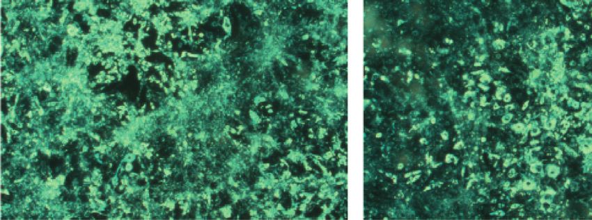
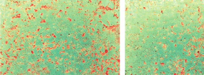
6 Evidence-Based Complementary and Alternative Medicine

 Table 3: IC50 values for the antioxidant activities of F2 and its ingredients.
Samples DPPH-IC50 (μg/mL) ABTS-IC50 (μg/mL)
Formulation F2 5.99 9.52
Orostachys japonicus 11.37 18.1
Geranium thunbergii 9.57 24.35
Rhus verniciflua 1.84 7.98
Royal jelly No activity No activity
Citrus limon (lemon) No activity No activity
Gallic acid 0.04 2.84
The percentage of DPPH and ABTS radical scavenging offered by F2, its components, and standard gallic acid plotted in logarithmic regression curve against
concentration and the IC50 was determined by interpolation from the logarithmic regression. The antioxidant activity is expressed in the form of IC50 value.

 100 100
 Cell viability (% of control)

 ∗∗
 ∗∗∗

 Lipid accumulation
 80 80 ∗∗

 (% of control)
 60 60

 40 40

 20 20

 0 0
 Control 5 10 20 40 60 80 Control 20 μg/mL 40 μg/mL 60 μg/mL
 Formulation F2 concentrations (μg/mL) Formulation F2
 (a) (b)

 Control 20 μg/mL Control 20 μg/mL

 40 μg/mL 60 μg/mL 40 μg/mL 60 μg/mL
 (c) (d)

Figure 2: Effect of F2 on cell viability and lipid production in 3T3-L1 adipocytes. The 3T3-L1 preadipocytes were treated with F2 and cell
viability (a) was assessed using MTT assay. The amount of lipid accumulation (b) on 3T3-L1 adipocytes was measured by Oil Red O (ORO)
assay. The lipid accumulating cells were visualized in 10X magnification before (c) and after (d) the ORO staining. The data shown are
presented as means ± SD of four separate experiments. Statistical significance was calculated using one-way ANOVA followed by Dunnett’s
multiple comparisons test. ∗ ∗ p < 0.01 and ∗ ∗ ∗ p < 0.001 vs. control.

3.5. Effect of F2 on Gene Expression of Adipogenic Tran- shown in Figure 3, the gene expressions of the aforemen-
scription Factors in 3T3-L1 Adipocytes. The effect of F2 on tioned transcription factors and adipogenic markers were
the gene expression level of adipogenic transcription factors markedly downregulated whereas the expression of an IL-6
was examined by real-time PCR. The 3T3-L1 adipocytes was upregulated by F2 in a concentration-dependent
were treated with 40 and 60 μg/mL concentrations of F2 and manner compared to the control.
total mRNA was extracted at day 8 of the sample treatment.
The mRNA expression level of specific adipogenic markers 3.6. Effect of F2 on Mice Body Weight, Food Intake, and Food
(PPARc, C/EBPα, SREBP-1c, aP2, leptin, LPL, and adipo- Efficiency Ratio during 8 Weeks. The mice were treated for 8
nectin) and inflammatory marker (IL-6) was determined. As weeks. Mice body weight gaining pattern over the treatment
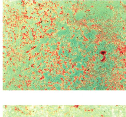
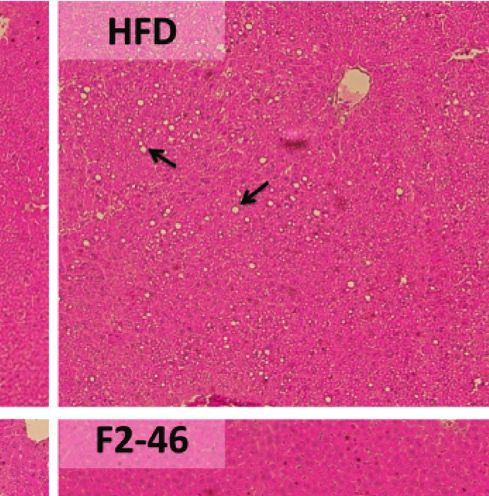
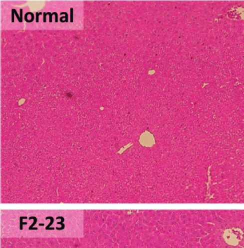
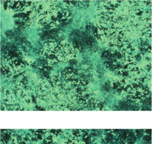
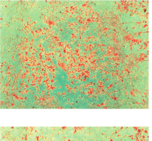
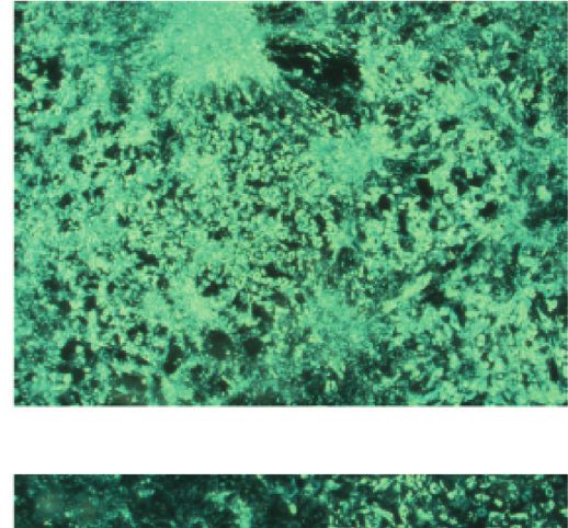
Evidence-Based Complementary and Alternative Medicine 7

 Table 4: Synergistic action of F2 on fat reduction in 3T3-L1cells.
F2 and ingredients Amount of ingredients in 60 μg/mL of F2 Lipid inhibition (%)
Control — 0.00 ± 3.08
GT 7.481 μg/ml 5.41 ± 6.59
RV 22.44 μg/ml 14.01 ± 2.36
OJ 29.93 μg/ml −7.57 ± 5.96
RJ 0.074 μg/ml −4.27 ± 4.58
Lemon 0.074 μg/ml −1.83 ± 3.93
F2 60 μg/ml 26.31 ± 4.73
Cumulative inhibition of lipid production by the ingredients of F2 � 5.75%
The synergistic effect of F2 by its ingredients on lipid production by 3T3-L1 adipocytes was determined by ORO assay. The lipid accumulations (expressed
as the percentage of control) are presented as means ± SD of triplicate experiments. GT: Geranium thunbergii; RV: Rhus verniciflua; OJ: Orostachys japonicus;
RJ: Royal jelly.

periods is shown in Figure 4. As presented in Table 5, the determined by H&E staining. The histological observation of
HFD control group significantly gained body weight com- liver tissue revealed the accumulation of lipid droplets into
pared with that of the normal group. However, weights of the liver in the HFD control group. Similarly, the sizes of
mice treated with high dose (46 mg/kg) of F2 and GC adipocytes in WAT were enlarged. Treatment with F2
(200 mg/kg) were found to be significantly reduced within 8 inhibited lipid deposition into the liver and reduced the size
weeks in comparison to the HFD control group. The food of adipocytes in WAT (Figure 7). The diameter of adipocytes
intake pattern was similar among the HFD fed groups. Food in the WAT was significantly reduced with the treatment of
efficiency ratio (FER) for F2 (46 mg/kg) and GC was found F2 and GC in comparison to the HFD control (Figure 8).
to be lowered in comparison to the HFD control group
indicating the lowest food was utilized for weight gain in 4. Discussion
those groups.
 Obesity, caused by the over deposition of lipids into WAT, is
 perceived as a global health risk in present days. Multiple
3.7. Effect of F2 on Lipid Excretion. The mice feces were metabolic maladies such as diabetes, cardiovascular diseases,
collected before one day of sacrifice for the measurement of fatty liver disease, mental disorders, and even certain cancers
lipid content. The feces lipid content represents the excretion resulting from a complication of obesity are seriously
of dietary fat without absorption from the alimentary canal. threatening human health [2, 36]. Secondarily to the dietary
In the current experiment, F2 and GC were found to en- restriction and exercise, several drug therapies are also
hance lipid excretion in feces compared with the HFD recommended for the management of obesity. However,
(Figure 5). The lipid excretion by F2 was increased in a dose- unexpected adverse effects such as cardiovascular, gastro-
dependent manner. This result suggests that the reduction of intestinal, and psychological effects associated with most of
body weight in the F2 group might be due to inhibition of the antiobesity drugs limit their use in the general pop-
lipid absorption in the gastrointestinal tract. ulation [7]. Therefore, developing alternative therapies for
 obesity with minimal adverse effects is warranted. Due to
3.8. Effect of F2 on Fasting Blood Glucose. The fasting blood having the aforementioned adverse effect along with high
glucose of mice was measured before sacrifice. As shown in cost and physical dependency by long-term use of the
Figure 6, the blood glucose was elevated in the HFD control pharmaceutical agents, plant-based remedies are gaining
group (173.17 ± 9.52 mg/dL) compared to the normal group attention globally for the management of obesity and
(155.00 ± 8.51 mg/dL). The blood glucose was significantly overweight [8]. The present study was designed to develop a
reduced in F2 treated groups. 23 mg/kg and 46 mg/kg treated novel, safe, and effective herbal formulation for the man-
groups showed blood glucose level of 139.60 ± 13.52 mg/dL agement of obesity. We developed a polyherbal formulation
and 147.80 ± 17.61 mg/dL, respectively. But the GC treated (F2) by homogeneously mixing of trace amount of royal jelly
groups remained nonsignificant with the HFD control. The and lemon juice with ethanol extracts of Orostachys japo-
blood-glucose-lowering effect of F2 may have a beneficial nicus (OJ), Rhus verniciflua (RV), and Geranium thunbergii
effect on the management of diabetes and its complications. (GT). We assessed the antiobesity efficacy of the developed
 formulation in 3T3-L1 adipocytes and high-fat diet-fed
 C57BL/6J mice. The F2 was further analyzed using UPLC
3.9. Effect of F2 on Organ Weights and Histological and quantification was done for its major five marker
Observation. The epididymal white adipose tissue (WAT), compounds (astragalin, fustin, fisetin, sulfuretin, and ellagic
liver, kidney, and spleen were isolated and weight was taken acid).
in situ. As presented in Table 6, the weights of WAT were Free radicals or reactive oxygen species are generated in
increased significantly in the HFD group compared with that the body through multiple mechanisms. Chronic inflam-
in the normal group. However, WAT weight was markedly mation associated with obesity is one of the major causes of
decreased by F2 in a dose-dependent manner. Moreover, the systemic oxidative stress in the body [37], which results in
histological morphology of epididymal WAT and liver was the development of metabolic disorders such as insulin
8 Evidence-Based Complementary and Alternative Medicine

 mPPARγ mc/EBPα mSREBP-1c
 ∗ ∗∗ ∗∗ ∗∗ ∗∗ ∗∗
 1.1 1.1 1.2
 1.0 1.0 1.1

 Relative expression level
 Relative expression level
 Relative expression level

 0.9 1.0
 0.9 0.9
 0.8 0.8
 0.7 0.8
 0.7 0.7
 0.6 0.6 0.6
 0.5 0.5 0.5
 0.4 0.4 0.4
 0.3 0.3 0.3
 0.2 0.2 0.2
 0.1 0.1 0.1
 0.0 0.0 0.0
 NC Con 40 60 NC Con 40 60 NC Con 40 60

 Formulation F2 (μg/mL) Formulation F2 (μg/mL) Formulation F2 (μg/mL)

 (a) (b) (c)
 maP2 mLeptin mLPL
 ∗∗ ∗∗ ∗∗ ∗∗ ∗ ∗
 1.2 1.2 1.1
 1.1 1.1 1.0
 Relative expression level

 Relative expression level

 Relative expression level
 1.0 1.0 0.9
 0.9 0.9 0.8
 0.8 0.8 0.7
 0.7 0.7 0.6
 0.6 0.6
 0.5 0.5 0.5
 0.4 0.4 0.4
 0.3 0.3 0.3
 0.2 0.2 0.2
 0.1 0.1 0.1
 0.0 0.0 0.0
 NC Con 40 60 NC Con 40 60 NC Con 40 60
 Formulation F2 (μg/mL) Formulation F2 (μg/mL) Formulation F2 (μg/mL)

 (d) (e) (f )
 mAdiponectin
 ∗
 mIL-6
 1.2 2.5 ∗ ∗∗
 1.1
 Relative expression level

 Relative expression level

 1.0 2.0
 0.9
 0.8
 0.7 1.5
 0.6
 0.5 1.0
 0.4
 0.3
 0.2 0.5
 0.1
 0.0 0.0
 NC Con 40 60 NC Con 40 60
 Formulation F2 (μg/mL) Formulation F2 (μg/mL)

 (g) (h)

Figure 3: Effects of F2 on the gene expression level of adipogenic transcription factors during adipocyte differentiation. (a) Peroxisome
proliferator-activated receptor gamma (PPARc), (b) CAAT/enhancer-binding protein alpha (C/EBPα), (c) sterol response element-binding
protein-1c (SREBP-1c), (d) adipocyte protein 2 (aP2), (e) leptin, (f ) lipoprotein lipase (LPL), (g) adiponectin, and (h) interleukin-6 (IL-6).
Each gene’s expression levels were quantified and normalized to β-actin. The data shown are presented as means ± SD of triplicate ex-
periments. Statistical significance was calculated using one-way ANOVA followed by Dunnett’s multiple comparisons test. ∗p < 0.05 and
∗ ∗ p < 0.01 vs. control. NC: negative control (undifferentiated); Con: control (MDI).

resistance, hypertension, asthma, etc. [38]. Herbal remedies animal tissues making it possible to extrapolate results to
with antioxidant potentials can counteract such obesity- humans [41]. The results revealed that F2 treatment sig-
associated oxidative damage and prevent comorbidities. The nificantly reduced the lipid accumulation in 3T3-L1 adi-
F2 with synergistic antioxidant activity could be a better pocytes, demonstrating the ability of F2 to inhibit adipocyte
alternative for preventing and protecting the body from differentiation (Figure 2). Previous reports mentioned that
obesity and related complications. the extracts or isolated compounds from O. japonicus [14],
 A commitment of stem cell and terminal differentiation R. verniciflua [15, 16, 26, 31], and royal jelly [30] had the
of adipocytes into a fat-storing mature adipose cell is a antiadipogenic effect in 3T3-L1 adipocytes. The present
crucial step in obesity [39]. Being a reliable cellular model to study revealed the synergistic antiadipogenic activity of these
study adipogenesis and antiadipogenic activity [40], we ingredients treated even in a very small concentration in
selected 3T3-L1 fibroblast cells for the measurement of the formulation F2 (Table 4).
antiadipogenic activity of F2. Also, most of the physiology of Differentiation of preadipocytes into mature adipocytes
3T3-L1 preadipocytes differentiation resembles that in is guided by various biochemical regulators such as insulin,
Evidence-Based Complementary and Alternative Medicine 9

 30

 Mice body weight (g)
 25

 20

 Week 0 Week 1 Week 2 Week 3 Week 4 Week 5 Week 6 Week 7 Week 8

 Normal F2-46
 HFD GC
 F2-23

Figure 4: Patterns of body weight gain observed in mice during 8 weeks. Mice body weights were measured every week. Values are expressed
as mean ± SD (n � 5). Normal: standard diet; HFD: high-fat diet control; F2-23: F2 (23 mg/kg); F2-46: F2 (46 mg/kg); GC: Garcinia cambogia
(200 mg/kg).

Table 5: Effect of F2 on mice body weight (initial and final), weight gain, food intake, and food efficiency ratio (FER) of different groups
during 8 weeks of the experiment.
 Body weight (g)
Groups Weight gain (g) Food intake (g/mice) FER (%)
 Initial Final
Normal 20.40 ± 1.21 24.10 ± 1.10 3.79 ± 0.68∗∗∗ 180.08 2.11 ± 0.38∗∗∗
HFD 18.85 ± 0.99 28.46 ± 0.84 9.61 ± 0.56 113.3 8.48 ± 0.51
F2-23 19.16 ± 0.90 28.15 ± 1.88 8.99 ± 1.19 117.85 7.63 ± 1.01
F2-46 19.35 ± 0.83 27.46 ± 1.57 8.11 ± 0.79∗ 120.25 6.75 ± 0.65∗∗
GC 18.37 ± 0.93 25.96 ± 0.60 7.586 ± 0.79∗∗ 110.23 6.88 ± 0.72∗∗
Food intake and body weight gains were measured every week. Food efficiency ratio (FER) was calculated as follows: FER% � gained body weight (g) × 100/
food intake (g). Results are presented as the mean ± SD (n � 5). Statistical significance was calculated using one-way ANOVA followed by Dunnett’s multiple
comparisons test. ∗p < 0.05, ∗ ∗ p < 0.01, and ∗ ∗ ∗ p < 0.001 vs. HFD group. Normal: standard diet; HFD: high-fat diet control; F2-23: F2 (23 mg/kg); F2-46:
F2 (46 mg/kg); GC: Garcinia cambogia (200 mg/kg).

 40 200 ∗∗ ∗
 Fasting blood glucose (mg/dL)
 Fecal fat content (mg/g)

 30 175

 20 150

 10 125

 0 100
 Normal HFD F2-23 F2-46 GC Normal HFD F2-23 F2-46 GC

Figure 5: Effect of F2 on lipid excretion. Feces were collected from Figure 6: Effect of F2 on fasting blood glucose. Mice were kept
each group cage and lipid contents were measured collectively. The fasting overnight. The levels of blood glucose were measured before
fat content was extracted with methanol-chloroform solvent and mice sacrifice. Values are expressed as mean ± SD (n � 5). Statistical
presented as percentage of dry stool weight. Normal: standard diet; significance was calculated using one-way ANOVA followed by
HFD: high-fat diet control; F2-23: F2 (23 mg/kg); F2-46: F2 (46 mg/ Dunnett’s multiple comparisons test. ∗p < 0.05 and ∗ ∗ p < 0.01 vs.
kg); GC: Garcinia cambogia (200 mg/kg). HFD. Normal: standard diet; HFD: high-fat diet control; F2-23: F2
 (23 mg/kg); F2-46: F2 (46 mg/kg); GC: Garcinia cambogia (200 mg/
 kg).
expressions of adipogenic genes (PPARc, C/EBPα, SREBP-
1c, aP2, leptin, LPL, FAS, adiponectin, etc.), and accumu-
lations of TG and free fatty acids in cells [39, 42, 43]. restriction of adipogenesis (Figure 3). In another way, it is
Therefore, downregulation of the adipogenic transcription understood that the expression of pro-inflammatory cyto-
factors may restrict initial as well as terminal differentiation, kines like interleukin-6 (IL-6) is greater in preadipocytes
leading to inhibition of lipid accumulation in adipocytes. In than in 3T3-L1 adipocytes [44]. Hence, overexpression on
the present study, our observations revealed that the gene mRNA level of IL-6 in F2 treated adipocytes suggested that
expression level of the adipogenic factors was significantly the differentiation of 3T3-L1 preadipocyte was inhibited by
downregulated suggesting the crucial role of F2 in the F2. But the IL-6 expression was downregulated by
10 Evidence-Based Complementary and Alternative Medicine

 Table 6: Effect of F2 on organ weight.
Groups WAT (g) Liver (g) Kidney (g) Spleen (mg)
Normal 0.46 ± 0.09∗∗∗ 0.94 ± 0.05∗ 0.29 ± 0.02 53.17 ± 5.71
HFD 1.38 ± 0.09 0.82 ± 0.08 0.28 ± 0.02 60.59 ± 8.99
F2-23 1.10 ± 0.32∗ 0.75 ± 0.07 0.29 ± 0.02 59.50 ± 5.32
F2-46 0.75 ± 0.09∗∗∗ 0.80 ± 0.06 0.30 ± 0.03 63.30 ± 5.68
GC 0.92 ± 0.15∗∗ 0.77 ± 0.08 0.30 ± 0.02 57.88 ± 10.49
Results are presented as the mean ± standard deviation (n � 5). Statistical significance was calculated using one-way ANOVA followed by Dunnett’s multiple
comparisons test. ∗p < 0.05, ∗ ∗ p < 0.01, and ∗ ∗ ∗ p < 0.001 vs. HFD group. Normal: standard diet; HFD: high-fat diet control; F2-23: F2 (23 mg/kg); F2-46:
F2 (46 mg/kg); GC: Garcinia cambogia (200 mg/kg).

 (a)

 (b)

Figure 7: Effect of F2 on liver and white adipose tissue (WAT) histology. Histological evaluation of (a) liver and (b) WAT was done by H&E
staining (magnification ×10). Lipid deposition into the liver and size of adipocytes in WAT were evaluated. The fat droplets deposited in the
liver (arrows) are more noticeable in HFD control group mice. Normal: standard diet; HFD: high-fat diet control; F2-23: F2 (23 mg/kg); F2-
46: F2 (46 mg/kg); GC: Garcinia cambogia (200 mg/kg).

O. japonicas in inflammatory human THP-1 cells [45]. Also, might only be associated with inhibition of adipocyte dif-
R. verniciflua decreased the level of hepatic IL-6 in LPS ferentiation but not inflammatory action. All those obser-
induced rats [46]. Other scientific reports also declared that vations confirm the potential of the antiadipogenic and anti-
the F2 ingredients employ potential anti-inflammatory ac- inflammatory effects of F2.
tivity [47–50]. In our study, treatment of F2 on LPS induced The exciting outcomes from in vitro experiments en-
RAW264.7 macrophages revealed significant inhibition of couraged us to conduct an in vivo antiobesity study. In order
nitrite production (Supplementary data, Figure S1), indi- to confirm the potential of F2 for preventing obesity, C57BL/
cating the anti-inflammatory response of F2. Hence, the IL-6 6J mice were fed with a high-fat diet to induce obesity and
expression upregulating effect of F2 in 3T3-L1 adipocyte the F2 was treated simultaneously. A well-accepted
Evidence-Based Complementary and Alternative Medicine 11

 90 ∗∗∗ ∗∗∗ ∗∗∗ ∗∗∗ lowering blood glucose in streptozotocin-induced dia-
 betic rats [10]. Higher blood glucose level is also a con-
 80 sequence of lower storage of glucose in the form of
 WAT adipocyte diameter (μm)

 70 glycogen due to reduction of glycogen synthase in liver
 60 and muscle tissue [58]. Besides, the reduction of glycogen
 synthase accelerates hepatic insulin resistance and liver
 50
 steatosis, and vice versa. Accumulation of lipid compo-
 40 nents in the liver activates diacyl glycerol activated protein
 30 kinase (PKCε), which further impairs the activation of
 insulin receptors and insulin-stimulated glycogen syn-
 20
 thesis [59, 60]. Previous studies already exposed the
 10 scientific evidence that dietary high fat can lead to liver
 0 steatosis which is associated with obesity and liver dys-
 Normal HFD F2-23 F2-46 GC function [61]. In our study, feeding a high-fat diet to mice
 for 8 weeks led to the accumulation of lipid globules in the
Figure 8: Effect of F2 on size of adipocytes in the epididymal white
 liver causing liver steatosis in the HFD control group.
adipose tissue (WAT). The images of WATs from H&E staining
(magnification ×10) were analyzed and diameters of the adipocytes However, F2 was effective in protecting the liver from
were evaluated using ImageJ software. Results are presented as the steatosis (Figure 7). Obesity is primarily associated with
mean ± standard deviation. Statistical significance was calculated the accumulation of lipids into white adipose tissues [62].
using one-way ANOVA followed by Dunnett’s multiple compar- The visceral fat accumulation in epididymal adipocytes in
isons test. ∗ ∗ ∗ p < 0.001 vs. HFD group. Normal: standard diet; the HFD control groups resulted in a significant increase
HFD: high-fat diet control; F2-23: F2 (23 mg/kg); F2-46: F2 (46 mg/ in the white adipose tissue (WAT) weight. In our ob-
kg); GC: Garcinia cambogia (200 mg/kg). servations, F2 significantly reduced the tissue weight
 (Table 6) as well as adipocytes size (Figure 8) of WAT in
antiobesity agent, Garcinia cambogia, was used for standard dose-dependent manner supporting the effectiveness of
comparison. Several scientific pieces of the literature dem- F2 to prevent obesity in C57BL/6J mice.
onstrated that a high-fat diet induces obesity and diabetes in
animals [51–53]. Obesity in animals can be measured by 5. Conclusions
considering some criteria such as body weight gain, body fat
content, and food efficiency [51, 54]. The body weight gain The present study demonstrated the synergistic anti-
has been amplified in the HFD group while F2 slowed down adipogenic activity of F2 in 3T3-L1 adipocytes. The re-
the level of weight gaining. Previous studies revealed that duction of adipogenesis might be a result of the
ethyl acetate fraction of O. japonicus and 70% ethanol extract downregulation of various transcription factors and adi-
of G. thunbergii showed better antiobesity activity in high-fat pogenic markers in the adipocytes. Further treatment of F2
diet-induced rodents [13, 19]. During the process of di- in high-fat diet-fed C57BL/6J mice confirmed its preventing
gestion, dietary fat gets absorbed from the intestine once it is effect on obesity and its comorbidities. From the overall
subjected to emulsification with pancreatic lipase [55]. results, it may be concluded that the mechanisms of anti-
Therefore, alteration of pancreatic lipase may decrease ab- obesity activity of F2 include inhibition of adipocyte dif-
sorption of intestinal fat. Food efficiency ratio (FER) is ferentiation, restriction of dietary fat absorption, and
considered as the relation between total food intake and the reduction of free fatty acids accumulation in tissues. Fur-
amount available for anabolism to increase body weight [56]. thermore, F2 showed potential antioxidant activity, which
The lower FER in F2 treated mice was due to lower weight may alleviate obesity-related oxidative stress. In addition, F2
gain on a similar amount of food consumption. This ob- exhibited potent blood glucose reducing activity in high-fat
servation suggests that F2 may impair dietary fat absorption diet-induced obese mice. Therefore, the novel polyherbal
by altering pancreatic lipase. Elevation of lipid excretion in formulation (F2) can be a potential medication for the
feces by F2 also supports this finding. management of obesity and its comorbidities without ad-
 Hyperglycemia or diabetes is one of the major com- verse effects.
plications of obesity. Previous studies also revealed that
the HFD fed mice developed hyperglycemia as well as
 Data Availability
insulin resistance [51, 57]. A high-fat diet causes a de-
crease in glucose transporter, insulin receptors, and The data will be made available upon reasonable request to
glucose metabolism, and reduction in glycogen synthesis the corresponding author.
in the liver and muscle, which results in elevation of blood
glucose [51]. The decreased level of fasting blood glucose
by F2 in comparison to the HFD control indicates that the Disclosure
F2 might have a protecting effect against insulin resistance
 The funding body had no implication in the design of the
and related complications. A previous study showed that
 study, analysis, and interpretation of data, and writing of the
the 80% ethanol extract of O. japonicus was effective in
 manuscript.
12 Evidence-Based Complementary and Alternative Medicine

Conflicts of Interest [4] I. M. Faust, P. R. Johnson, J. S. Stern, and J. Hirsch, “Diet-
 induced adipocyte number increase in adult rats: a new model
The authors declare no conflicts of interest. of obesity,” American Journal of Physiology-Endocrinology
 and Metabolism, vol. 235, no. 3, p. E279, 1978.
Authors’ Contributions [5] B. J. Klyde and J. Hirsch, “Increased cellular proliferation in
 adipose tissue of adult rats fed a high-fat diet,” Journal of Lipid
H. J. Jung contributed to conceptualization. P. R. Pandeya, Research, vol. 20, no. 6, pp. 705–715, 1979.
R. Lamichhane, and S. G. Kim contributed to methodology. [6] B. J. Klyde and J. Hirsch, “Isotopic labeling of DNA in rat
P. R. Pandeya, G. Lamichhane, and K. H. Lee contributed to adipose tissue: evidence for proliferating cells associated with
formal analysis. P. R. Pandeya, R. Lamichhane, K. H. Lee, mature adipocytes,” Journal of Lipid Research, vol. 20, no. 6,
and G. Lamichhane contributed to investigation. P. R. pp. 691–704, 1979.
Pandeya, G. Lamichhane, and H. J. Jung contributed to data [7] J. G. Kang and C.-Y. Park, “Anti-obesity drugs: a review about
 their effects and safety,” Diabetes & Metabolism Journal,
curation. P. R. Pandeya and G. Lamichhane contributed to
 vol. 36, no. 1, pp. 13–25, 2012.
writing original draft. P. R. Pandeya, H. J. Jung, [8] K. Sengupta, A. T. Mishra, M. K. Rao, K. V. S. Sarma,
R. Lamichhane, and G. Lamichhane contributed to review A. V. Krishnaraju, and G. Trimurtulu, “Efficacy and tolera-
and editing. H. J. Jung and S. G. Kim contributed to project bility of a novel herbal formulation for weight management in
administration and funding acquisition. H. J. Jung con- obese subjects: a randomized double blind placebo controlled
tributed to supervision. clinical study,” Lipids in Health and Disease, vol. 11, no. 1,
 p. 122, 2012.
Acknowledgments [9] S. Parasuraman, G. Thing, and S. Dhanaraj, “Polyherbal
 formulation: concept of ayurveda,” Pharmacognosy Reviews,
The authors give their sincere thanks to the Bio & Medical vol. 8, no. 16, p. 73, 2014.
Technology Development Program of the National Research [10] S. J. Lee, G. F. Zhang, and N. J. Sung, “Hypolipidemic and
Foundation (NRF) and the Korean Government for funding hypoglycemic effects of Orostachys japonicus A. Berger ex-
this study. This research was supported and funded by the tracts in streptozotocin-induced diabetic rats,” Nutrition
Bio & Medical Technology Development Program of the Research and Practice, vol. 5, no. 4, pp. 301–307, 2011.
National Research Foundation (NRF) of Korean Govern- [11] H.-J. Jung, J. Choi, J.-H. Nam, and H.-J. Park, “Anti-ul-
ment (MSIT) (NRF-2015M3A9A5031098). cerogenic effects of the flavonoid-rich fraction from the ex-
 tract of Orostachys japonicus in mice,” Journal of Medicinal
 Food, vol. 10, no. 4, pp. 702–706, 2007.
Supplementary Materials [12] H.-J. Park, H. J. Yang, K. H. Kim, and S. H. Kim, “Aqueous
In vitro anti-inflammatory activity of F2 on RAW264.7 extract of Orostachys japonicus A. Berger exerts immunos-
 timulatory activity in RAW 264.7 macrophages,” Journal of
macrophage: the Raw264.7 macrophages (ATCC TIB-
 ® Ethnopharmacology, vol. 170, pp. 210–217, 2015.
 ™
71 ), used to determine the anti-inflammatory efficacy of
F2, were purchased from American Type Culture Collection
 [13] S. G. Kim, A. Poudel, J. W. Choi et al., “Anti-obesitic effect of
 Orostachys japonicus in rats model fed a hyperlipidemic diet,”
(ATCC). The macrophages were induced by lipopolysac- Natural Product Sciences, vol. 17, no. 2, pp. 117–122, 2011.
charide (LPS) and different concentrations of F2 were [14] M. Jang, H.-Y. Choi, and G.-H. Kim, “Phenolic components
treated. The absorbance of nitrites produced by LPS induced rich ethyl acetate fraction of Orostachys japonicus inhibits
RAW264.7 macrophages was measured spectrometrically lipid accumulation by regulating reactive oxygen species
and the amount was calculated using linear regression curve generation in adipogenesis,” Journal of Food Biochemistry,
of sodium nitrite standard. The result showed that nitrite vol. 43, no. 8, Article ID e12939, 2019.
production was significantly higher in LPS induced control [15] N.-J. Song, H.-J. Yoon, K. H. Kim et al., “Butein is a novel anti-
wells compared to the noninduced control. The treatments adipogenic compound,” Journal of Lipid Research, vol. 54,
of F2 at the concentration of 20, 40, and 80 μg/mL signifi- no. 5, pp. 1385–1396, 2013.
cantly reduced LPS nitrite production. This result suggested [16] R. Lamichhane, S.-G. Kim, S. Kang, K.-H. Lee, P. R. Pandeya,
that the F2 may exhibit anti-inflammatory activity. (Sup- and H.-J. Jung, “Exploration of underlying mechanism of
plementary Materials) anti-adipogenic activity of sulfuretin,” Biological and Phar-
 maceutical Bulletin, vol. 40, no. 9, pp. 1366–1373, 2017.
 [17] S.-J. Jeong, J.-G. Park, S. Kim et al., “Extract of Rhus verni-
References ciflua stokes protects the diet-induced hyperlipidemia in
 [1] World Health Organization, Obesity and Overweight, World mice,” Archives of Pharmacal Research, vol. 38, no. 11,
 Health Organization, Geneva, Switzerland, 2020. pp. 2049–2058, 2015.
 [2] T. Ananthakumar, N. R. Jones, L. Hinton, and P. Aveyard, [18] M.-Y. Song, G.-S. Jeong, K.-B. Kwon et al., “Sulfuretin pro-
 “Clinical encounters about obesity: systematic review of pa- tects against cytokine-induced β-cell damage and prevents
 tients’ perspectives,” Clinical Obesity, vol. 10, no. 1, Article ID streptozotocin-induced diabetes,” Experimental and Molec-
 e12347, 2020. ular Medicine, vol. 42, no. 9, pp. 628–638, 2010.
 [3] UNICEF, WHO, World Bank Group, Levels and Trends in [19] Y.-Y. Sung, T. Yoon, W.-K. Yang, S. J. Kim, and H. K. Kim,
 Child Malnutrition in UNICEF/WHO/World Bank Group “Anti-obesity effects of Geranium thunbergii extract via im-
 Joint Child Malnutrition Estimates Key Findings of the 2016 provement of lipid metabolism in high-fat diet-induced obese
 Edition, World Bank, World Health Organization, World- mice,” Molecular Medicine Reports, vol. 4, no. 6,
 BankGroup, Washington, DC, USA, 2016. pp. 1107–1113, 2011.
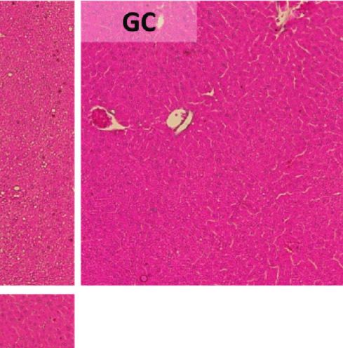
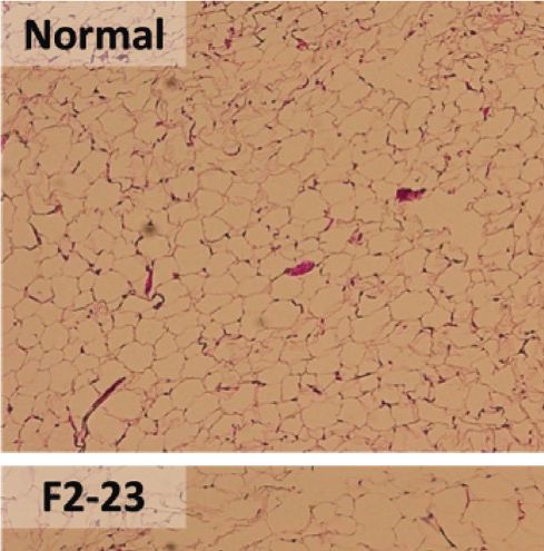
Evidence-Based Complementary and Alternative Medicine 13

[20] T.-H. Kwon, S.-J. Lee, Y.-J. Kim, J.-J. Park, T. Kim, and [34] K. J. Livak and T. D. Schmittgen, “Analysis of relative gene
 N.-H. Park, “Anti-inflammatory effect of Geranium thun- expression data using real-time quantitative PCR and the
 bergii on lipopolysaccharide-stimulated RAW 264.7 cells,” 2−ΔΔCT method,” Methods, vol. 25, no. 4, pp. 402–408, 2001.
 Korean Journal of Food Science and Technology, vol. 48, no. 6, [35] J. Folch, M. Lees, and G. H. S. Stanley, “A simple method for
 pp. 618–621, 2016. the isolation and purification of total lipides from animal
[21] A.-S. Cho, S.-M. Jeon, M.-J. Kim et al., “Chlorogenic acid tissues,” Journal of Biological Chemistry, vol. 226, no. 1,
 exhibits anti-obesity property and improves lipid metabolism pp. 497–509, 1957.
 in high-fat diet-induced-obese mice,” Food and Chemical [36] R. K. Baboota, M. Bishnoi, P. Ambalam et al., “Functional
 Toxicology, vol. 48, no. 3, pp. 937–943, 2010. food ingredients for the management of obesity and associ-
[22] M. Khazaei, A. Ansarian, and E. Ghanbari, “New findings on ated co-morbidities—a review,” Journal of Functional Foods,
 biological actions and clinical applications of royal jelly: a vol. 5, no. 3, pp. 997–1012, 2013.
 review,” Journal of Dietary Supplements, vol. 15, no. 5, [37] G.-S. Liu, E. Chan, M. Higuchi, G. Dusting, and F. Jiang,
 pp. 757–775, 2017. “Redox mechanisms in regulation of adipocyte differentia-
[23] A. Petelin, S. Kenig, R. Kopinc, M. Dezelak, M. Cernelic tion: beyond a general stress response,” Cells, vol. 1, no. 4,
 Bizjak, and Z. Jenko Praznikar, “Effects of royal jelly ad- pp. 976–993, 2012.
 ministration on lipid profile, satiety, inflammation, and an- [38] P. Manna and S. K. Jain, “Obesity, oxidative stress, adipose
 tioxidant capacity in asymptomatic overweight adults,” tissue dysfunction, and the associated health risks: causes and
 Evidence-Based Complementary and Alternative Medicine, therapeutic strategies,” Metabolic Syndrome and Related
 vol. 2019, Article ID 4969720, 11 pages, 2019. Disorders, vol. 13, no. 10, pp. 423–444, 2015.
[24] T. Yoneshiro, R. Kaede, K. Nagaya et al., “Royal jelly ame- [39] F. M. Gregoire, “Adipocyte differentiation: from fibroblast to
 liorates diet-induced obesity and glucose intolerance by endocrine cell,” Experimental Biology and Medicine, vol. 226,
 promoting brown adipose tissue thermogenesis in mice,” no. 11, pp. 997–1002, 2001.
 Obesity Research & Clinical Practice, vol. 12, no. 1, pp. 127– [40] Y.-C. Tung, P.-H. Hsieh, M.-H. Pan, and C.-T. Ho, “Cellular
 137, 2016. models for the evaluation of the antiobesity effect of selected
[25] S. Pourmoradian, R. Mahdavi, M. Mobasseri, E. Faramarzi, phytochemicals from food and herbs,” Journal of Food and
 and M. Mobasseri, “Effects of royal jelly supplementation on Drug Analysis, vol. 25, no. 1, pp. 100–110, 2017.
 body weight and dietary intake in type 2 diabetic females,” [41] S. P. Poulos, M. V. Dodson, and G. J. Hausman, “Cell line
 Health Promotion Perspectives, vol. 2, no. 2, p. 231, 2012. models for differentiation: preadipocytes and adipocytes,”
[26] Y.-A. Rha, M.-S. Choi, and S.-J. Park, “Antioxidant and anti- Experimental Biology and Medicine, vol. 235, no. 10,
 adipogenic effects of fermented Rhus verniciflua,” The Korean pp. 1185–1193, 2010.
 Journal of Culinary Research, vol. 20, no. 3, pp. 137–147, 2014. [42] D. A. Bernlohr, M. A. Bolanowski, T. J. Kelly, and M. D. Lane,
[27] S. R. Cheong, R. Kim, Y. K. Park, S. Baek, S.-H. Yeo, and “Evidence for an increase in transcription of specific mRNAs
 C. Lee, “Anti-obesity effect of fermented detoxified Rhus during differentiation of 3T3-L1 preadipocytes,” Journal of
 verniciflua vinegar supplementation in diet-induced obese Biological Chemistry, vol. 260, no. 9, pp. 5563–5567, 1985.
 rats,” Journal of the Korean Society of Food Science and Nu- [43] H. Green and O. Kehinde, “Spontaneous heritable changes
 trition, vol. 44, no. 12, pp. 1771–1778, 2015. leading to increased adipose conversion in 3T3 cells,” Cell,
[28] S.-G. Kim, J. Choi, H.-J. Park, S.-M. Lee, and H.-J. Jung, “Anti- vol. 7, no. 1, pp. 105–113, 1976.
 hyperlipidemic effects of the flavonoid-rich fraction from the [44] J. M. Harkins, N. Moustaid-Moussa, Y.-J. Chung et al.,
 methanol extract of orostachy japonicus in rats,” Korean “Expression of interleukin-6 is greater in preadipocytes than
 Journal of Pharmacognosy, vol. 40, no. 1, pp. 51–58, 2009. in adipocytes of 3T3-L1 cells and C57BL/6J and ob/ob mice,”
[29] S.-G. Kim, R. Lamichhane, D. K. Sharma, K.-H. Lee, J. Choi, The Journal of Nutrition, vol. 134, no. 10, pp. 2673–2677, 2004.
 and H.-J. Jung, “Anti-obesity and anti-hyperlipidemic effects [45] Y.-K. Yoon, H.-J. Woo, and Y. Kim, “Orostachys japonicus
 of butanol soluble fraction from methanol extract of Gera- inhibits expression of the TLR4, NOD2, iNOS, and COX-2
 nium thunbergii in sprague-dawley rats,” Korean Journal of genes in LPS-stimulated human PMA-differentiated THP-1
 Pharmacognosy, vol. 45, no. 1, pp. 69–76, 2014. cells by inhibiting NF-κB and MAPK activation,” Evidence-
[30] P. R. Pandeya, R. Lamichhane, K.-H. Lee et al., “Bioassay- Based Complementary and Alternative Medicine, vol. 2015,
 guided isolation of active anti-adipogenic compound from Article ID 682019, 9 pages, 2015.
 royal jelly and the study of possible mechanisms,” BMC [46] J. E. Moon, J.-H. Shin, O. Kwon, and J. Y. Kim, “A stan-
 Complementary and Alternative Medicine, vol. 19, no. 1, p. 33, dardized extract of Rhus verniciflua Stokes protects wistar rats
 2019. against lipopolysaccharide-induced acute inflammation,”
[31] S.-G. Kim, D.-Y. Rhyu, D.-K. Kim et al., “Inhibitory effect of Journal of Medicinal Food, vol. 18, no. 11, pp. 1223–1230, 2015.
 heartwood of Rhus verniciflua stokes on lipid accumulation in [47] H.-J. Choi, H.-J. Choi, M.-J. Park et al., “The inhibitory effects
 3T3-L1 cells,” Korean Journal of Pharmacognosy, vol. 41, no. 1, of Geranium thunbergii on interferon-c- and LPS-induced
 pp. 21–25, 2010. inflammatory responses are mediated by Nrf2 activation,”
[32] S.-J. Jeong, S.-R. Yoo, O.-S. Kim, C.-S. Seo, and H.-K. Shin, International Journal of Molecular Medicine, vol. 35, no. 5,
 “Antioxidant and antiadipogenic activities of galkeun-tang, a pp. 1237–1245, 2015.
 traditional Korean herbal formula,” Evidence-Based Com- [48] B.-G. Kim, Y. Song, M.-G. Lee et al., “Macrophages from mice
 plementary and Alternative Medicine, vol. 2014, Article ID administered Rhus verniciflua Stokes extract show selective
 763494, 9 pages, 2014. anti-inflammatory activity,” Nutrients, vol. 10, no. 12, p. 1926,
[33] S. I. Choi, J. S. Lee, S. Lee et al., “Radical scavenging-linked 2018.
 anti-adipogenic activity of Alnus firma extracts,” Interna- [49] J.-D. Lee, J.-E. Huh, Y.-H. Baek, K.-C. Cho, D.-Y. Choi, and
 tional Journal of Molecular Medicine, vol. 41, no. 1, pp. 119– D.-S. Park, “The efficacy and mechanism action of RvCSd, a
 128, 2018. new herbal agent, on immune suppression and cartilage
You can also read