Supplementation of polyunsaturated fatty acids (PUFAs) and aerobic exercise improve functioning, morphology, and redox balance in prostate obese ...
←
→
Page content transcription
If your browser does not render page correctly, please read the page content below
www.nature.com/scientificreports
OPEN Supplementation
of polyunsaturated fatty acids
(PUFAs) and aerobic exercise
improve functioning, morphology,
and redox balance in prostate
obese rats
Allice Santos Cruz Veras1,8, Rayana Loch Gomes4, Maria Eduarda Almeida Tavares2,8,
Inês Cristina Giometti6, Ana Paula Mattoso Miskulin Cardoso8,
Beatriz da Costa Aguiar Alves7, Sabrina Alves Lenquiste4, Luiz Carlos Marques Vanderlei3 &
Giovana Rampazzo Teixeira 1,2,5,8*
The high-fat diet (HFD) stimulates an increase in lipids and can be prejudicial for harmful to prostatic
morphogenesis. Polyunsaturated fatty acid (PUFAs) have anti-inflammatory and antioxidant action
in some types of cancer. The combination of aerobic physical exercise and PUFA can be more effective
and reduce the risk of death. The study evaluates the effects of aerobic physical exercise associated
with omega-3 (fish and chia oils), on the ventral prostate of Wistar rats those fed with HFD. Here,
we report that HFD modified the final body weight and the weight gain, decreased the expression of
the androgen receptor and increased prostatic inflammation via TNF-α produced damage prostatic
like intraepithelial neoplasia. The supplementation with fish oil decreases final body weight, reduced
BCL-2 and inflammation compared to chia oil; aerobic physical exercise associated with fish oil
reduced lipids circulant and prostatic, increased proteins pro-apoptotic expression and reduced IL-6
(p < 0.0001) and TNF-α potentiating the CAT (p = 0.03) and SOD-1 (p = 0.001) expression. Additionally,
the chia oil increased the NRF-2 (p < 0.0001) and GSS (p = 0.4) genes. PUFAs reduced the damage
caused by excessive high-fat diet in the prostate so that there is greater effectiveness in omega-3
intake, it is necessary to associate with aerobic physical exercise.
The prostate is an accessory gland that secretes several nutrients that make up the seminal fluid, essential for the
nutrition and motility of sperm. Prostate alterations affect men frequently, it is estimated by National Cancer
Institute1 that approximately 65,840 new cases of prostate cancer occurred in Brazil in 2020, equivalent to 29.2%
of the population. According to American Cancer Society (ACS), researchers estimate that in the US in 2021,
almost 1.9 million new cancer cases will be diagnosed, and more than 600,000 people will die from cancer. Sys-
temic metabolic alterations associated with increased consumption of saturated fat and obesity are linked with
increased risk of prostate cancer progression and mortality, but the molecular underpinnings of this association
are poorly understood1.
1
Postgraduate Program in Movement Sciences, São Paulo State University (UNESP), Presidente Prudente,
SP, Brazil. 2Department of Physical Education, School of Technology and Sciences, São Paulo State University
(UNESP), 19060‑900, Street Roberto Simonsen, Presidente Prudente, SP 305, Brazil. 3Department of
Physiotherapy, São Paulo State University (UNESP), Presidente Prudente, SP, Brazil. 4Faculty of Nutrition,
University of Oeste Paulista (UNOESTE), Presidente Prudente, SP, Brazil. 5Multicentre Graduate Program in
Physiological Sciences, São Paulo State University (UNESP), Araçatuba, SP, Brazil. 6Faculty of Veterinary Medicine,
Universidade Do Oeste Paulista (UNOESTE), Presidente Prudente, SP, Brazil. 7Faculty of Medicine of ABC
(FMABC), Santo André, São Paulo, Brazil. 8Experimental Laboratory of Exercise Biology (LEBioEx), São Paulo State
University (UNESP), Presidente Prudente, SP, Brazil. *email: giovana.rampazzo@unesp.br
Scientific Reports | (2021) 11:6282 | https://doi.org/10.1038/s41598-021-85337-9 1
Vol.:(0123456789)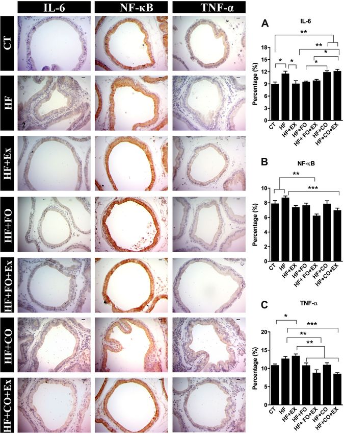
www.nature.com/scientificreports/
The omega-3 PUFAs is a metabolite that has anti-inflammatory p roperties2 are extensively investigated
throughout the body and shown a low correlation with cardiovascular diseases in obesity3 and which may be
effective in either prevention or prostate cancer t reatment4. The omega-3 fatty acids in fish include eicosapen-
taenoic acid and docosahexaenoic acid (300 mg/g/oil)5 and chia seeds contains the highest proportion of alpha-
linolenic acid (ALA, 0.6 g/g/oil) from any known vegetable s ource6. When analyzing the effects of omega-3
PUFAs on cell proliferation and s urvival7, these fatty acids exhibit anti-inflammatory properties through their
impact on prostaglandin synthesis and eicosapentaenoic acid (EPA) and docosahexaenoic acid DHA have inhibi-
tory effects on prostate cancer growth and p rogression8. Additionally, a wide range of mechanisms by which
omega-3 fatty acids affect cancer development have been clarified. Additionally, is possible that activation of
EPA-induced gamma peroxisome proliferation receptors (PPAR-γ), which can interfere with the translocation
of factor nuclear kappa B (NF-κB) to the nucleus, reducing associated cytokines, tumor necrosis factor (TNF-
α) and interleukin-6 (IL-6). Notably, pro‑inflammatory cytokines such as interleukin (IL)-1, IL-6 and TNF are
able to affect cancer risk. The mechanism antitumor activity of omega-3 PUFAs is tightly linked to their ability
to trigger autophagy and apoptosis, reducing expression of BCL-2 and stimulation of the BAX and BAD mito-
chondrial and set the stage for an effective treatment of tumors possessing functional p53; however, since p53
is frequently mutated in human c ancers8.
In the prostate, the panorama of alterations caused by the fish or chia oil supplementation, rich diets, EPA
and DHA or just ALA, is controversial and there is much to be related to the high-fat diet. Fatty acids are the
primary energy source for prostate cancer cells and androgens upregulate fatty acid synthase (FASN), the enzyme
responsible for the de novo synthesis of fatty acids which is linked to an increase in prostatic a denocarcinoma9.
Sterol response element binding protein-1 (SREBP-1) is a positive regulator of FASN expression through bind-
ing elements in the FASN promoter and it is possible that diets rich in omega-3 PUFAs inhibit the cleavage of
SREBP-1 and consequent downregulation of FASN. Additionally, was demonstrated that SREBP-1 regulated
AR promoter activity and expression, and cell viability in p rostate10. Furthermore, SREBP-1 increased reactive
oxygen species (ROS) levels via increased NADPH oxidase 5 (Nox5) expression in prostate cancer cells. ROS
has been shown to induce signal transduction, survival and progression of cancer c ells11. We trust that omega-3
PUFAs supplementation can regulate lipogenesis and ROS signaling by increasing the production of antioxidant
defenses and regulation of AR in the prostate, however it is not clear in the literature about the best proportion
of EPA, DHA and ALA and their potentials effects.
Aerobic physical exercise regulates body energy expenditure helping to decrease body f at12, predominantly
using fatty acid oxidation (AG) as an energy source13, regulating the profile and metabolism of lipids and
glucose14, and reducing plasma lipid levels15, chronic inflammation16 and antioxidant e nzymes17. Although the
most optimal intensity, volume, and modality of exercise to combat disease have yet to be established in the
literature, there are reports that moderate physical exercise increases apoptosis in prostate cancer cells18. Differ-
ent types of exercises can modulate the negative effects of poor lifestyle, obesity, smoking and in the obesogenic
environment, thus, It has already been demonstrated that physical training modulates positively the prostate of
rats submitted to high-fat d iet19. Therefore, we investigated in the present study the ability of fish and chia oil
supplementation associated with aerobic exercise to improve metabolic changes and inhibit prostate diseases
associated with a high-fat diet.
Results
Aerobic physical exercise is associated with PUFA supplementation and effects on body and
adiposity. As expected, the high-fat diet increased weight gain, when compared to the CT group; and the
supplementation of fish and chia oil, alone or associated with physical exercise significantly reduces the weight
gain when compared to the HF group (Table 1). Adipose reserves differed significantly across groups as shown
by differences in epididymal fat (p = 0.004; Table 1), mesenteric fat (p = 0.0008; Table 1), retroperitoneal fat
(p = 0.002; Table 1), and fat index (p = 0.0001; Table 1). Post-hoc analysis revealed that the HF group had signifi-
cantly greater epididymal fat, mesenteric, and retroperitoneal fat, and a greater fat index compared to the CT
and exercise groups (Table 1). Adipose tissue and fat index in animals subjected to aerobic training and HF were
comparable to the CT group. Though non-significant, retroperitoneal fat and fat index were lower in animals
following aerobic physical training with fish oil and chia oil supplementation compared with the HF + FO and
HF + CO groups (Table 1). The absolute prostate weight and relative prostate weights were reduced in the HF,
HF + CO, and HF + FO groups, but no significant differences across groups (Table 1).
PUFA supplementation associated with aerobic training regulates food consumption, and
body weight of rats fed with HFD. During the six-week induction period, significant differences in
weight gain, food consumption (p = 0.0001), and energy intake (p = 0.0001) were observed across groups. Rats
in the CT groups demonstrated greater weight gain at weeks three through six (Fig. 1A), and greater food con-
sumption and energy expenditure at weeks two through six (Fig. 1C,E) compared to the HF groups. Significant
differences in weight gain (p = 0.015) and feed efficiency (p = 0.03) were also observed across groups and time,
with both measures greater in the CT group at weeks two and three, but greater in the HFD groups at week four
and six (Fig. 1D,M).
After the diet acclimatization period, the animals given HF were divided into six subgroups (HF, HF + Ex,
HF + FO, HF + FO + Ex, HF + CO, and HF + CO + Ex) and began oil supplement and aerobic physical training until
week 14. Though nominal, the groups supplemented with fish oil with and without physical exercises showed the
lowest weight gain relative to all other groups (Fig. 1B). The CT group showed significantly greater food consump-
tion than all other treatment groups (p = 0.0001; Fig. 1D) but the lowest feed efficiency of all treatments (p = 0.3;
Fig. 1H). Physical exercise combined with HF did not reduce the energy intake and feed efficiency compared to
Scientific Reports | (2021) 11:6282 | https://doi.org/10.1038/s41598-021-85337-9 2
Vol:.(1234567890)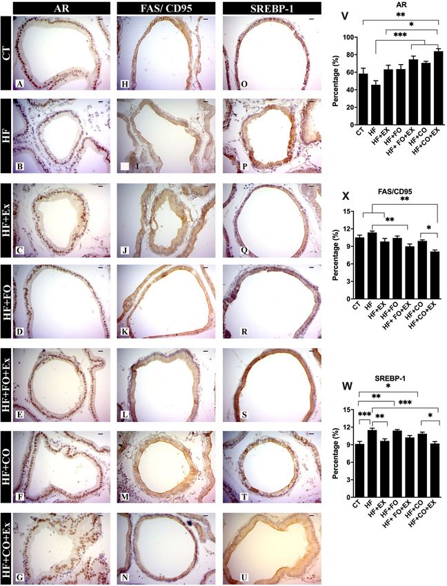
www.nature.com/scientificreports/

                                  Variables                   CT              HF              HF + Ex         HF + FO         HF + FO + Ex    HF + CO         HF + CO + Ex
                                  Initial body weight (g)     209.80 ± 5.22   199.80 ± 9.29   194.10 ± 8.25   191.70 ± 7.80   193.70 ± 8.40   195.60 ± 6.34   198.8 ± 5.57
                                  Final body weight (g)       409.60 ± 7.27   410.30 ± 6.26   395.20 ± 8.89   382.50 ± 9.16   372.20 ± 9.21   380.10 ± 12.2   374.60 ± 8.413
                                  Weight gain (g)             189.90 ± 6.69   224.00 ± 4.21   201.10 ± 3.71   190.80 ± 6.94   175.10 ± 5.42   188.10 ± 9.24   182.20 ± 9.15
                                  Absolute prostate weight
                                                              0.19 ± 0.01     0.15 ± 0.009    0.18 ± 0.01     0.16 ± 0.01     0.18 ± 0.02     0.14 ± 0.01     0.17 ± 0.01
                                  (g)
                                  Relative prostate weight
                                                              0.04 ± 0.005    0.03 ± 0.004    0.04 ± 0.005    0.03 ± 0.005    0.04 ± 0.007    0.03 ± 0.005    0.04 ± 0.004
                                  (g)
                                  Total fat (g)               18.77 ± 1.09    28.08 ± 1.66b   22.42 ± 2.12    23.16 ± 1.87    19.66 ± 1.30    22.75 ± 2.54    19.24 ± 1.47
                                  Relative fat (%)            4.56 ± 0.18     7.08 ± 0.28ac   5.18 ± 0.33     6.42 ± 0.25ae   4.98 ± 0.27b    5.91 ± 0.46     4.59 ± 0.26b
                                  Epididimal adipose
                                                              6.45 ± 0.33     9.46 ± 0.36a    6.99 ± 0.66     7.10 ± 0.69     6.50 ± 0.49b    7.18 ± 0.96     5.76 ± 0.42b
                                  tissue (g)
                                  Mesenteric adipose
                                                              5.21 ± 0.31     7.77 ± 0.24a    6.02 ± 0.33b    6.31 ± 0.42     5.90 ± 0.24b    6.39 ± 0.57     5.45 ± 0.39b
                                  tissue (g)
                                  Retroperitoneal adipose
                                                              7.10 ± 0.75     11.34 ± 0.85a   8.06 ± 0.70     9.97 ± 0.76     7.24 ± 0.69b    9.44 ± 0.92     7.84 ± 0.79b
                                  tissue (g)

                                  Table 1.  Data on initial and final body weight, weight gain, absolute and relative prostate weights, absolute
                                  and relative fats, epididimal. mesenteric and retroperitoneal adipose tissues of trained animals, fish and chia oil
                                  intake, submitted to high-fat diet for 14 weeks. Data are presented as the mean ± SEM (n = 7). The significance
                                  of p < 0.05 is indicated by lowercase letters indicating a difference between the groups. a Referring to the control
                                  group. b Referring to the high-fat diet group. c Referring to a diet rich in fat + ω-3 group. d Referring to a high-fat
                                  diet + physical exercise group. e Referring to a diet rich in fat + ω-3 + aerobic physical exercise group. f Referring
                                  to a high-fat diet + chia group. g Referring to high-fat diet + chia + physical exercise group. The Two-Way
                                  ANOVA test was used to compare the means. with the Tukey post-test.

                                  the HF group (Fig. 1F,H). On the other hand, fish oil supplementation reduced energy intake compared to HF
                                  treatment (Fig. 1F,H), and physical exercise in combination with fish oil supplementation reduced feed efficiency
                                  from 10 weeks compared fish oil supplementation alone (Fig. 1H). Similarly, chia oil with and without physical
                                  exercise reduced energy intake and feed efficiency when compared to the HF + CO, HF, and HF + Ex treatments
                                  (Fig. 1F,H). Food consumption was also lower in all HFD groups compared to the CT group (Fig. 1D), however,
                                  HFD considerably increased feed efficiency and reduced energy intake throughout the experimental period.

                                  Effect of PUFA associated with aerobic training altering lipid profile of rats fed with HFD. The
                                  plasma lipid composition was analyzed the following sacrifice at 14 weeks of post-dietary initiation (Fig. 2).
                                  Comparisons across groups revealed a significant main effect of treatment on plasma TAG levels (p = 0.0001).
                                  A 2.24-fold reduction in plasma TAG was observed in the HF + FO + Ex treatment group when compared with
                                  CT (95% CI 23.63–107.7; p = 0.0003); similarly, a 3.09-fold reduction in TAG in the HF + FO + Ex group was
                                  observed relative to the HF treatment group (95% CI 68.50–152.6; p < 0.0001; Fig. 2A). In addition, TAG levels
                                  in serum from the HF + CO (95% CI 9.529–93.58; p = 0.002) and HF + CO + Ex (95% CI 14.97–99.03; p = 0.002)
                                  groups were also significantly lower than levels from the CT group (Fig. 2A). The HF group increased TAG levels
                                  when compared to CT group (95% CI − 86.90 to − 2.84; p = 0.03; Fig. 2A). Similar effects, TAG levels in serum
                                  from the HF + Ex (95% CI 27.94 to 112.0; p = 0.0001), HF + FO (95% CI 42.50 to 126.6; p < 0.0001), HF + CO
                                  (95% CI 54.40 to 138.5; p < 0.0001) and HF + CO + Ex (95% CI 59.84 to 143.9; p < 0.0001; 2.66-fold) groups were
                                  also significantly lower than levels from the HF group (Fig. 2A). The HF + FO (95% CI 0.438 to 33.62; p = 0.03),
                                  HF + FO + Ex (95% CI 0.210 to 33.39; p = 0.03), and HF + CO (95% CI 2.624 to 35.80; p = 0.03) groups exhibited
                                  statistically significant reduced in TC levels when compared to the HF group (Fig. 2B).
                                      The main effect of treatment on serum low-density lipoprotein (LDL) was also apparent, with a 4.78-fold and
                                  5.48-fold reduction in LDL observed following HF + FO + Ex treatment compared to CT (p < 0.0001) and HF
                                  (p < 0.0001) groups, respectively (Fig. 2D). Additionally, HF + CO and HF + CO + Ex reduce LDL levels when
                                  compared with CT (p < 0.0001) and HF (p < 0.0001) groups, respectively (Fig. 2D). The HF + Ex, HF + CO, and
                                  HF + FO statistically significantly reduced compared to CT and HF groups, respectively (p = 0.0001, Fig. 2D).
                                  The VLDL levels were also reduced in HF + FO (p < 0.0001), HF + FO + Ex (p < 0.0001), HF + CO (p < 0.0001),
                                  and HF + CO + Ex (p < 0.0001) and HF + Ex (p = 0.0001) groups when compared with HF treatment, and VLDL
                                  levels were lower in the HF + CO (p = 0.007), HF + CO + Ex (p = 0.002) and HF + FO + Ex (p = 0.003) groups
                                  compared to CT (Fig. 2E). However, VLDL levels following HF treatment increased significantly VLDL levels
                                  when compared to CT (p = 0.02; Fig. 2E).
                                      In contrast to TAG, LDL, and VLDL levels, the CT group demonstrated significantly lower levels of HDL com-
                                  pared with all other groups (Fig. 2C). Nevertheless, significant changes to the ratio of TC/HDL associated with
                                  oil supplementation and aerobic physical training suggest these treatments alter plasma lipid profile (p = 0.0001)
                                  the PUFAs supplementation and PUFAs supplementation alongside exercise (HF + Ex, HF + FO, HF + FO + Ex,
                                  HF + CO, and HF + CO + Ex) groups reduced the TC/HDL ratios compared to the CT group (Fig. 2F). The TC/
                                  HDL ratio reduced significantly in the HF + FO, HF + FO + Ex, HF + CO, HF + CO + Ex groups compared to the
                                  HF group (Fig. 2F).

Scientific Reports |   (2021) 11:6282 |                      https://doi.org/10.1038/s41598-021-85337-9                                                                        3

                                                                                                                                                                      Vol.:(0123456789)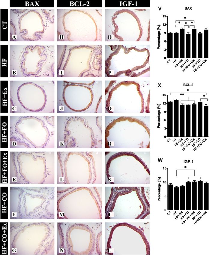
www.nature.com/scientificreports/

                                            Figure 1.  Nutritional data of weight gain, food consumption, energy intake and feed efficiency of the animals
                                            for the 14-week protocol.

                                            Effect of PUFA diet composition associated with aerobic physical exercise altering lipid oxida-
                                            tive stresses of the prostate of rats fed with HFD. To investigate the effects of fish oil and chia supple-
                                            mentation on the balance of oxidative stress production and antioxidant capacity, we analyzed the gene expres-
                                            sions of SOD1, CAT, GSS, NRF-2, and NOS2 in the prostate of rats that consumed a high-fat diet (Fig. 2G–K).
                                            The expression of SOD1 and CAT mRNA was lower in the HF + CO group compared to the HF + FO group,
                                            however, the group supplemented with chia oil showed higher expression of GSS, NRF2, and NOS2 mRNA to
                                            the other groups (Fig. 2). Aerobic physical exercise increased the expression of SOD1 and CAT mRNA associ-

          Scientific Reports |   (2021) 11:6282 |               https://doi.org/10.1038/s41598-021-85337-9                                                   4

Vol:.(1234567890)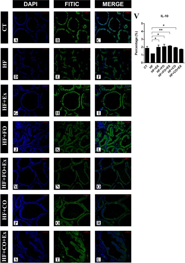
www.nature.com/scientificreports/

                                  Figure 2.  The significance of p < 0.05 is indicated by * and p > 0.05 ** indicating a difference between the
                                  groups. CT control, HF high-fat diet, HF + Ex high fat and aerobic exercise, HF + FO high fat and fish oil,
                                  HF + FO + Ex high fat, fish oil and aerobic exercise, HF + CO high fat and chia oil, HF + CO + Ex high fat, chia oil
                                  and aerobic exercise. (A) tryacilgricerol, (B) total cholesterol, (C) HDL levels, (D) LDL levels, (E) VLDL levels,
                                  (F) TC/HDL levels, (G) amount of the superoxide dismutase gene (SOD-1) in the ventral prostate; (H) amount
                                  of catalase (CAT) in the ventral prostate; (I) amount of NRF-2 in the ventral prostate; (J) amount of glutathione
                                  (GSS) in the ventral prostate; (K) amount of nitric oxide synthase 2 (NOS-2) in the ventral prostate. The Two-
                                  Way ANOVA test was used to compare the means, with the Tukey post-test.

Scientific Reports |   (2021) 11:6282 |                https://doi.org/10.1038/s41598-021-85337-9                                                    5

                                                                                                                                                Vol.:(0123456789)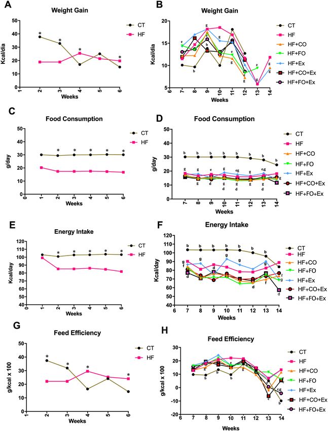
www.nature.com/scientificreports/
ated with fish oil and chia oil supplementation, however, the groups that practiced aerobic physical exercise
showed low GSS and NRF2 mRNA expressions (Fig. 2). The groups supplemented with chia oil (HF + CO and
HF + CO + Ex, respectively) significantly increased the expression of NOS2 mRNA when compared to the other
groups (Fig. 2). In another perspective, the intervention with chia and physical exercise in the HF + CO + Ex
group raised NOS 2 levels in relation to the HF, HF + Ex, and HF + FO + Ex groups (Fig. 2).
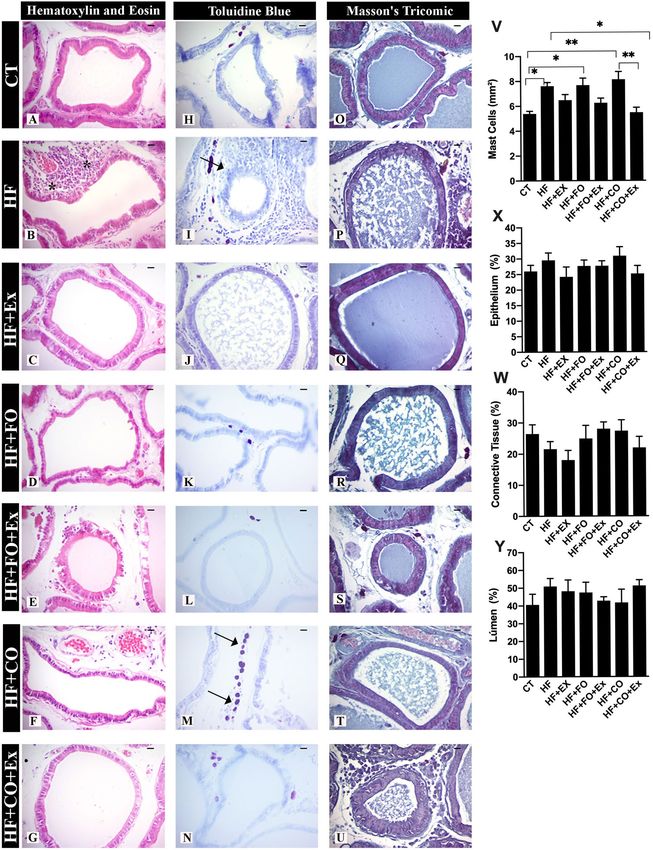
Changes of the histopathological, mast cells and stereological analysis in the prostate of
rats submitted the aerobic physical exercise and PUFFA supplementation across feeding
HFD. The ventral prostate structure in the CT group presented the prevalence of acini with simple cylindri-
cal epithelium, polarized nuclei in the basal part of the cells, and a clear supranuclear region, the latter of which
corresponds to the Golgi Apparatus area (Fig. 3A). Stereological analysis show the reduction of epithelium and
connective tissue in the HF + Ex group compared with groups, but without significant differences (Fig. 3X,Y), the
other groups showed no differences in the stereological analysis.
It was possible to observe epithelial cell nuclei to apical areas, where they presented different heights to give
a pseudo-stratified aspect to the tissue, which was frequently observed in the HF group (Fig. 3B). The presence
of cells in division moving to the apical part of the epithelium indicates proliferative activity in this tissue and
in some areas, the epithelium had become thick with agglomerated nuclei of various heights similar to prostatic
intraepithelial neoplasia (PIN) and was showed an increase of 37% of PIN in HF group (Table 2). The prolifera-
tive inflammatory atrophy (PIA) characterized by agglomerated epithelial cells with heterogeneous phenotypes,
stratified epithelial patterns, and different compacted chromatin nuclear patterns show epithelial inflammatory
reactive atypia was observed in 24% and 39% of the animals of HF and HF + CO groups respectively (Table 2).
Inflammatory foci observed in the animals of HF (39%) and HF + CO (24%) groups presented similar character-
istics including a prevalence of lymphocytes and plasmatic cells (Table 2). The HF + FO sowed lowed alteration
of how PIN and inflammation foci than compared to other groups (Table 2). On the other hand, the aerobic
physical exercise showed reduced histopathological alteration in the prostate associated with supplementation or
alone (Table 2). The HF (95% CI − 4.198 to − 0.2576; p = 0.01), HF + FO (95% CI − 4.264 to − 0.3236; p = 0.01) and
HF + CO (95% CI − 4.720 to − 0.7796; p = 0.0023) had a higher number of mast cells in the CT group (Fig. 3V).
The aerobic physical exercise reduced mast cells in HF + CO + Ex (95% CI 0.6978 to 4.638; p = 0.003) group when
compared to HF + CO (Fig. 3V).
Effect of PUFA diet composition associated aerobic physical exercise on the modulation andro-
genic, lipogenic, and apoptotic prostatic. We investigated the immunoreactivity of the AR, SREBP-1,
IGF-1, BCL-2, BAX, and FAS/CD95 effects of fish and chia oil supplementation and physical exercise after HFD
and is shown in Fig. 4. The HFD (as compared to the CT group) significantly reduced the immunoreactivity of de
AR in the prostate, there was no significant difference. The HF + FO + Ex (95% CI − 47.64 to − 10.47; p = 0.0006),
HF + CO (95% CI − 43.64 to − 6.474; p = 0.003) and HF + CO + Ex (95% CI − 58.01 to − 18.59; p < 0.0001) show
increased AR in prostate when compared to HF group (Fig. 4A–G and V). Likewise, the mean of AR was higher
in rats fed the chia oil and submitted the aerobic physical exercise (HF + CO + Ex), were significant difference
in the CT (95% CI − 46.30 to − 4.745; p = 0.009) group (Fig. 4V). To compare the effects of the aerobic physical
exercise and aerobic physical exercise associated with chia oil in the expression of prostatic AR it was possible to
verify the difference in AR expression stimulated by chia oil in the HF + CO + Ex group when compared to the
HF + Ex group (95% CI − 40.4 − 0.985; p = 0.01).
To verify the prostatic effects of the high-fat diet and the addition of fish oil and chia in the diet associated
with physical exercise, we analyzed lipogenesis through the expression of SREBP-1. There was a significant
increase in the SREBP-1 expression on HF (95% CI − 3.90 to − 0.83; p = 0.001), HF + FO (95% CI − 3.92 to
− 0.66; p = 0.01) and HF + CO (95% CI − 3.27 to − 0.21; p = 0.01), when compared to CT group (Fig. 4W). On
the other hand, aerobic physical exercise reduced the SREBP-1 expression alone (HF + Ex, 95% CI 0.39 to 3.29;
p = 0.001) and associated with chia (HF + CO + Ex 95% CI 0.84 to 3.73; p = 0.001) oil when compared to the HF
group (Fig. 4W). Physical exercise reduced effects lipogenic in prostate associated chia oil vs. HF + CO group
(95% CI 0.22 to 3.11; p = 0.01).
To identify the effect of the high-fat diet on epithelial progression and development of prostatic lesions and
possible effects of n-3 PUFA supplementation associated with aerobic exercise, we investigated the expression of
IGF-1. There was a significant increase in IGF-1 in fish and chia oil supplementation, HF + FO (95% CI − 3.43 to
− 0.14; p = 0.01) and HF + CO (95% CI − 4.47 to − 0.55; p = 0.01) respectively, compared to the HF group (Fig. 5).
The HF + FO + Ex increased IGF-1 compared to HF (95% CI − 3.76 to − 0.27; p = 0.05, Fig. 5).
We check the state of different proteins related to cell death and survival concerning the mitochondrial
pathway after consumption of HFD and interaction of dietary n-3 PUFA and aerobic physical exercise. The
HF + Ex (95% CI 0.27 to 3.14; p = 0.05), HF + FO (95% CI 0.33 to 3.09; p = 0.01) and HF + FO + Ex (95% CI 0.25
to 2.96; p = 0.05) groups showed lower BCL-2 when compared to HF group (Fig. 5). There was a more reduced
level of BCL-2 in HF + CO + Ex when compared to HF + CO (95% CI 0.13 to 2.83; p = 0.05), HF (95% CI 0.78
to 3.49; p = 0.001), and CT (95% CI 0.16 to 2.87; p = 0.05) groups (Fig. 5). On the other hand, aerobic physical
exercise upregulation of the BAX expression in the prostate showed in the HF + Ex group when compared with
HF (95% CI − 3.19 to − 0.09; p = 0.05), HF + FO (95% CI 0.32 to 3.43; p = 0.05) and HF + CO (95% CI 0.12 to
3.23; p = 0.05, Fig. 5). The addition of fish oil to the diet associated with aerobic physical exercise, HF + FO + Ex
group, significantly reduced BAX in the prostate compared to HF (95% CI − 3.14 to − 0.04; p = 0.05), HF + FO
(95% CI − 3.38 to − 0.27; p = 0.05, Fig. 5).
The activity of FAS/CD95 death receptors was investigated, where the HF group showed the highest expres-
sion when compared to the HF + FO + Ex (95% CI 0.76 to 3.98; p = 0.01) and HF + CO + Ex (95% CI 1.74 to 4.79;
Scientific Reports | (2021) 11:6282 | https://doi.org/10.1038/s41598-021-85337-9 6
Vol:.(1234567890)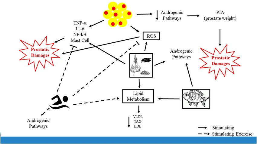
www.nature.com/scientificreports/
Figure 3. The data are presented as the mean ± SEM (n = 7). The significance of p < 0.05 is indicated by *
indicating a difference between the groups. The asterisk * in H&E technique represents shows rounded acini
with polymorphonuclear cells around prostate epithelium and the arrows in toluidine’s blue demonstrate the
mast cells in connective tissue of prostate. CT control, HF high-fat diet, HF + Ex high fat and aerobic exercise,
HF + FO high fat and fish oil, HF + FO + Ex high fat, fish oil and aerobic exercise, HF + CO high-fat diet and
chia oil, HF + CO + Ex high fat, chia oil and exercise group. Graph V: referring to the number of mast cells per
mm2; Graph W: referring to the prostatic epithelium; Graph X: referring to prostatic connective tissue; Graph Y:
referring to the amount of prostatic lumen. Bar = 20 µm; Resolution = ×40. The Two-Way ANOVA test was used
to compare the means, with the Tukey post-test.
Scientific Reports | (2021) 11:6282 | https://doi.org/10.1038/s41598-021-85337-9 7
Vol.:(0123456789)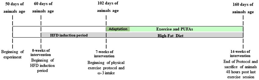
www.nature.com/scientificreports/
Groups PIN PIA Inflammation
CT 17 (85)–20,76%f,e,g 13 (85)–14,08%b,f,g 13 (85)–14,28%f
e,g e,f,g
HF 43 (114)–37,30% 27 (114)–24,20% 46 (114)–39,97%d,f,g
HF + Ex 16 (83)–20,30% 7 (83)–7,60% 3 (83)–3,41%
HF + FO 13 (68)–18,16% 14 (68)–21,93% 1 (68)–1,32%
HF + FO + Ex 22 (101)–21,60%g 20 (101)–19,42%g 5 (101)–5,00%
g
HF + CO 23 (103)–21,53% 39 (103)–39,06% 26 (103)–24,69%
HF + CO + Ex 37 (100)–35,65% 22 (100)–22,01% 14 (100)–12,73%
Table 2. Occurrence of histopathological data of experimental animals. CT Control Group, HF high-fat diet,
HF + Ex high-fat diet and aerobic physical exercise, HF + FO high-fat diet and fish oil, HF + FO + Ex high-fat
diet, fish oil and aerobic physical exercise, HF + CO high-fat diet and chia oil, HF + CO + Ex high-fat diet, chia
oil and aerobic physical exercise. The results were expressed in absolute values and occurrence of percentages.
The significative differences were adopted based on p < 0,05. Test Mann–Whitney.
p = 0.05) groups, respectively (Fig. 4H–N,X). Chia supplementation, HF + CO group, showed higher values of
FAS/CD95 when compared to the HF + CO + Ex group (95% CI 0.28 to 3.33; p = 0.05, Fig. 4X). The HF + Ex group
showed higher FAS/CD95 values when compared to the HF + CO + Ex group (95% CI 0.18 to 3.24; p = 0.05,
Fig. 4X). The HF + CO + Ex group showed reduced FAS/CD95 compared to CT (95% CI 0.87 to 3.93; p = 0.001).
It was evident that the stimulated pathway of apoptosis by aerobic exercise was greater expression of BAX and
reduced the expression of FAS/CD95.
Effect PUFA supplementation associated with aerobic physical exercise on inflammation after
HF diet in the prostate. We investigated the immunoreactivity of the inflammatory markers IL-6, TNF-α,
and NF-κB. The chia oil supplementation increased IL-6 (95% CI − 5.03 to − 0.84; p = 0.01) than compared to
the CT group (Fig. 6). The HF + CO and HF + CO + Ex groups significantly increased IL-6 immunoreactivity
compared to the HF + Ex (95% CI − 4.92 to − 0.74; p = 0.01) and HF + FO + Ex groups (95% CI − 5.19 to − 1.00;
p = 0.01), respectively (Fig. 6). The HF + FO groups showed less IL-6 in the prostate compared to the HF + CO
(95% CI − 4.48 to − 0.29; p = 0.05) group (Fig. 6). The association of supplementation of fish oil and aerobic
physical exercise reduced IL-6 than compared to the chia oil and physical exercise group (95% CI − 4.43 to
− 0.24; p = 0.05, Fig. 6).
There was a significant increase in the TNF-α immunostaining in the HF + Ex group compared to the CT
group (95% CI − 5.00 to − 0.13; p = 0.05). The results showed that HF + FO + Ex (95% CI 1.27 to 6.45; p = 0.01)
and HF + CO + Ex groups (95% CI 1.55 to 6.72; p = 0.001) significantly reduced TNF-α labeling compared to HF
(Fig. 6). The association of the supplementation of the fisher (95% CI 2.20 to 7.07; p = 0.0001) and chia (95% CI
2.47 to 7.35; p = 0.0001) oil more physical exercise reduced TNF-α labeling in the HF + Ex (Fig. 6). Additionally,
the group HF + CO + Ex was significantly reduced TNF-α when compared to HF + CO (95% CI 0.0016 to 4.87;
p = 0.05). The association of fish oil supplementation with aerobic physical exercise showed a significantly reduced
NF-κB when compared to HF (95% CI 0.71 to 4.10; p = 0.01) and HF + Ex (95% CI 0.19 to 3.77; p = 0.05) groups
(Fig. 6). There was no significance between the other groups.
The anti-inflammatory effects of FO and CO intake associated with physical exercise in the prostate were
evaluated by the expression of cytokine IL-10. We observed that the animals that were submitted to HFD showed
lower expression of prostatic IL-10 when compared to the other groups. The groups supplemented with fish oil
(HF + FO) showed significantly higher expression of IL-10 when compared to the HF group (Fig. 7). The groups
that were submitted to physical exercise, HF + Ex, HF + FO + Ex and HF + CO + Ex, showed no difference between
them, however, all were significantly different from the HF group (Fig. 7).
Discussion
We compared the biological effects of supplementing with fish and chia oils alone or in combination with aerobic
physical exercise in rats submitted to a high-fat diet. A potential mechanism by which obesity can promote severe
cancer prognosis is through the induction of functional metabolic abnormalities, altering the metabolic profile,
promoting inflammation and oxidative stress. The HFD exposure increased LDL-cholesterol levels, TC, and
TAG, which are predominantly synthesized in the liver, are important markers of lipid metabolic disorders. Chia
oil has been described as a cholesterol regulator owing to the effect of PUFA in lipid m etabolism20. When rats
were fed HFD and supplemented with fish or chia oil, HDL cholesterol levels in the plasma were not noticeably
different from those not receiving oil, whereas TC levels were marginally lower relative to non-supplemented
groups. Aerobic physical exercise has been reported to reduced serum levels of LDL and VLDL. Similarly, fish
supplementation has been reported to reduce LDL levels21. The incorporation of fish and chia oils with the
physical exercise clearly has more effect on lipid profile compared with fish oil supplementation or chia oil
alone, as demonstrated by reduced LDL, VLDL, and TAG levels by chia oil supplementation alone. These results
provide evidence that aerobic physical exercise and supplementation with fish oil may synergistically improve
the amount of circulating lipoproteins and reduce lipid stocks in adipose tissue despite no apparent changes in
weight during HFD consumption.
Scientific Reports | (2021) 11:6282 | https://doi.org/10.1038/s41598-021-85337-9 8
Vol:.(1234567890)www.nature.com/scientificreports/
Figure 4. The data are presented as the mean ± SEM (n = 7). The significance of p < 0.05 is indicated by * and
p > 0.05 ** indicating a difference between the groups. (A) CT—control; (B) HF—high-fat diet; (C) HF + Ex—
high fat and aerobic exercise; (D) HF + FO—high fat and fish oil; (E) HF + FO + Ex—high fat, fish oil and aerobic
exercise; (F) HF + CO—high fat and chia oil; (G) HF + CO + Ex—high fat, chia oil and aerobic exercise. Graph
I: androgen receptor expression (AR); Graph II: death receptor expression (FAS/CD95); Graph III: Sterol
regulatory element-binding transcription factor-1 (SREBP-1). The Two-Way ANOVA test was used to compare
the means, with the Tukey post-test. Bar = 20 µm; Resolution = ×40.
Scientific Reports | (2021) 11:6282 | https://doi.org/10.1038/s41598-021-85337-9 9
Vol.:(0123456789)www.nature.com/scientificreports/
Figure 5. The data are presented as the mean ± SEM (n = 7). The significance of p < 0.05 is indicated by * and
p > 0.05 ** indicating a difference between the groups. (A) CT—control; (B) HF—high-fat diet; (C) HF + Ex—
high fat and aerobic exercise; (D) HF + FO—high fat and fish oil; (E) HF + FO + Ex—high fat, fish oil and aerobic
exercise; (F) HF + CO—high fat and chia oil; (G) HF + CO + Ex—high fat, chia oil and aerobic exercise. Graph
V: data related to BAX expression, Graph X: data related to BCL-2 expression, Graph W: data related to IGF-1
expression.
Scientific Reports | (2021) 11:6282 | https://doi.org/10.1038/s41598-021-85337-9 10
Vol:.(1234567890)www.nature.com/scientificreports/
Figure 6. The data are presented as the mean ± SEM (n = 7). The significance of p < 0.05 is indicated by * and
p > 0.05 ** indicating a difference between the groups. (A) CT—control; (B) HF—high-fat diet; (C) HF + Ex—
high fat and aerobic exercise; (D) HF + FO—high fat and fish oil; (E) HF + FO + Ex—high fat, fish oil and aerobic
exercise; (F) HF + CO—high fat and chia oil; (G) HF + CO + Ex—high fat, chia oil and aerobic exercise. Graph
I: data related to IL-6 expression. Graph II: data related to NF-κB expression; Graph III: data related to TNF-α
expression.
Scientific Reports | (2021) 11:6282 | https://doi.org/10.1038/s41598-021-85337-9 11
Vol.:(0123456789)www.nature.com/scientificreports/
Figure 7. The data are presented as the mean ± SEM (n = 7). The significance of p < 0.05 is indicated by * and
p > 0.05 ** indicating a difference between the groups. Immunofluorescence of IL-10 in ventral prostate of
animals submitted to high-fat diet, supplemented with ω-3 PUFAs, and performed aerobic physical exercise.
(A) CT—control; (B) HF—high-fat diet; (C) HF + Ex—high fat and aerobic exercise; (D) HF + FO—high fat
and fish oil; (E) HF + FO + Ex—high fat, fish oil and aerobic exercise; (F) HF + CO—high fat and chia oil; (G)
HF + CO + Ex—high fat, chia oil and aerobic exercise.
Scientific Reports | (2021) 11:6282 | https://doi.org/10.1038/s41598-021-85337-9 12
Vol:.(1234567890)www.nature.com/scientificreports/
PUFAs are associated with reduced risk of several types of carcinogenesis, have been evidence in the pros-
tate, however, this depends on numerous factors, including the source of omega-3 PUFAs. The consumption of
the high-fat diet and obesity cause a reduction in testosterone and even so promote prostatic changes such as
prostatitis, BHP, HGPIN22 until c ancer23. A review study organized by Aucoin4 showed an association between
increased consumption of fish oil and reduced risk of prostate cancer, however more research is needed to
demonstrate the potential effects of the treatment of omega-3 and its relationship with prostate. Fish oil has
higher concentrations of EPA and DHA and exceptionally, seeds of chia (Salvia Hispanic) are abundant in ALA,
and the omega-3 PUFAs are considered to be activators of cholesterol esterification, an important mechanism
for cholesterol reduction24. We associated the increase in the expression of SREBP-1 with the consumption of
HFD and increased of the BHP, HGPIN in prostate independent of the AR. SREBP-1 induced prostate cancer
cell proliferation, migration and invasion in vitro and promoted prostate tumor growth through the induction
of FASN expression and lipid droplet formation and accumulation in prostate cells9. Physical exercise aerobic
alone and associated with chia oil intake, a-linolenic acid (EPA and DHA precursor) reduced the levels of pros-
tate SREBP-1 reduced PPAR activation regulated the lipogenic effects concomitant with the increase in AR. The
effects of physical training promoted an increase in AR in the prostate, thus regulating the expression of SREBP-1,
exhibited different efficiencies in the inhibition of proliferation.
To determine whether supplementation of chia and fish oil would cause cellular apoptosis, we checked the
intrinsic and extrinsic pathways. HFD is often accompanied by decreased levels of omega-3 P UFAs25 and is
believed to be prejudicial for the prostate. The Fas/FasL pathway is an important extrinsic apoptotic pathway
and the Fas/CD95 membrane receptor initiates intracellular signaling of the apoptosis pathway by activating
caspases 8 and 9. Thus, it has been shown that Fas ligand (FasL) is secreted by prostatic carcinoma cells and
together with Fas/CD95 plays a key role in the development of abnormal cells26. Jiang27 reported that Fas/CD95
is more expressed in a high-grade PIN. The activation of Fas/CD95 occurs by TNF-a initiating the proteolytic
cleavage pathways, in the absence of TNF-α this pathway is minimized27. We found that omega-3 PUFAs reduced
expression of Fas/CD95 and BCL-2, and increased BAX when associated with aerobic physical exercise. It is
already well documented that physical exercise promotes alteration of apoptosis in the prostate cell increases the
BAX reduce proliferative ratios in the ventral p rostate28 even in animals submitted to a high-fat d iet18. Thus, it is
possible to relate that supplementation of omega-3 PUFAs associated with aerobic exercise promotes prostatic
cell apoptosis intrinsically.
Oxidative load is strongly implicated in the pathogenesis of age-related diseases, including the formation of
prostate cancer tumor, and omega-3 fatty acids have antioxidant and anti-inflammatory properties, we investi-
gated the component effects of fish oil with higher concentrations of EPA and DHA, and chia oil components
with greater composition of ALA in reducing the effects of oxidative damage to DNA induced by obesity. The
high-fat diet increases lipid peroxidation and higher lipid accumulation probably was related to increasing of
omega-3 PUFAs with fish and chia oil supplementation. Such omega-3 is metabolized primarily at the peroxi-
some fraction, a well-known site of H2O2 generation, due to long-chain fatty acid structure. Therefore, the anti-
oxidant effects of fish oil supplementation (concentration of EPA and DHA) were mainly at the mitochondrial
compartment since, despite not recovering to control levels, such promoted decreased O 2·− directly modifying
29
the levels of enzymatic expression of NOS2 and increased the antioxidant c apacity . Physical exercise is clinically
associated with a reduction in lipid peroxidation by increasing the expression of antioxidant enzymes30. On the
other hand, supplementation with chia oil increased the levels of GSS, NOS 2, and NRF-2 in the ventral prostate.
Like other exogenous stimuli, chia oil can promote the NRF-2 activation pathway, to control the pro-oxidative
response31, once activated, participates in the regulation of programmatic functions stimulated by oxidants,
including autophagy, reticulum stress, and mitochondrial biogenesis.
Omega-3 fatty acids exhibit known inflammatory properties that suppress prostate carcinogenesis, we investi-
gate the potential role of fish oil and chia in reducing the inflammatory effects on HFD-induced prostate epithelial
cells. Statements have been published in the literature that omega-3 PUFAS are important in the quantity and
quality of immune r esponses32. Fish oil intake, containing a mixture of omega-3 PUFAS, reduces the expression
of IL-6, TNF-α, and NF-κB in the ventral prostate and was more efficient when associated with aerobic physical
exercise (Fig. 8). NF-κB is a pro-survival nuclear transcription factor activated by a variety of stimuli, including
oxidative stress. Evidence suggests that fish oil components such as DHA can attenuate the transcriptional activity
of NF-κB by inhibiting translocation to the nucleus in obesity-induced prostate c ells33. Omega-3 PUFAS -acti-
vated PPAR α can also directly interfere with the NF-κB (p50–p65 dimer) and consequently inhibit expression
of the gene encoding pro-inflammatory cytokineIL-6. Possibly the higher concentration of omega-3 PUFAS in
fish oil inhibited AR/NF-κB promoted down-regulation in TNF-α and COX-2, and this modification reduced
PIN. Physical exercise associated with fish oil supplementation (concentration of EPA and DHA) significantly
reduced prostate inflammation for increased IL-10 in the prostate. Another described effect of EPA and DHA is
COX inhibition that reduces inflammation and ROS p roduction34.
Conclusions
Physical exercise and encouraging PUFA consumption are of utmost importance in the treatment of obesity
and related diseases, which are characterized by variations in adipose tissue deposition, lipoprotein profiles.
Employing both strategies concurrently provides additional benefits for reducing the negative effects of obesity
and prostatic diseases. When we incorporate fish and chia oil supplementation into a high-fat diet, we verify
the ability to prevent prostatic damage by reducing the circulating lipid profile, increasing antioxidant activity
and its anti-inflammatory capabilities. This protection was more effective when associated with aerobic exercise,
suggesting regulation of antioxidant activity such as higher expression of CAT and SOD-1, lower expression of
Scientific Reports | (2021) 11:6282 | https://doi.org/10.1038/s41598-021-85337-9 13
Vol.:(0123456789)www.nature.com/scientificreports/
Figure 8. Effects of aerobic physical exercise associated with ω-3 supplementation in the prostatic obesogenic
environment occurs due to an excess of high-fat diet in Wistar rats during 14 weeks of experimental protocol.
The high-fat diet hypertrophy adipocytes, which, in turn, signal inflammatory cytokines such as TNF-α, IL-6,
transcription factor NF-κB and mast cells recruited by the inflammatory process, cause prostatic changes.
Adipocytes cause an androgenic signaling (RA), potentiating pro-oxidant factors in reactive oxygen species
(ROS), which can lead to an inflammatory prostatic atrophy (PIA). In turn, aerobic physical exercise inhibits
all the damage caused by the high fat diet, stimulating androgenic signaling, lipid metabolism, by reducing the
levels of VLDL, TAG, LDL lipoproteins and increase as a buffering “factor” stimulating as anti-oxidative proteins
during ROS. Finally, fish and chia oils enhance the effects initiated by physical exercise, improving androgenic
signaling, lipid metabolism and ROS signaling.
IL-6, TNF-α and NF-κB as well as an increase in anti-apoptotic proteins of BAX and FAS/CD95 and reduction
of BCL-2.
Materials and methods
Ethics statement. Experiments, all animal procedures, were conducted in accordance with the ethical
principles in animal research adopted by the Brazilian College of Animal Experimentation (COBEA) and the
study protocol was approved by the Ethics Committee on Animal Use (CEUA) of the Universidade do Oeste
Paulista-Unoeste, Presidente Prudente (protocol number 3962).
Animals and experimental procedures. Forty-nine adult Wistar rats (60 days old) were individually
housed and maintained at 22 ± 1 °C, 60–70% humidity, and kept on a 12-h light/dark cycle for the duration of
the experiment. Animals were randomized into seven treatment groups (n = 7): Adaptation phase (1st to 6th
week) of the high-fat diet (HFD) was realized with all groups and control group (CT) treatment that received
ad libitum standard diet and water; HFD (HF) treatment that received ad libitum high-fat diet and water; HFD
with fish oil supplement (HF + FO) treatment; HFD with physical exercise (HF + Ex) treatment; HFD with fish
oil supplement and physical exercise (HF + FO + Ex) treatment; HFD with chia oil supplement (HF + CO) treat-
ment; and HFD with chia oil supplement and physical exercise (HF + CO + Ex) treatment. Beginning at 60 days
old, all animals in the HFD groups underwent an HFD induction period in which they had ad libitum access
to HFD, standard ration and water. At seven weeks post-adaptation (102 days old), rats began the experimental
phase in which the fish oil, chia oil, and physical exercise groups began oil supplement intake and the physical
exercise protocol (Fig. 9). All procedures with the animals were carried out from 1 to 6 pm.
Dietary composition. At 60 days of age, rats were maintained on standard rat chow (commercial Supralab)
or began the HFD induction period. The HFD used in this research was previously described by Estadella et al.35
and consisted of a hypercaloric mixture (normoproteic and HFD) containing ground and mixed commercial
Supralab ration, roasted peanuts, and milk chocolate and cornstarch in a 3:2:2:2. The high-fat diet was composed
by lipids (59%), carbohydrates (28%) and proteins (13%). The proximate composition of the experimental diets
was evaluated according to the analytical methods recommended by the Association of Official Analytical Chem-
ists. The commercial diet was composed of 24.11% of proteins, 4.27% of lipids and 52.20% of carbohydrates,
Scientific Reports | (2021) 11:6282 | https://doi.org/10.1038/s41598-021-85337-9 14
Vol:.(1234567890)www.nature.com/scientificreports/
Figure 9. Timeline of intervention protocol for 14 weeks of a high-fat diet, fish oil supplementation, chia oil,
and aerobic physical exercise.
Fish oil Chia oil
Saturated fatty acids (g/day) 0.13 0.11
Monounsaturated fatty acids (g/day) 0.20 0.07
Linoleic (LA) – 58.4
Linolenic (ALA) – 170.84
Polyunsaturated fatty acids (mg/day)
Eicosapentaenoic (EPA) 129.33 –
Docosahexaenoic (DHA) 113,33 –
Cholesterol (mg/day) 3.33 –
Calories oil (Kcal/1 g/day) 6 9
Table 3. Components of chia and fish oils.
while the high-fat diet was composed of 18.84% of proteins, 23.80% of lipids and 50, 4% of carbohydrates in the
mixture, being 4.9 kcal/g.
PUFAs supplementation. The PUFAs were supplemented with fish and chia oil from RSBLUMOS
(RSBLUMOS Comercial Produtos Alimentícios LTDA), through gavage at 1 mL/day fish and chia oils based
on prior investigation by Marinelli et al.36, three times a week, for 8 weeks and the animals in the other groups
received the daily water gavage as a placebo so that they were submitted to the same type of stress in the animals.
The fish oil was composed of saturated fatty acids (g/day) in 0.13; while chia oil was composed of 0.11, mono-
unsaturated fatty acids (g/day) 0.20 of FO, and 0.07 in the CO. Neither oils were composed of linolenic acid and
α-linolenic acid in your composition, the chia oil nor composed by 170.84 mg/day of ALA and 58.4 mg/day
of linolenic acid. On the other hand, the FO was composed of 129.33 mg/day of EPA and 113.33 of DHA, and
finally 3.33 mg/day of cholesterol, different from CO (Table 3).
Aerobic physical exercise protocol. Rats in the aerobic physical exercise groups were subjected to 30 min
of swimming while wearing a weighted vest in a tank divided into sections using plastic dividers. The swimming
tank was filled with water maintained at 29 °C. During the 1-week adaption phase, the rat was habituated to the
swimming protocol and the vest without weights attached (Fig. 9). The training protocol was conducted three
times per week. Following the adaptation phase, a weight corresponding to 3.5% of the animal’s total mass was
attached to the vest at the posterior region of the thorax that corresponds to a 70% exercise intensity moderate37.
This intensity was adjusted to 70% exercise intensity at the end of the first four weeks of training in order to avoid
adaptation to the protocol.
Samples collection. At 172 days of age, 48 h after the last physical training session, after sacrifice, the ani-
mals remained overnight fasting for 12 h, had their blood collected under intraperitoneal anesthesia of ketamine
(60 mg/kg) and xylazine hydrochloride (1 mg/kg), abdominal-pelvic laparotomy was performed, the ventral
prostate and epididymal, retroperitoneal and mesenteric adipose tissues were removed, weighed, and processed
for future analysis. The slides were analyzed and photographed in a light microscope, model AxioCam ECR5s
Zeiss.
Blood sample. Serum levels of total cholesterol (TC), high-density lipoprotein (HDL), and TAG were ana-
lyzed by blinded experimenters using a colorimetric method with Cobas C111 equipment (Roche Diagnostics-
Scientific Reports | (2021) 11:6282 | https://doi.org/10.1038/s41598-021-85337-9 15
Vol.:(0123456789)www.nature.com/scientificreports/
Brazil) and the ROCHE commercial kit according to the manufacturer’s instructions. The TC/HDL levels were
calculated based on total cholesterol (mg/dl) divided by HDL (mg/dl), the VLDL levels were calculated by TAG
divided per five, the LDL was calculated using total cholesterol values minus HDL mg/dl minus VLDL ratios.
Body weight and nutritional analyses. Body weight was also measured, to evaluate weight gain, we cal-
culated body mass gain (Δ = final weight—starting weight). Relative prostate weight, used to evaluate the growth
of prostate in different interventions, was determined as the ratio between absolute prostate weight and total ani-
mal bodyweight (g). During the experimental period, weekly consumption of water and food and changes in rat
body mass were monitored. Food intake value and caloric value of ration for rodents (3 kcal/g for standard ration
and 9 kcal/g for HFD) were used to obtain total energy consumption (TEI, kcal/day = average food consumption
per day [g] × 3) and (ii) feed efficiency (FE, g/kcal = mean bodyweight gain/total TEI mean)38.
Histologic analysis of prostate. The ventral prostate was removed, weighed, and embedded in Paraplast
and the 5 µm sections were cut. These sections were then stained with hematoxylin and eosin (H&E), five histo-
logical sections by intermediate ventral prostate were evaluated and the histological fields were photographed,
resulting in 10 or more fields per group. The slides were analyzed and photographed in an optical light micro-
scope, model AxioCam ECR5s Zeiss. The ventral prostate was collected, fixed, prepared, and processed for histo-
logical slides, stained with hematoxylin and eosin (H&E), toluidine blue, and Masson trichrome techniques. For
masson trichomic data, quantification was performed using the Weibel m ethod39.
Immunohistochemistry analysis. To block the endogenous peroxidase, the sections were subjected to a
solution of hydrogen peroxide + methanol and peroxidase block. Protein blockade was performed by incubation
in a blocking solution. In the next step, the sections were subjected to reaction with specific primary antibodies
AR (N-20, sc-816); BAX (P-19, sc-526), BCL-2 (N-19, sc-492), NF-κB (p-65, A, sc-109); IL-6 (E-4, sc-28343);
TNF-α (52B83, sc-257); SREBP-1 (H-160, sc-8984); FAS/CD95 (X-20, sc-1024), IGF-1 (G-17, sc-1422) and incu-
bated in a humid chamber overnight. The sections were incubated with secondary antibodies, anti-rabbit HRP
(IgG-HRP, sc-2030 conjugate), anti-goat antibody (IgG-HRP, sc-2354 conjugate, or m-IgGK (IgG-HRP, sc-516
conjugate) at room temperature, developed with diaminobenzidine (DAB), stained with Harris hematoxylin and
with a Zeiss Axiophoto photomicroscope (Zeiss, Munich, Germany). The intensity of immunoreactivity of IL-6,
NF-κB, TNF-α, SREBP-1, FAS/CD95, IGF-1, BAX e BCL-2 antigens were examined in 10 fields per animal using
Image-J software version 1.50i (National Institutes of Health, Bethesda, MD, USA), and the percentage of tissue
marking was quantified for each image and immunopositivity cells were used for percentage for the area. For the
AR quantification, the labeling indices for each group were estimated as the percentage of stained-positive epi-
thelial cells. The average GR index in each group was then obtained in 1,000 secretory epithelial cells per animal
(from 10 fields of the intermediate region of the ventral prostate).
Immunofluorescence analysis. To block the endogenous peroxidase, the sections were subjected to a
solution of hydrogen peroxide + methanol and peroxidase block. Protein blockade was performed by incubation
in a blocking solution with bovine serum albumin for 1 h diluted with PBS buffer. The sections were subjected to
reaction with specific primary antibody IL-10 (NYRm, sc-73309) and incubated in a humid chamber overnight.
After wash, all sections were incubated at room temperature with FITIC (goat anti-mouse (626511), Invitro-
gen NOVEX), and the DAPI was applied (DAPI:DI306). The sections were mounted with Vectashield (H-1000,
CA94010 Burlingame, Vector Laboratories) and examined using an inverted confocal microscope. The intensity
of immunoreactivity of IL-10, antigens was examined in 10 fields per animal using Image-J software version
1.50i (National Institutes of Health, Bethesda, MD, USA), and the percentage of tissue marking was quantified
for each image and used for percentage for the area.
Quantitative RT‑PCR. The prostate samples were stored in the freezer at -80ºC, immersed in trizol,
crushed in the tissue homogenizer, and submitted to the Trizol extraction protocol, following the protocol of the
manufacturer. The concentration of the total RNA recovered was measured by spectrophotometry, all samples
of total RNA were treated with DNAse before being submitted to RT-qPCR, according to the instructions of the
DNAse I-Amplification Grade. Reverse transcription was performed according to the high capacity protocol
using random primers as a primer oligonucleotide. The expression of NOS2, GSS, NFR2, SOD1, and CAT genes
was evaluated by real-time PCR (Table 4), and for the normalization of the relative expression of the target
genes, mean expression values of the GAPDH gene were u sed40. The initial standardization of real-time PCR
amplification occurred on an Applied Biosystems 7500 Real-Time PCR Systems thermocycler. The calibration
curve for each gene under study was made with serial dilutions of a pool of cDNA synthesized from 20 μg of
prostate mRNA.
Statistical analysis. Analysis of variance was performed for repeated measures with a 95% confidence
interval adjusted by the control variables group and time. Mann–Whitney and Chi-Square statistical analysis
were performed to compare the histopathological data of the ventral prostate, the analysis of variance of bidi-
rectional estimated marginal means (ANOVA) was used to compare the mobility of the seven groups analyzed,
followed by the post-test Tukey’s. For nutritional analyzes up to 6 weeks samples was used between groups a
Student’s t-test for independent. All analyzes were performed using the SPSS version 25 statistical program, the
p-value < 0.05 was considered statistically significant.
Scientific Reports | (2021) 11:6282 | https://doi.org/10.1038/s41598-021-85337-9 16
Vol:.(1234567890)www.nature.com/scientificreports/
Gene Primer Forward primer Reverse
F- TATCTCTGCCAGCTTTGGGG
GSS 95
R- TCTTGGAAGCTTCGTTGGTCT
F- TCCATTCCCGAGTTACAGTGTC
NFR2 91
R- TCTCTGTCAGTGTGGCCTCT
F- AAACAACAGGAACCTACCAGCT
NOS2 100
R- GACCACTGAATCCTGCCGAT
F- GCGTCATTCACTTCGAGCAG
SOD1 191
R- CCTCTCTTCATCCGCTGGAC
F- GCGGATTCCTGAGAGAGTGG
CAT 188
R- GAGGGTCACGAACTGTGTCA
F- CCATCACCATCTTCCAGGAG
GAPDH 102
R- TCTCCATGGTGGTGAAGACA
Table 4. Sequences of forward and reverse primers used for RT-PCR. Determined by the formula 2(-ΔCq)
(Pfaffl, 2001). GSS Glutathione synthetase, NRF-2 Nuclear factor erythroid 2-related factor-2, NOS-2 Nitric
Oxide Synthase-2, SOD-1 Superoxide Dismutase, CATcatalase, GAPDH Glyceraldehyde 3‐phosphate
dehydrogenase gene.
Received: 2 November 2020; Accepted: 1 March 2021
References
1. Labbé, D. P. et al. PTP1B deficiency enables the ability of a high-fat diet to drive the invasive character of PTEN-deficient prostate
cancers. Cancer Res. 76, 3130–3135 (2017).
2. Bhangle, S. & Kolasinski, S. L. Fish oil in rheumatic diseases. Rheum. Dis. Clin. NA. 37, 77–84 (2011).
3. Oliveira-Alves, S. C. et al. Characterization of phenolic compounds in chia (Salvia hispanica L.) seeds, fiber flour and oil. Food
Chem. 232, 295–305 (2017).
4. Aucoin, M. et al. Fish-derived omega-3 fatty acids and prostate cancer: A systematic review. Integr. Cancer Therap. Integr. Cancer
Ther. 16, 32–62 (2017).
5. Calder, P. C. Omega-3 fatty acids and inflammatory processes. Nutrients 2, 355–374 (2010).
6. Ayerza, R. & Coates, W. Composition of chia (Salvia hispanica) grown in six tropical and subtropical ecosystems of South America.
Trop. Sci. 44, 131–135 (2004).
7. Bratton, B. A. et al. Effect of polyunsaturated fatty acids on proliferation and survival of prostate cancer cells. PLoS ONE 14,
e0219822 (2019).
8. Shin, S. et al. The omega-3 polyunsaturated fatty acid DHA induces simultaneous apoptosis and autophagy via mitochondrial
ROS-mediated Akt-mTOR signaling in prostate cancer cells expressing mutant p53. Biomed Res. Int. 20, 13 (2013).
9. Baron, A. et al. Fatty acid synthase: A metabolic oncogene in prostate cancer ?. J. Cell Biochem. 53, 47–53 (2004).
10. Huang, W. et al. Androgen receptor survival signaling is blocked by anti-beta2-microglobulin monoclonal antibody via a MAPK/
lipogenic pathway in human prostate cancer cells. J. Biol. Chem. 285, 7947–7956 (2010).
11. Khandrika, L. et al. Oxidative stress in prostate cancer. Cancer Lett. 282, 125–136 (2009).
12. Foucaut, A. M. et al. Feasibility of an exercise and nutritional intervention for weight management during adjuvant treatment for
localized breast cancer: The PASAPAS randomized controlled trial. Support. Care Cancer. 27, 3449–3461 (2019).
13. Krout, D. et al. Paternal exercise protects mouse offspring from high-fat-diet-induced type 2 diabetes risk by increasing skeletal
muscle insulin signaling. J. Nutr. Biochem. 57, 35–44 (2018).
14. Monteiro, P. A. et al. Concurrent and aerobic exercise training promote similar benefits in body composition and metabolic profiles
in obese adolescents. Lipids Health Dis. 26, 153 (2015).
15. Mann, S. et al. Differential effects of aerobic exercise, resistance training and combined exercise modalities on cholesterol and the
lipid profile: Review, synthesis and recommendations. Sport. Med. 44, 211–221 (2014).
16. Liao, M. T. et al. Intradialytic aerobic cycling exercise alleviates inflammation and improves endothelial progenitor cell count and
bone density in hemodialysis patients. Medicine 95, e4134 (2016).
17. Urso, M. L. & Clarkson, P. M. Oxidative stress, exercise, and antioxidant supplementation. Toxicology 189, 2 (2003).
18. Veras, A. S. C. et al. Strength training modulates prostate of wistar rats submitted to high-fat diet. Reprod. Sci. 27, 2187–2196
(2020).
19. Veras, A. S. C. et al. Impact of cigarette smoke and aerobic physical training on histological and molecular markers of prostate
health in rats. Braz. J. Med. Biol. Res. 53, e9108 (2020).
20. Okada, K. et al. Nrf2 inhibits hepatic iron accumulation and counteracts oxidative stress-induced liver injury in nutritional stea-
tohepatitis. J. Gastroenterol. 47, 924–935 (2012).
21. Cazzola, R. et al. Age- and dose-dependent effects of an eicosapentaenoic acid-rich oil on cardiovascular risk factors in healthy
male subjects. Atherosclerosis 193, 159–167 (2007).
22. Ferrucci, D. et al. Dietary fatty acid quality affects systemic parameters and promotes prostatitis and pre-neoplastic lesions. Sci.
Rep. 9, 19233 (2019).
23. Ribeiro, D. L. et al. High-fat diet obesity associated with insulin resistance increases cell proliferation, estrogen receptor, and PI3K
proteins in rat ventral prostate. J. Androl. 33, 854–865 (2012).
24. Bargut, T. C. L. et al. Effects of a diet rich in n-3 polyunsaturated fatty acids on hepatic lipogenesis and beta-oxidation in mice.
Lipids 49, 431–444 (2014).
25. Bermúdez-cardona, J. & Velásquez-rodríguez, C. Profile of free fatty acids and fractions of phospholipids, cholesterol esters and
triglycerides in serum of obese youth with and without metabolic syndrome. Nutrients. 8, 54 (2016).
26. Sharma, S. et al. Apoptotic signalling targets the post-endocytic sorting machinery of the death receptor Fas/CD95. Nat. Commun.
10, 3105 (2019).
Scientific Reports | (2021) 11:6282 | https://doi.org/10.1038/s41598-021-85337-9 17
Vol.:(0123456789)You can also read