AIN COMMUNICATIONS - Oxford Academic ...
←
→
Page content transcription
If your browser does not render page correctly, please read the page content below
doi:10.1093/braincomms/fcz039                                                                                  BRAIN COMMUNICATIONS 2020: Page 1 of 14                          | 1

BRAIN
  AIN COMMUNICATIONS

Exercise alters cerebellar and cortical activity
related to working memory in phenotypes of

                                                                                                                                                                                      Downloaded from https://academic.oup.com/braincomms/article-abstract/2/1/fcz039/5673581 by guest on 08 February 2020
Gulf War Illness
Stuart D. Washington,1 Rakib U. Rayhan,2 Richard Garner,1 Destie Provenzano,1
Kristina Zajur,1 Florencia Martinez Addiego,1 John W. VanMeter3 and James N. Baraniuk1
1 Department of Medicine, Georgetown University Medical Center, Washington, DC 20057, USA
2 Department of Physiology and Biophysics, Howard University College of Medicine, Washington, DC 20059, USA
3 Center for Functional and Molecular Imaging, Georgetown University Medical Center, Washington, DC 20057, USA

Gulf War Illness affects 25–32% of veterans from the 1990–91 Persian Gulf War. Post-exertional malaise with cognitive dysfunc-
tion, pain and fatigue following physical and/or mental effort is a defining feature of Gulf War Illness. We modelled post-exertional
malaise by assessing changes in functional magnetic resonance imaging at 3T during an N-Back working memory task performed
prior to a submaximal bicycle stress test and after an identical stress test 24 h later. Serial trends in postural changes in heart rate
between supine and standing defined three subgroups of veterans with Gulf War Illness: Postural Orthostatic Tachycardia
Syndrome (GWI-POTS, 15%, n ¼ 11), Stress Test Associated Reversible Tachycardia (GWI-START, 31%, n ¼ 23) and Stress Test
Originated Phantom Perception (GWI-STOPP, no postural tachycardia, 54%, n ¼ 46). Before exercise, there were no differences in
blood oxygenation level-dependent activity during the N-Back task between control (n ¼ 31), GWI-START, GWI-STOPP and
GWI-POTS subgroups. Exercise had no effects on blood oxygenation level-dependent activation in controls. GWI-START had
post-exertional deactivation of cerebellar dentate nucleus and vermis regions associated with working memory. GWI-STOPP had
significant activation of the anterior supplementary motor area that may be a component of the anterior salience network. There
was a trend for deactivation of the vermis in GWI-POTS after exercise. These patterns of cognitive dysfunction were apparent in
Gulf War Illness only after the exercise stressor. Mechanisms linking the autonomic dysfunction of Stress Test Associated
Reversible Tachycardia and Postural Orthostatic Tachycardia Syndrome to cerebellar activation, and Stress Test Originated
Phantom Perception to cortical sensorimotor alterations, remain unclear but may open new opportunities for understanding, diag-
nosing and treating Gulf War Illness.

Correspondence to: Stuart D. Washington, PhD
Department of Medicine
Georgetown University Medical Center
3900 Reservoir Road NW
Washington, DC 20057, USA
E-mail: sdw4@georgetown.edu

Keywords: Gulf War Illness; verbal working memory; cerebellum; supplementary motor area; post-exertional malaise

Received June 04, 2019. Revised October 04, 2019. Accepted October 17, 2019. Advance Access publication December 12, 2019
C The Author(s) (2019). Published by Oxford University Press on behalf of the Guarantors of Brain.
V
This is an Open Access article distributed under the terms of the Creative Commons Attribution Non-Commercial License (http://creativecommons.org/licenses/by-nc/4.0/), which
permits non-commercial re-use, distribution, and reproduction in any medium, provided the original work is properly cited. For commercial re-use, please contact
journals.permissions@oup.com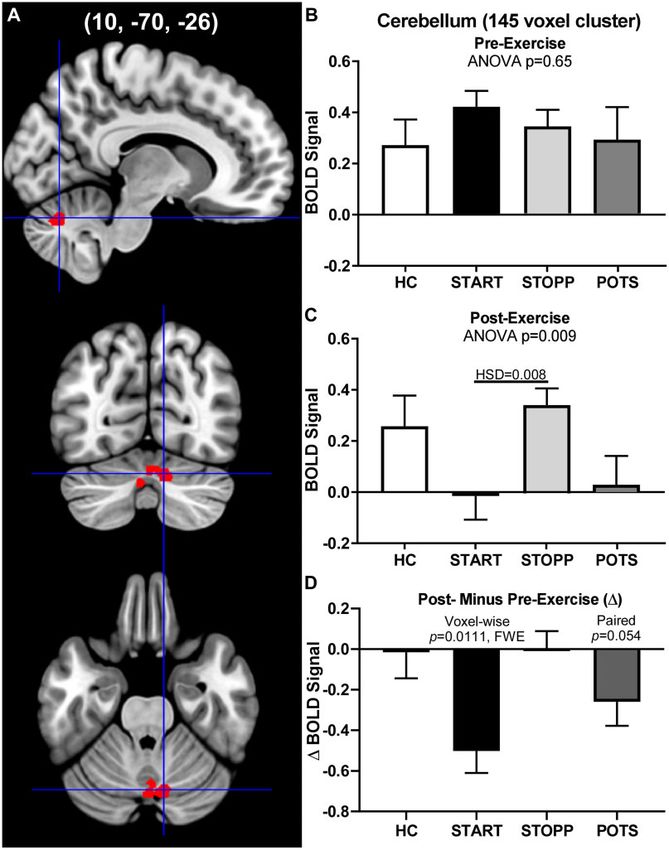
2   | BRAIN COMMUNICATIONS 2020: Page 2 of 14                                                                    S. D. Washington et al.

Abbreviations: AAL = automated anatomical labelling atlas; BOLD = blood oxygenation level dependent; bpm = beats per mi-
nute; fMRI = functional magnetic resonance imaging; FoV = field of view; EPI = echo-planar imaging; FWE = family-wise error cor-
rection; GWI = Gulf War illness; GWI-START = Gulf War Illness with stress test associated reversible tachycardia; GWI-STOPP =
Gulf War Illness with stress test originated phantom perception; GWI-POTS = Gulf War Illness with postural orthostatic tachycar-
dia syndrome; HC = healthy control; HR = heart rate; HSD = Tukey’s honest significant difference; MNI = Montreal Neurological
Institute; ROI = region of interest; SD = standard deviation; SUIT = spatially unbiased infra-tentorial template atlas; T1 = longitu-
dinal relaxation time; TE = echo time; TI = inversion time; TR = repetition time

Graphical Abstract

                                                                                                                                           Downloaded from https://academic.oup.com/braincomms/article-abstract/2/1/fcz039/5673581 by guest on 08 February 2020
Introduction                                                         Working memory tasks activate frontal-parietal executive
                                                                     control networks. We employed a continuous, verbal ver-
Gulf War Illness (GWI) affects approximately a quarter               sion of the N-Back in which subjects performed the sim-
of veterans from the 1990–91 Persian Gulf War (Fukuda                ple 0-Back stimulus matching task (‘see the letter, push
et al., 1998; Steele, 2000; Research Advisory Committee              the button’), and the high cognitive load 2-Back task that
on Gulf War Veterans’ Illnesses U.S. Department of                   involves viewing a series of nine letters at 2 s intervals,
Veterans Affairs, 2008; Steele et al., 2012). Symptoms in-           remembering their order, then pressing buttons corre-
clude debilitating fatigue, chronic widespread pain,                 sponding to the letter seen two places (4 s) previously.
gastrointestinal complaints and cognitive impairment.                Regional brain activation was determined by contrasting
Diagnosis is based on subjective symptoms of fatigue,                the blood oxygenation level-dependent (BOLD) signals of
mood/cognition and bodily pain using the 1998 Center                 the whole brain between the 2-Back and 0-Back trials in
for Disease Control criteria (Fukuda et al., 1998), and              order to identify regions that were differentially activated
moderate to severe complaints in at least three out of               in this 2-Back > 0-Back condition. Preliminary qualitative
these six categories: fatigue/sleep, neurological/mood/cog-          analysis found that when all GWI subjects were com-
nition, pain, gastrointestinal, respiratory and skin symp-           bined into a single group, there were no differences in
tom domains of the 2000 Kansas criteria (Steele, 2000).              BOLD between GWI and control groups, or between pre-
Although GWI has been attributed to somatoform, de-                  exertion and post-exertion MRI studies. However, two
pressive and other psychiatric causes (Fiedler et al., 2006;         patterns of activation and exercise-induced BOLD altera-
Dursa et al., 2016; Janulewicz et al., 2017), evidence is            tions were found when GWI subjects were stratified by
accumulating for chronic neurotoxic pathologies that                 exercise-induced    changes    in    postural    orthostatic
were induced by exposures during the Gulf War (White                 tachycardia.
et al., 2016; Georgopoulos et al., 2017). An important                  In that study, because orthostatic intolerance com-
clinical finding in GWI is post-exertional malaise with se-          plaints are common in GWI veterans, we assessed pos-
vere symptom exacerbations following physical, cognitive,            tural changes in heart rate (HR) before exercise and the
emotional or other effort (Steele, 2000).                            time course of potential alterations after exercise. Before
   We previously studied the physical and cognitive                  exercise, GWI veterans had normal elevation of HRs be-
domains of post-exertional malaise by having subjects                tween supine and standing [DHR ¼ 12 6 5 beats per min
perform two submaximal bicycle exercise stress tests on              (bpm), mean6SD]. However, after the first stress test,
consecutive days with functional magnetic resonance                  one-third developed postural tachycardia with DHR
imaging (fMRI) before the first and after the second exer-           exceeding 30 bpm at least twice during their 5 min peri-
cise (Rayhan et al., 2013). We evaluated cognitive dys-              ods of standing. This exaggerated orthostatic effect was
function using the N-Back verbal working memory task                 transient as DHR returned to normal within 24 to 48 h
during fMRI data acquisition (Owen et al., 2005).                    after exercise (Rayhan et al., 2013; Garner et al., 2018).
Exercise alters neural activity in GWI                                    BRAIN COMMUNICATIONS 2020: Page 3 of 14    | 3

The phenomenon was termed Stress Test Activated                Research Unit of the Georgetown–Howard Universities
Reversible Tachycardia (GWI-START). START was dis-             Center for Clinical and Translational Science. From the
tinct from Postural Orthostatic Tachycardia Syndrome           original study, we included the data of 10 control (5
(GWI-POTS), which is defined by persistent postural            deployed Gulf War-era veterans), 10 GWI-START, 18
tachycardia with DHR 30 bpm during virtually all peri-        GWI-STOPP and no POTS subjects (Rayhan et al.,
ods of standing (Sheldon et al., 2015). The other two-         2013). The additional recruits included 21 control (3
thirds of GWI subjects did not develop postural tachycar-      deployed veterans), 13 GWI-START, 28 GWI-STOPP and
dia at any time and were termed the Stress Test                11 GWI-POTS subjects (Garner et al., 2018). Subjects
Originated Phantom Perception (GWI-STOPP) group be-            had history and physical examinations to ensure their in-
cause their perceptions of pain were reminiscent of phan-      clusion by meeting Chronic Multisymptom Illness

                                                                                                                            Downloaded from https://academic.oup.com/braincomms/article-abstract/2/1/fcz039/5673581 by guest on 08 February 2020
tom limb pain (Romero-Romo et al., 2010; De Ridder             (Fukuda et al., 1998) and Kansas (Steele, 2000) criteria
et al., 2014).                                                 for GWI, confirmation of sedentary lifestyle for control
   Qualitative analysis of BOLD outcomes for the 2-Back        subjects ( 0-Back condition suggested differences in patterns of        clusion because of medical or psychiatric conditions
brain activation between the GWI-START and GWI-                (Steele, 2000; Reeves et al., 2003; Jones et al., 2009).
STOPP groups and generated the hypothesis that exertion        Subjects completed Chronic Fatigue Syndrome Symptom
induced distinct pathological mechanisms of cognitive          Severity (Baraniuk et al., 2013), SF-36 quality of life
dysfunction in each subgroup (Rayhan et al., 2013).            (Ware and Sherbourne, 1992), Chalder Fatigue (Cella
Here, this hypothesis was tested by recruiting a second
                                                               and Chalder, 2010) and McGill Pain (Dworkin et al.,
wave of healthy control (HC) and GWI subjects to per-
                                                               2009) questionnaires and were examined for systemic
form the identical exercise-MRI protocol. Quantitative
                                                               hyperalgesia by dolorimetry (Naranch et al., 2002).
analysis used paired t-tests to identify regions of interest
(ROIs) with significantly different pre- and post-exertion
BOLD responses for any of the HC, GWI-START, GWI-              Protocols
STOPP or GWI-POTS subgroups. This approach identi-             Exercise
fied ROIs in each subgroup that were either recruited for
                                                               Two submaximal bicycle exercise tests were performed
cognitive compensation (significantly increased BOLD ac-
                                                               24 h apart. Subjects cycled for 25 min at 70% predicted
tivation after exercise) or had reduced activation indicat-
                                                               maximum HR (220-patient’s age), followed by a climb to
ing exercise-induced cognitive dysfunction and exertional
                                                               85% maximum HR to reach anaerobic threshold (Garner
exhaustion. Each ROI was tested in post hoc fashion in
                                                               et al., 2018).
all subgroups for differences between pre-exercise and
post-exercise BOLD response. The pre-exercise data deter-
                                                               Postural tachycardia protocol and exercise-induced
mined if there were significant pre-existing differences in
                                                               postural tachycardia subgroups
BOLD activation between groups. The post-exercise com-
                                                               All subjects were assessed for postural tachycardia before
parison determined if exercise caused significant differen-
                                                               and after exercise (Garner et al., 2018). Subjects rested
ces in BOLD activation between subgroups.
                                                               supine for 5 min and had HR measured by continuous
                                                               ECG monitor. After standing up, HR and blood pressure
Methods and materials                                          were measured every minute for 5 min. The incremental
                                                               changes in HR after standing up (DHR) were calculated
Ethics                                                         to identify episodes of postural tachycardia defined as
                                                               DHR  30 bpm. The normal DHR was 12 6 5 bpm
The protocol was approved by the Georgetown
                                                               (mean 6 SD).
University Institutional Review Board (IRB 2009-229,
                                                                 POTS was defined by DHR 30 bpm at two or more
2013-0943 and 2015-0579) and Department of Defense
                                                               measurements prior to exercise during the 5-min standing
Congressionally Directed Medical Research Program
                                                               periods. POTS criteria were met before and after exercise.
(CDMRP) Human Research Program Office (HRPO) (A-
                                                               Exercise did not exacerbate the postural tachycardia, so
15547 and A-18479), and listed in clinicaltrials.gov
                                                               POTS subjects were distinct from the START and STOPP
(NCT01291758 and NCT00810225). All clinical investi-
                                                               groups.
gations were conducted according to the principles
                                                                 START was defined by having a normal DHR before
expressed in the Declaration of Helsinki.
                                                               exercise, but at least two measurements of postural tachy-
                                                               cardia with DHR 30 bpm after exercise. This phenom-
Subjects                                                       enon was transient as DHR returned to normal 24–48 h
Veterans of the 1990–91 Persian Gulf War with GWI,             following exercise (Garner et al., 2018).
and healthy, non-military control subjects were recruited        STOPP was defined by normal DHR (DHR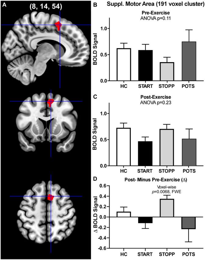
4   | BRAIN COMMUNICATIONS 2020: Page 4 of 14                                                            S. D. Washington et al.

Verbal working memory task                                     ‘scrubbing’ based on artefact detection tools, (iv) segmen-
Each subject performed the continuous 2-Back verbal            tation and spatial normalization of the anatomic scan to
working memory task during fMRI data acquisition               the Montreal Neurological Institute (MNI) template and
(Fig. 1). Each 18-s stimulus block had instruction (REST,      (v) spatial smoothing with a stationary Gaussian filter of
0-BACK, 2-BACK, 2 s each), fixation (i.e. cross-hairs,         6 mm full width at half maximum. Voxel size was 2.0
8 s), 0-Back and 2-Back components. For the 0-Back and         mm3 (isotropic) after spatial normalization. Pre-processed
2-Back tasks, subjects viewed nine individual, pseudo-ran-     EPI data from each individual were modelled with the
domly selected upper-case letters (A, B, C, D) for 0.8 s       following events: instruction, fixation, 0-Back and 2-
each followed by an inter-stimulus interval (blank screen)     Back. The contrast of interest, 2-Back > 0-Back, was
of 1.2 s. For the 0-Back task, they pressed a button corre-    modelled using a one-sample t-test with motion parame-
                                                               ters (i.e. translation and rotation) included as covariates

                                                                                                                                   Downloaded from https://academic.oup.com/braincomms/article-abstract/2/1/fcz039/5673581 by guest on 08 February 2020
sponding to the letter viewed. For the 2-Back task, they
remembered the first two letters, then began to press the      of no interest. The resulting 2-Back > 0-Back contrast
button corresponding to the letter seen two presentations      maps from every subject were then used in second-level
(4 s) previous. These blocks were repeated for five cycles.    analyses.
Responses were collected from an MRI-compatible fibre-
                                                               Second level
optic button box that subjects held with both hands.
Response times between onset of each letter presentation       To identify regions where BOLD response changes related
and button pressing were recorded. There were 45 button        to working memory were significantly altered by exertion,
responses for the 0-Back and 35 for the 2-Back tasks.          for each subgroup we performed two-tailed, paired t-tests
The complete task (i.e. all five cycles) lasted a total of 5   for 2-Back > 0-Back and 2-Back < 0-Back contrasts
min (300 s).                                                   [cluster level: P < 0.05, family-wise error correction
                                                               (FWE), kE50]. Each ROI was then applied to all sub-
                                                               groups. The mean BOLD response within each significant
MRI data acquisition                                           ROI was extracted from the contrast maps of all subjects
All structural and functional MRI data were acquired on        using MarsBaR 0.44 (https://www.fil.ion.ucl.ac.uk/spm/
a Siemens 3T Tim Trio scanner located within the Center        ext/#MarsBar; Brett et al., 2002). MarsBaR output was
for Functional and Molecular Imaging at Georgetown             compared between the HC, GWI-START, GWI-STOPP
University Medical Center equipped using a transmit-re-        and GWI-POTS groups on the pre- and post-exercise
ceive body coil and a commercial 12-element head coil          days by one-way ANOVA (P < 0.05) followed by post
array. Structural 3D T1-weighted Magnetization Prepared        hoc Tukey’s Honest Significant Difference (HSD) to cor-
Rapid Acquisition Gradient Echo images parameters              rect for multiple comparisons. Exercise effects within
were: TR/TE ¼ 1900/2.52 ms, flip angle ¼ 9 , TI ¼ 900 ms,     each group were determined from 2-tailed paired t-tests
FoV ¼ 250 mm, 176 slices, slice resolution ¼ 1.0 mm and        between pre- and post-exercise data.
voxel size ¼ 1  1  1 mm. Magnetization Prepared Rapid           To minimize the effects of ‘double-dipping’ from the
Acquisition Gradient Echos were all processed in SPM12         use of the same data set for ROI selection (Kriegeskorte
(http://www.fil.ion.ucl.ac.uk/spm/software/spm12/; Friston     et al., 2009; Button, 2019), significant HSDs between
et al., 1996). fMRI data consisted of T2*-weighted gradi-      contrast groups that yielded ROIs were disregarded and
ent echo-planar images (EPIs) acquired during the 5-min        FWE cluster-level significance was reported instead. Effect
task. EPI data acquisition parameters were: TR/                sizes for changes between the pre- and post-exertional
TE ¼ 2500/30 ms, flip angle ¼ 90 , FoV ¼ 205 mm2, ma-         fMRI sessions were estimated by Cohen’s d (paired stud-
trix size ¼ 64  64, number of slices ¼ 47 and voxel           ies) (Cohen, 1988, 1992; Ellis, 2010) and between groups
size ¼ 3.2 mm3 (isotropic).                                    on pre- and post-exercise days by Hedges’ g (Stangroom,
                                                               2019). In brief, effect sizes larger than 0.8 indicate strong
                                                               studies and allow new studies of appropriate numbers of
fMRI statistical analysis                                      subjects to reproduce the effects in significant fashion
Both the first-level (fixed effects) and second-level (ran-    (P < 0.05) with adequate power (>0.8) and so control
dom effects) statistical analyses were performed in            false-positive and false-negative rates. These analyses were
SPM12.                                                         performed in SPSS (version 25).
                                                                  The precise location and ‘regional compositional per-
First level                                                    centage’ (e.g. 10% anterior cingulate, 13% medial frontal
The first six scans from each block were removed to ac-        gyrus, etc.) of each ROI in cortical, subcortical and cere-
count for T1 saturation (Fig. 1). Data were pre-processed      bellar structures were found using a custom
through the default pipeline within the CONN version           MATLAB script that employed functions from both SPM
17 toolbox (https://web.conn-toolbox.org/; Whitefield-         and     xjView    9.6     (http://www.alivelearn.net/xjview/).
Gabrieli and Nieto-Castanon, 2012). In brief, steps were:      Anatomical labels for every ROI voxel were identified
(i) slice-timing correction, (ii) realignment and unwarping    using the AAL (Automated Anatomic Labelling) atlas.
of the functional images, (iii) outlier detection for          Cerebellar regions were confirmed with the spatially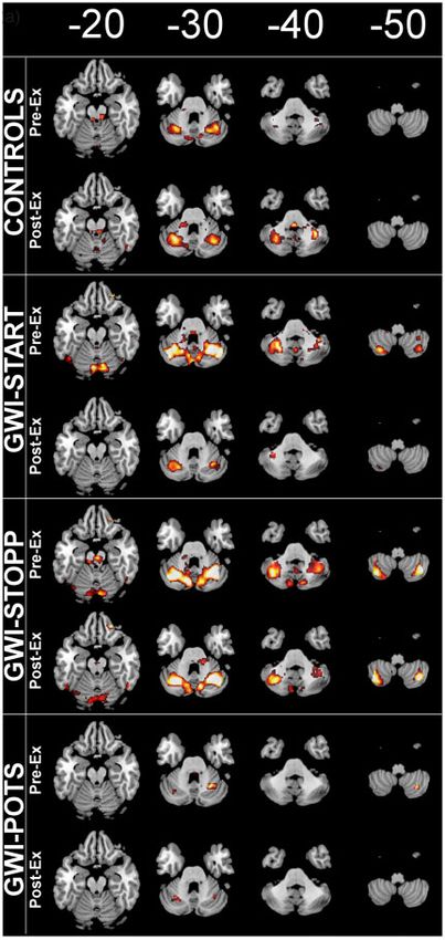
Exercise alters neural activity in GWI                                                        BRAIN COMMUNICATIONS 2020: Page 5 of 14               | 5

                                                                                                                                                           Downloaded from https://academic.oup.com/braincomms/article-abstract/2/1/fcz039/5673581 by guest on 08 February 2020
 Figure 1 Continuous N-Back paradigm. (Top) One complete 60-s cycle of the N-Back task consisted for 0-Back and 2-Back task blocks (18
 s each) separated by an instruction block advising subjects to REST (2 s) followed by 8 s of fixation (i.e. cross-hairs). For the 0-Back task, subjects
 pressed a button corresponding to the letter they viewed. For the 2-Back task, subjects had to remember the string of letters and press the
 button corresponding to the letter seen two presentations (4 s) earlier. (Bottom) The numbers in circles indicate the five complete cycles of
 fixation, 0-back and 2-back tasks in the stimulus paradigm. The task lasted 5 min (300 s).

unbiased     infra-tentorial  template     (SUIT)                  atlas       groups had equivalent results that, when combined,
(Diedrichsen, 2006; Diedrichsen et al., 2009).                                 were significantly different from HC (n ¼ 31) for CFS
                                                                               Symptom Severity Questionnaire, pain (McGill Total
Data availability                                                              Pain Score) and tenderness (dolorimetry), fatigue
                                                                               (Chalder Fatigue Score) and quality of life (SF-36;
Data are available from the authors upon request and
                                                                               ANOVA P < 0.004; HSD P < 0.05). All three GWI sub-
will be uploaded to OpenfMRI and/or the Boston
                                                                               groups had significantly greater pain and tenderness
Biorepository, Recruitment, and Integrative Network
                                                                               than HC.
(BBRAIN) for GWI.

Results                                                                        N-Back task performance
                                                                               There were no significant differences between groups for
Demographics                                                                   response times or numbers of correct responses on the 0-
Age, percentage of males and body mass index were not                          Back or 2-Back tasks either before or after exercise
different between groups (Table 1). GWI-START                                  (Fig. 2). Response times were faster for the 2-Back than
(n ¼ 23), GWI-STOPP (n ¼ 46) and GWI-POTS (n ¼ 11)                             0-Back task.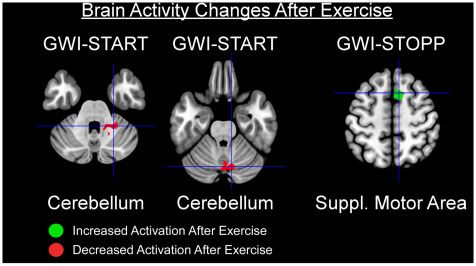
6 | BRAIN COMMUNICATIONS 2020: Page 6 of 14 S. D. Washington et al.
Table 1 Demographics
HC GWI-START GWI-STOPP GWI-POTS
N 31 23 46 11
Age 43.9 6 15.9 45.1 6 8.1 48.6 6 7.4 45.7 6 6.0
Male 0.645 6 0.486 0.818 6 0.395 0.756 6 0.435 0.818 6 0.405
Body mass index 28.7 6 4.5 27.9 6 4.3 29.8 6 5.9 30.4 6 6.1
McGill total pain score 4.2 6 7.0a 26.8 6 8.6 23.2 6 8.7 23.0 6 10.8
Dolorimetry (kg) 6.0 6 1.9a 3.3 6 2.1 3.6 6 1.9 4.0 6 2.3
Chalder Fatigue Score 12.4 6 5.4a 26.1 6 4.4 25.6 6 4.7 24.8 6 5.3
CFS Symptom Severity Questionnaire
Fatigue 1.4 6 1.3a 3.8 6 0.4 3.4 6 0.8 3.4 6 0.7
Downloaded from https://academic.oup.com/braincomms/article-abstract/2/1/fcz039/5673581 by guest on 08 February 2020
Short term memory and concentration 1.3 6 1.3a 3.2 6 0.9 3.2 6 0.8 2.9 6 0.7
Sore throat 0.3 6 0.7a 1.9 6 1.2 1.3 6 1.2 1.3 6 1.2
Sore lymph nodes 0.1 6 0.4a 2.0 6 1.3 1.3 6 1.2 1.6 6 1.2
Muscle pain 0.8 6 1.1a 3.4 6 0.6 3.1 6 1.0 3.0 6 1.2
Joint pain 0.9 6 1.2a 3.5 6 0.9 3.2 6 0.9 2.8 6 1.1
Headaches 1.0 6 1.2a 2.9 6 1.1 2.6 6 1.2 3.0 6 1.1
Sleep 1.8 6 1.4a 3.7 6 0.5 3.5 6 0.8 3.2 6 1.0
Exertional exhaustion 0.7 6 1.2a 3.8 6 0.5 3.2 6 1.0 3.4 6 1.0
SF-36
Physical functioning 83.8 6 24.4a 42.4 6 25.7 47.0 6 22.6 45.5 6 22.7
Role physical 76.7 6 39.4a 6.0 6 22.2 11.3 6 25.3 0.0 6 0.0
Bodily pain 80.9 6 21.2a 22.8 6 16.5 28.9 6 18.4 34.1 6 15.0
General health 67.6 6 23.3a 20.7 6 15.5 29.0 6 20.3 23.4 6 16.4
Vitality 58.0 6 23.0a 13.3 6 11.0 17.9 6 17.1 20.5 6 13.9
Social functioning 77.1 6 27.7a 22.6 6 17.9 33.1 6 25.4 33.0 6 18.8
Role emotional 84.4 6 32.4a 25.4 6 36.4 30.8 6 39.5 30.3 6 40.7
Mental health 73.9 6 16.7a 49.7 6 21.5 55.5 6 22.3 54.5 6 23.7
a
ANOVA with P < 0.05 followed by P < 0.05 by HSD for HC compared with the combined GWI subgroups.
Neuroimaging P ¼ 0.0028, FWE; Fig 5C). Post hoc analyses examined
In cortex, all groups activated the frontal-parietal execu- differences between subgroups on each day. Prior to exer-
tive control network before and after exercise (Fig. 3). cise, the HC, GWI-START, GWI-STOPP and GWI-POTS
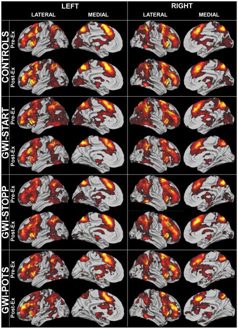
Likewise, all groups activated several regions within the groups had equivalent activation levels [ANOVA: F(3,
cerebellum before and after exercise, but a marked de- 110) ¼ 0.343, P ¼ 0.80]. After exercise, levels were sig-
cline in cerebellar activity is evident after exercise in the nificantly different [ANOVA: F(3, 110) ¼ 5.64,
GWI-START group (Fig. 4). As the GWI-POTS group P ¼ 0.001], and GWI-START had significant deactivation
had fewer subjects relative to the other groups, we kept relative to HC (HSD, P ¼ 0.001) and GWI-STOPP (HSD,
height thresholds in Figs 3 and 4 at P < 0.001 (uncorrect- P ¼ 0.041). Paired comparisons of pre- versus post-exer-
ed) for visualization purposes. tional activity revealed a trend towards deactivation in
ROI analysis identified three clusters that were signifi- GWI-POTS (paired t-test, P ¼ 0.051; Fig. 5D). The effects
cantly altered (P < 0.05, FWE) in response to the exercise of exercise were large with Cohen’s d of 1.7 for GWI-
stressor in the GWI-START and GWI-STOPP subgroups. START and 0.89 for GWI-POTS. Effect sizes were small
Contrasting pre-exercise > post-exercise in the GWI- for GWI-STOPP (d ¼ 0.28) and HC (d ¼ 0.02). Hedges’ g
START group resulted in two significant cerebellar clus- indicated strong effect sizes to detect differences between
ters. Contrasting post-exercise > pre-exercise in the GWI- GWI-START and the post-exercise HC (g ¼ 1.16), GWI-
STOPP group resulted in one significant cortical STOPP (g ¼ 0.92) and GWI-POTS (g ¼ 0.73) results
cluster. Reversing either of these contrasts (GWI-START: (Stangroom, 2019).
post-exercise > pre-exercise; GWI-STOPP: pre-exercise > The second cerebellar ROI (145 voxels) was more
post-exercise) yielded no significant clusters. medioposterior and included vermis lobules VII (51/145
The larger of the two cerebellar ROIs (186 voxels) was voxels, 35%) and VI (30/145, 21%), and right cerebellar
in the right hemisphere and included voxels in the right lobule VI (27/145, 19%; Fig. 6A). Activation in this se-
cerebellar lobule VI (43/186 voxels, 23%), dentate nu- cond cerebellar ROI closely paralleled that of the first,
cleus (43/186, 23%), lobule IV (7/186, 4%) and lobule with equivalent activity between groups prior to exercise
III (5/186, 3%; Fig. 5A). The remaining voxels (88/186) (Fig. 6B) and deactivation after exercise in the GWI-
were undefined in the AAL and SUIT atlases. Activation START group (cluster level: P ¼ 0.011, FWE) with a simi-
in this ROI was equivalent between groups prior to exer- lar trend in GWI-POTS (P ¼ 0.054; Fig. 6C). The
cise (Fig. 5B). This ROI resulted from deactivation after responses before exercise were similar [ANOVA: F(3,
exercise in the GWI-START group (cluster level: 110) ¼ 0.648, P ¼ 0.65]. The responses after exerciseExercise alters neural activity in GWI BRAIN COMMUNICATIONS 2020: Page 7 of 14 | 7
Downloaded from https://academic.oup.com/braincomms/article-abstract/2/1/fcz039/5673581 by guest on 08 February 2020
Figure 2 Response times and accuracies. Responses times for 0-Back (A) and 2-Back (B) and the total number of correct responses,
respectively (C and D), were not different between groups or pre-exertion (white bars) and post-exertion (grey bars) periods. Mean 6 SEM.
were significantly different from each other [ANOVA: exercise (cluster-level P ¼ 0.0068, FWE; Cohen’s d ¼ 0.55).
F(3, 110) ¼ 5.21, P ¼ 0.0087], with negative BOLD sig- The 191-voxel ROI mapped to the anterior right supple-
nal in GWI-START compared with GWI-STOPP (HSD, mentary motor area (163/191, 85%). MNI co-ordinates
P ¼ 0.008). Thus, these results like those for the larger for this third ROI overlap with the dorsal anterior cingu-
cerebellar ROI reflect a significant relative decrease in late cortex regions of the anterior salience network as
GWI-START activity, not an increase in HC and GWI- described by Shirer et al. (2012). Activation in this ROI
STOPP activity, after exercise. was equivalent between groups before and after exercise.
One GWI-START subject was an outlier for the cere- HC, GWI-START, GWI-STOPP and GWI-POTS groups
bellar ROIs following exercise. Pre-exercise values were had similar activation levels before [ANOVA: F(3, 110) ¼
comparable with all other subjects. However, after exer- 2.08, P ¼ 0.11] and after [ANOVA: F(3, 110) ¼ 1.47,
cise, the BOLD response increased to more than two P ¼ 0.23] exercise. GWI-STOPP was predicted to have
standard deviations greater than the mean for the other higher BOLD responses following exercise provocations in
GWI-START group members, reaching 1.26 and 0.61 for future studies when compared with GWI-START (Hedges’
the 145- and 186-voxel ROIs, respectively. This was a g ¼ 1.01) and GWI-POTS (g ¼ 0.73).
focal finding because all of this subject’s other BOLD Table 2 contains regional comparisons between the
responses were comparable with the other GWI-START AAL and SUIT atlases.
subjects. No abnormality was seen on structural scans.
The subject’s data were excluded from the analysis of the
cerebellar clusters but were used for all other analyses.
Discussion
The third ROI (Fig. 7) was characterized by a significant Exercise caused alterations of regional brain blood
increase in activation in the GWI-STOPP group following flow measured by BOLD activity during the continuous8 | BRAIN COMMUNICATIONS 2020: Page 8 of 14 S. D. Washington et al.
Downloaded from https://academic.oup.com/braincomms/article-abstract/2/1/fcz039/5673581 by guest on 08 February 2020
Figure 3 Cortical BOLD activation for 2-Back > 0-Back.
Frontal parietal executive control network regions were activated
in the control and GWI START, STOPP and POTS groups during the
pre-exertion and post-exertion scans.
N-back working memory task (2-Back > 0-Back condi-
tion), and autonomic changes leading to the transient
postural tachycardia of the START group. Before exer-
cise, BOLD responses were positive and equivalent
Figure 4 Cerebellar BOLD activation for 2-Back > 0-Back.
among the four groups. Exercise had no effect on BOLD
Brain slices at the 20, 30, 40 and 50 MNI axial planes.
responses in HC subjects. Cerebellar regions were activated in the Control, GWI-START,
GWI-START had significant deactivation of right cere- GWI-STOPP and GWI-POTS groups during the pre-exertion and
bellar dentate nucleus and vermis lobules VI and VII post-exertion scans.
(Figs 5 and 6). GWI-POTS had a trend for deactivation
in the cerebellar ROIs after exercise (P 0.054 by paired
t-tests, Fig. 5) that was limited by the small sample size
(n ¼ 11, Cohen’s d ¼ 0.89). The cerebellar findings (Figs 5 et al., 2009; Stoodley and Schmahmann, 2009; Kirschen
and 6) confirmed previous studies showing activation of et al., 2010; Cooper et al., 2012; Stoodley et al., 2012;
cerebellar lobules VI, VII (Stoodley et al., 2012), crus I Thurling et al., 2012). Other roles include sensorimotor
and dentate nucleus (Thurling et al., 2012) with various control (Sokolov et al., 2017), reward circuits, social be-
other N-Back task designs. Increasing task difficulty and haviour and dysfunction in autism spectrum disorder,
working memory cognitive load recruit additional areas schizophrenia and addiction (Carta et al., 2019).
in the dentate nucleus and vermis VI and VII for cogni- GWI-STOPP had a gain of function response after ex-
tive compensation (Kuper et al., 2016). Roles for cerebel- ercise with a significant increase in BOLD response in the
lar dentate and vermis lobules VI and VII in verbal right anterior supplementary motor area (Fig. 7). The
working memory are well attested from numerous brain MNI co-ordinates for this right anterior supplementary
lesion and neuroimaging studies (Desmond et al., 1997; motor area ROI overlap with one atlas’s dorsal anterior
Chen and Desmond, 2005; Kirschen et al., 2005; Hautzel cingulate cortex subdivision of the anterior salienceExercise alters neural activity in GWI BRAIN COMMUNICATIONS 2020: Page 9 of 14 | 9
Downloaded from https://academic.oup.com/braincomms/article-abstract/2/1/fcz039/5673581 by guest on 08 February 2020
Figure 5 Right cerebellar cluster. The 186-voxel ROI was identified from the exercise-induced decrease in BOLD activation in the GWI-
START subgroup (cluster-level P ¼ 0.0028, FWE). (A) Sagittal (top), coronal (middle) and transverse (bottom) slices of an MNI-standard brain,
where cross-hairs indicate the cluster’s most active voxel (24, 46, 32). BOLD response for the 2-Back > 0-Back condition (mean 6 SEM) are
shown for (B) pre-exercise and (C) post-exercise changes in BOLD responses for the control (white bars), GWI-START (black bars), GWI-
STOPP (light grey bars) and GWI-POTS (dark grey bars). The ANOVA P-values refer to the difference between the HC and the combined three
GWI subgroups. (D) Post-minus pre-exercise BOLD response for the 2-Back > 0-Back condition for the HC, GWI-START, GWI-STOPP and
GWI-POTS groups.
network (Shirer et al., 2012). This ROI is also immedi- Coombes, 2015). The latter study found functional con-
ately adjacent to ‘pre-supplementary motor area’ and nectivity between the supplementary motor area and cere-
‘midcingulate’ regions that are activated during pain in bellar lobules VI and VII indicating simultaneous
HC children (Hohmeister et al., 2010) and by thermal multimodal processing of motor control and pain (Misra
pain during a motor task in young adults (Misra and and Coombes, 2015; Coombes and Misra, 2016).10 | BRAIN COMMUNICATIONS 2020: Page 10 of 14 S. D. Washington et al.
Downloaded from https://academic.oup.com/braincomms/article-abstract/2/1/fcz039/5673581 by guest on 08 February 2020
Figure 6 Medial cerebellar cluster. The 145-voxel ROI was identified from the exercise-induced incremental BOLD
deactivation in the GWI-START subgroup (cluster-level P 5 0.011, FWE). (A) Sagittal (top), coronal (middle) and transverse (bottom)
slices of an MNI-standard brain, where cross-hairs indicate the cluster’s most active voxel (10, 70, 26). BOLD response for the 2-Back > 0-
Back condition (mean 6 SEM) are shown for (B) pre-exercise and (C) post-exercise changes in BOLD responses for the control (white bars),
GWI-STOPP (light grey bars), GWI-POTS (dark grey bars) and GWI-START (black bars). The ANOVA P-values refer to the difference between
the HC and the combined three GWI subgroups. (D) Post-minus pre-exercise BOLD response for the 2-Back > 0-Back condition for the HC,
GWI-START, GWI-STOPP and GWI-POTS groups.
Limitations analysis (Rayhan et al., 2013). The exercise was required
The quantitative ROI analysis verified deactivation in the to unveil the transient postural tachycardia in GWI-
cerebellar vermis in the GWI-START group after exertion START and patterns of BOLD activation in GWI subsets,
but did not substantiate the qualitative impression of indicating that resting state or baseline studies may not
basal ganglia and anterior insula activation in the GWI- distinguish between GWI-START, GWI-STOPP and con-
STOPP group that was suggested by the earlier interim trol groups. GWI-POTS subjects had cerebellarExercise alters neural activity in GWI BRAIN COMMUNICATIONS 2020: Page 11 of 14 | 11
Downloaded from https://academic.oup.com/braincomms/article-abstract/2/1/fcz039/5673581 by guest on 08 February 2020
Figure 7. Right Supplementary Motor Area Cluster. The 191-voxel ROI was identified from the exercise-induced increased
BOLD activation in the GWI-STOPP subgroup (cluster-level: P 5 0.0068, FWE). (A) Sagittal (top), coronal (middle) and transverse
(bottom) slices of an MNI-standard brain, where cross-hairs indicate the cluster’s most active voxel (8, 14, 54). BOLD response for the 2-
Back > 0-Back condition (mean 6 SEM) are shown for (B) pre-exercise and (C) post-exercise changes in BOLD responses for the control
(white bars), GWI-START (black bars), GWI-POTS (dark grey bars) and GWI-STOPP (light grey bars). The ANOVA P-values refer to the
difference between the HC and the combined three GWI subgroups. (D) Post-minus pre-exercise BOLD response for the 2-Back > 0-Back
condition for the HC, GWI-START, GWI-STOPP and GWI-POTS groups.
deactivation after exercise comparable to the GWI- were identical. All scans were analysed as a single batch
START group, but larger studies will be needed to exam- with extensive computational efforts to reduce the effects
ine the statistical significance. Effects sizes were provided of head motion. This was a particular problem for veterans
to help power future studies. with severe pain and substantial systemic hyperalgesia.
The study spanned two campaigns of recruitment and Our ‘continuous’ version of the N-Back task had the
testing, but the exercise, N-Back and analytical procedures unusual effect of eliciting faster response times from12 | BRAIN COMMUNICATIONS 2020: Page 12 of 14 S. D. Washington et al.
Table 2 Comparison of cerebellar ROI voxel co-ordin- two groups, and to determine if there were significant
ate labels in the SUIT and AAL atlases intergroup differences on the pre- and post-exercise days.
Cerebellar SUIT Voxel AAL Voxel
Furthermore, our ROI analyses employed only contiguous
ROI (Peak labels count labels count clusters, which are less susceptible to serious result distor-
Voxel) tion from selective analyses of selected data (Kriegeskorte
145-Voxel Left Left et al., 2009). Critics of circular analysis in systems neuro-
Cluster (10, Crus I 3 Lobule 8 4 science concede that selective in-depth analysis of ROIs
-70, -26) Lobule VI 3 Crus1 3 can provide additional insights to analyses employing
Vermis Crus II 6 Crus2 8
non-selective mapping (Saxe et al., 2006; Kriegeskorte
Vermis VI 6 Vermis 7 9
Vermis VIIb 4 et al., 2009). Lastly, as we have addressed the need for
Downloaded from https://academic.oup.com/braincomms/article-abstract/2/1/fcz039/5673581 by guest on 08 February 2020
Right Right larger studies, one can leverage larger subject pools to
Crus I 11 Lobule 6 27 address issues with double-dipping. Specifically, provided
Crus II 1 Lobule 8 1
a sufficiently large population, one can divide the popula-
Lobule VI 36 Crus1 13
Vermis Crus II 7 Crus2 4 tion into two contrast groups, one for ROI detection and
Vermis VI 64 Vermis 6 30 the other for quantitation within that ROI (Kriegeskorte
Vermis 7 42 et al., 2009).
Undefined 2 Undefined 4 The entire period of the two submaximal bicycle exer-
186-Voxel Left Left
Cluster (24, Lobule V 1 Vermis 4/5 1
cise stress test, postural tachycardia testing and overnight
-46, -32) Right Right stay in the Clinical Research Unit constituted the physio-
Dentate 7 Lobule 3 5 logical stressor. Our design did not determine if a single
Lobule I/IV 34 Lobule 4/5 43 exercise stress test was sufficient to alter BOLD
Lobule V 21 Vermis 1/2 4
responses. Regional cerebellar deactivation was inferred
Lobule VI 26 Vermis 4/5 26
Vermis 6 1 to indicate cognitive decompensation, while the enhanced
Undefined 97 Undefined 96 activation in the right anterior supplementary motor area
may indicate cognitive compensation by the GWI-STOPP
group.
subjects during the 2-Back condition than during the 0- Lastly, the data presented here are restricted to the
Back condition. We designed the continuous N-Back to BOLD responses elicited by verbal working memory (i.e.
be more challenging than the typical N-Back as the con- 2-Back > 0-Back). It is possible to differentiate veterans
tinuous version demands that subjects retain and update with GWI from HCs using other neural processes. For
a string of letters in working memory over the entirety of example, contrasting 0-Back > 2-Back revealed that the
the task block. We suggest that the shorter reaction time magnitude of deactivation in the default mode networks
in the 2-Back condition reflects the subjects’ desire to use of veterans with GWI is greater than that of HCs
information in memory (i.e. the letter from two presenta- (Rayhan et al., 2019). Thus, we plan future studies that
tions prior) and expunge it quickly, thereby reducing the will report the results of other contrasts in these data
amount of information (i.e. the number of letters in the (e.g. 0-Back > 2-Back, 2-Back > Fixation, etc.) so that we
string) needing to be stored. The continuous N-Back was can better differentiate autonomic subgroups of GWI
adequate for the purposes of this study, but its differen- from each other and HCs.
ces from other version of the N-Back raises concerns
about comparability to previous working memory studies.
However, as there are over 50 variants of the N-Back Conclusion
task targeting verbal working memory alone (Owen
et al., 2005), the continuous N-Back is simply one vari- The exercise stressor protocol caused distinct changes in
ation within a highly diverse field. the patterns of dysfunction in veterans with GWI. After
The discovery of the three ROIs was based on the hy- exercise, GWI-STOPP activated the right supplementary
pothesis that each subgroup would have incremental dif- motor area/dorsal anterior cingulate cortex while GWI-
ferences in brain activation induced by exercise. The START had cerebellar deactivation. GWI-POTS had a
incremental changes within groups and differences be- trend towards cerebellar deactivation similar to GWI-
tween groups after exercise had large effect sizes suggest- START. Thus, patterns of cerebellar and cortical somato-
ing the outcomes are likely to be reproduced in sensory activation could be used to quantitatively distin-
reasonably sized groups of subjects using the same exer- guish the GWI-START from the GWI-STOPP phenotype.
cise and cognitive testing paradigm. Testing for effect size These results suggest that the exercise stressor paradigm
in this way was not ‘double-dipping’ in two of the four affected different neural mechanisms in the GWI pheno-
groups because the ROIs that were discovered by intra- types and that distinct neural networks may mediate the
group cluster analysis were used as masks to test for the cognitive dysfunction, post-exertional malaise and exer-
consequences of exercise between the 2 days in the other tional exhaustion in these veterans.Exercise alters neural activity in GWI BRAIN COMMUNICATIONS 2020: Page 13 of 14 | 13
and finger-tapping tasks as revealed by functional MRI. J Neurosci
Acknowledgements 1997; 17: 9675–85.
Diedrichsen J. A spatially unbiased atlas template of the human cere-
We would like to acknowledge our participants and their bellum. Neuroimage 2006; 33: 127–38.
families, whose co-operation made this research possible. We Diedrichsen J, Balsters JH, Flavell J, Cussans E, Ramnani N. A prob-
would also like to acknowledge Drs. Adeen Flinker and Kyle abilistic MR atlas of the human cerebellum. Neuroimage 2009; 46:
39–46.
Shattuck for helpful advice regarding data analyses.
Dursa EK, Barth SK, Schneiderman AI, Bossarte RM. Physical and
mental health status of Gulf War and Gulf Era veterans: results
from a large population-based epidemiological study. J Occup
Funding Environ Med 2016; 58: 41–6.
Dworkin RH, Turk DC, Revicki DA, Harding G, Coyne KS, Peirce-
The study was supported by funding from the Sergeant Sandner S, et al. Development and initial validation of an expanded
Downloaded from https://academic.oup.com/braincomms/article-abstract/2/1/fcz039/5673581 by guest on 08 February 2020
and revised version of the Short-form McGill Pain Questionnaire
Sullivan Circle, Dr. Barbara Cottone, Dean Clarke Bridge
(SF-MPQ-2). Pain 2009; 144: 35–42.
Prize, Department of Defense Congressionally Directed Ellis PD. The essential guide to effect sizes: statistical power, meta-ana-
Medical Research Program (CDMRP) W81XWH-15-1-0679 lysis, and the interpretation of research results. Cambridge and New
and W81-XWH-09-1-0526, and the National Institute of York: Cambridge University Press; 2010.
Neurological Disorders and Stroke R21NS088138 and Fiedler N, Ozakinci G, Hallman W, Wartenberg D, Brewer NT,
Barrett DH, et al. Military deployment to the Gulf War as a risk fac-
RO1NS085131. This project has been funded in whole or in tor for psychiatric illness among US troops. Br J Psychiatry 2006;
part with Federal funds (Grant #UL1TR000101 previously 188: 453–9.
UL1RR031975) from the National Center for Advancing Friston KJ, Williams S, Howard R, Frackowiak RS, Turner R.
Translational Sciences (NCATS), National Institutes of Movement-related effects in fMRI time-series. Magn Reson Med
1996; 35: 346–55.
Health (NIH), through the Clinical and Translational Fukuda K, Nisenbaum R, Stewart G, Thompson WW, Robin L,
Science Awards Program (CTSA), a trademark of DHHS, Washko RM, et al. Chronic multisymptom illness affecting Air
part of the Roadmap Initiative, ‘Re-Engineering the Clinical Force veterans of the Gulf War. JAMA 1998; 280: 981–8.
Research Enterprise’. Garner RS, Rayhan RU, Baraniuk JN. Verification of exercise-induced
transient postural tachycardia phenotype in Gulf War Illness. Am J
Transl Res 2018; 10: 3254–64.
Georgopoulos AP, James LM, Carpenter AF, Engdahl BE, Leuthold
Competing interests AC, Lewis SM. Gulf War illness (GWI) as a neuroimmune disease.
Exp Brain Res 2017; 235: 3217–25.
The authors report no competing interests. Hautzel H, Mottaghy FM, Specht K, Muller HW, Krause BJ. Evidence
of a modality-dependent role of the cerebellum in working memory?
An fMRI study comparing verbal and abstract n-back tasks.
Neuroimage 2009; 47: 2073–82.
References Hohmeister J, Kroll A, Wollgarten-Hadamek I, Zohsel K, Demirakca
S, Flor H, et al. Cerebral processing of pain in school-aged children
Baraniuk JN, Adewuyi O, Merck SJ, Ali M, Ravindran MK, Timbol
with neonatal nociceptive input: an exploratory fMRI study. Pain
CR, et al. A Chronic Fatigue Syndrome (CFS) severity score based
2010; 150: 257–67.
on case designation criteria. Am J Transl Res 2013; 5: 53–68.
Janulewicz PA, Krengel MH, Maule A, White RF, Cirillo J, Sisson E,
Brett M, Anton J-L, Valabregue R, Poline J. Region of interest analysis
et al. Neuropsychological characteristics of Gulf War illness: a meta-
using an SPM toolbox. In: 8th International Conferance on
analysis. PLoS One 2017; 12: e0177121.
Functional Mapping of the Human Brain, Sendai, Japan, 2002.
Jones JF, Lin JM, Maloney EM, Boneva RS, Nater UM, Unger ER,
Button KS. Double-dipping revisited. Nat Neurosci 2019; 22: 688–90.
et al. An evaluation of exclusionary medical/psychiatric conditions
Carta I, Chen CH, Schott AL, Dorizan S, Khodakhah K. Cerebellar
in the definition of chronic fatigue syndrome. BMC Med 2009; 7:
modulation of the reward circuitry and social behavior. Science 57.
2019; 363: eaav0581. Kirschen MP, Chen SH, Desmond JE. Modality specific cerebro-cere-
Cella M, Chalder T. Measuring fatigue in clinical and community set- bellar activations in verbal working memory: an fMRI study. Behav
tings. J Psychosom Res 2010; 69: 17–22. Neurol 2010; 23: 51–63.
Chen SH, Desmond JE. Cerebrocerebellar networks during articulatory Kirschen MP, Chen SH, Schraedley-Desmond P, Desmond JE. Load-
rehearsal and verbal working memory tasks. Neuroimage 2005; 24: and practice-dependent increases in cerebro-cerebellar activation in
332–8. verbal working memory: an fMRI study. Neuroimage 2005; 24:
Cohen J. Statistical power analysis for the behavioral sciences. 2nd 462–72.
edn. Hillsdale, NJ: L. Erlbaum Associates; 1988. Kriegeskorte N, Simmons WK, Bellgowan PS, Baker CI. Circular ana-
Cohen J. A power primer. Psychol Bull 1992; 112: 155–9. lysis in systems neuroscience: the dangers of double dipping. Nat
Coombes SA, Misra G. Pain and motor processing in the human cere- Neurosci 2009; 12: 535–40.
bellum. Pain 2016; 157: 117–27. Kuper M, Kaschani P, Thurling M, Stefanescu MR, Burciu RG,
Cooper FE, Grube M, Von Kriegstein K, Kumar S, English P, Kelly Goricke S, et al. Cerebellar fMRI activation increases with increas-
TP, et al. Distinct critical cerebellar subregions for components of ing working memory demands. Cerebellum 2016; 15: 322–35.
verbal working memory. Neuropsychologia 2012; 50: 189–97. Misra G, Coombes SA. Neuroimaging evidence of motor control and
De Ridder D, Vanneste S, Freeman W. The Bayesian brain: phantom pain processing in the human midcingulate cortex. Cereb Cortex
percepts resolve sensory uncertainty. Neurosci Biobehav Rev 2014; 2015; 25: 1906–19.
44: 4–15. Naranch K, Park YJ, Repka-Ramirez MS, Velarde A, Clauw D,
Desmond JE, Gabrieli JD, Wagner AD, Ginier BL, Glover GH. Baraniuk JN. A tender sinus does not always mean rhinosinusitis.
Lobular patterns of cerebellar activation in verbal working-memory Otolaryngol Head Neck Surg 2002; 127: 387–97.14 | BRAIN COMMUNICATIONS 2020: Page 14 of 14 S. D. Washington et al.
Owen AM, McMillan KM, Laird AR, Bullmore E. N-back working Steele L. Prevalence and patterns of Gulf War illness in Kansas
memory paradigm: a meta-analysis of normative functional neuroi- veterans: association of symptoms with characteristics of person,
maging studies. Hum Brain Mapp 2005; 25: 46–59. place, and time of military service. Am J Epidemiol 2000; 152:
Rayhan RU, Stevens BW, Raksit MP, Ripple JA, Timbol CR, Adewuyi 992–1002.
O, et al. Exercise challenge in Gulf War Illness reveals two sub- Steele L, Sastre A, Gerkovich MM, Cook MR. Complex factors in
groups with altered brain structure and function. PLoS One 2013; 8: the etiology of Gulf War illness: wartime exposures and risk
e63903. factors in veteran subgroups. Environ Health Perspect 2012; 120:
Rayhan RU, Washington SD, Garner R, Zajur K, Martinez Addiego F, 112–8.
VanMeter JW, et al. Exercise challenge alters default mode network Stoodley CJ, Schmahmann JD. Functional topography in the human
dynamics in Gulf War Illness. BMC Neurosci 2019; 20: 7. cerebellum: a meta-analysis of neuroimaging studies. Neuroimage
Reeves WC, Lloyd A, Vernon SD, Klimas N, Jason LA, Bleijenberg G, 2009; 44: 489–501.
et al. Identification of ambiguities in the 1994 chronic fatigue syn- Stoodley CJ, Valera EM, Schmahmann JD. Functional topography of
drome research case definition and recommendations for resolution. the cerebellum for motor and cognitive tasks: an fMRI study.
Downloaded from https://academic.oup.com/braincomms/article-abstract/2/1/fcz039/5673581 by guest on 08 February 2020
BMC Health Serv Res 2003; 3: 25. Neuroimage 2012; 59: 1560–70.
Romero-Romo JI, Bauer CC, Pasaye EH, Gutierrez RA, Favila R, Thurling M, Hautzel H, Kuper M, Stefanescu MR, Maderwald S,
Barrios FA. Abnormal functioning of the thalamocortical system Ladd ME, et al. Involvement of the cerebellar cortex and nuclei in
underlies the conscious awareness of the phantom limb phenom- verbal and visuospatial working memory: a 7 T fMRI study.
enon. Neuroradiol J 2010; 23: 671–9. Neuroimage 2012; 62: 1537–50.
Saxe R, Brett M, Kanwisher N. Divide and conquer: a defense of func- Research Advisory Committee on Gulf War Veterans’ Illnesses U.S.
tional localizers. Neuroimage 2006; 30: 1088–96; discussion Department of Veterans Affairs. Gulf War illness and the health of
1097–9. Gulf War veterans: scientific findings and recommendations.
Sheldon RS, Grubb BP 2nd, Olshansky B, Shen WK, Calkins H, Washington, DC: Research Advisory Committee on Gulf War
Brignole M, et al. 2015 heart rhythm society expert consensus state- Veterans’ Illnesses; 2008.
ment on the diagnosis and treatment of postural tachycardia syn- Ware JE, Sherbourne CD. The MOS 36-item short-form health survey
drome, inappropriate sinus tachycardia, and vasovagal syncope. (SF-36): I. Conceptual framework and item selection. Med Care
Heart Rhythm 2015; 12: e41–63. 1992; 30: 473–83.
Shirer WR, Ryali S, Rykhlevskaia E, Menon V, Greicius MD. White RF, Steele L, O’Callaghan JP, Sullivan K, Binns JH, Golomb
Decoding subject-driven cognitive states with whole-brain connectiv- BA, et al. Recent research on Gulf War illness and
ity patterns. Cereb Cortex 2012; 22: 158–65. other health problems in veterans of the 1991 Gulf War:
Sokolov AA, Miall RC, Ivry RB. The cerebellum: adaptive prediction effects of toxicant exposures during deployment. Cortex 2016; 74:
for movement and cognition. Trends Cogn Sci 2017; 21: 313–32. 449–75.
Stangroom J. Social Science Statistics. 2019 [cited 2019]; Effect Size Whitefield-Gabrieli S, Nieto-Castanon A. Conn: a functional connect-
Calculator for T-Test]. https://www.socscistatistics.com/effectsize/de ivity toolbox for correlated and anticorrelated brain networks. Brain
fault3.aspx (18 April 2019, date last accessed). Connect 2012; 2: 125–41.You can also read