Early Astigmatism Can Alter Myopia Development in Chickens - IOVS
←
→
Page content transcription
If your browser does not render page correctly, please read the page content below
Anatomy and Pathology/Oncology

              Early Astigmatism Can Alter Myopia Development in
              Chickens
              Sonal Aswin Vyas and Chea-su Kee
              School of Optometry, The Hong Kong Polytechnic University, Hong Kong SAR, China

              Correspondence: Chea-su Kee,              PURPOSE. To determine the effects of optically imposed astigmatism on myopia develop-
              School of Optometry, The Hong             ment in chickens.
              Kong Polytechnic University, Hong
              Kong SAR, China;                          METHODS. Chicks were randomly assigned to wear either spherical (−10D, “LIM”,
              c.kee@polyu.edu.hk.                       n = 14) or sphero-cylindrical lenses (n ≥ 19 in each group) monocularly for a week from
                                                        5 days of age. All lenses imposed the same magnitude of spherical-equivalent hyperopic
              Received: October 1, 2020
                                                        defocus (−10D), with the two astigmatic magnitudes (−8D or −4D) and four axes (45°,
              Accepted: January 25, 2021
              Published: February 19, 2021              90°, 135°, or 180°) altered to simulate four subtypes of clinical astigmatism. At the end of
                                                        the treatment, refractive state was measured for all birds, whereas ocular axial dimensions
              Citation: Vyas SA, Kee C-s. Early         and corneal curvature were measured for subsets of birds.
              astigmatism can alter myopia
              development in chickens. Invest           RESULTS. Sphero-cylindrical lens wear produced significant impacts on nearly all refrac-
              Ophthalmol Vis Sci. 2021;62(2):27.        tive parameters (P < 0.001), resulting in myopic-astigmatic errors in the treated eyes.
              https://doi.org/10.1167/iovs.62.2.27      Compared to LIM, the presence of astigmatic blur induced lower myopic error (all except
                                                        L180 group, P < 0.001) but with higher refractive astigmatism (all P < 0.001) in birds
                                                        treated with sphero-cylindrical lenses. Distributions of the refractive, axial, and corneal
                                                        shape parameters in the sphero-cylindrical lens-wear groups indicated that the astig-
                                                        matic blur had directed the eye growth toward the least hyperopic image plane, with
                                                        against-the-rule (ATR) and with-the-rule (WTR) astigmatisms typically inducing differen-
                                                        tial biometric changes.
                                                        CONCLUSIONS. The presence of early astigmatism predictably altered myopia development
                                                        in chicks. Furthermore, the differential effects of WTR and ATR astigmatisms on anterior
                                                        and posterior segment changes suggest that the eye growth mechanism is sensitive to
                                                        the optical properties of astigmatism.
                                                        Keywords: myopia development, astigmatism, chicken

                                                                                     tivity,22,24 and stereopsis.22,24 Alarmingly, in a population
              A     stigmatism is a highly prevalent refractive error world-
                    wide, frequently coexisting with myopia (near-/short-
              sightedness) and hyperopia (far-/long-sightedness).1–3 The
                                                                                     of school-age children with a high prevalence of signifi-
                                                                                     cant astigmatism, early intervention with cylindrical spec-
              prevalence of astigmatism in school-aged children is gener-            tacle lenses improved visual function (grating and vernier
              ally higher in East Asian populations (18.4%–42.7%)4–8 than            acuities) only in the first 6 weeks of lens wear, as their
              in other ethnicities (3.5%–9.5%).9–12 Several studies involv-          visual function measured after 1 year of treatment was still
              ing multiethnic populations have also reported a signif-               lower than that of nonastigmatic children.26 Despite the high
              icantly higher prevalence of astigmatism in residents of               prevalence and the multiple visual impairments associated
              Asian13 and East Asian origin.14,15 The association of astig-          with astigmatism in school-age children, there is no consen-
              matism with increasing myopia in school-age Asian popu-                sus among clinical practitioners as to when astigmatism
              lations, including Hong Kong Chinese, raises the question              should be corrected in young children.27
              of whether the presence of early astigmatism contributes                   The axis orientation of astigmatism has been asso-
              to the development of myopia.6,8,16,17 Similar to the major-           ciated with ametropia development. In particular, previ-
              ity of epidemiologic studies, astigmatism in Hong Kong                 ous studies have shown that children with against-the-
              Chinese school-age children is related to spherical errors             rule (ATR) astigmatism, in which the horizontal meridian
              (i.e., myopia and hyperopia), corneal in nature, and predom-           has stronger refractive power than vertical meridian, were
              inantly with-the-rule (WTR), in which the vertical meridian            more likely to become myopic compared to those who
              has stronger refractive power than the horizontal merid-               had WTR astigmatism.28–30 In addition, analysis of the rela-
              ian.6,8,18 Because of the optical aberration, patients with            tionship between astigmatic axis and ametropia in patients
              astigmatism have degraded vision that can be improved                  from a large optometry practice revealed that high myopes
              only by ophthalmic aids or corneal surgery. Significant astig-         and low myopes have increased odds of WTR and ATR
              matism has been associated with abnormalities in retinal               astigmatism, respectively.31 A recent study in young adults
              electrophysiology,19 amblyopia,20,21 and permanent visual              also showed a high prevalence of WTR astigmatism in
              deficits in grating acuity,22–24 visual acuity,23,25 contrast sensi-   subjects with high myopia or hyperopia,32 and a similar

              Copyright 2021 The Authors
              iovs.arvojournals.org | ISSN: 1552-5783                                                                                             1

                 This work is licensed under a Creative Commons Attribution-NonCommercial-NoDerivatives 4.0 International License.

Downloaded from iovs.arvojournals.org on 05/24/2021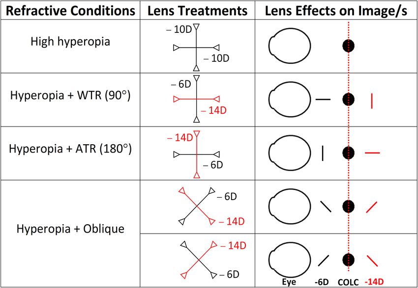
Astigmatism and Myopia Development                                          IOVS | February 2021 | Vol. 62 | No. 2 | Article 27 | 2

              association has been reported in the Hong Kong Chinese            imposed by the sphero-cylindrical lens. To determine the
              clinical population.8 While long-term, longitudinal studies       effects of astigmatism on myopia development, a total of
              are lacking to confirm the pattern of change in ocular            eight combinations of hyperopic-astigmatic defocus were
              components during the course of astigmatic development,           employed: two astigmatic magnitudes (−4DC or −8DC) and
              the disappearance of infantile astigmatism before school age      four axes (45°, 90°, 135°, or 180°) were imposed using two
              has led to the hypothesis that astigmatism acts as a visual       sphero-cylindrical lenses (low magnitude = −8DS/−4DC;
              error signal during early eye growth. It has been proposed        high magnitude = −6DS/−8DC). Each of these eight treat-
              that the presence of astigmatism may improve the efficiency       ment groups was denoted with a letter indicating the astig-
              of the mechanism that regulates emmetropization by inte-          matic magnitude (H, high; L, low), followed by a number
              grating the optical signals associated with the two principal     indicating the astigmatic axis (e.g., H45). Table 1 describes
              power meridians of astigmatic eyes.33,34 Alternatively, it has    how the different refractive conditions were achieved by
              been suggested that astigmatism may come about as a conse-        four of these eight combinations imposing the higher magni-
              quence of ametropic development,31 or the two may share a         tude of astigmatic errors (−8DC). Note that the two principal
              common etiology.3                                                 powered meridians had −6D and −14D, respectively, impos-
                  To date, the impact of astigmatism on refractive develop-     ing astigmatic magnitude of 8DC and spherical equivalent
              ment has been studied only in monkeys35,36 and chicks.37–42       of −10D. All lenses (PMMA material, base curve = 7.5mm,
              However, the important question of whether and how the            diameter = 10.8 mm, optical zone = 10 mm; Conforma, VA,
              presence of early astigmatism can alter the endpoint of           USA) were first verified for power using a digital focimeter
              myopia development has not been answered. This study              (Auto Lensmeter LM-1800PD/1800P; Nidek Co., Japan). Each
              tested this hypothesis using a chick model wearing sphero-        lens was glued firmly to a Velcro ring using Norland Opti-
              cylindrical lenses imposing hyperopic-astigmatic blur. The        cal adhesive (Norland Products, New Brunswick, NJ, USA),
              effects on refractive, axial, and corneal components were         and the axis of the negative cylinder (i.e., the least negative-
              measured after a week and compared to a control group             power meridian) was labeled clearly with a fine-tip marker
              wearing spherical lenses imposing the same magnitude of           on each side of the ring. The Velcro mate was then glued to
              spherical equivalent errors but no astigmatic error. It was       the feathers around the right orbit for the attachment of the
              found that the presence of astigmatism altered the endpoints      Velcro ring with the lens, and the space between the hooks
              of myopia development in chicks depending on the astig-           and loops of this Velcro mating pair ensured enough air
              matic properties.                                                 ventilation to prevent a foggy lens. To accurately orient the
                                                                                axis of the negative cylinder, a protractor was used to mark
                                                                                the intended treatment axis on this Velcro mate attached
              MATERIALS      AND   METHODS                                      to the feathers by referring to the palpebral fissure as the
              Animals                                                           horizontal reference line (0° or 180°). By matching the line
                                                                                on the Velcro ring (with lens) and the line on the Velcro
              In total, 193 White Leghorn chicks (Gallus gallus domes-          mate (on the feathers), the accuracy of the cylindrical axis
              ticus) were obtained from the Centralized Animal Facility         could be controlled within ±5°. During the treatment period
              of the Hong Kong Polytechnic University. They were reared         (P5−P12), lenses were cleaned frequently and checked for
              in the animal facility in a 12-hour/12-hour light/dark cycle      any scratches or defects, and all lenses with defects were
              (lights on from 0700 to 1900) under controlled tempera-           replaced immediately. If the lens was found detached during
              ture (20−22°C) with unlimited access to food and water.           the treatment period, the data were excluded from further
              The illumination level was about 150 lux at the chick’s eye       analysis.
              level. Care and use of animals were in compliance with the
              ARVO Statement for the Use of Animals in Ophthalmic and
              Vision Research. The experimental protocol was reviewed
                                                                                Measurement
              and approved by the Animal Subjects Ethics subcommittee           At the end of the treatment period, refractive state, corneal
              of the Hong Kong Polytechnic University (ASESC#15-16/41).         curvature, and ocular axial dimensions were measured
                                                                                by a modified Hartinger coincidence refractometer47 ( Jena
              Visual Manipulations                                              coincidence Refractometer, Model 110; Carl Zeiss Meditec,
                                                                                Jena, Germany), custom-made Placido-ring videokeratog-
              On posthatching day 5 (P5), chicks were randomly assigned         raphy,48 and high-resolution A-scan ultrasonography,49,50
              to wear either spherical (−10D, n = 14) or sphero-cylindrical     respectively. The refractive state was measured for all birds,
              lenses (n = 179) of identical spherical-equivalent power          whereas corneal curvature and ocular axial dimensions
              (−10D) on the right eye for 7 days (P5–P12); the left eye was     were measured for subsets of birds. All measurements were
              left untreated. Previous studies have shown that the mecha-       performed in the morning, within a ±1-hour time frame to
              nism regulating eye growth in chicks is sensitive to spherical    avoid any variation due to diurnal rhythms.49
              defocus of –10D to +20D43,44 within a short period of time
              and with less intersubject variability when compared to the       Hartinger Coincidence Refractometer
              ocular responses induced by form deprivation myopia. In
              addition, obvious refractive and axial dimensional changes        After the animal was anesthetized using isoflurane (1.0%–
              (see also Table 3) have consistently been reported in previ-      1.5% in oxygen), the palpebral fissure was aligned horizon-
              ous studies using the –10D LIM paradigm41,43,45 ; we thus         tally and the eye was held open using a lid retractor (tooth-
              selected –10D spherical defocus to induce myopia devel-           proof stainless steel wire; American Fishing, Coatesville,
              opment in this study. We adopted the same starting point          PA, USA). The lid retractor was inserted gently so that
              of lens treatment (P5) as our previous study41 to minimize        it did not come into contact with the cornea and distort
              the potential optical effects of the naturally occurring astig-   the mires as described in a previous study.41 The pres-
              matism in chicks39,46 from interfering with the astigmatism       ence of a lid retractor and anesthesia did not prevent the

Downloaded from iovs.arvojournals.org on 05/24/2021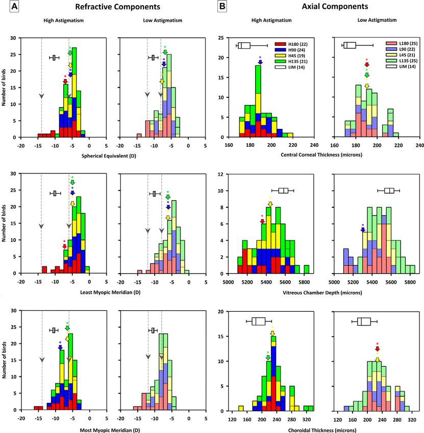
Astigmatism and Myopia Development                                                    IOVS | February 2021 | Vol. 62 | No. 2 | Article 27 | 3

              TABLE 1. Optically Imposed Hyperopic-Astigmatic Defocus Conditions

                  Spherical and sphero-cylindrical lenses were used to impose different refractive conditions. Left: refractive conditions imposed; middle:
              pictorial representation of principal power meridians of spherical lens and sphero-cylindrical lenses of high astigmatic magnitude; right:
              optical effects on image properties of a point source. Note that all refractive conditions imposed identical spherical-equivalent errors (−10D),
              as indicated by the identical locations of circle of least confusion (blurred circle aligned with red dashed line).

              eye from blinking through the nictitating membrane (the                   lated by subtracting corneal shape parameters from refrac-
              “third eyelid”), allowing corneal hydration and optical clar-             tive parameters.
              ity to be maintained during the measurement (2–3 minutes
              per eye). Three measurements for both principal meridians
              were recorded and averaged using power vector analysis.51
                                                                                        A-Scan Ultrasonography
              Refractive components used for further analysis included                  High-resolution A-scan ultrasonography consisted of a
              spherical-equivalent refractive error (M), refractive astigma-            50-MHz focused polymer with a manually adjustable pulser-
              tism (RA), least myopic meridian (LMM), most myopic merid-                receiver (model 176599; Panametrics), employing sampling
              ian (MMM), and the two astigmatic components, R-J0 and                    signals at 500 MHz. Eyelids of anesthetized chicks were
              R-J45.51                                                                  held open with a speculum while three measurements were
                                                                                        taken along the pupillary axis. Each measurement consisted
                                                                                        of 50 echograms, and these were averaged as previously
              Placido Ring Videokeratography System                                     described49,50 to obtain the axial dimensions of individual
              A Placido ring–based videokeratography system was used                    ocular components. The axial length was defined as the sum
              to measure corneal curvature in alert chicks. Once the pupil              of the distance from anterior cornea to the posterior sclera.
              was aligned concentrically with the Placido rings, the CCD
              camera captured 500–800 frames via multiple-shot mode. Six                Statistical Analysis
              high-quality images with complete Placido ring images were
              selected manually for each eye, based on the pupil align-                 Data were analyzed using SPSS statistical software (version
              ment and diameter.48 These images were analyzed using a                   23.0.0; IBM, Chicago, IL, USA). The effect of lens wear
              custom-written MATLAB algorithm to obtain corneal curva-                  on biometric parameters was first tested by comparing the
              tures of the principal meridians. These parameters were used              treated and fellow untreated eyes using paired t-tests. To
              to derive corneal shape parameters using power vector anal-               test the effect of the presence of astigmatism on myopia
              ysis: average corneal radius (CR), flattest curvature (FCR),              development, one-way ANOVAs with Bonferroni’s pairwise
              steepest curvature (SCR), corneal astigmatism (CA), and the               post hoc comparisons were used to compare parameters
              two astigmatic components, C-J0 and C-J45. Likewise, the                  across all nine groups of birds (spherical and sphero-
              refractive components of internal astigmatism were calcu-                 cylindrical lenses). To test the effect of characteristics of

Downloaded from iovs.arvojournals.org on 05/24/2021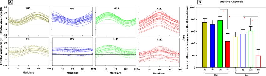
Astigmatism and Myopia Development                                            IOVS | February 2021 | Vol. 62 | No. 2 | Article 27 | 4

              astigmatism on ocular biometric parameters, two-way                 −10D lens-wearing chicks, and the two dashed lines mark
              ANOVAs (first factor: astigmatic magnitude, two levels;             the locations of line focus with magnitudes correspond-
              second factor: astigmatic axis, four orientations) followed         ing to the sphero-cylindrical lens-wear chicks. Compared
              by Bonferroni’s multiple post hoc comparisons were                  to −10D group, groups treated with sphero-cylindrical
              used to compare the eight groups of birds treated with              lenses developed significantly lower M (all except L180
              sphero-cylindrical lenses. Pearson correlation analyses were        group; one-way ANOVA with Bonferroni’s post hoc tests, all
              performed for refractive, corneal, and axial parameters. In all     P ≤ 0.01), LMM (all except L180 group; one-way ANOVA
              tests, the significance level was set at the 95% level of confi-    with Bonferroni’s post hoc tests, all P < 0.001), and MMM
              dence. Unless otherwise stated, all data were expressed in          (H45, H90, and H135; one-way ANOVA with Bonferroni’s
              terms of mean ± SE.                                                 post hoc tests, all P < 0.05). The presence of astigmatic
                                                                                  blur with sphero-cylindrical lens wear also induced signif-
                                                                                  icantly higher refractive astigmatism in all groups (one-way
              RESULTS                                                             ANOVAs with Bonferroni’s post hoc tests, all P < 0.001;
              Effects of Visual Manipulations on Treated Eyes                     Supplementary Table S1) and had significant impacts on the
                                                                                  two vector components (one-way ANOVAs, all P < 0.001):
                  Refractive Parameters. At the end of the 7-day treat-           compared to the −10D group, higher R-J0 (L45, H90, L90,
              ment period (P12), all nine visual manipulations produced           H135, and L135; Bonferroni’s post hoc tests, all P ≤ 0.01)
              significant impacts on virtually all refractive parameters of       and higher R-J45 (H45, L45, and H135; Bonferroni’s post
              the treated eyes (Table 2). Wearing −10D spherical lenses           hoc tests, all P < 0.01) were found in individual sphero-
              (LIM) induced significant changes in all refractive parame-         cylindrical lens-wear groups (Supplementary Table S1).
              ters except R-J45 (paired t-tests, all P < 0.01). Similarly, all       Axial Parameters. Sphero-cylindrical lens wear had
              eight groups that received sphero-cylindrical-lens treatment        a significant impact on corneal thickness, vitreous cham-
              showed significant changes in most refractive parameters in         ber depth, and choroidal thickness when compared to
              the treated eyes (all P < 0.001); only the two groups treated       −10D lens wear (Supplementary Table S2). Figure 1B shows
              with the axis oriented at 180° (H180 and L180) showed               frequency distributions of corneal thickness (top), vitreous
              nonsignificant change in both vector components (R-J0 and           chamber depth (middle), and choroidal thickness (bottom)
              R-J45) in the treated eyes.                                         in eyes treated by sphero-cylindrical lens wear. Compared
                  Axial Parameters. Table 3 shows a comparison of                 to the −10D group, individual groups treated with sphero-
              ocular axial dimensions between treated and fellow eyes.            cylindrical lenses had thicker central corneal thickness (L45,
              Individual treatments resulted in a thinner cornea (H45,            H90, L135, and L180; one-way ANOVAs with Bonferroni’s
              H180, and L180, paired t-tests, all P < 0.05), a thinner crys-      post hoc tests, all P ≤ 0.05), shorter vitreous chamber depth
              talline lens (H90, P < 0.05), a deeper anterior chamber,            (H45, L90 and H180; one-way ANOVAs with Bonferroni’s
              (all groups, all P < 0.05), a deeper vitreous chamber, and          post hoc tests, all P < 0.05), and thicker choroidal thickness
              a longer axial length (all groups, all P < 0.01). Interest-         (H45, L45, H135, and L180; one-way ANOVAs with Bonfer-
              ingly, while wearing −10D spherical lens induced a thinner          roni’s post hoc tests, all P < 0.05). There were no signifi-
              choroid as reported in previous studies (paired t-test, P <         cant differences in the remaining axial components (anterior
              0.01), wearing sphero-cylindrical lenses induced a thicker          chamber depth [ACD], lens thickness [LT], retinal thickness
              choroid with statistical significance in all (paired t-test, P <    [RT], scleral thickness [ST], and axial length [AXL]).
              0.01) except H90, L90, and L135 groups (thicker choroid but            Corneal Shape Parameters. Sphero-cylindrical lens
              did not reach statistical significance).                            wear did not result in a significant effect on the majority
                  Corneal Shape Parameters. The effects of spherical              of corneal shape parameters when compared to −10D lens
              and sphero-cylindrical lens wear on corneal shape parame-           wear (Supplementary Table S3); the only exception was a
              ters were in general less pronounced than those on refrac-          more positive C-J45 in both H45 and L45 groups (one-way
              tive parameters. As presented in Table 4, significantly higher      ANOVA with Bonferroni’s post hoc tests, P < 0.01).
              corneal astigmatisms were observed in the treated eyes of
              LIM, L45, H90, L90, H135, and L135 groups compared to the
              fellow untreated eyes (paired t-test, all P < 0.05). In contrast,
                                                                                  Effects of Astigmatic Characteristics on Myopia
              wearing a sphero-cylindrical lens induced a steeper cornea
              (SCR and CR) in the L180 group but a flatter cornea (FCR            Development
              and CR) in the H135 group when compared to their fellow                 Refractive Parameters. The impacts of characteris-
              eyes (all P < 0.05).                                                tics of astigmatism (i.e., orientation and magnitude) on
                                                                                  myopia development were tested among the eight groups
              Effects of Presence Versus Absence of                               of birds receiving sphero-cylindrical lens wear. While
              Astigmatism on Myopia Development                                   astigmatic orientation had significant main effects on all
                                                                                  six refractive parameters (M, LMM, MMM, RA, R-J0, and
                 Refractive Parameters. Compared to spherical lens                R-45; two-way ANOVAs, all P < 0.01), astigmatic magni-
              wear, the presence of astigmatic blur imposed by sphero-            tude showed a significant impact on three spherical compo-
              cylindrical lens wear induced less myopic errors (Fig. 1A           nents (M, LMM, and MMM; two-way ANOVAs, all P < 0.001).
              and Supplementary Table S1) in the treated eyes. Figure             In addition, significant interaction effects of astigmatic axis
              1A shows the frequency distributions of spherical equiva-           with magnitude were observed on RA and R-J45 (two-way
              lent (top), the least myopic meridian (middle), and the most        ANOVAs, both P < 0.05). Figure 2 shows three parame-
              myopic meridian (bottom) in treated eyes receiving differ-          ters (two refractive and one axial) that were affected the
              ent magnitudes (left: high cylinder, 8DC; right: low cylin-         most by the characteristics of sphero-cylindrical lens wear.
              der, 4DC) and axes (yellow: 45°; blue: 90°; green: 135°;            As shown in Figure 2A, both H180 and L180 groups devel-
              red: 180°). In each plot, the boxplot represents the data of        oped significantly more myopic LMM compared to the other

Downloaded from iovs.arvojournals.org on 05/24/2021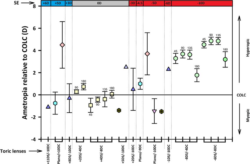
Downloaded from iovs.arvojournals.org on 05/24/2021
                                                                                                                                                                                                                                                                                                         Astigmatism and Myopia Development

                                                      TABLE 2. Effects of Spherical and Sphero-Cylindrical Lens Wear on Refractive Parameters in Treated Eyes
                                                                                                  45 (≥19)                                    90 (22)                                    135 (≥21)                                  180 (≥22)                                –10DS (14)

                                                      Refractive (D)   Group (n)           RE                 LE                     RE                     LE                     RE                 LE                     RE                     LE                  RE                LE
                                                      M                  H         −5.84   ±    0.35***   −0.36   ±   0.10   −5.97   ±    0.36***   −0.83   ±    0.14†††   −5.66   ±    0.36***   −0.51   ±   0.10   −7.56   ±    0.73***   −0.75   ±    0.17††   −10.29 ± 0.22***   −0.06 ± 0.07
                                                                         L         −7.71   ±    0.45***   −0.44   ±   0.08   −6.91   ±    0.40***   −0.60   ±    0.12      −6.63   ±    0.41***   −0.24   ±   0.08   −8.85   ±    0.58***   −0.63   ±    0.10†
                                                      LMM                H         −4.43   ±    0.37***   −0.28   ±   0.10   −4.18   ±    0.34***   −0.79   ±    0.13†††   −4.12   ±    0.36***   −0.44   ±   0.10   −6.77   ±    0.72***   −0.64   ±    0.16††   −10.06 ± 0.24***   −0.02 ± 0.08
                                                                         L         −5.88   ±    0.47***   −0.39   ±   0.08   −5.35   ±    0.45***   −0.48   ±    0.11      −5.11   ±    0.41***   −0.19   ±   0.07   −7.89   ±    0.54***   −0.52   ±    0.09
                                                      MMM                H         −7.24   ±    0.36***   −0.43   ±   0.10   −7.85   ±    0.45***   −0.94   ±    0.15†††   −7.20   ±    0.38***   −0.58   ±   0.11   −8.37   ±    0.75***   −0.82   ±    0.19††   −10.51 ± 0.20***   −0.10 ± 0.07
                                                                         L         −8.46   ±    0.44***   −0.50   ±   0.08   −8.46   ±    0.36***   −0.64   ±    0.13      −8.15   ±    0.42***   −0.30   ±   0.08   −9.94   ±    0.62***   −0.74   ±    0.11†
                                                      RA                 H         −2.82   ±    0.19***   −0.14   ±   0.04   −3.67   ±    0.23***   −0.15   ±    0.05      −3.08   ±    0.17***   −0.15   ±   0.03   −1.62   ±    0.12***   −0.18   ±    0.06      −0.46 ± 0.08**    −0.09 ± 0.03
                                                                         L         −2.57   ±    0.13***   −0.11   ±   0.03   −3.10   ±    0.20***   −0.16   ±    0.04      −3.04   ±    0.13***   −0.12   ±   0.03   −2.06   ±    0.21***   −0.22   ±    0.05
                                                      R-J0               H         −0.86   ±    0.15***   −0.07   ±   0.02   −1.50   ±    0.18***   −0.06   ±    0.02      −1.28   ±    0.07***   −0.07   ±   0.01   +0.20   ±    0.16      −0.09   ±    0.03      −0.20 ± 0.04**    −0.04 ± 0.01
                                                                         L         −1.06   ±    0.07***   −0.05   ±   0.01   −1.39   ±    0.13***   −0.07   ±    0.02      −1.41   ±    0.06***   −0.06   ±   0.01   −0.13   ±    0.22      −0.10   ±    0.02
                                                      R-J45              H         +0.91   ±    0.10***   +0.00   ±   0.01   +0.33   ±    0.10**    +0.00   ±    0.01      −0.65   ±    0.13***   +0.00   ±   0.00   +0.03   ±    0.09      −0.01   ±    0.01      −0.02 ± 0.03      −0.01 ± 0.00
                                                                         L         +0.66   ±    0.07***   +0.00   ±   0.00   +0.23   ±    0.09**    −0.01   ±    0.00      −0.27   ±    0.10**    −0.01   ±   0.00   −0.07   ±    0.09      +0.01   ±    0.01
                                                         Comparisons of six refractive parameters between treated right eyes (RE) versus the untreated left eyes (LE) by student paired t-tests and across fellow untreated left eyes from the
                                                      –10DS group versus fellow untreated left eyes from the eight sphero-cylindrical lens-wear groups using one-Way ANOVA with Bonferroni’s post hoc tests. Data for groups treated with
                                                      sphero-cylindrical lenses were arranged according to the astigmatic axis (45°, 90°, 135°, and 180°) and magnitude (high, H, –6DS/–8DC; low, L, –8DS/–4DC). Data for group treated with
                                                      spherical lenses (–10DS) are presented on the right for easy comparison. The levels of significance are represented by different symbols: *P < 0.05, **P < 0.01, and ***P < 0.001 for
                                                      paired t-tests and † P < 0.05, †† P < 0.01, and ††† P < 0.001 for post hoc tests.
                                                                                                                                                                                                                                                                                                    IOVS | February 2021 | Vol. 62 | No. 2 | Article 27 | 5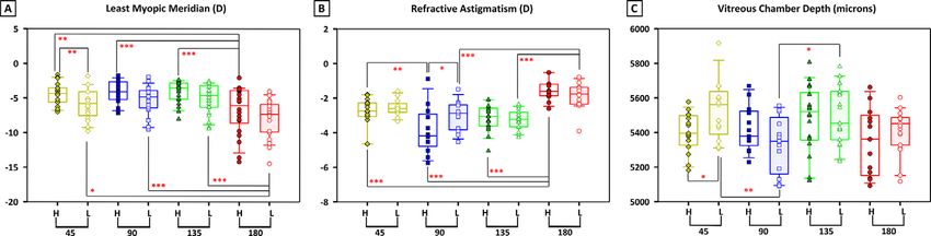
Downloaded from iovs.arvojournals.org on 05/24/2021
                                                      TABLE 3. Effects of Spherical and Sphero-Cylindrical Lens Wear on Axial Parameters in Treated Eyes
                                                                                                                                                                                                                                                                                                   Astigmatism and Myopia Development

                                                                                           45 (≥13)                                    90 (≥13)                                   135 (≥18)                                    180 (≥13)                               –10DS (14)

                                                      Groups (n)                   RE                     LE                   RE                     LE                   RE                     LE                   RE                     LE                  RE                LE
                                                      CCT (μm)     H       186.2   ±    3.05*     190.6   ±    2.39    189.4   ±    3.08      194.8   ±    4.31†   183.7   ±    1.66      185.4   ±    2.01    186.5   ±    2.61*     198.1   ±    4.60††    175.5 ± 2.62      178.1 ± 2.99
                                                                   L       190.4   ±    3.07      190.3   ±    2.55    186.2   ±    3.08      194.2   ±    3.62    189.9   ±    2.80      188.5   ±    2.84    189.5   ±    2.47***   203.6   ±    4.21†††
                                                      ACD (mm)     H        1.28   ±    0.02***    1.19   ±    0.02     1.29   ±    0.03***    1.15   ±    0.02†    1.28   ±    0.02**     1.21   ±    0.01     1.34   ±    0.04**     1.17   ±    0.02       1.35 ± 0.03**     1.26 ± 0.02
                                                                   L        1.29   ±    0.03*      1.21   ±    0.02     1.32   ±    0.03**     1.21   ±    0.02     1.28   ±    0.02**     1.21   ±    0.01     1.32   ±    0.02***    1.14   ±    0.03†††
                                                      LT (mm)      H        2.07   ±    0.01       2.06   ±    0.02     2.05   ±    0.02*      2.08   ±    0.02     2.07   ±    0.02       2.09   ±    0.02     2.08   ±    0.02       2.08   ±    0.02       2.06 ± 0.02       2.07 ± 0.02
                                                                   L        2.07   ±    0.01       2.07   ±    0.02     2.07   ±    0.02       2.05   ±    0.02     2.06   ±    0.01       2.06   ±    0.01     2.08   ±    0.03       2.10   ±    0.03
                                                      VCD (mm)     H        5.40   ±    0.03***    5.13   ±    0.03     5.42   ±    0.04***    5.11   ±    0.04     5.48   ±    0.04***    5.15   ±    0.03     5.35   ±    0.05***    5.11   ±    0.04       5.58 ± 0.02***    5.18 ± 0.02
                                                                   L        5.53   ±    0.05***    5.24   ±    0.04     5.33   ±    0.05***    5.06   ±    0.04     5.50   ±    0.04***    5.17   ±    0.02     5.42   ±    0.03***    5.12   ±    0.04
                                                      RT (mm)      H        0.25   ±    0.00       0.26   ±    0.01     0.25   ±    0.00       0.25   ±    0.00     0.25   ±    0.00*      0.26   ±    0.00†    0.25   ±    0.01*      0.24   ±    0.01       0.23 ± 0.01       0.23 ± 0.01
                                                                   L        0.24   ±    0.01       0.25   ±    0.01     0.25   ±    0.01       0.26   ±    0.01     0.25   ±    0.00       0.25   ±    0.00     0.25   ±    0.01*      0.26   ±    0.01
                                                      CT (mm)      H        0.23   ±    0.01**     0.21   ±    0.01     0.22   ±    0.01       0.20   ±    0.01     0.23   ±    0.01**     0.20   ±    0.01     0.23   ±    0.00**     0.21   ±    0.01       0.19 ± 0.01**     0.20 ± 0.00
                                                                   L        0.23   ±    0.01***    0.20   ±    0.01     0.22   ±    0.01       0.21   ±    0.01     0.20   ±    0.01       0.19   ±    0.01     0.23   ±    0.01**     0.21   ±    0.01
                                                      ST (mm)      H        0.12   ±    0.00*      0.10   ±    0.01†    0.11   ±    0.01       0.10   ±    0.01     0.12   ±    0.01       0.11   ±    0.00     0.12   ±    0.00       0.10   ±    0.01       0.12 ± 0.00       0.13 ± 0.00
                                                                   L        0.12   ±    0.01*      0.11   ±    0.00     0.12   ±    0.00       0.12   ±    0.01     0.13   ±    0.01**     0.11   ±    0.01     0.11   ±    0.00*      0.10   ±    0.01
                                                      AXL (mm)     H        9.54   ±    0.04***    9.13   ±    0.03     9.53   ±    0.05***    9.08   ±    0.04     9.61   ±    0.04***    9.21   ±    0.03     9.56   ±    0.08***    9.10   ±    0.05       9.71 ± 0.02***    9.25 ± 0.02
                                                                   L        9.68   ±    0.06***    9.27   ±    0.05     9.50   ±    0.07***    9.09   ±    0.06     9.61   ±    0.04***    9.18   ±    0.03     9.58   ±    0.03***    9.13   ±    0.05
                                                         Comparisons of eight axial parameters were made for treated right eyes (RE) versus the untreated left eyes (LE) by student paired t-tests and for fellow untreated left eyes from the
                                                      –10DS group versus fellow untreated left eyes from the eight sphero-cylindrical lens-wear groups using one-Way ANOVA with Bonferroni’s post hoc tests. Data for groups treated with
                                                      sphero-cylindrical lenses were arranged according to the astigmatic axis (45°, 90°, 135°, and 180°) and magnitude (high, H, –6DS/–8DC; low, L, –8DS/–4DC). Data for group treated with
                                                      spherical lenses (–10DS) are presented on the right for easy comparison. CCT, central corneal thickness; CT, choroidal thickness. The levels of significance are represented by different
                                                      symbols: *P < 0.05, **P < 0.01, and ***P < 0.001 for paired t-tests and † P < 0.05, †† P < 0.01, and ††† P < 0.001 for post hoc tests.
                                                                                                                                                                                                                                                                                              IOVS | February 2021 | Vol. 62 | No. 2 | Article 27 | 6
Downloaded from iovs.arvojournals.org on 05/24/2021
Astigmatism and Myopia Development
TABLE 4. Effects of Spherical and Sphero-Cylindrical Lens Wear on Corneal Shape Parameters in Treated Eyes
45 (≥13) 90 (≥13) 135 (≥18) 180 (≥13) –10DS (14)
Groups (n) RE LE RE LE RE LE RE LE RE LE
FCR (mm) H 3.15 ± 0.02 3.13 ± 0.01 3.17 ± 0.03 3.17 ± 0.02 3.21 ± 0.02* 3.18 ± 0.01 3.16 ± 0.03 3.16 ± 0.02 3.15 ± 0.01 3.16 ± 0.01
L 3.21 ± 0.02 3.21 ± 0.02 3.14 ± 0.03 3.13 ± 0.02 3.19 ± 0.02 3.18 ± 0.02 3.14 ± 0.01 3.16 ± 0.01
SCR (mm) H 3.12 ± 0.01 3.11 ± 0.02 3.12 ± 0.03 3.14 ± 0.02 3.17 ± 0.02 3.15 ± 0.01 3.13 ± 0.03 3.14 ± 0.02 3.11 ± 0.02 3.13 ± 0.02
L 3.16 ± 0.02 3.18 ± 0.02 3.10 ± 0.02 3.10 ± 0.02 3.14 ± 0.02 3.15 ± 0.01 3.11 ± 0.02* 3.13 ± 0.02
CR (mm) H 3.14 ± 0.01 3.12 ± 0.01 3.14 ± 0.03 3.16 ± 0.02 3.19 ± 0.02* 3.16 ± 0.01 3.14 ± 0.03 3.15 ± 0.02 3.13 ± 0.02 3.15 ± 0.02
L 3.18 ± 0.02 3.19 ± 0.02 3.12 ± 0.02 3.12 ± 0.02 3.17 ± 0.02 3.17 ± 0.02 3.12 ± 0.01* 3.14 ± 0.02
CA (D) H −1.21 ± 0.12 −0.91 ± 0.13 −1.62 ± 0.25* −1.11 ± 0.12 −1.64 ± 0.13*** −0.96 ± 0.13 −1.09 ± 0.12 −0.83 ± 0.09 −1.40 ± 0.14* −0.97 ± 0.09
L −1.47 ± 0.10** −1.00 ± 0.08 −1.56 ± 0.18* −1.05 ± 0.10 −1.61 ± 0.12** −1.09 ± 0.10 −1.06 ± 0.12 −0.89 ± 0.07
C-J0 (D) H −0.48 ± 0.07 −0.43 ± 0.07 −0.71 ± 0.11* −0.49 ± 0.06 −0.37 ± 0.08 −0.30 ± 0.07 −0.08 ± 0.09 −0.34 ± 0.06 −0.43 ± 0.07 −0.43 ± 0.06
L −0.52 ± 0.13 −0.47 ± 0.04 −0.56 ± 0.13 −0.44 ± 0.07 −0.54 ± 0.06 −0.50 ± 0.06 −0.24 ± 0.07 −0.35 ± 0.03
C-J45 (D) H +0.10 ± 0.09 +0.06 ± 0.04 −0.23 ± 0.11* +0.08 ± 0.07 −0.67 ± 0.07*** +0.09 ± 0.08 −0.33 ± 0.10** +0.09 ± 0.05 −0.48 ± 0.08*** +0.01 ± 0.05
L +0.00 ± 0.09 +0.08 ± 0.04 −0.25 ± 0.10* +0.08 ± 0.07 −0.55 ± 0.06*** −0.07 ± 0.04 −0.30 ± 0.07** −0.04 ± 0.06
Comparisons of six corneal shape parameters were made for treated right eyes (RE) versus the untreated left eyes (LE) by Student’s paired t-tests and for fellow untreated left eyes
from the –10DS group versus fellow untreated left eyes from the eight sphero-cylindrical lens-wear groups using one-way ANOVA with Bonferroni’s post hoc tests. Data for groups treated
with sphero-cylindrical lenses were arranged according to the astigmatic axis (45°, 90°, 135°, and 180°) and magnitude (high, H, –6DS/–8DC; low, L, –8DS/–4DC). Data for group treated
with spherical lenses (–10DS) are presented on the right for easy comparison. The levels of significance are represented by different symbols: *P < 0.05, **P < 0.01, and ***P < 0.001 for
paired t-tests, and † P < 0.05, †† P < 0.01, and ††† P < 0.001 for post hoc tests.
IOVS | February 2021 | Vol. 62 | No. 2 | Article 27 | 7Astigmatism and Myopia Development IOVS | February 2021 | Vol. 62 | No. 2 | Article 27 | 8
FIGURE 1. Effects of the presence of astigmatism on refractive (A) and axial (B) components in the treated eyes. Frequency distributions
of three refractive (A) and axial (B) parameters of chicks treated with sphero-cylindrical lenses of two astigmatic magnitudes (high, H;
low, L) and four astigmatic axes (45°, yellow; 90°, blue; 135°, green; and 180°, red). The darker (left column) and lighter (right column)
shades represent the higher and lower magnitudes of astigmatism imposed, respectively. The sample size for each group is shown in the
parentheses in the legend. In each plot, the horizontal boxplot presents the descriptive statistics (the solid line and box margins represent the
median and interquartile range, respectively) of the group wearing the –10D lens (LIM). Significant differences between LIM with individual
sphero-cylindrical lens-wear groups (marked by colored arrows) were found in these refractive and axial parameters (one-way ANOVA with
Bonferroni-corrected pairwise post hoc comparisons). The level of significance (from LIM) is indicated by *P < 0.05.
FIGURE 2. Effects of astigmatic magnitude and axis on the least myopic meridian (A), refractive astigmatism (B), and vitreous chamber
depth (C) in the treated eyes. Comparisons of three parameters at the end of the treatment period among the eight groups of birds treated
with sphero-cylindrical lenses. The eight groups of birds are organized from left to right according to the magnitude (high, H; low, L) and
cylindrical axis (45°, 90°, 135°, and 180°). Each symbol represents data from one bird; the solid line in the box and the box margins represent
the median and interquartile range, respectively, for each group. Significant differences between groups (two-way ANOVA with Bonferroni’s
multiple post hoc comparisons) are indicated by *P < 0.05, **P < 0.01, and ***P < 0.001. Birds treated with 180 cylindrical axes developed
higher LMM (A) but lower refractive astigmatism (B) compared to birds treated with the other three orientations. In addition, the L90 group
had a significantly shorter vitreous chamber depth than the L45 and L135 groups.
Downloaded from iovs.arvojournals.org on 05/24/2021Astigmatism and Myopia Development IOVS | February 2021 | Vol. 62 | No. 2 | Article 27 | 9
FIGURE 3. Effective ametropia at the end of the treatment period in birds treated with sphero-cylindrical lenses. (A) Effective ametropia
(ocular ametropia–lens power) as a function of meridian for individual birds (colored lines) at the end of the treatment period. Group data
are arranged according to cylindrical magnitude (top, high magnitude, solid line; bottom, low magnitude, dashed line) and axis (left to right:
45°, 90°, 135°, and 180°). In each plot, the black solid line represents the emmetropic condition; the area enclosed by each curve (bird) from
this black line is calculated as an arbitrary unit to quantify the effective ametropia. (B) Effective ametropia (mean and SEM), calculated as
an area departing from the emmetropic line (summation of effective ametropia [ocular ametropia–lens power] across the 180° meridians),
for the eight groups of birds treated with sphero-cylindrical lenses. In general, with the same magnitude of astigmatism imposed (i.e., high
or low), birds treated with 180 cylindrical axes developed the lowest effective ametropia compared to the other three groups. Significant
differences between groups (two-way ANOVA with Bonferroni’s multiple post hoc comparisons) are indicated by *P < 0.05.
three groups receiving the same magnitude but different matism (top panel). To quantify the magnitude of departure
axes (two-way ANOVAs with Bonferroni’s multiple post hoc of effective ametropia from emmetropia as a whole, the area
comparisons, all P ≤ 0.05). In addition, the H180 group enclosed by the effective ametropia curve and emmetropia
also developed significantly lower RA when compared to (black line in Fig. 3A) was calculated for individual birds and
the other three groups with the same magnitude but differ- this magnitude compared across groups. Two-way ANOVAs
ent axes (two-way ANOVAs with Bonferroni’s multiple post revealed that both the axis (P < 0.001) and magnitude (P <
hoc comparisons, all P ≤ 0.001), whereas the L180 group 0.001) of astigmatism had significant effects on the effective
had significantly lower RA when compared to L90 and L135 ametropia (area), but no interaction effects were found. As
groups (two-way ANOVA with Bonferroni’s multiple post presented in Figure 3B, L180 induced significantly less effec-
hoc comparisons, all P ≤ 0.05). Significant difference in tive ametropia compared to L90, L135, and H180, whereas
RA was also observed between H90 versus H45 and L90 H180 had less effective ametropia compared to H135 (two-
groups (two-way ANOVAs with Bonferroni’s multiple post way ANOVA with Bonferroni’s multiple post hoc compar-
hoc comparisons, all P ≤ 0.05). For the vector components, isons, all P ≤ 0.05).
H180 and L180 groups resulted in positive R-J0, which was Axial Parameters. Astigmatic axis produced signifi-
significantly different from the other three orientations of cant main effects on the vitreous chamber depth (two-
the same astigmatic magnitude. In contrast, both H135 and way ANOVA, P < 0.01), but astigmatic magnitude had no
L45 developed significantly higher R-J45 than the other three significant effects on axial parameters. Figure 2C shows
groups of different orientations but with same astigmatic the effects of astigmatic axis on vitreous chamber depth.
magnitude (two-way ANOVAs with Bonferroni’s multiple The L90 group had shorter vitreous chamber depth when
post hoc comparisons, all P ≤ 0.05). In addition, R-J45 in compared to L45 and L135 groups (two-way ANOVAs with
the H45 group was significantly higher than those in H180 Bonferroni’s multiple post hoc comparisons, P < 0.05). In
and H90 groups (two-way ANOVA with Bonferroni’s multi- addition, the H45 group had a significantly shorter vitre-
ple post hoc comparisons, all P ≤ 0.001), whereas R-J45 in ous chamber depth when compared to the L45 group (two-
the L135 group was significantly lower than those in H135 way ANOVA with Bonferroni’s multiple post hoc compar-
and L90 groups (two-way ANOVA with Bonferroni’s multiple isons, P < 0.05). Among these sphero-cylindrical lens-wear
post hoc comparisons, all P ≤ 0.01). groups, the L135 group showed a thinner choroid when
To compare the effect of sphero-cylindrical lens wear on compared to the H135 and L180 groups (two-way ANOVA
emmetropization, Figure 3A shows the effective ametropia with Bonferroni’s multiple post hoc comparisons, P < 0.05).
profile at the end of the treatment period for individual birds There were no significant differences in the remaining axial
in each group. Using the calculation method from an earlier components (central corneal thickness [CCT], anterior cham-
study involving monkeys,35 the effective ametropia for indi- ber depth [ACD], lens thickness [LT], retinal thickness [RT],
vidual meridians was derived by subtracting the lens refrac- scleral thickness [ST], and axial length [AXL]) among these
tive power from the ocular refraction. The refractive power eight groups of birds.
at a given meridian θ is derived by Corneal Shape Parameters. Astigmatic axis
produced significant main effects on corneal astigma-
tism (two-way ANOVA, P < 0.001) and the two vector
Fθ = FS ph − FCyl sin(θ − α)2
components (C-J0 and C-J45; both P < 0.001). In contrast,
astigmatic magnitude had no significant effects on corneal
where FSph and FCyl are the spherical and cylindrical powers, shape parameters. When groups treated with the same
respectively, and α is the axis of the minus cylinder. As magnitude were compared, significantly higher corneal
shown in Figure 3A, imposing a lower magnitude of astig- astigmatism was found in birds treated with 135° compared
matism (bottom panel) usually resulted in a smaller effec- to 180° cylindrical axis (H135 versus H180 and L135 versus
tive ametropia (i.e., a smaller departure from emmetropia) L180; two-way ANOVA with Bonferroni’s multiple post hoc
when compared to imposing a higher magnitude of astig- comparisons, P < 0.05; Table 4). For C-J0, the H90 group was
Downloaded from iovs.arvojournals.org on 05/24/2021Astigmatism and Myopia Development IOVS | February 2021 | Vol. 62 | No. 2 | Article 27 | 10
TABLE 5. Correlations Between Refractive Components and Axial were correlated with LMM in birds treated with the 135°
Parameters in Different Treatment Groups cylindrical axis. There were no other significant correlations
LIM 180 90 45 between refractive and corneal components.
Corneal and Axial Components. Both the flattest
Characteristic LT VCD AXL RT LT VCD and steepest corneal radii were correlated with axial length
M −0.63* −0.54* −0.52* −0.39* −0.52**
ns in all (P < 0.01) except the 180º cylindrical axis–treated
LMM −0.57* −0.55* ns −0.41* −0.49* −0.40* group. In contrast, both corneal radii were correlated with
MMM −0.67** ns −0.55* −0.41* −0.51** ns vitreous chamber depth in all groups (all P < 0.05). Figure
4 shows the positive correlations between these parameters;
Significant correlations were found between three spherical
ametropia (M, LMM, and MMM) and individual axial parameters (LT, only Pearson’s r with statistical significance is inserted. The
VCD, AXL, and RT). The levels of significance (Pearson’s correla- relationship between the ratio of vitreous chamber depth
tion coefficient) are represented by *P < 0.05 and **P < 0.01. ns, to corneal radius (VCD/CR) and the ratio of axial length to
nonsignificant. corneal radius (AL/CR) with spherical ametropia was further
investigated. Table 6 presents the Pearson’s correlation anal-
yses between three spherical ametropia with VCD/CR and
significantly more negative than the H180 group (two-way AL/CR. In the LIM group, there were no significant correla-
ANOVA with Bonferroni’s multiple post hoc comparisons, tions between spherical ametropia with either ratio (VCD/CR
P < 0.05). For C-J45, the H135 group had a significantly and AL/CR). All three spherical ametropia were significantly
more negative value compared to the H45, H90, and H180 correlated with VCD/FCR (all P < 0.05) and VCD/SCR (all P
groups (two-way ANOVA with Bonferroni’s multiple post < 0.05) in birds treated with 180º and 45º cylindrical axes,
hoc comparisons, all P < 0.05). There were no significant and significant correlation was noted with AL/FCR (all P <
differences in the other corneal components. 0.05) and AL/SCR (all P < 0.05) in birds treated with 180º
and 135º cylindrical axes. There were no other significant
Correlations Between Refractive, Corneal, and correlations between corneal and axial components.
Axial Components
Subsets of birds, which had all biometric parameters DISCUSSION
measured, were used for correlation analyses. Because the
Using chicken as an animal model, it was shown that
imposed astigmatic magnitude did not produce significant
(1) imposing hyperopic-astigmatic defocus induced myopic-
effects on these parameters, the data from both groups (high
astigmatic error in the treated eyes; (2) with the same level
and low magnitudes) receiving the same astigmatic axis
of spherical-equivalent hyperopic blur (−10D), the presence
were pooled. Results from these five groups of birds (LIM,
of astigmatism in the sphero-cylindrical lens-wear groups
45°, 90°, 135°, and 180°) are presented below.
induced less myopia than the spherical lens-wear group; and
Refractive and Axial Components. Table 5 (3) myopia development was sensitive to the magnitude and
presents significant Pearson’s correlation coefficients found
axis of astigmatic blur.
between refractive and axial parameters. While M and LMM
were significantly correlated with vitreous chamber depth
(VCD) in LIM (all P < 0.05), only LMM was significantly Effects of Sphero-Cylindrical Lens Wear on
correlated with VCD in birds treated with the cylindrical Treated and Fellow Untreated Eyes
axis oriented at 45º (all P < 0.05). In the LIM group, M and
MMM were mildly correlated with axial length (all P < 0.05). While wearing −10D spherical lenses induced typical
The three spherical ametropia were significantly correlated changes in refractive and axial parameters of the treated
with lens thickness in LIM (all P < 0.05) and birds treated eyes as previously reported,41,43 sphero-cylindrical lens wear
with 90º cylindrical axis (all P < 0.05). In contrast, the three produced significant refractive (Table 1), axial (Table 2), and
spherical ametropia were significantly correlated with retinal corneal changes (Table 3) that depended on the astigmatic
thickness in birds treated with 180º cylindrical axis (all P < characteristics (see further discussion below) in both treated
0.05). There were no other significant correlations observed and fellow untreated eyes. The eyes treated with sphero-
between refractive and axial components. cylindrical lenses not only developed significantly deeper
Refractive and Corneal Components. Supplemen- anterior and vitreous chamber depths, similar to those
tary Figure S2 shows the results of Pearson’s correlation wearing spherical lenses, but also developed idiosyncratic
analyses between refractive astigmatism and corneal (top) changes in anterior axial components (e.g., central corneal
and internal astigmatisms (bottom). Only correlation coef- thickness) and corneal refractive parameters (e.g., steeper
ficients showing statistical significance are inserted in each corneal radius) in individual groups of birds. Interestingly,
plot. In general, internal astigmatic components were more the untreated eyes of birds wearing sphero-cylindrical lenses
strongly correlated with refractive than with corneal compo- also showed a variety of changes from anterior to poste-
nents. While refractive and internal astigmatic magnitudes rior segments compared to the untreated eyes of −10D birds
were significantly correlated in all groups (all P < 0.01), (Supplementary Materials section). To the best of our knowl-
corneal and refractive astigmatic magnitudes were signifi- edge, this is the first study to report significant impacts of
cantly correlated only in groups treated with 90° and 135° sphero-cylindrical lenses on a wide range of ocular param-
axes (both P < 0.05). For J0 and J45 astigmatic components, eters. Although one previous study used a combination of
while refractive and internal components were significantly spherical lenses (±3DS and ±6DS) and crossed-cylindrical
correlated in all groups (all P < 0.01), corneal and refractive lenses (+5DS/−10DC) to create a sphero-cylindrical lens
J0 were only mildly correlated in groups treated with 90º and effect, comparison with the current study is difficult
135º axes (both P < 0.01). In contrast, the flattest (r = 0.34, because the closest match to our lenses was −1DS/−10DC52
P = 0.04) and mean (r = 0.33, P = 0.05) corneal curvatures (further discussion below). In this respect, the inclusion of
Downloaded from iovs.arvojournals.org on 05/24/2021Astigmatism and Myopia Development IOVS | February 2021 | Vol. 62 | No. 2 | Article 27 | 11
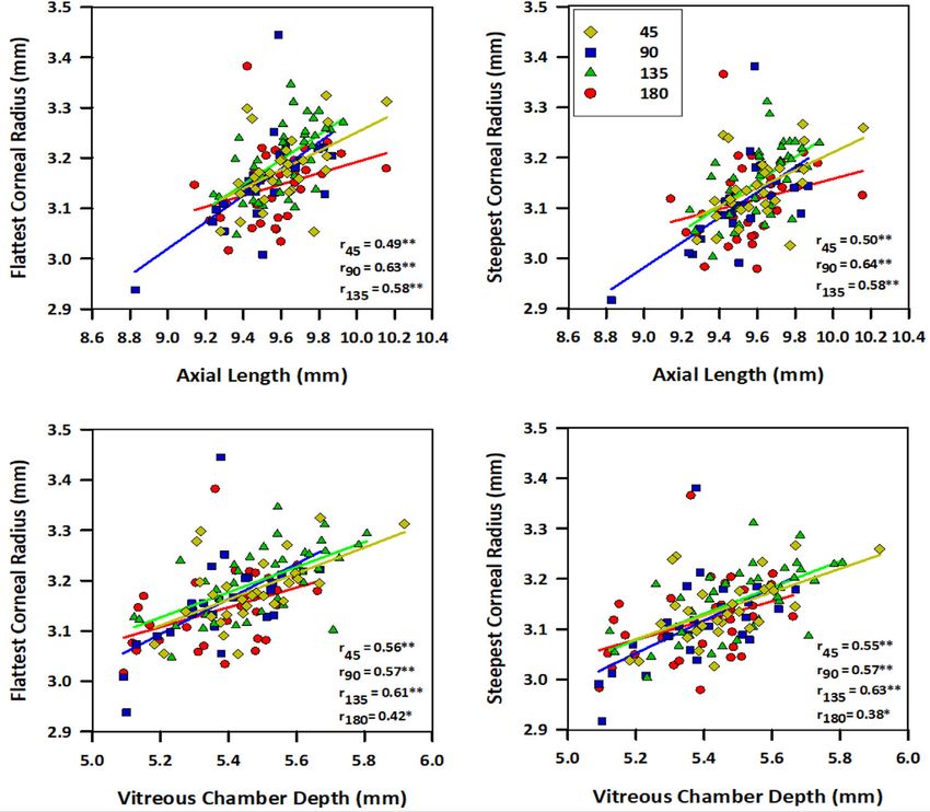
FIGURE 4. Correlations between corneal radius of curvature with axial length and vitreous chamber depth. Corneal radii along the flattest
(left column) and steepest (right column) meridians are plotted against the axial length (top row) and vitreous chamber depth (bottom row)
in eyes treated with sphero-cylindrical lenses of different cylindrical axes. Different-colored symbols represent groups receiving different
cylindrical axes, as shown in the legends. The significant level of correlation is represented by *P < 0.05 and **P < 0.01. All except one
group showed significant correlations between both corneal radii (flattest and steepest) with axial length and vitreous chamber depth. The
only exception was the group treated with 180° axis, in which no correlation was found between both corneal radii with the axial length.
TABLE 6. Correlations Between the Spherical Ametropia With Ratios of VCD/CR and AL/CR in Different Treatment Groups
45 135 180
Ratios VCD/FK VCD/SK AL/FK AL/SK VCD/FK VCD/SK AL/FK AL/SK
M −0.40* −0.41* −0.38* −0.37* −0.38* −0.40* −0.44* −0.45**
LMM −0.38* −0.39* −0.40* −0.37* −0.37* −0.39* −0.43* −0.44*
MMM −0.41* −0.41* −0.35* −0.35* −0.35* −0.37* −0.44* −0.45**
Significant correlations were found between three spherical ametropia (M, LMM, and MMM) and ratios of VCD/corneal radii (VCD/FCR,
ratio of vitreous chamber depth to flattest corneal curvature; VCD/SCR, ratio of vitreous chamber depth to steepest corneal curvature)
and ratios of AL/corneal radii (AL/FCR, ratio of axial length to flattest corneal curvature; AL/SCR, ratio of axial length to steepest corneal
curvature). The levels of significance (Pearson’s correlation coefficient) are represented by *P < 0.05 and **P < 0.01.
refractive, axial, and corneal measurements in this study chicks (Fig. 1 and Supplementary Tables S1–S3). In contrast
provides a strong foundation for future work in this area. to developing approximately −10D of myopia, as in the
Nevertheless, because children rarely encounter this magni- spherical lens-wear group, both principal refractive merid-
tude of hyperopic-astigmatic blur during the normal course ians in the sphero-cylindrical lens-wear group developed
of refractive development, the question remains if these lower magnitudes of myopia equal to or less than the
results can be translated into human studies. magnitude sufficient to compensate for the least hyper-
opic image plane (Fig. 1 and Table 1). Comparison of the
Astigmatism Interferes With Myopia Development distributions between the two principal refractive meridians
(Fig. 1A, middle versus bottom plots) indicates that, while
Although both spherical and sphero-cylindrical lenses there are subtle differences between groups (Fig. 1A, arrow-
imposed the same magnitude of −10D spherical-equivalent heads), overall the most myopic meridian appears to be
error (Table 1), the coexistence of astigmatic blur in the latter more successful than the least myopic meridian in devel-
treatment clearly alters refractive and ocular endpoints in oping myopic magnitude that closely matches the image
Downloaded from iovs.arvojournals.org on 05/24/2021Astigmatism and Myopia Development IOVS | February 2021 | Vol. 62 | No. 2 | Article 27 | 12
FIGURE 5. Impacts of optically imposed astigmatism on the effective endpoint in chicks. Comparisons of effective endpoints, with respect
to the plane of COLC, across different studies. The effective endpoint on the y-axis is calculated by subtracting the COLC (x-axis, bottom:
cylindrical format; x-axis, top: spherical-equivalent format) from the resultant ametropia for individual treatment groups reported in the
literature. A positive value indicates a relative hyperopic endpoint and vice versa (y-axis, right). Different symbols represent different studies;
number above the symbol indicates the axis of the cylindrical lens. For each study, the starting day of treatment (e.g., P6, posthatching day
6) and the duration of lens treatment are given for references; , McLean and Wallman, 200352 : P6, 2 days; , Irving et al., 199538 : P0/P2,
7 days; , Schmid and Wildsoet, 199739 : P0, 12 days; , Chu and Kee, 201542 : P5, 7 days; , Phillips and Collins, 200053 : P3, 7 days; ,
Thibos L, et al. IOVS 2001;42:ARVO Abstract 324: P4, 7 days; , current study: P5, 7 days. The effective endpoint appears to be directed
toward the hyperopic image plane relative to COLC in chicks receiving a high magnitude of hyperopic spherical-equivalent errors (–10D).
plane imposed by the weaker lens meridian (dashed lines in Figure 5, it is clear that the presence of different degrees
on the right in Fig. 1A). Consequently, the average spherical- of spherical equivalent combined with constant astigmatic
equivalent values (Fig. 1A, top plots) are more hyperopic blurs did not result in extreme myopia similar to that
than this image plane. Thus, results from all three refrac- induced by form deprivation conditions,45 a finding consis-
tive parameters support the notion that the endpoint is tent with the monkey study using a relatively large number
directed toward the least hyperopic image plane in the of animals.36 Rather, the endpoints in these studies appeared
sphero-cylindrical lens-wear groups. to be directed toward the image plane that were near the
Whether astigmatism alters the endpoints of refractive COLC plane (Thibos L, et al. IOVS 2001;42:ARVO Abstract
development is an important question and has been inves- 324)38,53 or the more hyperopic plane than COLC.39,42,52 In
tigated only in chick and monkey models. Nevertheless, this respect, the current study, which involved a relatively
interpreting results across these studies requires caution large number of chicks, provides the first evidence that in the
because different astigmatic magnitude, cylindrical lens presence of hyperopic-astigmatic blur, the emmetropization
design (plano-/crossed-/sphero-cylindrical), and experimen- process for both principal refractive meridians is directed
tal protocols (age of onset, treatment period, axis orienta- toward the least hyperopic image plane (Fig. 1A). These
tion) were used.3,45 In contrast to the only monkey study, results are supported by the hypothetical optical effects on
which used a crossed-cylindrical lens with zero spherical the astigmatic focal planes in an earlier monkey study36 :
equivalent (+1.50D and −1.50D in the two principal merid- regardless of the pupil size, computations of modulation
ians), the chick studies used different lens designs with transfer function (MTF) for a monochromatic point source
spherical equivalents ranging from 0D to ±6D (Thibos L, et indicate that the volumes underneath the three-dimensional
al. IOVS 2001;42:ARVO Abstract 324).38,39,42,52,53 In order to MTF (taking into account different spatial frequencies and
incorporate this information into the comparison of chick orientations) were much higher at the astigmatic line foci
studies, Figure 5 illustrates the effects of imposing astig- than that at the COLC. Hence, directing the developing reti-
matic blur on the resultant ametropia relative to the location nal plane to the least hyperopic image plane (the closest
of circle of least confusion (COLC), organized along the x- focal plane) would facilitate the overall neuronal activities
axis (top) by the magnitude of spherical equivalent received. with respect to retinal contrast and orientation.
Note that the induced ametropia is expressed as a relative
value to the COLC plane (i.e., eye’s ametropia–lens refrac- Differential Effects of Astigmatic Orientation on
tion). Thus, a value closer to zero on the y-axis indicates Eye Growth
that the refractive endpoint attempts to match the COLC
plane, whereas a positive value indicates that the endpoint The axis of astigmatism produced an extensive impact on
is more hyperopic than the COLC and vice versa. As shown myopia development (Figs. 2–4, Tables 2–4). Among the
Downloaded from iovs.arvojournals.org on 05/24/2021You can also read