PROCEEDINGS OF SPIE Front Matter: Volume 7139
←
→
Page content transcription
If your browser does not render page correctly, please read the page content below
PROCEEDINGS OF SPIE
       SPIEDigitalLibrary.org/conference-proceedings-of-spie

                   Front Matter: Volume 7139

                                   , "Front Matter: Volume 7139," Proc. SPIE 7139, 1st Canterbury Workshop on
                                   Optical Coherence Tomography and Adaptive Optics, 713901 (3 March 2009);
                                   doi: 10.1117/12.823527

                                   Event: 1st Canterbury Workshop and School in Optical Coherence
                                   Tomography and Adaptive Optics, 2008, Canterbury, United Kingdom

Downloaded From: https://www.spiedigitallibrary.org/conference-proceedings-of-spie on 07 Jul 2021 Terms of Use: https://www.spiedigitallibrary.org/terms-of-use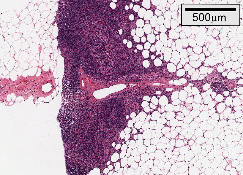
PROCEEDINGS OF SPIE

                   1st Canterbury Workshop on
                   Optical Coherence Tomography
                   and Adaptive Optics
                   Adrian Podoleanu
                   Editor

                   8–10 September 2008
                   Canterbury, Kent, United Kingdom

                   Organised by
                   School of Physical Sciences, University of Kent (United Kingdom)

                   Coorganised by
                   HIRESOMI—Marie Curie Early Stage Training Consortium • National University of Ireland, Galway (Ireland)
                   University of Porto (Portugal) • Imagine Eyes (France) • Multiwave Photonics (Portugal)

                   Sponsored by
                   European Commission, Marie Curie Actions • Imagine Eyes (France) • Michelson Diagnostic (United
                   Kingdom) • Multiwave Photonics (Portugal) • The Ratiu Family Charitable Foundation (United Kingdom)
                   Santec (Japan) • Superlum, Diodes, Ltd. (Russia) • Thorlabs, Inc. (United Kingdom) • University of Kent
                   (United Kingdom)

                   Published by
                   SPIE

                                                                                                                                     Volume 7139

                                                                 Proceedings of SPIE, 0277-786X, v. 7139

                               SPIE is an international society advancing an interdisciplinary approach to the science and application of light.

                                              1st Canterbury Workshop on Optical Coherence Tomography and Adaptive Optics
                                                edited by Adrian Podoleanu, Proc. of SPIE Vol. 7139, 713901 · © 2008 SPIE
                                                           CCC code: 0277-786X/08/$18 · doi: 10.1117/12.823527

                                                                          Proc. of SPIE Vol. 7139 713901-1

Downloaded From: https://www.spiedigitallibrary.org/conference-proceedings-of-spie on 07 Jul 2021
Terms of Use: https://www.spiedigitallibrary.org/terms-of-use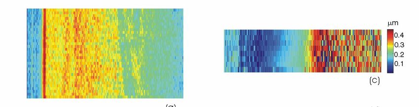
The papers included in this volume were part of the technical conference cited on the cover and
title page. Papers were selected and subject to review by the editors and conference program
committee. Some conference presentations may not be available for publication. The papers
published in these proceedings reflect the work and thoughts of the authors and are published
herein as submitted. The publisher is not responsible for the validity of the information or for any
outcomes resulting from reliance thereon.
Please use the following format to cite material from this book:
Author(s), "Title of Paper," in 1st Canterbury Workshop on Optical Coherence Tomography and
Adaptive Optics, edited by Adrian Podoleanu, Proceedings of SPIE Vol. 7139 (SPIE, Bellingham, WA,
2008) Article CID Number.
ISSN 0277-786X
ISBN 9780819473806
Published by
SPIE
P.O. Box 10, Bellingham, Washington 98227-0010 USA
Telephone +1 360 676 3290 (Pacific Time)· Fax +1 360 647 1445
SPIE.org
Copyright © 2008, Society of Photo-Optical Instrumentation Engineers
Copying of material in this book for internal or personal use, or for the internal or personal use of
specific clients, beyond the fair use provisions granted by the U.S. Copyright Law is authorized by
SPIE subject to payment of copying fees. The Transactional Reporting Service base fee for this
volume is $18.00 per article (or portion thereof), which should be paid directly to the Copyright
Clearance Center (CCC), 222 Rosewood Drive, Danvers, MA 01923. Payment may also be made
electronically through CCC Online at copyright.com. Other copying for republication, resale,
advertising or promotion, or any form of systematic or multiple reproduction of any material in this
book is prohibited except with permission in writing from the publisher. The CCC fee code is
0277-786X/08/$18.00.
Printed in the United States of America.
Publication of record for individual papers is online in the SPIE Digital Library.
SPIEDigitalLibrary.org
Paper Numbering: Proceedings of SPIE follow an e-First publication model, with papers published
first online and then in print and on CD-ROM. Papers are published as they are submitted and meet
publication criteria. A unique, consistent, permanent citation identifier (CID) number is assigned to
each article at the time of the first publication. Utilization of CIDs allows articles to be fully citable as
soon they are published online, and connects the same identifier to all online, print, and electronic
versions of the publication. SPIE uses a six-digit CID article numbering system in which:
The first four digits correspond to the SPIE volume number.
The last two digits indicate publication order within the volume using a Base 36 numbering
system employing both numerals and letters. These two-number sets start with 00, 01, 02, 03, 04,
05, 06, 07, 08, 09, 0A, 0B … 0Z, followed by 10-1Z, 20-2Z, etc.
The CID number appears on each page of the manuscript. The complete citation is used on the first
page, and an abbreviated version on subsequent pages. Numbers in the index correspond to the
last two digits of the six-digit CID number.
Proc. of SPIE Vol. 7139 713901-2
Downloaded From: https://www.spiedigitallibrary.org/conference-proceedings-of-spie on 07 Jul 2021
Terms of Use: https://www.spiedigitallibrary.org/terms-of-use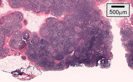
Contents

                                  ix      Conference Committees
                                  xi      Sponsors
                                 xiii     Introduction

                                 xv       Can normal lymph node architecture be characterised by optical coherence
                                          tomography? (Abstract Only)
                                          R. A. McLaughlin, L. Scolaro, B. R. Klyen, Univ. of Western Australia (Australia); S. Hamza, Sir
                                          Charles Gairdner Hospital (Australia); P. Robbins, PathWest QEII Medical Ctr. (Australia);
                                          C. Saunders, Sir Charles Gairdner Hospital (Australia) and Univ. of Western Australia
                                          (Australia); D. D. Sampson, Univ. of Western Australia (Australia)

                                 xix      A first demonstration of audio-frequency optical coherence elastography of tissue
                                          (Abstract Only)
                                          S. G. Adie, S. A. Alexandrov, J. J. Armstrong, B. F. Kennedy, D. D. Sampson, Univ. of Western
                                          Australia (Australia)

                                          OPTICAL SOURCES

                          7139 02         Femtosecond lasers for optical coherence tomography (Invited Paper) [7139-01]
                                          H. M. Crespo, C. C. Rosa, Univ. of Porto (Portugal)

                          7139 03         Optical fiber sources for measurement and imaging (Invited Paper) [7139-02]
                                          A. B. Lobo Ribeiro, M. Melo, J. R. Salcedo, Multiwave Photonics, S.A. (Portugal)

                          7139 04         Towards 1.0 W CW reliable SLD at 840 nm [7139-03]
                                          Yu. O. Kostin, P. I. Lapin, V. V. Prokhorov, V. R. Shidlovsky, S. D. Yakubovich, Superlum Diodes
                                          Ltd. (Russian Federation)

                          7139 05         Towards 100 nm Band NIR SLDs [7139-04]
                                          Yu. O. Kostin, P. I. Lapin, V. V. Prokhorov, V. R. Shidlovsky, S. D. Yakubovich, Superlum Diodes
                                          Ltd. (Russian Federation)

                          7139 06         Development of fibre optic broadband sources at 1 µm region for optical coherence
                                          tomography [7139-05]
                                          I. Trifanov, M. O. Berendt, J. R. Salcedo, Multiwave Photonics S.A. (Portugal);
                                          A. G. Podoleanu, Univ. of Kent (United Kingdom); A. B. Lobo Ribeiro, Multiwave Photonics
                                          S.A. (Portugal)

                          7139 07         Improvement of the mode quality in large mode area (LMA) fibres [7139-06]
                                          S. Grünsteidl, Multiwave Photonics S.A. (Portugal) and National Univ. of Ireland (Ireland);
                                          J. M. Sousa, Multiwave Photonics S.A. (Portugal); G. O'Connor, T. Glynn, National Univ. of
                                          Ireland (Ireland)

                                                                                                                                             iii

                                                                          Proc. of SPIE Vol. 7139 713901-3

Downloaded From: https://www.spiedigitallibrary.org/conference-proceedings-of-spie on 07 Jul 2021
Terms of Use: https://www.spiedigitallibrary.org/terms-of-use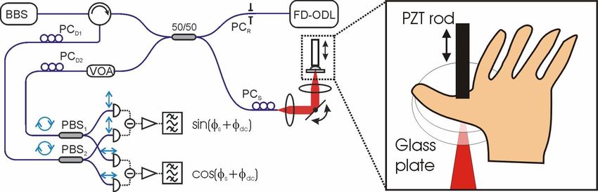
OCT TECHNOLOGY

                          7139 08         Theory and applications of multi-beam OCT [7139-07]
                                          J. Holmes, Michelson Diagnostics Ltd. (United Kingdom)

                          7139 09         Multi-channel time domain spectroscopic optical coherence tomography system [7139-08]
                                          A. Meadway, S. H. H. Darbrazi, R. Cernat, G. Dobre, A. G. Podoleanu, Univ. of Kent (United
                                          Kingdom); R. B. Rosen, The New York Eye and Ear Infirmary (United States)

                         7139 0A          Multiple delay lines full-field optical coherence tomography [7139-09]
                                          J. Wang, Univ. of Kent (United Kingdom); C. Dainty, National Univ. of Ireland (Ireland);
                                          A. G. Podoleanu, Univ. of Kent (United Kingdom)

                          7139 0B         En face OCT system at 1060 nm [7139-10]
                                          L. Neagu, Univ. of Kent (United Kingdom); A. B. Lobo Ribeiro, Multiwave Photonics S.A.
                                          (Portugal); R. G. Cucu, A. Bradu, L. Ma, A. G. Podoleanu, Univ. of Kent (United Kingdom)

                         7139 0C          Application of optical coherence tomography for imaging of scaffold structure and
                                          micro-flows characterization [7139-11]
                                          B. Veksler, E. Kobzev, M. Bonesi, I. Meglinski, Cranfield Univ. (United Kingdom)

                         7139 0D          Theoretical approach on a galvanometric scanner with an enhanced duty cycle [7139-12]
                                          V.-F. Duma, Aurel Vlaicu Univ. of Arad (Romania); A. G. Podoleanu, Univ. of Kent (United
                                          Kingdom)

                          7139 0E         Denoising based on noise parameter estimation in speckled OCT images using neural
                                          network [7139-13]
                                          M. R. N. Avanaki, P. P. Laissue, A. G. Podoleanu, A. Hojjat, Univ. of Kent (United Kingdom)

                                          OCT MICROSCOPY

                          7139 0F         Gabor domain optical coherence microscopy (Invited Paper) [7139-14]
                                          J. P. Rolland, P. Meemon, S. Murali, A. Jain, N. Papp, College of Optics & Photonics, Univ. of
                                          Central Florida (United States); K. P. Thompson, Optical Research Associates (United States);
                                          K. Lee, College of Optics & Photonics, Univ. of Central Florida (United States)

                         7139 0G          Using en face optical coherence tomography to analyse gene function in Drosophila
                                          melanogaster larval heart [7139-15]
                                          A. Bradu, L. Ma, J. Bloor, A. Podoleanu, Univ. of Kent (United Kingdom)

                         7139 0H          In vivo imaging of adult zebrafish using optical coherence tomography [7139-16]
                                          Y. Verma, K. Divakar Rao, P. K. Gupta, Raja Ramanna Ctr. for Advanced Technology (India)

                                          OCT IN THE CLINIC

                           7139 0I        Doppler optical coherence tomography in cardiovascular physiology [7139-17]
                                          M. Bonesi, Univ. of Sheffield (United Kingdom); I. Meglinski, Cranfield Univ. (United Kingdom);
                                          S. Matcher, Univ. of Sheffield (United Kingdom)

                iv

                                                                          Proc. of SPIE Vol. 7139 713901-4

Downloaded From: https://www.spiedigitallibrary.org/conference-proceedings-of-spie on 07 Jul 2021
Terms of Use: https://www.spiedigitallibrary.org/terms-of-use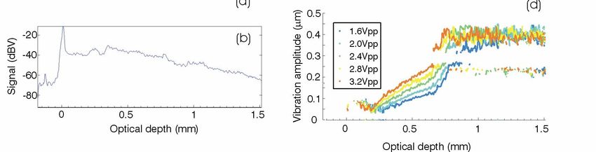
7139 0J         Imaging of basal cell carcinoma tissue using en face OCT [7139-18]
                                          B. R. Penmetsa, Univ. of Kent (United Kingdom); M. Khandwala, Maidstone and Tunbridge
                                          Wells NHS Trust (United Kingdom); A. Bradu, M. Hughes, Univ. of Kent (United Kingdom);
                                          C. A. Jones, J. Schofield, Maidstone and Tunbridge Wells NHS Trust (United Kingdom);
                                          A. G. Podoleanu, Univ. of Kent (United Kingdom)

                          7139 0L         Structural characterization of hair fiber by optical coherence tomography (OCT) [7139-20]
                                          A. Z. Freitas, Instituto de Pesquisas Energéticas e Nucleares (Brazil); M. V. Robes Velasco,
                                          Univ. de São Paulo (Brazil); M. P. Raele, Instituto de Pesquisas Energéticas e Nucleares
                                          (Brazil); T. M. Kaneko, Univ. de São Paulo (Brazil); N. Dias Vieira, Jr., Instituto de Pesquisas
                                          Energéticas e Nucleares (Brazil); A. R. Baby, Univ. de São Paulo (Brazil)

                         7139 0M          En-face OCT microleakage investigation after laser-assisted dental hard tissue treatment
                                          [7139-21]
                                          C. Todea, C. Balabuc, C. Sinescu, M. Negrutiu, L. Filip, Victor Babeş University of Medicine
                                          and Pharmacy, Timişoara (Romania); A. Bradu, A. G. Podoleanu, Univ. of Kent (United
                                          Kingdom)

                         7139 0N          Optical coherence tomography and confocal microscopy investigations of dental
                                          prostheses [7139-22]
                                          M. L. Negrutiu, C. Sinescu, Victor Babeş University of Medicine and Pharmacy, Timişoara
                                          (Romania); M. Hughes, A. Bradu, Univ. of Kent (United Kingdom); M. Rominu, C. Todea,
                                          Victor Babeş University of Medicine and Pharmacy, Timişoara (Romania); G. Dobre,
                                          A. G. Podoleanu, Univ. of Kent (United Kingdom)

                         7139 0O          An innovative approach for investigating the ceramic bracket-enamel interface: optical
                                          coherence tomography and confocal microscopy [7139-23]
                                          R. O. Romînu, C. Sinescu, M. Romînu, M. Negruţiu, Victor Babeş University of Medicine and
                                          Pharmacy, Timişoara (Romania); P. Laissue, Univ. of Kent (United Kingdom); S. Mihali, Victor
                                          Babeş University of Medicine and Pharmacy, Timişoara (Romania); L. Cuc, Aurel Vlaicu Univ.
                                          (Romania); M. Hughes, A. Bradu, A. G. Podoleanu, Univ. of Kent (United Kingdom)

                                          IMAGING THE EYE

                          7139 0P         Simultaneous SLO/OCT imaging of the human retina in vivo with high speed axial eye
                                          motion correction (Invited Paper) [7139-24]
                                          M. Pircher, E. Götzinger, B. Baumann, H. Sattmann, C. K. Hitzenberger, Medical Univ. of
                                          Vienna (Austria)

                         7139 0Q          Revealing fine microstructural morphology in the living human retina using optical
                                          coherence tomography with pancorrection (Invited Paper) [7139-25]
                                          C. Torti, B. Považay, B. Hofer, A. Unterhuber, B. Hermann, W. Drexler, Cardiff Univ. (United
                                          Kingdom)

                          7139 0R         High-speed high-resolution optical coherence tomography at 800 and 1060 nm [7139-26]
                                          B. Považay, B. Hofer, B. Hermann, C. Torti, V. Kajić, A. Unterhuber, W. Drexler, Cardiff Univ.
                                          (United Kingdom)

                                                                                                                                             v

                                                                          Proc. of SPIE Vol. 7139 713901-5

Downloaded From: https://www.spiedigitallibrary.org/conference-proceedings-of-spie on 07 Jul 2021
Terms of Use: https://www.spiedigitallibrary.org/terms-of-use
MODULATION OF OPTICAL REFLECTIVITY

                          7139 0S         Retinal intrinsic optical signal and optical coherence tomography (Invited Paper) [7139-27]
                                          A. R. Tumlinson, B. Hermann, T. H. Margrain, B. Hofer, B. Považay, W. Drexler, Cardiff Univ.
                                          (United Kingdom)

                          7139 0T         En face differential absorption optical coherence tomography with gold nanorods as the
                                          contrast agent [7139-28]
                                          M. Leitner, Univ. of Porto (Portugal), INESC Porto (Portugal), and Univ. of Kent (United
                                          Kingdom); A. Henkel, C. Sönnichsen, Univ. of Mainz (Germany); C. C. Rosa, Univ. do Porto
                                          (Portugal) and INESC Porto (Portugal); A. G. Podoleanu, Univ. of Kent (United Kingdom)

                         7139 0V          Nanoparticles for enhanced contrast optical coherence tomography [7139-30]
                                          C. D. Maule, Univ. of Porto (Portugal) and INESC Porto (Portugal); P. Quaresma, Univ. of Porto
                                          (Portugal) and Univ. Nova de Lisboa (Portugal); P. A. Carvalho, Instituto Superior Técnico
                                          (Portugal); P. Jorge, INESC Porto (Portugal); E. Pereira, C. C. Rosa, Univ. of Porto (Portugal)

                         7139 0W          Ex vivo and in vivo OCT image contrast [7139-31]
                                          N. Krstajić, Univ. of Sheffield (United Kingdom); J. Jacobs, M. Bonesi, L. E. Smith,
                                          P. Deshpande, S. MacNeil, Kroto Research Institute (United Kingdom); R. Smallwood,
                                          Univ. of Sheffield (United Kingdom); S. J. Matcher, Kroto Research Institute (United Kingdom)

                                          ADAPTIVE OPTICS

                          7139 0X         Challenges and possibilities for developing adaptive optics: ultra-high resolution optical
                                          coherence tomography for clinical in vivo retinal imaging (Invited Paper) [7139-32]
                                          R. J. Zawadzki, S. S. Choi, J. W. Evans, J. S. Werner, Univ. of California, Davis (United States)

                          7139 0Y         Optical coherence tomography for the investigation of posterior and anterior eye segments
                                          (Invited Paper) [7139-33]
                                          Y. Yasuno, Univ. of Tsukuba (Japan) and Tokyo Medical Univ. (Japan)

                          7139 0Z         Does transverse chromatic aberration limit performance of AO-OCT retinal imaging?
                                          [7139-34]
                                          D. T. Miller, B. Cense, E. Koperda, R. S. Jonnal, W. Gao, Indiana Univ. (United States)

                          7139 10         Adaptive optics loop for en face optical coherence tomography and laser scanning
                                          confocal microscopy [7139-35]
                                          S. Tuohy, A. Bradu, Univ. of Kent (United Kingdom); F. Harms, N. Chateau, Imagine Eyes
                                          (France); A. G. Podoleanu, Univ. of Kent (United Kingdom)

                          7139 11         Performance assessment of a pupil tracking system for adaptive optics retinal imaging
                                          [7139-36]
                                          B. Sahin, Imagine Eyes (France) and National Univ. of Ireland, Galway (Ireland); F. Harms,
                                          B. Lamory, Imagine Eyes (France)

                          7139 12         Optimization of the temporal performance of a deformable mirror for use in ophthalmic
                                          applications [7139-37]
                                          E. Odlund, Univ. of Kent (United Kingdom) and Imagine Eyes (France); M. Navarro, Imagine
                                          Eyes (France); E. Lavergne, Imagine Optics (France); F. Martins, X. Levecq, Imagine Eyes
                                          (France); A. Dubra, Univ. of Rochester (United States)

                vi

                                                                          Proc. of SPIE Vol. 7139 713901-6

Downloaded From: https://www.spiedigitallibrary.org/conference-proceedings-of-spie on 07 Jul 2021
Terms of Use: https://www.spiedigitallibrary.org/terms-of-use
7139 13 First steps toward 3D high resolution imaging using adaptive optics and full-field optical
coherence tomography [7139-38]
L. Blanco, LESIA, Observatoire de Paris, CNRS, UPMC, Univ. Paris Diderot (France); M. Blavier,
LESIA, Observatoire de Paris, CNRS, UPMC, Univ. Paris Diderot (France) and Ctr.
d’Investigations Cliniques du CHNO des Quinze-Vingts, Univ. Paris VI (France); M. Glanc,
LESIA, Observatoire de Paris, CNRS, UPMC, Univ. Paris Diderot (France) and Groupement
d’Intérêt Scientifique PHASE, ONERA, Observatoire de Paris, CNRS, and Univ. Paris Diderot
(France); F. Pouplard, S. Tick, Ctr. d’Investigations Cliniques du CHNO des Quinze-Vingts,
Univ. Paris VI (France); I. Maksimovic, LESIA, Observatoire de Paris, CNRS, UPMC, Univ. Paris
Diderot (France); G. Chenegros, L. Mugnier, DOTA ONERA (France) and Groupement
d’Intérêt Scientifique PHASE, ONERA, Observatoire de Paris, CNRS, and Univ. Paris Diderot
(France); F. Lacombe, LESIA, Observatoire de Paris, CNRS, UPMC, Univ. Paris Diderot
(France) and Mauna Kea Technologies (France); G. Rousset, LESIA, Observatoire de Paris,
CNRS, UPMC, Univ. Paris Diderot (France) and Groupement d’Intérêt Scientifique PHASE,
ONERA, Observatoire de Paris, CNRS, and Univ. Paris Diderot (France); M. Pâques,
J. Le Gargasson, J. Sahel, Ctr. d’Investigations Cliniques du CHNO des Quinze-Vingts, Univ.
Paris VI (France)
7139 14 Full-range, high-speed, high-resolution 1 µm spectral-domain optical coherence
tomography with BM-scan method for the human posterior eye imaging [7139-39]
S. Makita, Univ. of Tsukuba (Japan) and Computational Optics and Opthamology Group
(Japan); T. Fabritius, Univ. of Tsukuba (Japan) and Univ. of Oulu (Finland); M. Miura, Tokyo
Medical Univ. (Japan) and Computational Optics and Opthamology Group (Japan);
Y. Yasuno, Univ. of Tsukuba (Japan) and Computational Optics and Opthamology Group
(Japan)
OCT FOR ART
7139 15 Optical coherence tomography in archaeological and conservation science: a new
emerging field (Invited Paper) [7139-40]
H. Liang, B. Peric, Nottingham Trent Univ. (United Kingdom); M. Hughes, A. Podoleanu, Univ.
of Kent (United Kingdom); M. Spring, The National Gallery (United Kingdom); S. Roehrs, The
British Museum (United Kingdom)
7139 16 Optical coherence tomography for non-destructive investigations of structure of easel
paintings [7139-41]
E. Kwiatkowska, M. Sylwestrzak, B. J. Rouba, L. Tymińska-Widmer, M. Iwanicka, P. Targowski,
Nicolaus Copernicus Univ. (Poland)
7139 17 A swept source OCT at 1300 nm with angular compounding for art and archaeological
conservation [7139-42]
M. Hughes, D. A. Jackson, A. G. Podoleanu, Univ. of Kent (United Kingdom)
Author Index
vii
Proc. of SPIE Vol. 7139 713901-7
Downloaded From: https://www.spiedigitallibrary.org/conference-proceedings-of-spie on 07 Jul 2021
Terms of Use: https://www.spiedigitallibrary.org/terms-of-useProc. of SPIE Vol. 7139 713901-8 Downloaded From: https://www.spiedigitallibrary.org/conference-proceedings-of-spie on 07 Jul 2021 Terms of Use: https://www.spiedigitallibrary.org/terms-of-use
Conference Committees

                              Conference Chair and Marie Curie Coordinator
                                                Adrian Podoleanu, University of Kent (United Kingdom)

                              International Organising Committee

                                                Peter E. Andersen, Technical University of Denmark (Denmark)
                                                Nicolas Chateau, Imagine Eyes, Paris (France)
                                                Tatyana Cherezova, Active Optics Ltd, Moscow (Russian Federation)
                                                Chris Dainty, National University of Ireland, Galway (Ireland)
                                                Aristide Dogariu, University of Central Florida, Orlando, Florida (United
                                                  States)
                                                Wolfgang Drexler, Cardiff University, Cardiff (United Kingdom)
                                                Costel Flueraru, National Research Council Canada, Ottawa
                                                  (Canada)
                                                Thomas Glynn, National University of Ireland, Galway (Ireland)
                                                Fabrice Harms, Imagine Eyes, Paris (France)
                                                Joseph Izatt, Duke University (United States)
                                                Dean Johnson, Haag-Streit (United Kingdom)
                                                Peter Koch, Thorlabs Luebeck (Germany)
                                                Antonio Lobo, Multiwave Photonics, Porto (Portugal)
                                                Donald Miller, Indiana University (United States)
                                                Carla Rosa, University of Porto (Portugal)
                                                Richard Rosen, New York Eye and Ear Infirmary (United States)
                                                Jose Salcedo, Multiwave Photonics, Porto (Portugal)
                                                David Sampson, University of Western Australia, Crawley (Australia)
                                                Jose Luis Santos, University of Porto (Portugal)
                                                Ruikang Wang, Oregon Health and Science University (United States)
                                                Maciej Wojtkowski, Torun University (Poland)

                              Local Organising Committee Chair
                                                George Dobre, University of Kent (United Kingdom)

                              Local Organising Committee
                                                Pat Douglas, University of Kent (United Kingdom)
                                                Maggie Francis, University of Kent (United Kingdom)
                                                Michael Hughes, University of Kent (United Kingdom)
                                                David Jackson, University of Kent (United Kingdom)
                                                James Redmond, University of Kent (United Kingdom)
                                                Simon Tuohy, University of Kent (United Kingdom)

                                                                                                                            ix

                                                                          Proc. of SPIE Vol. 7139 713901-9

Downloaded From: https://www.spiedigitallibrary.org/conference-proceedings-of-spie on 07 Jul 2021
Terms of Use: https://www.spiedigitallibrary.org/terms-of-use
Session Chairs
1 OCT Microscopy
Robert Huber, Ludwig-Maximilians-Universität München (Germany)
2 OCT in the Clinic
Jannick Rolland, University of Central Florida (United States)
3 OCT Technology
David Sampson, University of Western Australia (Australia)
4 Optical Sources I
Antonio B. Lobo Ribeiro, Multiwave Photonics S.A. (Portugal)
5 Optical Sources II
Thomas Glynn, National University of Ireland, Galway (Ireland)
6 Modulation of Optical Reflectivity
Yoshiaki Yasuno, University of Tsukuba (Japan)
7 OCT for Art
Michael Pircher, Medizinische Universität Wien (Austria)
8 General Skills
Adrian Podoleanu, University of Kent (United Kingdom)
9 Adaptive Optics I
Robert Zawadzki, University of California, Davis (United States)
10 Imaging the Eye I
Christopher Dainty, National University of Ireland, Galway (Ireland)
11 Adaptive Optics II,
Donald T. Miller, Indiana University (United States)
12 Imaging the Eye II
Wolfgang Drexler, Cardiff University (United Kingdom)
x
Proc. of SPIE Vol. 7139 713901-10
Downloaded From: https://www.spiedigitallibrary.org/conference-proceedings-of-spie on 07 Jul 2021
Terms of Use: https://www.spiedigitallibrary.org/terms-of-useSponsors
U)
z
U
4
MARIE CURIE
httpJ/ec.europeuIm?riecurieathon
-iiagine eyes
Michelson
DIAGNOSTICS
V muI11wcve
I
THE RATIU FAMILY
CHARITABLE FOUNDATION
santec
SUPERLUM
ThOR]
University of
Kent
xi
Proc. of SPIE Vol. 7139 713901-11
Downloaded From: https://www.spiedigitallibrary.org/conference-proceedings-of-spie on 07 Jul 2021
Terms of Use: https://www.spiedigitallibrary.org/terms-of-useProc. of SPIE Vol. 7139 713901-12 Downloaded From: https://www.spiedigitallibrary.org/conference-proceedings-of-spie on 07 Jul 2021 Terms of Use: https://www.spiedigitallibrary.org/terms-of-use
Introduction

                              The 1st Canterbury Workshop on Optical Coherence Tomography and Adaptive
                              Optics has been initiated as a reporting meeting of the Marie Curie Training Site
                              (MCTS) for early stage training (EST) researchers, MEST-2005-020353 supported by
                              the European Commission (EC) with participants from National University of
                              Ireland, Galway, University of Porto, Imagine Eyes, Multiwave Photonics, and
                              coordinated by the University of Kent. The reporting meeting has the generous
                              support of the EC to gather over 20 supervisors and EST researchers to review the
                              achievements in research of the Marie Curie Training Site.
                              In opening participation to this meeting to the outside world we aimed not only
                              for a wider scientific communication exercise, but also to add education to our
                              EST researchers by involving them in an international exchange of ideas with
                              other specialists in their fields: optical coherence tomography (OCT) and
                              adaptive optics (AO). At the same time, we hope that other researchers and
                              specialists in these fields take advantage of the educational component of this
                              meeting.
                              We are grateful to the sponsors who agreed to offer their generous support to
                              transform the Marie Curie meeting into a larger size workshop with wide
                              international participation. These industrial sponsors are innovative companies
                              active in the field of systems and devices for OCT and AO: Imagine Eyes
                              (France), Michelson Diagnostics (United Kingdom), Multiwave Photonics
                              (Portugal), Thorlabs (United Kingdom), Santec (Japan), and Superlum (Russia).
                              We also appreciate the continuing support given by the Ratiu Family Charitable
                              Foundation to the University of Kent in the form of bursaries enabling Romanian
                              students to further their education in the field of biomedical imaging and we
                              thank the Foundation for co-sponsoring this workshop.
                              The site of the event is the University of Kent where research in OCT dates back to
                              1991, when some of the precursors of spectral domain OCT, Talbot bands, and
                              channelled spectrum low coherence interferometry for sensing were
                              investigated.
                              The field of non-invasive high resolution imaging has progressed considerably in the
                              last three years. Among the participants to the workshop we were fortunate to
                              have leaders in the field, who have been pushing the limits of acquisition speed.
                              We will all be interested in finding out where those limits stand today. In the last few
                              years, spectral domain OCT led to a significant increase in the number of Mega-
                              voxels acquired from the tissue volume, 100 times more than in time domain OCT.
                              Using swept sources, we expect this limit to exceed 350 Mega-voxels, while good

                                                                                                                         xiii

                                                                         Proc. of SPIE Vol. 7139 713901-13

Downloaded From: https://www.spiedigitallibrary.org/conference-proceedings-of-spie on 07 Jul 2021
Terms of Use: https://www.spiedigitallibrary.org/terms-of-use
images of the retina at 122 Mega-voxels have been demonstrated so far1. The time
to generate an en-face image in spectral domain OCT has also decreased, but
we have not achieved as yet the capability of resonant scanning time domain
OCT. A steady flow of research is manifest in improved resolution: in depth by OCT,
and lateral by AO. In a B-scan generated by spectral OCT, we have recently seen
the first demonstration of AO correction on shrinking the depth profile of the
confocal microscope in the retina, however we are still far from achieving an
ultimate resolution of 50 microns in the in vivo retina.
How will the two technologies, OCT and AO, look when this limit is finally
achieved? How will they be best combined? These are stimulating challenges for
our continuing research in pushing the limits of our capabilities.
We hope that the 1st Canterbury Workshop in Optical Coherence Tomography
and Adaptive Optics will contribute towards advancing the frontier of knowledge
in OCT and AO beyond the ‘natural’ limits which appear to be imposed by
current technology.
Adrian Podoleanu
Conference Chair and Marie Curie Coordinator
1 A. Gh. Podoleanu, R. B. Rosen, “Combinations of techniques in imaging the retina
with high resolution,“ Progress in Retinal and Eye Research, Vol. 27, No. 4, 464-499
(2008).
xiv
Proc. of SPIE Vol. 7139 713901-14
Downloaded From: https://www.spiedigitallibrary.org/conference-proceedings-of-spie on 07 Jul 2021
Terms of Use: https://www.spiedigitallibrary.org/terms-of-useCan normal lymph node architecture be characterised by optical
coherence tomography?
R.A.McLaughlina, L.Scolaroa, B.R.Klyena, S.Hamzab, P.Robbinsc, C.Saundersb,d, D.D.Sampsona
a
Optical+Biomedical Engineering Laboratory, Uni. Western Australia, Crawley WA 6009, Australia
b
Sir Charles Gairdner Hospital, Crawley WA 6009, Australia
c
PathWest QEII Medical Centre, Crawley WA 6009, Australia
d
School of Surgery, Uni. Western Australia, Crawley WA 6009, Australia
ABSTRACT
Assessment of lymph node involvement is a key prognostic marker in early breast cancer. This paper demonstrates the
ability of optical coherence tomography (OCT) to characterise the micro-architecture of healthy, non-cancerous lymph
nodes. OCT is shown to differentiate stroma, cortex and adipose tissue. Characteristic patterns are also identified for
germinal centres and blood vessels within the node. Results are correlated against a histopathological gold standard.
Keywords: breast cancer, lymph node, optical coherence tomography, OCT.
1. INTRODUCTION
Breast cancer is the most commonly diagnosed cancer in women in the USA, with an incidence rate of 117.7 per 100,000
women [1]. In England, over 38,000 cases are diagnosed each year [2]. A critical stage in the management of cancer is to
assess the spread (metastasis) of the malignancy to the lymphatic system. The lymphatic system is a network of vessels
and lymph nodes throughout the body which forms a key part of the immune system.
A common surgical option to evaluate lymph node involvement is axillary clearance. This involves removal of some or
all of the 25-30 lymph nodes from the axilla (armpit). The excised lymph nodes are examined using histopathological
techniques to identify the presence of malignant cells. However, this results in the excision of many healthy (non-
cancerous) lymph nodes and approximately 26% of patients will suffer lymphoedema [3]. Lymphoedema is caused by an
accumulation of lymph (fluid) in the tissue, and manifests as a chronic swelling of the arm, breast or chest wall.
Optical coherence tomography is an optical imaging modality, using coherence-gated light in the near infrared spectrum.
It has been used to obtain high-resolution images of the structural changes caused by cancer in the gastrointestinal tract
[4][5], breast [6], biliary system [7] and cervix [8]. Recently, Luo et al. [9] proposed using OCT in the assessment of
axillary lymph nodes. OCT provides the possibility of an imaging-based optical biopsy method for determining lymph
node involvement. If performed in vivo, such a tool could potentially avoid the unnecessary excision of healthy lymph
nodes; thus, reducing the incidence of complications such as lymphoedema.
When cancer metastasises to a lymph node, it disrupts the node’s normal micro-architecture. In this paper, we describe
the use of OCT to characterise the micro-architecture of excised normal (non-cancerous) lymph nodes. This work is an
important preliminary step in establishing the feasibility of using OCT to optically identify cancerous lymph nodes.
2. EXPERIMENT
For this study, 18 lymph nodes were obtained from 13 patients undergoing either axillary clearance or sentinel lymph
node biopsy. Within 10 minutes of excision, the fresh (unfixed) tissue was dissected into 2mm slices. Each slice was
imaged using a time-domain OCT system with a source centre wavelength of 1320nm and full-width-half-maximum
bandwidth of 101nm. Axial and lateral resolutions of the system were ~11µm. After scanning, each sample was
immediately fixed in formalin and underwent histological assessment using Haematoxylin and Eosin (H&E) staining.
Image registration was then performed between the 3D OCT and 2D histology. Custom visualisation software was
implemented in C++ on a Windows platform to enable the arbitrary rotation, translation and zooming of the 3D OCT
data. This enabled the H&E image to be matched against the optimal 2D plane extracted from the 3D OCT data.
Proc. of SPIE Vol. 7139 713901-15
Downloaded From: https://www.spiedigitallibrary.org/conference-proceedings-of-spie on 07 Jul 2021
Terms of Use: https://www.spiedigitallibrary.org/terms-of-use
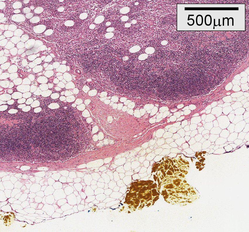
xvCapsule Capsule
Cortex Cortex (stroma)
(stroma)
Adipose
Adipose
Ink Ink
Figure 1: (Left) OCT image of lymph node. (Right) H&E histology: showing lymph cortex, adipose and stroma.
3. RESULTS AND DISCUSSION
Figure 1 demonstrates the ability of OCT to differentiate the primary tissue types found in lymph nodes: stroma, cortex
and adipose (fat). The stroma consists of fibrous tissue forming the lymph node capsule, and also the delicate trabeculae
which extend into the node parenchyma. The cortex is the outer portion of the lymph node. The dark staining visible in
the H&E stained image is due to the high concentration of lymphocytes found in the cortex. Lymphocytes are a type of
white blood cell produced in the lymph node and play a critical role in the body’s immune system. The adipose tissue
appears as a white area with a characteristic honeycomb structure, which is also evident in the histological images.
Figure 2 shows a cross-sectional image through two node-related blood vessels. The lymph node contains a network of
lymphovascular spaces that allow the various cellular components of the immune system to enter and depart the node.
Blood vessels are characterised by a dark ring surrounding an area of low back-reflection.
A non-cancerous reactive lymph node is shown in Figure 3, with prominent ‘secondary’ lymphoid follicles containing
germinal centres within the cortex. Broadly speaking, secondary ‘active’ lymphoid follicles form in the cortex as part of
the B-cell immune response to antigens. Under H&E staining, they are characterised by a pale germinal centre, encircled
by a darker mantle zone of lymphocytes. Under OCT scanning, the circular structure of the germinal centre can be seen.
Several artefacts were observed during imaging. Adipose tissue often resulted in an increased back-reflection signal from
deeper tissue. This was evidenced as dark spots (high reflection) further into the A-scan. This artefact can also be seen in
the images of [9]. The mechanism for this effect has not yet been verified and will require further investigation.
Additionally, the image quality was noted to deteriorate deeper in the tissue due to the cumulative effects of scattering,
absorption and beam spread.
Blood
Blood vessels
vessels Blood
Blood vessels
vessels
Figure 2: (left) OCT and (right) H&E histology showing blood vessels.
Proc. of SPIE Vol. 7139 713901-16
Downloaded From: https://www.spiedigitallibrary.org/conference-proceedings-of-spie on 07 Jul 2021
Terms of Use: https://www.spiedigitallibrary.org/terms-of-use
xviGerminal
Germinal Germinal
Germinal
centers
centers centers
centers
Figure 3: (left) OCT and (right) H&E histology showing lymph germinal centres and mantles.
4. CONCLUSION
In this paper, we have demonstrated the ability of OCT to image the micro-architecture of normal lymph nodes, with
correlation against a histological gold standard. OCT was seen to differentiate tissue types such as cortex, stroma and
adipose. In addition, characteristic patterns have been identified for microvasculature and germinal centres. Future work
will focus on characterising changes in the micro-architecture due to the presence of cancer.
REFERENCES
[1]
U.S. Cancer Statistics Working Group, “United States cancer statistics: 2004 incidence and mortality”. Atlanta: U.S.
Department of Health and Human Services, Centers for Disease Control and Prevention and National Cancer
Institute (2007).
[2]
National Statistics, “Cancer statistics registrations: registrations of cancer diagnosed in 2005, England”, Series
MB1(36), (2006).
[3]
Erickson V.S., Pearson M.L., Ganz P.A., Adams J., Kahn K.L., “Arm edema in breast cancer patients”, Journal of
the National Cancer Institute 93(2), 96-111 (2001)
[4]
Bouma B.E., Tearney G.J., Compton C.C., Nishioka N.S., "High-resolution imaging of the human esophagus and
stomach in vivo using optical coherence tomography", Gastrointestinal Endoscopy 51(4), 467-474 (2000).
[5]
Evans J.A. et al., "Optical coherence tomography to identify intramucosal carcinoma and high-grade dysplasia in
Barrett’s Esophagus", Clinical Gastroenterology and Hepatology 4, 38-43 (2006).
[6]
Boppart S.A., Luo W., Marks D.L., Singletary K.W., “Optical coherence tomography: feasibility for basic research
and image-guided surgery of breast cancer”, Breast Cancer Research and Treatment 84, 85–97 (2004)
[7]
Poneros J.M., Tearney G.J., Shiskov M., Kelsey P.B., Lauwers G.Y., Nishioka N.S., Bouma B.E., “Optical
coherence tomography of the biliary tree during ERCP”, Gastrointestinal Endoscopy 55(1), 84-88 (2002)
[8]
Escobar P.F., Belinson J.L., White A., Shakhova N.M., Feldchtein F.I., Kareta M.V., Gladkova N.D., “Diagnostic
efficacy of optical coherence tomography in the management of preinvasive and invasive cancer of uterine cervix
and vulva”, International Journal of Gynecological Cancer 14, 470-474 (2004)
[9]
Luo W., Nguyen F.T., Zysk A.M., Ralston T.S., Brockenbrough J., Marks D.L., Oldenburg A.L., Boppart S.A.,
"Optical biopsy of lymph node morphology using optical coherence tomography”, Technology in Cancer Research
& Treatment 4(5), 539-547 (2005).
Proc. of SPIE Vol. 7139 713901-17
Downloaded From: https://www.spiedigitallibrary.org/conference-proceedings-of-spie on 07 Jul 2021
Terms of Use: https://www.spiedigitallibrary.org/terms-of-use
xviiProc. of SPIE Vol. 7139 713901-18 Downloaded From: https://www.spiedigitallibrary.org/conference-proceedings-of-spie on 07 Jul 2021 Terms of Use: https://www.spiedigitallibrary.org/terms-of-use
A first demonstration of audio-frequency optical coherence
                                               elastography of tissue
                  Steven G. Adie, Sergey A. Alexandrov, Julian J. Armstrong, Brendan F. Kennedy and David D. Sampson
                 Optical+Biomedical Engineering Laboratory, School of Electrical, Electronic & Computer Engineering, The
                     University of Western Australia, 35 Stirling Highway, Crawley, Western Australia 6009, Australia

                                                                                   ABSTRACT

                Optical elastography is aimed at using the visco-elastic properties of soft tissue as a contrast mechanism, and could be
                particularly suitable for high-resolution differentiation of tumour from surrounding normal tissue. We present a new
                approach to measure the effect of an applied stimulus in the kilohertz frequency range that is based on optical coherence
                tomography. We describe the approach and present the first in vivo optical coherence elastography measurements in
                human skin at audio excitation frequencies.
                Keywords: Elastography, optical coherence tomography, tissue mechanical properties

                1. Introduction
                Optical coherence elastography (OCE) reported to date has been based on speckle-tracking techniques, and has
                employed predominantly quasi-static mechanical loading of tissue to quantitatively assess local tissue motion [1-4]. In
                this paper, we present a new approach to OCE suitable for the quantitative measurement of tissue mechanical properties
                in the hundred Hertz to kilohertz frequency range.

                2. Theory and experimental method
                Consider an interferometric signal generated by a reference light beam combining with light backscattered from particles
                undergoing harmonic displacement along the optical (z) axis. At frequencies up to several kHz (with corresponding
                sound wavelength Λ ~ 1m), particles in a medium (of typical thickness ~ 1mm) can be expected to move in phase with
                each other. The dynamic interferometric signal amplitude of interest depends not only on the scatterer’s vibration
                amplitude but also on the quasi-static phase of the interferometer, which in turn is governed by the precise differential
                axial position of the particle relative to the reference path in the interferometer. This unwanted dependence of the
                dynamic displacement on the quasi-static displacement is generally known as interferometric signal fading [5,6]. It can
                be overcome by various means, e.g., by polarization-based optical quadrature detection [7].
                A schematic of the experimental fibre-based, time-domain OCT system utilizing balanced optical quadrature detection is
                presented in Fig. 1. A broadband source with a near-Gaussian spectrum centred at 1334nm with 3dB bandwidth of 42nm
                was employed, and the sample arm utilized a triplet lens (f = 30mm) to focus the beam through a glass window that
                provided a rigid platform upon which the samples were placed. Dynamic compression was applied to the sample, as
                indicated in Fig. 1 (inset), with a PZT actuator. The frequency-domain optical delay line (FD-ODL) was operated off-
                pivot to acquire conventional OCT images using a carrier frequency of 1150 Hz [8] and on-pivot when the vibration of
                sample scatterers generated the carrier frequency.

                                                                                                        PZT rod

                                                                                                       Glass
                                                                                                       plate

                Figure 1: Schematic of the fibre-based OCT system employing balanced optical quadrature detection and the
                experimental geometry.

                                                                                                                                    xix

                                                                         Proc. of SPIE Vol. 7139 713901-19

Downloaded From: https://www.spiedigitallibrary.org/conference-proceedings-of-spie on 07 Jul 2021
Terms of Use: https://www.spiedigitallibrary.org/terms-of-use
The detected photocurrents in the orthogonal polarization channels (see Fig. 1), denoted by the subscripts 1 and 2 can be
expanded as a series of Bessel functions [6], resulting in the following equation:
⎧ ⎡ ∞ ⎤ ⎡ ∞ ⎤⎫
i ( z, t) = 2 ρ I I
1 R S ⎨ cos φ DC ⎢ J 0 (φ s ) + 2 ∑ J 2 n (φ s ) cos( 2 n Ω t ) ⎥ + sin φ DC ⎢ 2 ∑ J 2 n + 1 (φ s ) sin (( 2 n + 1) Ω t )⎥ ⎬ , (1)
⎩ ⎢⎣ n =1 ⎥⎦ ⎢⎣ n = 0 ⎥⎦ ⎭
⎧ ⎡ ∞ ⎤ ⎡ ∞ ⎤⎫
i ( z , t ) = 2 ρ I I ⎨sin φ ⎢ J (φ ) + 2 ∑ J (φ ) cos( 2 nΩt ) ⎥ + cos φ ⎢2 ∑ J (φ ) sin (( 2 n + 1) Ωt )⎥ ⎬ , (2)
2 R S DC ⎢ 0 s 2n s ⎥⎦ DC ⎢ 2n + 1 s ⎥⎦ ⎭
⎩ ⎣ n =1 ⎣ n=0
where ρ is the detector responsivity, IS and IR are the sample and reference optical intensities, respectively, φ is the
DC
4π
quasi-static interferometric phase (modulo 2π) governed by the axial position of the particle, φ = d ( z , Ω) , where λ
s λ
is the mean optical wavelength in the medium, d is the local, generally frequency-dependent, amplitude of vibration, Ω
is the angular vibration frequency and Jn is the nth order Bessel function of the 1st kind corresponding to the nth order
harmonic of the excitation frequency. It is readily seen from Eqs. (1) and (2) that the value of φ alters the detected
DC
signal and has the potential to produce distortions in the dynamic signal. In channel 1 (Eq. (1)), the even harmonics of
the signal fade when φ = π / 2 ; conversely, in channel 2, the odd harmonics of the signal fade when φ =π /2.
DC DC
The objective of the OCE scheme presented here is to deduce the absolute vibration amplitude (i.e., dynamic
displacement) of the scatterer through an interferometric measurement of φ . In our approach, the theoretical values of
S
J3/J1 are used to map the experimentally measured J3/J1 ratios to vibration amplitude, as described previously [6].
Furthermore, we use the ratio of J4/J2 to extend the unambiguous range of operation to beyond approximately λ/3; the
first calibration range given by J4/J2 < −4.696dB and the second by J4/J2 ≥ −4.696dB. At an operating mean wavelength
of λ =1300 nm, this permits measurement of absolute vibration amplitudes of up to about 0.6µm (optical depth).
Analysis of the full-fringed digitized data was carried out in post-processing, separately for each channel. We calculated
intensity images for four separate harmonic frequencies by applying bandpass filters centred at Ω, 2Ω, 3Ω and 4Ω
(producing in total eight frames from the two detection channels). These frames were then separately demodulated
utilizing the Hilbert transform and the resulting orthogonal envelopes at each vibration harmonic were incoherently
combined. The resulting four frames, immune to signal fading, were used to produce a frame containing local vibration
amplitude using the ratio J3/J1 as described above.
3. Results and discussion
Figure 2 presents results of in vivo measurements of human thick skin (on the palm) obtained by compressing the
“webbing” between the thumb and index finger between the PZT rod and the sample-arm window. The OCT image
(without applied vibration) consists of 32 A-scans taken over a lateral scan range of 0.8 mm. The lateral spacing was
relatively large compared to the lateral resolution in order to obtain an adequate field of view with the relatively slow A-
scan rate. The vibration amplitude measurement consisted of 8 A-scans centred at the same lateral position, but over a
0.4-mm range, again to avoid boundary effects. A relatively uniform signal from the skin surface to z = 0.2 mm optical
depth can be distinguished in both the OCT image (Figure 2a) and the average A-scan (Figure 2b), which corresponds to
a layer with low vibration amplitude in Figure 2c. This layer is attributed to the stratum corneum, which displays
negligible vibration amplitude due to its tight coupling to the rigid glass window and its low compressibility.
Below the stratum corneum, the OCT image does not show any clear boundary between skin layers until a depth of
approximately 0.85 mm. In contrast, the vibration amplitude traces show a distinct difference in slope at about 0.65 mm
that is replicated over the range of PZT drive voltages. The change in slope of the vibration amplitude traces indicates
two layers of different compressibility, with the deeper layer, with greater slope, being relatively more compressible. We
attribute these layers to the epidermis and dermis respectively, which are known to have distinct mechanical properties.
Beyond a depth of z = 0.85 mm, the signal is most likely due to the multiple scattering background. The discontinuity in
xx
Proc. of SPIE Vol. 7139 713901-20
Downloaded From: https://www.spiedigitallibrary.org/conference-proceedings-of-spie on 07 Jul 2021
Terms of Use: https://www.spiedigitallibrary.org/terms-of-usethe vibration traces of Figure 2d is a stitching artefact occurring at the intersection of the calibration curves (when the
vibration amplitude is approximately λ/3). The dynamic OCE method put forward here naturally detects the axial motion
of scatterers. Accordingly, our experimental geometry was designed so that, in each case, the PZT transducer applied
even pressure to the sample coaxially with the optical beam. Further work is required to establish the feasibility of
accurately detecting vibration amplitude in the presence of lateral scatterer motion, such as when shear waves are
excited.
Im
0.3
0.2
0.1
:0.4
(C
(d)
1.6Vpp
-20
0a 0.4 2.OVpp
o -40 0.3 2.4Vpp
a -60 E 2.8Vpp
a 0.2 3.2Vpp
=
-80
0
0.1
n
0.5
Optical depth (mm)
1 1.5
>0 0 0.5 1.5
Optical depth (mm)
Figure 2: OCT A-scan and OCE-derived vibration amplitude versus optical depth for human skin in vivo: (a)
OCT image without applied vibration; (b) Average A-scan; (c) Vibration amplitude image for PZT drive voltage
of 2.4Vpp; and (d) Average vibration amplitude for various PZT drive voltages.
4. Conclusions
In conclusion, we have presented a new dynamic OCE method suited to quantitative displacement assessment at
frequencies in the hundreds of Hertz to kilohertz regime. The method was able to distinguish the layers in a 3-layer
phantom with different elasticity based on differences in the measured vibration amplitude in each layer. The first
quantitative in vivo OCE measurements of human skin clearly distinguish layers based upon their mechanical response.
In these measurements, increased driving amplitude produced a higher gradient of vibration amplitude vs. depth,
indicating greater compression of compliant layers. The technique can be adapted to perform high-spatial resolution
vibration-based spectroscopy, where the excitation frequency is tuned over the 100 Hz to 10 kHz range. Such
spectroscopic measurements based on the frequency-dependent mechanical response of tissue could potentially enhance
the capability of OCE in diagnosis of a range of medical conditions, including tumours and arterial plaques.
References
[1] J. M. Schmitt. OCT elastography: imaging microscopic deformation and strain of tissue. Optics Express, 3(6):199–
211, 1998.
[2] R. C. Chan, A. H. Chau, W. C. Karl, S. Nadkarni, A. S. Khalil, N. Iftimia, M. Shishkov, G. J. Tearney, M. R.
Kaazempur-Mofrad, and B. E. Bouma. OCT-based arterial elastography: robust estimation exploiting tissue
biomechanics. Optics Express, 12(19):4558–4572, 2004.
[3] J. Rogowska, N. A. Patel, J. G. Fujimoto, and M. E. Brezinski. Optical coherence tomographic elastography
technique for measuring deformation and strain of atherosclerotic tissues. Heart, 90(5):556–562, 2004.
[4] H. J. Ko, W. Tan, R. Stack, and S. A. Boppart. Optical coherence elastography of engineered and developing tissue.
Tissue Engineering, 12(1):63–73, 2006.
[5] E. Udd. Fiber optic sensors : an introduction for engineers and scientists. Wiley, New York, 1991.
[6] O. Sasaki and H. Okazaki. Sinusoidal phase modulating interferometry for surface profile measurement. Applied
Optics, 25(18):3137–3140, 1986.
xxi
Proc. of SPIE Vol. 7139 713901-21
Downloaded From: https://www.spiedigitallibrary.org/conference-proceedings-of-spie on 07 Jul 2021
Terms of Use: https://www.spiedigitallibrary.org/terms-of-use[7] Y. Zhao, Z. Chen, Z. Ding, H. Ren, and J. S. Nelson. Real-time phase-resolved functional optical coherence
tomography by use of optical Hilbert transformation. Optics Letters 27(2): 98-100, 2002.
[8] A. V. Zvyagin, E. D. J. Smith, D. D. Sampson. Delay and dispersion characteristics of a frequency-domain optical
delay line for scanning interferometry. Journal of the Optical Society of America A. 20(2):333-341, 2003.
xxii
Proc. of SPIE Vol. 7139 713901-22
Downloaded From: https://www.spiedigitallibrary.org/conference-proceedings-of-spie on 07 Jul 2021
Terms of Use: https://www.spiedigitallibrary.org/terms-of-useYou can also read