Cyclic oligoadenylate signalling and regulation by ring nucleases during type III CRISPR defence
←
→
Page content transcription
If your browser does not render page correctly, please read the page content below

Downloaded from rnajournal.cshlp.org on August 22, 2021 - Published by Cold Spring Harbor Laboratory Press

 Cyclic oligoadenylate signalling and regulation by ring nucleases
                             during type III CRISPR defence

                         Januka S. Athukoralage* and Malcolm F. White

 Biomedical Sciences Research Complex, School of Biology, University of St Andrews, St Andrews,
                                              United Kingdom

                          *For correspondence e-mail: jsa8@st-andrews.ac.uk

Abstract
In prokaryotes, CRISPR-Cas immune systems recognise and cleave foreign nucleic acids to
defend against Mobile Genetic Elements (MGEs). Type III CRISPR-Cas complexes also
synthesise cyclic oligoadenylate (cOA) second messengers, which activate CRISPR ancillary
proteins involved in antiviral defence. In particular, cOA-stimulated nucleases degrade RNA
and DNA non-specifically, which slows MGE replication but also impedes cell growth,
necessitating mechanisms to eliminate cOA in order to facilitate cell recovery. Extant cOA is
degraded by a new class of enzyme termed a ‘ring nuclease’, which cleaves cOA specifically
and switches off CRISPR ancillary enzymes. Several ring nuclease families have been
characterised to date, including a family used by MGEs to circumvent CRISPR immunity, and
encompass diverse protein folds and distinct cOA cleavage mechanisms. In this review we
examine cOA signalling, discuss how different ring nucleases regulate the cOA signalling
pathway, and reflect on parallels between cyclic nucleotide-based immune systems to reveal
new areas for exploration.

1. Type III CRISPR systems and cyclic oligoadenylate signalling

CRISPR-Cas systems capture and store fragments of foreign DNA, transcribe these into
CRISPR RNA (crRNA), and carry out homology-directed nucleic acid cleavage to protect
prokaryotes from invading Mobile Genetic Elements (MGEs) 1. Type III CRISPR systems
comprise a multisubunit ribonucleoprotein effector complex and ancillary effector proteins,
which are recruited for defence by antiviral signalling (Figure 1). Activation of the immune
response relies on the lack of base pairing between the 8 nucleotide 5’-end of the crRNA and
                                                                                  2–5
the 3’-end of a target RNA, which is key to distinguishing non-self                  . When viral mRNA is
detected, the large Cas10 subunit is allosterically activated to carry out two enzymatic
                                                                                               6–10
activities: ssDNA cleavage by an HD (histidine-aspartate) nuclease domain                             and cyclic
oligoadenylate (cOA) synthesis by the Palm polymerase domains 11–15. cOA acts as a second

                                                       1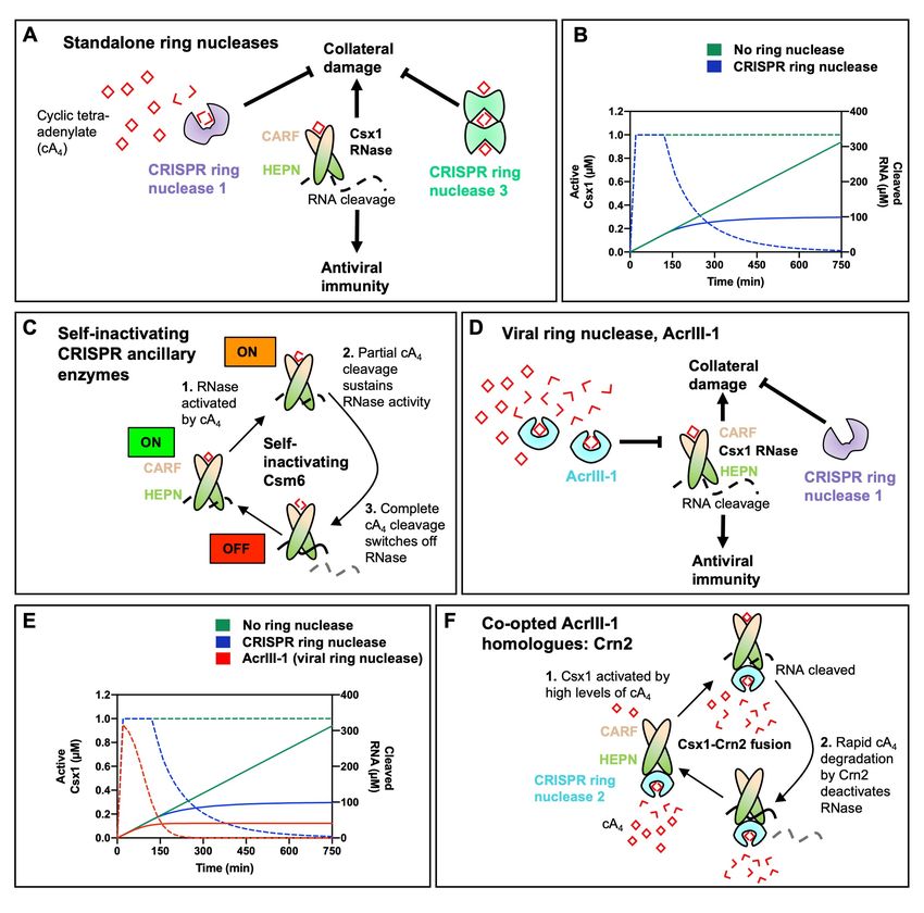
Downloaded from rnajournal.cshlp.org on August 22, 2021 - Published by Cold Spring Harbor Laboratory Press

messenger and binds to CRISPR-associated Rossmann Fold (CARF) domains of CRISPR
ancillary proteins, allosterically activating the adjacent effector domains, which are commonly
                               11,12,16–18
non-specific nucleases                       (Figure 2). Importantly, not all Cas10 proteins have a
                                                                             1,19,20
catalytically active HD domain and some lack it altogether                         , emphasising that these
systems likely rely on the cOA signalling pathway for antiviral defence.

Cas7 subunits comprise the backbone of the type III effector complex and cleave bound RNA,
regardless of host or viral genomic origin. This is predicted to allow for multiple-turnover
                                                                                 21
detection and continued surveillance by the effector complex                           . A conserved aspartate
                                                             22–25
residue in Cas7 is responsible for RNA cleavage                  , and while RNA cleavage by the effector
complex is not essential for resistance to phage infection in vivo 26, it switches off the ssDNase
and cOA synthesis activities of Cas10 that are activated by bona fide targets 11,12,21. Therefore
it is likely that RNA cleavage by the effector complex serves to exert control over the potent
                                                                                                                  21
HD nuclease and cOA signalling activities, rather than as a primary mode of defence                                .
Nevertheless, if extant cOA is not eliminated from cells it can sustain the immune response
27
 , and has the potential to compromise cell survival 28.

Cas10 can synthesise cOA of different sizes, containing between three and six 3’-5’ linked
AMP units, although in vitro most type III systems generate a single cOA species in greater
abundance 11,12,19,21,29. The mechanism for cOA synthesis has been elucidated using structural
biology, and involves a nucleophilic attack by the 3’-hydroxyl of one ATP molecule, held in
one Palm polymerase pocket of Cas10, on the α-phosphate of another ATP molecule held in
the other Palm polymerase pocket 13. Once the first pppApA intermediate is formed, it is then
                                                                                                  11–13
subjected to further rounds of AMP incorporation in the same manner                                    . Finally,
intermediates are cyclised by an intramolecular nucleophilic attack by the 3’-hydroxyl on the
                                        13
α-position of its 5’-triphosphate            . The factors governing the type(s) and abundance of cOA
molecules made, which may be determined by the protein architecture of Cas10 homologues
and/or cellular ATP levels, remain to be fully explored.

Most type III systems characterised to date employ cyclic tetra-adenylate (cA4) as the second
messenger 11,13,21,30–32, but several use cyclic hexa-adenylate (cA6) instead 12,33,34. Within these
systems, the CARF domains of cognate CARF family effector(s) specifically recognise one of
these signals 16,21,27,35. Csx1/Csm6 ribonucleases are the most common CARF family proteins
associated with type III CRISPR systems 1. When activated by cA4 or cA6, as determined by
the CARF domain architecture, Csx1/Csm6 cleaves RNA using its Higher Eukaryotes and
Prokaryotes Nucleotide-binding (HEPN) domain 11,12. The non-specific RNase activity of Csm6
has been shown to result in cell growth arrest, and cell growth is restored when infection is

                                                         2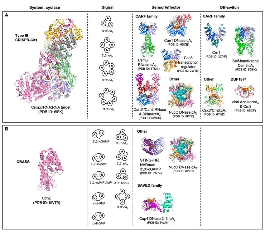
Downloaded from rnajournal.cshlp.org on August 22, 2021 - Published by Cold Spring Harbor Laboratory Press

                         36
cleared from cells         . cA4 has also been shown to bind to the CARF family transcription
regulator Csa3 , which upregulates CRISPR loci and cas gene expression 37–39, presumably
                    37

acting as a positive-feedback mechanism linked to MGE detection. Several cOA activated
DNA nucleases also feature in CRISPR immunity. CRISPR ancillary nuclease 1 (Can1) has
been shown to nick super-coiled DNA when activated by cA4, and is anticipated to slow phage
                                                       16
replication by collapsing replication forks                . The related Can2 enzyme, also termed cOA
activated RNase and DNase 1 (Card1), was demonstrated to be a dual-specificity nuclease
capable of both RNA and DNA cleavage 17,18. While one study showed that select Card1/Can2
                                                                                                                   18
enzymes cleaved ssRNA and nicked super-coiled DNA to provide antiphage immunity                                         ,
another identified a Card1/Can2 that cleaved ssRNA and exclusively ssDNA and provided
                           17
antiphage immunity            . Interestingly, the authors attributed the accompanying cell growth
arrest phenotype to the ssDNase activity, which is presumed to cause DNA lesions in the host
                                                                             17
chromosome, rather than the RNase activity of Card1/Can2                       . Another DNase, NucC, is a
cA3-activated enzyme associated with both type III CRISPR systems and cyclic
oligonucleotide-based anti-phage signalling systems (CBASS) 40,41. As a CBASS component,
NucC has been shown to degrade dsDNA non-specifically and trigger cell death before phage
replication is complete 40,41. NucC is expected to act similarly during type III CRISPR immunity,
and its activation may represent an instance when adaptive immunity leads to abortive
infection.

Figure 1. Cyclic oligoadenylate signalling by type III CRISPR systems. Multisubunit type III
CRISPR-Cas complexes (denoted Csm or Cmr) detect foreign RNA and carry out nucleic acid cleavage
directly and by recruiting CRISPR ancillary enzymes. RNA bound by the CRISPR-Cas complex, as a
result of complementary base pairing with the crRNA, is degraded by the Cas7 (Csm3/Cmr4) backbone
subunits 22–25. Bona fide RNA targets contain a 3’-region that is not complementary to the 8 nucleotide
5’-end of the crRNA, which allosterically activates the Cas10 subunit to synthesise cyclic
oligoadenylates (cOA) and cleave ssDNA            11,12.   Target RNA cleavage by Cas7 subunits switches off
both the ssDNase and cOA synthesis activities of Cas10 21. cOA can activate Csx1/Csm6 ribonucleases
that cleave RNA non-specifically, DNases such as NucC 40 and the CRISPR ancillary nuclease 1 (Can1)
16,   and the related dual-specificity cOA activated RNase and DNase (Card1)/Can2                  17,18,   which help
eliminate invading mobile genetic elements (MGE). cOA can also stimulate the transcription regulator
Csa3, which alters CRISPR loci and cas gene expression to promote MGE elimination 37.

Figure 2. Structures of type III CRISPR ancillary proteins. A. E. italicus (Eit) Csm6 dimer in complex
with its cyclic hexa-adenylate (cA6) activator (shown in sphere form). EitCsm6 is a non-specific
ribonuclease containing CARF (dark and light blue) and HEPN (dark and light green) domains 27. B. T.
onnurineus (Ton) Csm6 dimer in complex with its cyclic tetra-adenylate (cA4) activator. TonCsm6 is a
non-specific ribonuclease consisting of CARF and HEPN domains                     42.   C. S. islandicus (Sis) Csx1

                                                            3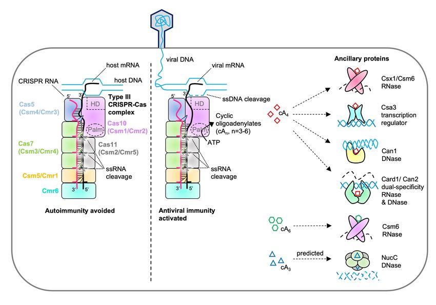
Downloaded from rnajournal.cshlp.org on August 22, 2021 - Published by Cold Spring Harbor Laboratory Press

hexamer in complex with its cA4 activator. SisCsx1 dimers form a hexamer upon cA4 binding and RNA
is cleaved at three distinct active sites within the interior of the hexamer 43. D. S. solfataricus (Sso) Csa3
dimer. SsoCsa3 is a cA4 stimulated transcription regulator consisting of CARF and a helix-turn-helix
DNA binding domains (yellow & orange)               44.   E. T. thermophilus (Tth) CRISPR ancillary nuclease 1
(Can1) monomer in complex with its cA4 activator. TthCan1 nicks super-coiled DNA and is comprised
of two CARF domains and a PD-D/ExK family nuclease domain (salmon coloured)                             16.   F. T.
succinifaciens cyclic oligoadenylate activated RNase and DNase 1 (Card1)/Can2 dimer in complex with
its cA4 activator. Card1/Can2 is related to Can1 and is a dual-specificity nuclease with CARF and PD-
D/ExK nuclease domains (red and salmon)               17,18.   G. E. coli (Eco) NucC hexamer in complex with its
cyclic tri-adenylate (cA3) activator. EcoNucC trimers assemble into a hexamer upon cA3 binding and
degrades dsDNA            40.   NucC is related to restriction endonucleases and binds cA3 at a protein domain
unrelated to the CARF family.

2. Regulation of cyclic oligoadenylate signalling

Non-specific degradation of DNA and RNA by cOA activated nucleases imbue type III CRISPR
systems with an intrinsic potential for self-destruction. While collateral cleavage of host nucleic
acids may instigate cell dormancy to slow viral propagation, as has been uncovered for the in
                                                                                            45
trans non-specific RNase activity of a type VI (Cas13) CRISPR system                         , it will compromise
cell survival if not carefully regulated. To overcome this problem, type III CRISPR systems
harbour two regulatory mechanisms. First, cOA synthesis is switched off by target RNA
                 11,19,21,30,33
cleavage                        , and second, extant cOA is eliminated within cells to switch off CRISPR
                             46
ancillary enzymes .

2.1 Switching off cOA synthesis is not enough

Although target RNA cleavage by the type III effector complex switches off cOA synthesis
11,19,21,30,33
             , extant cOA is predicted to sustain the immune response 47. In vitro, the Sulfolobus
solfataricus type III-D CRISPR system has been shown to synthesise approximately 1000
molecules of cA4 per viral mRNA 21,47,48, which equates to ~6 µM cA4 in a S. solfataricus cell,
                                                                                       47
in significant excess of what is required to activate the Csx1 RNase                    . While cOA synthesis
is ended by viral mRNA cleavage, the micromolar quantities of cOA already made must be
eliminated if the immune response is to be switched off in a timely manner. The need to control
the immune response and mitigate collateral damage has likely led to the evolution of cOA
degradative enzymes, which have been termed ‘ring nucleases’ 46.

2.2 Ring nucleases

                                                               4
Downloaded from rnajournal.cshlp.org on August 22, 2021 - Published by Cold Spring Harbor Laboratory Press
2.2.1 CRISPR ring nuclease 1
cOA degradative enzymes were first identified by activity-guided fractionation of S. solfataricus
cellular lysates and identification of enriched proteins by mass spectrometry 46. The identified
protein Sso2081, and the related protein Sso1393, were shown to act as standalone enzymes
46
that degraded cA4 specifically, and were named CRISPR ring nucleases 1 (Crn1) (Figure
3A). Crn1 enzymes are CARF domain only proteins that have evolved to catalyse cOA
cleavage, and do so by cleaving the symmetrical cA4 molecule as a protein dimer 46. The two
S. solfataricus Crn1 enzymes exhibited a 10-fold difference in the rate of cA4 cleavage, which
result in variable capacities for Csx1 deactivation, and may therefore be leveraged temporally
46
or synergistically for cA4 elimination . Protein sequence analyses and structural modelling
suggest that Sso1393, Sso2081, and the uncharacterised Sso1397, which is predicted to be
a ring nuclease, are orthologues and likely originated from gene duplication events 46,49. Crn1
enzymes are exclusive to the crenarchaea, and Sulfolobales typically encode several Crn1
50
orthologues alongside Csx1/Csm6 (Reviewed in ). For example, S. islandicus REY15A
encodes two Crn1 enzymes that degrade cA4 made by its type III-B CRISPR system, and like
Crn1 enzymes from S. solfataricus, one ring nuclease appears to degrade cA4 at a higher rate
35 46
compared to the other . As Crn1 enzymes degrade cOA slowly , the presence of multiple
Crn1 enzymes may reflect the regulatory needs of a given type III CRISPR system, particularly
where large quantities of cOA are made even at low levels of infection as per the S.
solfataricus type III-D CRISPR system 47.
While cyclic nucleotide phosphodiesterases typically coordinate metal ions to catalyse cyclic
51
nucleotide cleavage (Reviewed in ), Crn1 is metal-independent and yields cA4 cleavage
46
products containing 2’,3’-cyclic phosphates . Crn1 enzymes catalyse cA4 cleavage by
positioning and/or activating the 2’-hydroxyl group of a ribose for an in-line nucleophilic attack
on the adjacent scissile phosphodiester bond 46. The first nucleophilic attack generates a linear
tetra-adenylate containing a 2’,3’-cyclic phosphate (A4>P), which is then converted to two
molecules of di-adenylate containing a 2’,3’-cyclic phosphate (A2>P) by the second
46
nucleophilic attack on the other side of the molecule . The final A2>P products do not
35,46
stimulate CARF family CRISPR ancillary effectors such as Csx1 . Consequently, ring
nucleases are poised to act as crucial regulators of the type III CRISPR immune response.
Indeed, computational modelling demonstrates that Crn1 curtails RNA cleavage by
decreasing the active form of Csx1 over time, and without Crn1 cell death is the most probable
outcome 47 (Figure 3B).
2.2.2 Self-inactivating CRISPR ancillary ribonucleases
5Downloaded from rnajournal.cshlp.org on August 22, 2021 - Published by Cold Spring Harbor Laboratory Press
In the absence of standalone ring nucleases some Csm6 enzymes have adapted to
intrinsically degrade their cOA activators. Several Csm6 enzymes have a CARF domain that
acts as a dual cOA sensor and ring nuclease and this has been proposed to facilitate timed
27,29,42,52
deactivation of the RNase component . In this model, cOA is cleaved at the CARF
domain as it allosterically stimulates the HEPN RNase domain (Figure 3C). First, cOA is
cleaved to generate an A4>P intermediate, which is known to support HEPN RNase activation
12,21,48
, while the second cleavage event generates two A2>P products and deactivates the
enzyme, similar to the catalytic mechanism of Crn1 46. Notably, no standalone ring nucleases
have yet been identified that degrade cA6, although several Csm6 enzymes possess cA6
27,29
degradative CARF domains . Site-directed mutagenesis to abolish cOA cleavage at one
such CARF domain was found to result in sustained RNA cleavage by Csm6, which led to a
strong growth inhibition phenotype, highlighting the crucial role of ring nuclease activity in cell
recovery 27.
29,31,42
Some Csm6/Csx1 enzymes also degrade cA4 and cA6 by their HEPN RNase domain ,
although this may be a side reaction. For S. thermophilus Csm6, the HEPN domain has been
shown to exhibit higher affinity for single-stranded RNA substrates compared to cA6, and the
29
CARF domain was found to have higher affinity for cA6 compared to the HEPN domain .
These observations suggest that CARF ring nuclease activity will be particularly important at
low cA6 levels, as the lower cA6 binding affinity of the HEPN domain may prevent it from
eliminating cOA sufficiently to preclude Csm6 activation. Nevertheless, the S. thermophilus
Csm6 HEPN domain was highly efficient at cA6 degradation compared to its CARF domain 29,
and lesser conformational restraints associated with cA6 compared to other cOA species may
enable its rapid turnover at the HEPN active site. While single-stranded RNA substrates and
42
cOA appear to be cleaved similarly by the HEPN domain , it is worth noting that only
11,52 42,53
Csx1/Csm6 that lack cleavage preferences , or cleave selectively after adenosines ,
are likely to support cOA cleavage at the HEPN active site.
2.2.3 Viral ring nuclease AcrIII-1
Type III CRISPR immunity is capable of driving viruses to extinction 54. cOA activated effector
proteins that inhibit viral replication are therefore anticipated to drive viruses to evolve
mechanisms to inhibit cOA signalling. Indeed, diverse archaeal viruses, proviruses,
bacteriophages, prophages and plasmids encode a highly efficient cA4 degradative enzyme
55
. The DUF1874-family viral ring nuclease was shown to degrade cA4 at rate ~50-fold greater
than cellular Crn1 enzymes, enabling MGEs to circumvent type III CRISPR immunity 55 (Figure
6Downloaded from rnajournal.cshlp.org on August 22, 2021 - Published by Cold Spring Harbor Laboratory Press
3D). As an anti-CRISPR protein, the viral ring nuclease was named (AcrIII-1), in keeping with
56
established anti-CRISPR nomenclature , but incorporating a hyphen to denote that it is not
type III subtype-specific. In agreement with biochemistry and microbiology studies, kinetic
modelling has been used to show that AcrIII-1 is able to quickly decrease the level of activated
47
Csx1 in cells by rapidly degrading cA4, which underlies its potent anti-CRISPR function
(Figure 3E).
AcrIII-1 is a metal-independent ring nuclease that forms A2>P products similar to Crn1
enzymes 55. Both AcrIII-1 and Crn1 bind cA4 with high affinity 47, therefore its greater catalytic
efficiency is ascribed to conserved active site residues that better position the 2’-hydroxyl of
the ribose for an in-line nucleophilic attack, stabilise the transition state and protonate the
leaving group 55. In particular, a highly conserved active site histidine residue is critical for cA4
cleavage and is postulated to act as the general acid involved in protonating the oxyanion
leaving group 55.
2.2.4 Co-option of viral ring nucleases by bacteria: CRISPR ring nuclease 2
Intriguingly, AcrIII-1 is found in association with type III CRISPR systems in several bacteria,
independent of MGEs 55. Due to the wide distribution of AcrIII-1 among MGEs, it is plausible
that bacteria acquired AcrIII-1 by horizontal gene transfer from MGEs and harness the enzyme
to regulate cOA signalling. AcrIII-1 associated with type III CRISPR systems have been
termed CRISPR ring nuclease 2 (Crn2) 55.
In Marinitoga piezophila (Mpi) a Csx1 protein is fused to Crn2, directly implicating this Crn2 in
57
Csx1 regulation . The Crn2 domain was shown to exhibit constitutive cA4 degradation at a
rate comparable to AcrIII-1 enzymes 57. However, cA4 degradation by the Crn2 domain limited
but did not abolish RNA cleavage by Csx1, consistent with MpiCsx1-Crn2 acting as a self-
limiting ribonuclease 57. High micromolar levels of cA4 were required to detect MpiCsx1-Crn2
RNase activity, which is attributed to the higher cA4 affinity and potent ring nuclease activity
of the Crn2 domain 57. The Crn2 domain likely prevents spurious activation of Csx1, permitting
RNA degradation only once a set cA4 threshold - perhaps a molecular signature of a bona fide
infection, has been reached (Figure 3F). Interestingly, the majority of crn2 genes encode
standalone enzymes 55, and in these cases Crn2 expression may be temporally regulated so
as not to compromise the antiviral response by rapid elimination of cOA.
2.2.5 CRISPR ring nuclease 3
7Downloaded from rnajournal.cshlp.org on August 22, 2021 - Published by Cold Spring Harbor Laboratory Press
In prokaryotic phyla where crn1 genes are absent, csx3 is commonly found associated with
49,58
type III CRISPR systems, and csx1 or csm6 are its most common adjacent genes . Early
studies on Archaeoglobus fulgidus (Afu) Csx3 identified the protein as a manganese
59
dependent RNA exoribonuclease that specifically cleaved 3’ poly-A tails , although its
precise role in antiviral immunity remained unclear. Recently, the enzymatic activities of
AfuCsx3 were reassessed and the protein was demonstrated to be a cA4-specific ring
60,61
nuclease . Csx3 has been shown to regulate Csx1-mediated immunity in vivo and was
60
therefore renamed CRISPR ring nuclease 3 (Crn3) . Surprisingly, Crn3 degraded cA4 at a
rate comparable to AcrIII-1 enzymes in vitro, however in vivo studies showed that Crn3
provided only partial protection from Csx1 toxicity, whereas AcrIII-1 provided near complete
protection by fully deactivating the pathway 60.
Based on structural topology, Crn3 was initially postulated to be a divergent member of the
CARF family 62. However, recent structural analysis indicate that Crn3 is more closely related
49
to Sulfate Transporter and Anti-Sigma factor antagonist (STAS) domains . X-ray structures
revealed that Crn3 forms head to tail filaments containing cA4 bound between adjacent dimers
60
(Figure 3A). In contrast to the metal-independent Crn1 family, Crn3 relies on manganese
ions to cleave cA4 into A2>P and A2P (di-adenylate containing 5’-hydroxyl and 3’-phosphate
moieties) products. One face of Crn3 harbours three highly conserved histidine residues,
which may coordinate up to three manganese ions required for catalysis, while the other face
harbours a conserved aspartate that is also catalytically important. Dimer-dimer stacking leads
to the union of these two faces, otherwise over 20 Å apart, and forms the composite active
60
site . While the cOA cleavage mechanism is not fully understood, kinetic studies show
cooperativity in the Crn3 reaction cycle, which suggests that the rate of cA4 elimination may
be controlled by altering Crn3 levels in cells 60.
Intriguingly, cyanobacteria encode a protein family containing a N-terminal Crn3 domain and
58
a C-terminal AAA+ (ATPases Associated with diverse cellular activities) ATPase domain .
AAA+ ATPase domains hydrolyse ATP to drive a range of cellular processes including protein
63
unfolding and degradation and DNA repair, replication and recombination (Reviewed in ).
Hence the coupling of Crn3 and an AAA+ ATPase may indicate that cOA degradation is linked
to other cellular pathways. Additionally, recent work has uncovered a gene containing both
Csx1 and Crn3 domains in Thermodesulfobium narugense 49, directly implicating Crn3 in Csx1
regulation in bacteria.
Figure 3. Ring nucleases regulate the type III CRISPR immune response. A. Cyclic tetra-adenylate
(cA4) activated Csx1 ribonucleases cleave RNA non-specifically, which provides antiviral immunity but
8Downloaded from rnajournal.cshlp.org on August 22, 2021 - Published by Cold Spring Harbor Laboratory Press
also causes collateral damage to cells. cA4 binds the CRISPR-associated Rossmann Fold (CARF)
domain of Csx1 and allosterically activates its Higher Eukaryotes and Prokaryotes Nucleotide-binding
(HEPN) domain, which cleaves RNA. CRISPR ring nucleases 1 and 3 eliminate extant cA4 and
deactivate Csx1, mitigating sustained collateral damage to cells. B. Graph depicting kinetic modelling
of the type III CRISPR response in a cell where 60 µM of cA4 is generated upon infection, representative
of a medium-level infection. In the absence of ring nucleases Csx1 remains in an active state (dotted
green line, corresponding to left-hand side y-axis) and RNA cleavage (solid green line, corresponding
to right-hand side y-axis) continues unimpeded. Csx1 is slowly deactivated when CRISPR ring nuclease
1 is present (dotted blue line) and thus RNA cleavage is limited (solid blue line). Simulations were
carried out using KinTek Global Kinetic Explorer software 64, using a previously published model of the
S. solfataricus type III CRISPR defence pathway 47, and data were plotted using GraphPad Prism. C.
Some Csm6 enzymes act as bi-functional ribonucleases and ring nucleases. These enzymes cleave
cA4 at the CARF domain upon cA4 binding and activating the HEPN RNase. Some Csm6 enzymes also
cleave cOA at the HEPN domain. D. Prokaryotic viruses encode an anti-CRISPR viral ring nuclease
(AcrIII-1). AcrIII-1 rapidly degrades cA4 and attenuates RNA cleavage by swiftly deactivating Csx1. E.
Graph depicting the effect of having no ring nuclease (green), a host cell CRISPR ring nuclease 1
(blue), and both CRISPR ring nuclease 1 and AcrIII-1 on the active form of Csx1 (dotted lines, left-hand
side y-axis) and consequent RNA cleavage (solid lines, right-hand side y-axis). When AcrIII-1 is present
Csx1 is deactivated much more quickly. F. In some bacteria AcrIII-1 homologues are found associated
with type III CRISPR systems and these proteins have been named CRISPR ring nuclease 2 (Crn2). In
Marinitoga piezophila Csx1 is fused to Crn2, which limits Csx1 activity by rapidly and constitutively
degrading cA4. The Crn2 domain only permits Csx1 activation when a high cA4 threshold, determined
by the balance between cA4 affinity of the CARF domain of Csx1 and the high cA4 affinity and rate of
degradation by Crn2, is reached.
2.3 Predicted ring nucleases
Recent analysis of the CARF superfamily has led to identification of several genes which are
expected to comprise new ring nuclease families 49. Namely, Csx16, Csx20 and Unk_01 have
been identified to contain polar resides (histidine, arginine and aspartate or glutamate)
49
compatible with ring nuclease function . Csx16 and Csx20 share sequence identity, and
Csx16 has been noted as partially similar to the DUF1874 protein family which comprises both
AcrIII-1 and Crn2, suggesting homology to viral ring nucleases 49. Csx16, Csx20 and Unk_01
have also been detected fused to Csx1, and based on the characterised MpiCsx1-Crn2
protein, such fusions are good indicators of regulatory function. Csx14 is further predicted to
constitute a new ring nuclease family based on conservation of serine or threonine and lysine
residues which are crucial for cA4 cleavage in Crn1 enzymes 49. On the other hand, additional
cOA sensing and/or degradative enzymes unrelated to the CARF and DUF1874 protein
families should not be ruled out. Indeed, a membrane-associated DHH-DHHA1 (MAD) family
9Downloaded from rnajournal.cshlp.org on August 22, 2021 - Published by Cold Spring Harbor Laboratory Press
nuclease has recently been uncovered that non-specifically degrades cA4 65. In several cases,
CorA, which are associated with type III CRISPR systems and anticipated to be cOA activated
membrane channels, are found adjacent or fused to DHH family nucleases 58, and it is possible
that the DHH family nuclease may cleave cOA to regulate the CorA component.
3. Parallels between cyclic nucleotide-based immune systems in prokaryotes
Similar to type III CRISPR systems, CBASS systems generate cyclic nucleotide second
66
messengers in response to stimuli from phage infection (Reviewed in ) (Figure 4). Antiviral
signalling by CBASS comprises a greater repertoire of signals, which include cyclic
67
nucleotides containing non-canonical linkages . CBASS signals identified to date include
cyclic GMP-AMP (c-GAMP), cyclic di-GMP, cyclic UMP-UMP, cyclic UMP-AMP, cyclic AMP-
41,68–70 40,67
AMP-GMP, cA3 and cA4 . These activate NucC and Cap4 DNases , as well as
phospholipases 69 and NADases 70 that are involved in precipitating cell death as mechanisms
of antiphage immunity. In the Cap4 enzyme, cA3 is recognised by a SMODS-associated and
fused to various effectors sensor domain (SAVED), which structural study indicates is
67
evolutionarily linked to the CARF family , and SAVED domain containing effectors are also
71
associated with several type III CRISPR systems . Many of the characterised CBASS
associated effectors are cytotoxic 40,69,72, revealing another similarity between type III CRISPR
and CBASS immune responses. Indeed, when type III CRISPR-Cas and CBASS co-occur,
cOA synthesised by one or the other system might cross-activate ancillary effectors
associated with either system. Importantly, it remains to be elucidated whether CBASS
systems function exclusively via abortive infection, or if as of yet unidentified ring nucleases
or regulatory mechanisms play a role in deactivating these systems.
Figure 4. Comparisons of cyclic nucleotide-based immune systems in prokaryotes. A. Protein
Data Bank (PDB) identifiers are shown alongside all structures. Type III CRISPR immunity comprises
four key components; a detection and signalling platform, cyclic nucleotide signals, ancillary signal
sensors fused to effectors, and mechanisms to eliminate the signal. The type III CRISPR complex
detects viral mRNA and the Cas10 nucleotidyl cyclase generates cyclic oligoadenylates (cOA; cAn, n=3-
6) containing 3’-5’ phosphodiester linkages. cOA allosterically stimulates downstream CRISPR ancillary
effector proteins, typically by binding to a CRISPR-associated Rossmann Fold (CARF) domain
(coloured in marine and light blue in protein dimers). Finally, ring nucleases eliminate cOA and
deactivate CRISPR ancillary effectors, controlling the immune response. Some ring nucleases are
CARF family proteins, while others have unique cOA sensing domains. Viruses encode variant ring
nucleases (AcrIII-1) which rapidly degrade cOA and supress the type III CRISPR immune response. In
some bacteria AcrIII-1 is found associated with type III CRISPR systems and has been termed Crn2
because it appears to be harnessed by bacteria to regulate CRISPR immunity. B. The cyclic
10Downloaded from rnajournal.cshlp.org on August 22, 2021 - Published by Cold Spring Harbor Laboratory Press
oligonucleotide-based anti-phage signalling system (CBASS) resembles the type III CRISPR immune
system in some respects. The cyclase (CdnE) is activated by unknown stimuli during phage infection,
and different CdnE proteins synthesise different cyclic nucleotide molecules which activate downstream
effector proteins. CBASS can synthesise cyclic nucleotides containing both 3’-5’ and 2’-5’
phosphodiester linkages, giving rise to an enormous repertoire of possible cyclic nucleotide signals.
These signals are detected by diverse protein folds, including the SMODS-associated and fused to
various effectors sensor domain (SAVED), which is evolutionarily linked to the CARF domain. Although
ring nucleases may degrade cA4, no prokaryotic enzymes have been identified that degrade any of the
other cyclic nucleotide signals generated by CBASS. Some CBASS systems may function exclusively
via abortive infection while others may have novel regulatory mechanisms.
4. Concluding remarks and future perspectives
cOA signalling by type III CRISPR complexes to activate downstream effectors and eliminate
foreign intruders represents a highly potent antiviral defence strategy. Ring nucleases are a
crucial component in this pathway and deactivate CRISPR ancillary enzymes by degrading
cOA. CARF family ring nucleases exhibit significant sequence variation, highlighting
convergent evolution on a crucial catalytic function. From a microbiological stand-point ring
nucleases are undoubtedly important, this is emphasised by MGEs that use ring nucleases as
anti-immune weaponry. By eliminating extant cOA, ring nucleases have the potential to
regulate diverse cOA activated effectors, including putative proteases, RelE RNases predicted
to cleave mRNA at ribosomes, and WYL-domain containing transcription regulators that may
58,71,73
promote antiviral immunity and/or cell recovery . While rapid progress has been made
in understanding cOA signalling, many cOA activated enzymes await biochemical study, and
some may be particularly useful for biotechnological applications. For example, Csm6 has
been used to cleave reporter molecules as a second amplification step in several diagnostic
assays 74,75, including in recent studies using type III CRISPR-Cas systems and cOA signalling
32,76
as viral RNA diagnostic platforms . Likewise, newly predicted ring nucleases based on
49
CARF superfamily analysis may harbour new catalytic mechanisms of broad interest to
enzymology.
Importantly, the discovery of cOA elimination by ring nucleases should not exclude
consideration of other fates for cOA, such as the possibility that cOA is used to warn
neighbouring cells of infection. cOA may be disseminated within bacterial aggregates such as
biofilms by the action of efflux and influx transporters, or accompanying cell lysis. For example,
L. monocytogenes is known to secrete the second messenger c-di-AMP using a multidrug
efflux pump 77. In mammals, 2’,3’-cGAMP synthesised by cGAS (cyclic GMP-AMP synthase)
11Downloaded from rnajournal.cshlp.org on August 22, 2021 - Published by Cold Spring Harbor Laboratory Press
78
is disseminated between cells via gap junctions and confer immunity to bystander cells .
cGAS detects cytosolic DNA and synthesises 2’,3’-cGAMP, which stimulates STING
(Stimulator of Interferon Genes) signalosome assembly and downstream stimulation of
79,80
transcription regulators critical to the antiviral interferon response . 2’,3’-cGAMP has also
been found packaged into virions, whereby its transfer into newly infected cells triggers
81,82
STING-dependent innate immunity . As cOA levels reach high µM concentrations during
29
infection , cOA may similarly accumulate in new virions and serve to accelerate immune
activation in newly infected cells. Accelerated immune activation would be highly
advantageous because type III CRISPR immunity is transcription-dependent and effective
during middle to late viral gene expression 24,26,83,84.
Mobile genetic elements are known to encode multiple anti-CRISPR proteins to inhibit the
same CRISPR-Cas system 85,86. In addition to AcrIII-1, Sulfolobus islandicus rod-shaped virus
2 (SIRV2) encodes AcrIIIB1, a small protein that binds the S. islandicus LAL14/1 type III-B
Cmr complex and blocks cOA synthesis 87. However, AcrIIIB1 is subtype-specific, while AcrIII-
55
1 is effective against any type III subtype that employs cA4 as a second messenger .
Consequently, AcrIII-1 may drive type III CRISPR systems to select alternative cOA activators
to precipitate the immune response. As AcrIII-1 is cA4-specific 55, viral enzymes that degrade
cA3 and cA6 are highly anticipated. In turn, cyclic nucleotide-based immune systems may
protect signals by synthesising cyclic nucleotides containing non-canonical linkages, as seen
for CBASS systems 67, that may be resistant to cleavage by virus encoded nucleases. Thus,
the continued study of cyclic nucleotide-based immune systems, particularly under viral
selection, should provide valuable insights into the dynamics of virus-host coevolution, and
uncover further troves of natural products and novel enzymes.
Interestingly, similar to MGEs targeting prokaryotes, eukaryotic viruses employ second
messenger degradative enzymes, indicating that it is a widely effective immune evasion
strategy. Poxviridae that infect eukaryotes produce a 2’,3’-cGAMP phosphodiesterase, termed
88
poxvirus immune nuclease or poxin, which interferes with cGAS-STING immunity .
Furthermore, mirroring bacterial co-option of AcrIII-1 homologues, Lepidoptera (moths and
butterflies) encode poxin homologues that degrade 2’,3’-cGAMP 88. Phosphodiesterases that
degrade 2’,3’-cGAMP have not yet been identified in a eukaryotic cell cytoplasm, therefore
akin to acquisition of AcrIII-1 by bacteria, Lepidopterans may have acquired poxins from
viruses in order to regulate cGAS-STING immunity. Considering current knowledge of cyclic
nucleotide-based virus-host conflicts, eukaryotic viruses are the best studied and are known
to harbour diverse mechanisms that block and counteract antiviral signalling. For example,
eukaryotic viruses are known to shield DNA to prevent detection by cGAS 89,90, degrade cGAS
12Downloaded from rnajournal.cshlp.org on August 22, 2021 - Published by Cold Spring Harbor Laboratory Press
using proteases 91, prevent cyclic nucleotide synthesis by disabling DNA sensing or promoting
92–95
DNA dissociation , and inhibit various stages of downstream signalling by proteolytic
degradation of STING 96,97. It is likely that prokaryotic viruses adopt similar strategies to inhibit
different stages of the type III CRISPR pathway, and continued study of virus-host interactions
promises to yield exciting discoveries at every turn.
Abbreviations
Acr Anti-CRISPR
Afu Archaeoglobus fulgidus
A4>P Tetra-adenylate containing 5’-hydroxyl and 2’3’-cyclic phosphate
A2P Di-adenylate containing 5’-hydroxyl and 3’-phosphoryl
cA3, 4 or 6 Cyclic tri-, tetra- or hexa-adenylate
Can1 CRISPR ancillary nuclease 1
CARF CRISPR Associated Rossmann Fold
Cas CRISPR-associated
CBASS Cyclic oligonucleotide-based anti-phage signalling system
cGAS cyclic GMP-AMP synthase
Cmr Cas Module RAMP
cOA Cyclic oligoadenylate
Csm Cas subtype Mtube
Csx CRISPR-associated of unknown function
CRISPR Clustered regularly interspaced short palindromic repeats
Crn1/2/3 CRISPR ring nuclease 1, 2 or 3
HEPN Higher Eukaryotes and Prokaryotes Nucleotide binding
MAD Membrane-associated DHH-DHHA1
MGE Mobile genetic element
Mpi Marinitoga piezophila
SAVED SMODS-associated and fused to various effector domain
SIRV1/2 Sulfolobus islandicus rudivirus 1/2
Sso Sulfolobus solfataricus
ssRNA Single-stranded RNA
STAS Sulfate Transporter and Anti-Sigma factor antagonist
STING Stimulator of interferon genes
3’3’-cGAMP cyclic 3’5’-linked GMP-AMP
2’3’-cGAMP cyclic 2’5’- and 3’5’-linked GMP-AMP
13Downloaded from rnajournal.cshlp.org on August 22, 2021 - Published by Cold Spring Harbor Laboratory Press
Acknowledgements
We thank the RNA society for an invitation to submit this work as part of a 2021 RNA
Society/Sacringe Graduate Student Award. The work in the authors’ lab described in this
article was supported by research grants from the Biotechnology and Biological Sciences
Research Council (REF: BB/S000313/1 and BB/T004789/1).
Disclosure statement
The authors declare no competing interests.
Authors’ contributions
Athukoralage J.S. and White M. F. conceptualised and prepared the manuscript.
ORCID
Januka S. Athukoralage http://orcid.org/0000-0002-1666-0180
Malcolm F. White http://orcid.org/0000-0003-1543-9342
References
1. Makarova KS, Wolf YI, Iranzo J, Shmakov SA, Alkhnbashi OS, Brouns SJJ,
Charpentier E, Cheng D, Haft DH, Horvath P, et al. Evolutionary classification
of CRISPR–Cas systems: a burst of class 2 and derived variants. Nat Rev
Microbiol 2020; 18:67–83.
2. Hale CR, Majumdar S, Elmore J, Pfister N, Compton M, Olson S, Resch AM,
Glover CVC, Graveley BR, Terns RM, et al. Essential Features and Rational
Design of CRISPR RNAs that Function with the Cas RAMP Module Complex to
Cleave RNAs. Mol Cell 2012; 45:292–302.
3. Zhang J, Rouillon C, Kerou M, Reeks J, Brugger K, Graham S, Reimann J,
Cannone G, Liu H, Albers SV, et al. Structure and Mechanism of the CMR
Complex for CRISPR-Mediated Antiviral Immunity. Mol Cell 2012; 45:303–13.
4. Johnson K, Learn BA, Estrella MA, Bailey S. Target sequence requirements of
a type III-B CRISPR-Cas immune system. J Biol Chem 2019; 294:10290–9.
5. Marraffini LA, Sontheimer EJ. Self versus non-self discrimination during
CRISPR RNA-directed immunity. Nature 2010; 463:568–71.
6. Liu TY, Iavarone AT, Doudna JA. RNA and DNA Targeting by a Reconstituted
Thermus thermophilus Type III-A CRISPR-Cas System. PLoS One 2017;
14Downloaded from rnajournal.cshlp.org on August 22, 2021 - Published by Cold Spring Harbor Laboratory Press
12:e0170552.
7. Kazlauskiene M, Tamulaitis G, Kostiuk G, Venclovas Č, Siksnys V.
Spatiotemporal Control of Type III-A CRISPR-Cas Immunity: Coupling DNA
Degradation with the Target RNA Recognition. Mol Cell 2016; 62:295–306.
8. Estrella MA, Kuo F-T, Bailey S. RNA-activated DNA cleavage by the Type III-B
CRISPR–Cas effector complex. Genes Dev 2016; 30:460–70.
9. Elmore JR, Sheppard NF, Ramia N, Deighan T, Li H, Terns RM, Terns MP.
Bipartite recognition of target RNAs activates DNA cleavage by the Type III-B
CRISPR–Cas system. Genes Dev 2016; 30:447–59.
10. Jia N, Mo CY, Wang C, Eng ET, Marraffini LA, Patel DJ. Type III-A CRISPR-
Cas Csm Complexes: Assembly, Periodic RNA Cleavage, DNase Activity
Regulation, and Autoimmunity. Mol Cell 2019; 73:264-277.e5.
11. Kazlauskiene M, Kostiuk G, Venclovas Č, Tamulaitis G, Siksnys V. A cyclic
oligonucleotide signaling pathway in type III CRISPR-Cas systems. Science
2017; 357:605–9.
12. Niewoehner O, Garcia-Doval C, Rostøl JT, Berk C, Schwede F, Bigler L, Hall J,
Marraffini LA, Jinek M. Type III CRISPR-Cas systems produce cyclic
oligoadenylate second messengers. Nature 2017; 548:543–8.
13. Jia N, Jones R, Sukenick G, Patel DJ. Second Messenger cA4 Formation within
the Composite Csm1 Palm Pocket of Type III-A CRISPR-Cas Csm Complex
and Its Release Path. Mol Cell 2019; 75:933-943.e6.
14. You L, Ma J, Wang J, Artamonova D, Wang M, Liu L, Xiang H, Severinov K,
Zhang X, Wang Y. Structure Studies of the CRISPR-Csm Complex Reveal
Mechanism of Co-transcriptional Interference. Cell 2019; 176:239-253.e16.
15. Sofos N, Feng M, Stella S, Pape T, Fuglsang A, Lin J, Huang Q, Li Y, She Q,
Montoya G. Structures of the Cmr-β Complex Reveal the Regulation of the
Immunity Mechanism of Type III-B CRISPR-Cas. Mol Cell 2020; 79:741-757.e7.
16. McMahon SA, Zhu W, Graham S, Rambo R, White MF, Gloster TM. Structure
and mechanism of a Type III CRISPR defence DNA nuclease activated by cyclic
oligoadenylate. Nat Commun 2020; 11:500.
17. Rostøl JT, Xie W, Kuryavyi V, Maguin P, Kao K, Froom R, Patel DJ, Marraffini
LA. The Card1 nuclease provides defence during type III CRISPR immunity.
Nature 2021; 590:624-29.
18. Zhu W, McQuarrie S, Grüschow S, McMahon SA, Graham S, Gloster TM, White
15Downloaded from rnajournal.cshlp.org on August 22, 2021 - Published by Cold Spring Harbor Laboratory Press
MF. The CRISPR ancillary effector Can2 is a dual-specificity nuclease
potentiating type III CRISPR defence. Nucleic Acids Res 2021; 49:2777-89.
19. Grüschow S, Athukoralage JS, Graham S, Hoogeboom T, White MF. Cyclic
oligoadenylate signalling mediates Mycobacterium tuberculosis CRISPR
defence. Nucleic Acids Res 2019; 47:9259–70.
20. Staals RHJ, Agari Y, Maki-Yonekura S, Zhu Y, Taylor DW, vanDuijn E,
Barendregt A, Vlot M, Koehorst JJ, Sakamoto K, et al. Structure and Activity of
the RNA-Targeting Type III-B CRISPR-Cas Complex of Thermus thermophilus.
Mol Cell 2013; 52:135–45.
21. Rouillon C, Athukoralage JS, Graham S, Grüschow S, White MF. Control of
cyclic oligoadenylate synthesis in a type III CRISPR system. Elife 2018;
7:e36734.
22. Benda C, Ebert J, Scheltema RA, Schiller HB, Baumgärtner M, Bonneau F,
Mann M, Conti E. Structural Model of a CRISPR RNA-Silencing Complex
Reveals the RNA-Target Cleavage Activity in Cmr4. Mol Cell 2014; 56:43–54.
23. Staals RHJ, Zhu Y, Taylor DW, Kornfeld JE, Sharma K, Barendregt A, Koehorst
JJ, Vlot M, Neupane N, Varossieau K, et al. RNA Targeting by the Type III-A
CRISPR-Cas Csm Complex of Thermus thermophilus. Mol Cell 2014; 56:518–
30.
24. Tamulaitis G, Kazlauskiene M, Manakova E, Venclovas Č, Nwokeoji AO,
Dickman MJ, Horvath P, Siksnys V. Programmable RNA Shredding by the Type
III-A CRISPR-Cas System of Streptococcus thermophilus. Mol Cell 2014;
56:506–17.
25. Ramia NF, Spilman M, Tang L, Shao Y, Elmore J, Hale C, Cocozaki A,
Bhattacharya N, Terns RM, Terns MP, et al. Essential Structural and Functional
Roles of the Cmr4 Subunit in RNA Cleavage by the Cmr CRISPR-Cas Complex.
Cell Rep 2014; 9:1610–7.
26. Samai P, Pyenson N, Jiang W, Goldberg GW, Hatoum-Aslan A, Marraffini LA.
Co-transcriptional DNA and RNA Cleavage during Type III CRISPR-Cas
Immunity. Cell 2015; 161:1164–74.
27. Garcia-Doval C, Schwede F, Berk C, Rostøl JT, Niewoehner O, Tejero O, Hall
J, Marraffini LA, Jinek M. Activation and self-inactivation mechanisms of the
cyclic oligoadenylate-dependent CRISPR ribonuclease Csm6. Nat Commun
2020; 11:1596.
16Downloaded from rnajournal.cshlp.org on August 22, 2021 - Published by Cold Spring Harbor Laboratory Press
28. Koonin E V., Makarova KS. Discovery of Oligonucleotide Signaling Mediated by
CRISPR-Associated Polymerases Solves Two Puzzles but Leaves an Enigma.
ACS Chem Biol 2018; 13:309–12.
29. Smalakyte D, Kazlauskiene M, F. Havelund J, Rukšėnaitė A, Rimaite A,
Tamulaitiene G, Færgeman NJ, Tamulaitis G, Siksnys V. Type III-A CRISPR-
associated protein Csm6 degrades cyclic hexa-adenylate activator using both
CARF and HEPN domains. Nucleic Acids Res 2020; 48:9204–17.
30. Han W, Stella S, Zhang Y, Guo T, Sulek K, Peng-Lundgren L, Montoya G, She
Q. A Type III-B Cmr effector complex catalyzes the synthesis of cyclic
oligoadenylate second messengers by cooperative substrate binding. Nucleic
Acids Res 2018; 46:10319–10330.
31. Foster K, Grüschow S, Bailey S, White MF, Terns MP. Regulation of the RNA
and DNA nuclease activities required for Pyrococcus furiosus Type III-B
CRISPR-Cas immunity. Nucleic Acids Res 2020; 48:4418–34.
32. Steens JA, Zhu Y, Taylor DW, Bravo JPK, Prinsen SHP, Schoen CD, Keijser
BJF, Ossendrijver M, Hofstra LM, Brouns SJJ, et al. SCOPE: Flexible targeting
and stringent CARF activation enables type III CRISPR-Cas diagnostics.
bioRxiv 2021; :2021.02.01.429135.
33. Nasef M, Muffly MC, Beckman AB, Rowe SJ, Walker FC, Hatoum-Aslan A,
Dunkle JA. Regulation of cyclic oligoadenylate synthesis by the Staphylococcus
epidermidis Cas10-Csm complex. RNA 2019; 25:948–62.
34. Sridhara S, Rai J, Whyms C, Woodside W, Terns MP, Li H. Structure and
Function of an in vivo Assembled Type III-A CRISPR-Cas Complex Reveal
Critical Roles of Dynamics in Activity Control. bioRxiv 2021;
:2021.01.27.428455.
35. Molina R, Stella S, Feng M, Sofos N, Jauniskis V, Pozdnyakova I, López-
Méndez B, She Q, Montoya G. Structure of Csx1-cOA4 complex reveals the
basis of RNA decay in Type III-B CRISPR-Cas. Nat Commun 2019; 10:4302.
36. Rostøl JT, Marraffini LA. Non-specific degradation of transcripts promotes
plasmid clearance during type III-A CRISPR–Cas immunity. Nat Microbiol 2019;
4:656–62.
37. Lawrence CM, Charbonneau A, Gauvin C. Cyclic Tetra-Adenylate (cA 4 )
Activates CRISPR Associated Transcription Factor Csa3, Providing Feedback
Activation of Protospacer Acquisition and crRNA Expression. FASEB J 2020;
17Downloaded from rnajournal.cshlp.org on August 22, 2021 - Published by Cold Spring Harbor Laboratory Press
34:1–1.
38. Liu T, Li Y, Wang X, Ye Q, Li H, Liang Y, She Q, Peng N. Transcriptional
regulator-mediated activation of adaptation genes triggers CRISPR de novo
spacer acquisition. Nucleic Acids Res 2015; 43:1044–55.
39. Ye Q, Zhao X, Liu J, Zeng Z, Zhang Z, Liu T, Li Y, Han W, Peng N. CRISPR-
Associated Factor Csa3b Regulates CRISPR Adaptation and Cmr-Mediated
RNA Interference in Sulfolobus islandicus. Front Microbiol 2020; 11:2038.
40. Lau RK, Ye Q, Birkholz EA, Berg KR, Patel L, Mathews IT, Watrous JD, Ego K,
Whiteley AT, Lowey B, et al. Structure and Mechanism of a Cyclic Trinucleotide-
Activated Bacterial Endonuclease Mediating Bacteriophage Immunity. Mol Cell
2020; 77:723-733.e6.
41. Ye Q, Lau RK, Mathews IT, Birkholz EA, Watrous JD, Azimi CS, Pogliano J,
Jain M, Corbett KD. HORMA Domain Proteins and a Trip13-like ATPase
Regulate Bacterial cGAS-like Enzymes to Mediate Bacteriophage Immunity. Mol
Cell 2020; 77:709-722.e7.
42. Jia N, Jones R, Yang G, Ouerfelli O, Patel DJ. CRISPR-Cas III-A Csm6 CARF
Domain Is a Ring Nuclease Triggering Stepwise cA4 Cleavage with ApA>p
Formation Terminating RNase Activity. Mol Cell 2019; 75:944-956.e6.
43. Molina R, Stella S, Feng M, Sofos N, Jauniskis V, Pozdnyakova I, López-
Méndez B, She Q, Montoya G. Structure of Csx1-cOA4 complex reveals the
basis of RNA decay in Type III-B CRISPR-Cas. Nat Commun 2019; 10:4302.
44. Lintner NG, Frankel KA, Tsutakawa SE, Alsbury DL, Copié V, Young MJ, Tainer
JA, Lawrence CM. The Structure of the CRISPR-Associated Protein Csa3
Provides Insight into the Regulation of the CRISPR/Cas System. J Mol Biol
2011; 405:939–55.
45. Meeske AJ, Nakandakari-Higa S, Marraffini LA. Cas13-induced cellular
dormancy prevents the rise of CRISPR-resistant bacteriophage. Nature 2019;
570:241–5.
46. Athukoralage JS, Rouillon C, Graham S, Grüschow S, White MF. Ring
nucleases deactivate type III CRISPR ribonucleases by degrading cyclic
oligoadenylate. Nature 2018; 562:277–80.
47. Athukoralage JS, Graham S, Rouillon C, Grüschow S, Czekster CM, White MF.
The dynamic interplay of host and viral enzymes in type iii crispr-mediated cyclic
nucleotide signalling. Elife 2020; 9:e55852.
18Downloaded from rnajournal.cshlp.org on August 22, 2021 - Published by Cold Spring Harbor Laboratory Press
48. Rouillon C, Athukoralage JS, Graham S, Grüschow S, White MF. Investigation
of the cyclic oligoadenylate signaling pathway of type III CRISPR systems.
Methods Enzymol 2019; 616:191–218.
49. Makarova KS, Timinskas A, Wolf YI, Gussow AB, Siksnys V, Venclovas Č,
Koonin E V. Evolutionary and functional classification of the CARF domain
superfamily, key sensors in prokaryotic antivirus defense. Nucleic Acids Res
2020; 48:8828–47.
50. Zink IA, Wimmer E, Schleper C. Heavily armed ancestors: CRISPR immunity
and applications in archaea with a comparative analysis of CRISPR types in
sulfolobales. Biomolecules 2020; 10:1–41.
51. Conti M, Beavo J. Biochemistry and Physiology of Cyclic Nucleotide
Phosphodiesterases: Essential Components in Cyclic Nucleotide Signaling.
Annu Rev Biochem 2007; 76:481–511.
52. Athukoralage JS, Graham S, Grüschow S, Rouillon C, White MF. A Type III
CRISPR Ancillary Ribonuclease Degrades Its Cyclic Oligoadenylate Activator.
J Mol Biol 2019; 431:2894–9.
53. Sheppard NF, Glover CVC, Terns RM, Terns MP. The CRISPR-associated
Csx1 protein of Pyrococcus furiosus is an adenosine-specific endoribonuclease.
RNA 2016; 22:216–24.
54. Pyenson NC, Gayvert K, Varble A, Elemento O, Marraffini LA. Broad Targeting
Specificity during Bacterial Type III CRISPR-Cas Immunity Constrains Viral
Escape. Cell Host Microbe 2017; 22:343-353.e3.
55. Athukoralage JS, McMahon SA, Zhang C, Grüschow S, Graham S, Krupovic M,
Whitaker RJ, Gloster TM, White MF. An anti-CRISPR viral ring nuclease
subverts type III CRISPR immunity. Nature 2020; 577:572–5.
56. Bondy-Denomy J, Davidson AR, Doudna JA, Fineran PC, Maxwell KL, Moineau
S, Peng X, Sontheimer EJ, Wiedenheft B. A Unified Resource for Tracking Anti-
CRISPR Names. Cris J 2018; 1:304–5.
57. Samolygo A, Athukoralage JS, Graham S, White MF. Fuse to defuse: a self-
limiting ribonuclease-ring nuclease fusion for type III CRISPR defence. Nucleic
Acids Re 2020; 48:6149–56.
58. Shah SA, Alkhnbashi OS, Behler J, Han W, She Q, Hess WR, Garrett RA,
Backofen R. Comprehensive search for accessory proteins encoded with
archaeal and bacterial type III CRISPR- cas gene cassettes reveals 39 new cas
19Downloaded from rnajournal.cshlp.org on August 22, 2021 - Published by Cold Spring Harbor Laboratory Press
gene families. RNA Biol 2019; 16:530–42.
59. Yan X, Guo W, Yuan YA. Crystal structures of CRISPR-associated Csx3 reveal
a manganese-dependent deadenylation exoribonuclease. RNA Biol 2015;
12:749–60.
60. Athukoralage JS, McQuarrie S, Grüschow S, Graham S, Gloster TM, White MF.
Tetramerisation of the CRISPR ring nuclease Crn3/Csx3 facilitates cyclic
oligoadenylate cleavage. Elife 2020; 9:e57627.
61. Brown S, Gauvin CC, Charbonneau AA, Burman N, Lawrence CM. Csx3 is a
cyclic oligonucleotide phosphodiesterase associated with type-III CRISPR-Cas
that degrades the second messenger cA4. J Biol Chem 2020;
:jbc.RA120.014099.
62. Topuzlu E, Lawrence CM. Recognition of a pseudo-symmetric RNA
tetranucleotide by Csx3, a new member of the CRISPR associated Rossmann
fold superfamily. RNA Biol 2016; 13:254–7.
63. Ogura T, Wilkinson AJ. AAA+ superfamily ATPases: common structure-diverse
function. Genes to Cells 2001; 6:575–97.
64. Johnson KA, Simpson ZB, Blom T. Global kinetic explorer: a new computer
program for dynamic simulation and fitting of kinetic data. Anal Biochem 2009;
387:20–9.
65. Zhao R, Yang Y, Zheng F, Zeng Z, Feng W, Jin X, Wang J, Yang K, Liang YX,
She Q, et al. A Membrane-Associated DHH-DHHA1 Nuclease Degrades Type
III CRISPR Second Messenger. Cell Rep 2020; 32:108133.
66. Millman A, Melamed S, Amitai G, Sorek R. Diversity and classification of cyclic-
oligonucleotide-based anti-phage signalling systems. Nat Microbiol 2020;
5:1608-15.
67. Lowey B, Whiteley AT, Keszei AFA, Morehouse BR, Mathews IT, Antine SP,
Cabrera VJ, Kashin D, Niemann P, Jain M, et al. CBASS Immunity Uses CARF-
Related Effectors to Sense 3′–5′- and 2′–5′-Linked Cyclic Oligonucleotide
Signals and Protect Bacteria from Phage Infection. Cell 2020; 182:38-49.e17.
68. Whiteley AT, Eaglesham JB, de Oliveira Mann CC, Morehouse BR, Lowey B,
Nieminen EA, Danilchanka O, King DS, Lee ASY, Mekalanos JJ, et al. Bacterial
cGAS-like enzymes synthesize diverse nucleotide signals. Nature 2019;
567:194–9.
69. Cohen D, Melamed S, Millman A, Shulman G, Oppenheimer-Shaanan Y, Kacen
20Downloaded from rnajournal.cshlp.org on August 22, 2021 - Published by Cold Spring Harbor Laboratory Press
A, Doron S, Amitai G, Sorek R. Cyclic GMP–AMP signalling protects bacteria
against viral infection. Nature 2019; 574:691–5.
70. Morehouse BR, Govande AA, Millman A, Keszei AFA, Lowey B, Ofir G, Shao
S, Sorek R, Kranzusch PJ. STING cyclic dinucleotide sensing originated in
bacteria. Nature 2020; 586:429-33.
71. Shmakov SA, Makarova KS, Wolf YI, Severinov K V., Koonin E V. Systematic
prediction of genes functionally linked to CRISPR-Cas systems by gene
neighborhood analysis. Proc Natl Acad Sci 2018; 115:E5307–16.
72. Severin GB, Ramliden MS, Hawver LA, Wang K, Pell ME, Kieninger A-K,
Khataokar A, O’Hara BJ, Behrmann L V., Neiditch MB, et al. Direct activation of
a phospholipase by cyclic GMP-AMP in El Tor Vibrio cholerae. Proc Natl Acad
Sci U S A 2018; 115:E6048–55.
73. Makarova KS, Anantharaman V, Grishin N V., Koonin E V., Aravind L. CARF
and WYL domains: ligand-binding regulators of prokaryotic defense systems.
Front Genet 2014; 5:102.
74. Gootenberg JS, Abudayyeh OO, Kellner MJ, Joung J, Collins JJ, Zhang F.
Multiplexed and portable nucleic acid detection platform with Cas13, Cas12a,
and Csm6. Science 2018; 360:439–44.
75. Gootenberg JS, Abudayyeh OO, Lee JW, Essletzbichler P, Dy AJ, Joung J,
Verdine V, Donghia N, Daringer NM, Freije CA, et al. Nucleic acid detection with
CRISPR-Cas13a/C2c2. Science 2017; 356:438–42.
76. Santiago-Frangos A, Hall LN, Nemudraia A, Nemudryi A, Krishna P, Wiegand
T, Wilkinson RA, Snyder DT, Hedges JF, Jutila MA, et al. Intrinsic Signal
Amplification by Type-III CRISPR-Cas Systems Provides a Sequence-Specific
Viral Diagnostic. medRxiv 2020; :2020.10.14.20212670.
77. Woodward JJ, Iavarone AT, Portnoy DA. c-di-AMP Secreted by Intracellular
Listeria monocytogenes Activates a Host Type I Interferon Response. Science
2010; 328:1703–5.
78. Ablasser A, Schmid-Burgk JL, Hemmerling I, Horvath GL, Schmidt T, Latz E,
Hornung V. Cell intrinsic immunity spreads to bystander cells via the intercellular
transfer of cGAMP. Nature 2013; 503:530–4.
79. Sun L, Wu J, Du F, Chen X, Chen ZJ. Cyclic GMP-AMP synthase is a cytosolic
DNA sensor that activates the type I interferon pathway. Science 2013;
339:786–91.
21Downloaded from rnajournal.cshlp.org on August 22, 2021 - Published by Cold Spring Harbor Laboratory Press
80. Ishikawa H, Ma Z, Barber GN. STING regulates intracellular DNA-mediated,
type I interferon-dependent innate immunity. Nature 2009; 461:788–92.
81. Gentili M, Kowal J, Tkach M, Satoh T, Lahaye X, Conrad C, Boyron M, Lombard
B, Durand S, Kroemer G, et al. Transmission of innate immune signaling by
packaging of cGAMP in viral particles. Science 2015; 349:1232–6.
82. Bridgeman A, Maelfait J, Davenne T, Partridge T, Peng Y, Mayer A, Dong T,
Kaever V, Borrow P, Rehwinkel J. Viruses transfer the antiviral second
messenger cGAMP between cells. Science 2015; 349:1228–32.
83. Deng L, Garrett RA, Shah SA, Peng X, She Q. A novel interference mechanism
by a type IIIB CRISPR-Cmr module in Sulfolobus. Mol Microbiol 2013; 87:1088–
99.
84. Goldberg GW, Jiang W, Bikard D, Marraffini LA. Conditional tolerance of
temperate phages via transcription-dependent CRISPR-Cas targeting. Nature
2014; 514:633–7.
85. Pawluk A, Bondy-Denomy J, Cheung VHW, Maxwell KL, Davidson AR. A new
group of phage anti-CRISPR genes inhibits the type I-E CRISPR-Cas system of
Pseudomonas aeruginosa. MBio 2014; 5:e00896.
86. Bondy-Denomy J, Pawluk A, Maxwell KL, Davidson AR. Bacteriophage genes
that inactivate the CRISPR/Cas bacterial immune system. Nature 2013;
493:429–32.
87. Bhoobalan-Chitty Y, Johansen TB, Di Cianni N, Peng X. Inhibition of Type III
CRISPR-Cas Immunity by an Archaeal Virus-Encoded Anti-CRISPR Protein.
Cell 2019; 179:448-458.e11.
88. Eaglesham JB, Pan Y, Kupper TS, Kranzusch PJ. Viral and metazoan poxins
are cGAMP-specific nucleases that restrict cGAS-STING signalling. Nature
2019; 566:259–63.
89. Lahaye X, Satoh T, Gentili M, Cerboni S, Conrad C, Hurbain I, El Marjou A,
Lacabaratz C, Lelièvre J-D, Manel N. The Capsids of HIV-1 and HIV-2
Determine Immune Detection of the Viral cDNA by the Innate Sensor cGAS in
Dendritic Cells. Immunity 2013; 39:1132–42.
90. Sun C, Schattgen SA, Pisitkun P, Jorgensen JP, Hilterbrand AT, Wang LJ, West
JA, Hansen K, Horan KA, Jakobsen MR, et al. Evasion of Innate Cytosolic DNA
Sensing by a Gammaherpesvirus Facilitates Establishment of Latent Infection.
J Immunol 2015; 194:1819–31.
22You can also read