A MOLECULAR TEST BASED ON RT LAMP FOR RAPID, SENSITIVE AND INEXPENSIVE COLORIMETRIC DETECTION OF SARS COV 2 IN CLINICAL SAMPLES
←
→
Page content transcription
If your browser does not render page correctly, please read the page content below
www.nature.com/scientificreports
OPEN A molecular test based
on RT‑LAMP for rapid, sensitive
and inexpensive colorimetric
detection of SARS‑CoV‑2 in clinical
samples
Catarina Amaral1, Wilson Antunes2, Elin Moe1, Américo G. Duarte1, Luís M. P. Lima1,
Cristiana Santos1, Inês L. Gomes2, Gonçalo S. Afonso1, Ricardo Vieira2, Helena Sofia S. Teles3,
Marisa S. Reis3, Manuel A. Ramalho da Silva4, Ana Margarida Henriques5, Miguel Fevereiro5,
M. Rita Ventura1, Mónica Serrano1* & Catarina Pimentel1*
Until there is an effective implementation of COVID-19 vaccination program, a robust testing strategy,
along with prevention measures, will continue to be the most viable way to control disease spread.
Such a strategy should rely on disparate diagnostic tests to prevent a slowdown in testing due to lack
of materials and reagents imposed by supply chain problems, which happened at the beginning of the
pandemic. In this study, we have established a single-tube test based on RT-LAMP that enables the
visual detection of less than 100 viral genome copies of SARS-CoV-2 within 30 min. We benchmarked
the assay against the gold standard test for COVID-19 diagnosis, RT-PCR, using 177 nasopharyngeal
RNA samples. For viral loads above 100 copies, the RT-LAMP assay had a sensitivity of 100% and a
specificity of 96.1%. Additionally, we set up a RNA extraction-free RT-LAMP test capable of detecting
SARS-CoV-2 directly from saliva samples, albeit with lower sensitivity. The saliva was self-collected
and the collection tube remained closed until inactivation, thereby ensuring the protection of the
testing personnel. As expected, RNA extraction from saliva samples increased the sensitivity of the
test. To lower the costs associated with RNA extraction, we performed this step using an alternative
protocol that uses plasmid DNA extraction columns. We also produced the enzymes needed for the
assay and established an in-house-made RT-LAMP test independent of specific distribution channels.
Finally, we developed a new colorimetric method that allowed the detection of LAMP products by the
visualization of an evident color shift, regardless of the reaction pH.
A robust population-scale testing strategy for SARS-CoV-2 based on rapid, reliable, decentralized and affordable
diagnostic tests is of utmost priority to guide public health interventions. This testing approach aligned with
measures such as mask wearing, frequent hand washing and social distancing may be enough to prevent and
contain major outbreaks while COVID-19 vaccination programs are in progress.
The gold standard of COVID-19 testing is RT-PCR, which detects the genetic material of SARS-CoV-2 in
nasopharyngeal (NP) samples. Although very reliable, RT-PCR diagnostics are complex, laborious and expen-
sive, and its worldwide use caused, in the early stages of the pandemic, a shortage of reagents needed for sample
collection and viral RNA extraction.
Loop-mediated isothermal amplification (LAMP) is a DNA amplification method that allows rapid and sensi-
tive detection of a specific g ene1–3. LAMP merged with reverse transcription (RT-LAMP) has been successfully
1
Instituto de Tecnologia Química e Biológica António Xavier, Universidade Nova de Lisboa, Av. República,
2780‑157 Oeiras, Portugal. 2Centro de Investigação da Academia Militar (CINAMIL), Unidade Militar Laboratorial
de Defesa Biológica e Química (UMLDBQ), Av. Dr. Alfredo Bensaúde, 1849‑012 Lisbon, Portugal. 3Centro
de Medicina Naval-Marinha Portuguesa, Alfeite, 2810‑001 Almada, Portugal. 4Hospital das Forças Armadas,
Azinhaga dos Ulmeiros, 1649‑020 Lisbon, Portugal. 5Instituto Nacional de Investigação Agrária e Veterinária,
I.P., Laboratório de Virologia, Av. República, Quinta do Marquês, 2780‑157 Oeiras, Portugal. *email: serrano@
itqb.unl.pt; pimentel@itqb.unl.pt
Scientific Reports | (2021) 11:16430 | https://doi.org/10.1038/s41598-021-95799-6 1
Vol.:(0123456789)
www.nature.com/scientificreports/
used for the detection of several respiratory RNA v iruses4–8, including SARS-CoV-2 (reviewed i n9). RT-LAMP is
a powerful alternative to RT-PCR due to its high specificity and sensitivity, cost-effectiveness, and fast turnaround
time (typically 30 min). In RT-LAMP, the amplification of the genetic material of the virus occurs at a constant
temperature and, therefore, diagnostic tests based on RT-LAMP can be carried out anywhere with basic resources,
as they only require a heat block or a water bath set to a single temperature. The reaction products can be ana-
lyzed by means of conventional DNA-intercalating dyes, agarose gel electrophoresis, UV-light illumination, or
real-time fluorescence10. Alternatively, end-point colorimetric readouts are also possible through the detection
of reaction by-products, such as pyrophosphate and protons, which are released during DNA polymerization,
after the incorporation of deoxynucleotide triphosphates. LAMP colorimetric methods detect the turbidity, trig-
gered by the accumulation of magnesium p yrophosphate1, or color changes, occurring when complexometric
indicators , pH sensitive dyes or even DNA-intercalating dyes13–15 are incorporated into the reaction. The
3,11 12
simple technical and instrumental requirements of colorimetric RT-LAMP tests make them extremely attractive
for point-of-care (POC) use and implementation in low-resource settings. Colorimetric RT-LAMP has been
successfully used for detection of SARS-CoV-2 in NP fluids from COVID-19 p atients15–24.
Recently, it was shown that SARS-CoV-2 could be detected in the saliva of infected individuals, highlighting
salivary tests as valuable alternatives for COVID-19 diagnosis25–27. Saliva-based testing has numerous advantages
over NP sampling, especially in a mass screening scenario. It can be performed easily and non-invasively, thus
minimizing patient discomfort, and it does not require specialized personnel or the use of protective equipment,
which saves time and reduces costs. For these reasons, saliva RT-PCR and RT-LAMP tests for SARS-CoV-2
detection have been widely explored in recent m onths28–31.
In the current study, we have established and evaluated a RT-LAMP colorimetric test for SARS-CoV-2 detec-
tion from RNA samples extracted from the NP fluid, or directly from the saliva, of COVID-19 patients. We have
also developed a new colorimetric detection method based on a complexometric indicator that, when merged to
LAMP, is capable of detecting SARS-CoV2 with great analytical sensitivity. In addition, we have produced the
enzymes needed for the test and implemented an in-house-made assay fully independent of commercial reagents.
With this work, we join efforts with many other authors who, in the last months, have been testing and vali-
dating alternative tests for the detection of SARS-CoV-2 in order to make the molecular diagnosis of COVID-19
more accessible and to facilitate its large-scale implementation, even in settings that lack economic or infra-
structural resources.
Results
Sensitivity of two different RT‑LAMP colorimetric setups. The main components of the RT-LAMP
colorimetric reaction are two enzymes (a reverse transcriptase (RT) and a strand displacement polymerase), a
colorimetric dye (phenol red) and a primer set (typically composed of six primers)12. To detect SARS-CoV-2
using RT-LAMP, we took advantage of the primer set previously validated in vitro by Zhang et al.24 and tested,
on clinical specimens from large cohorts of COVID-19 patients, by several other a uthors16–19. The primer set
(N-A) targeted the N gene, which encodes the nucleocapsid protein and has the most abundant expression of
subgenomic mRNA during infection32–35.
We tested two different assay formats. In one format, we used the WarmStart Colorimetric LAMP 2 × Master
Mix (New England Biolabs), which includes all the reagent components with the exception of the primers. In the
other, we purchased the separate enzymes (RTx and Bst 2.0) from New England Biolabs, while the reaction buffer
with the colorimetric dye (phenol red) were prepared in-house as described by Tanner et al.12. The analytical
sensitivities of these two setups were evaluated and compared by assaying in parallel tenfold serial dilutions of
an in vitro transcribed N-gene RNA standard (IVT RNA), starting from 105 copies down to 10 copies (per 20 μL
reaction), at tenfold intervals (Fig. 1A). Color changes from pink (negative) to yellow (positive) were registered
after a 30-min incubation period at 65 °C, as we found that for extended periods (up to 60 min), negative con-
trols often turned yellowish. The amplification of the IVT RNA was confirmed by agarose gel electrophoresis
(Fig. 1B). Ten replicates were analyzed per assay format (Fig. 1C) and IVT RNA dilutions were simultaneously
analyzed by RT-PCR (Fig. 1D). The limit of detection (LoD) was reliably found to be between 100–1000 viral
copies for the assay using the WarmStart Colorimetric LAMP 2 × Master Mix (Fig. 1A), whereas for that using
the separate components the LoD was consistently one Log10 lower (10–100 copies). However, for half of the
replicates, a tenfold lower LoD was achieved for both test formats (Fig. 1C). Such stochastic detection efficiency
has been reported by others (1), and therefore we defined 100–1000 (WarmStart Colorimetric LAMP 2 × Master
Mix) and 10–100 (reaction with separate components) as the robust limits of detection.
For the same serial dilution range, the RT-PCR assay was able to consistently detect down to 10 copies per
reaction (mean Ct = 35.22) (Fig. 1D). Compared to RT-PCR, the RT-LAMP assay, depending on the test setup,
detected up to ten- or one 100-fold less copies of viral RNA. As the RT-LAMP format using the separate com-
ponents was consistently more sensitive, we decided to choose this setup in subsequent assays.
Sensitivity and specificity of the colorimetric RT‑LAMP assay in detecting viral RNA from the
nasopharyngeal fluid. We investigated whether the RT-LAMP assay, using separate components, could be
used to accurately detect SARS-CoV-2 in clinical samples. For that purpose, we tested a set of surplus RNA sam-
ples extracted from the nasopharyngeal (NP) fluid of 177 individuals who were previously tested for COVID-19,
using the standard clinical RT-PCR testing. The samples comprised 126 RNA samples that tested positive (RT-
PCR positive, Ct ≤ 40) and 51 samples that tested negative (RT-PCR negative, Ct ≥ 40). As shown in Fig. 2A, after
incubation for 30 min at 65 °C, a pink to yellow color change was visualized in all RT-LAMP reactions estimated
to have more than 100 RNA molecules present in the reaction (RT-PCR positive, Ct ≤ 32, Fig. 1C), which is in
agreement with the observed experimental sensitivity (Fig. 1A). We found two false positives, i.e. two RT-PCR
Scientific Reports | (2021) 11:16430 | https://doi.org/10.1038/s41598-021-95799-6 2
Vol:.(1234567890)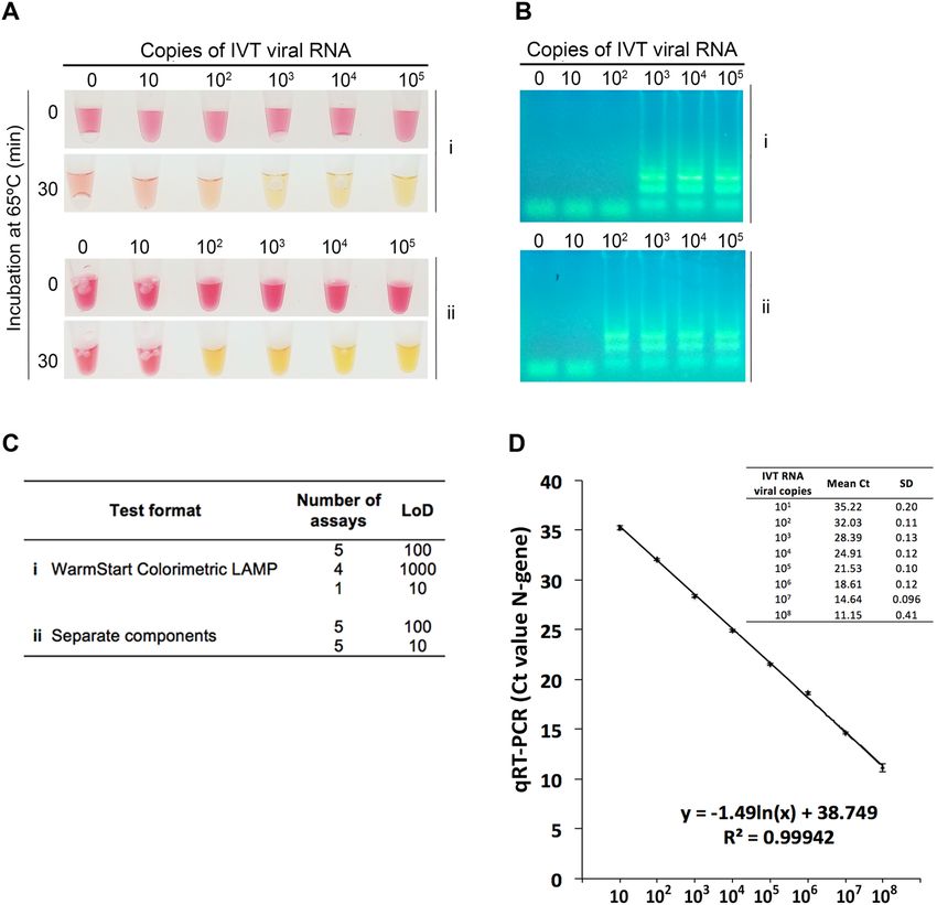
www.nature.com/scientificreports/
Figure 1. Limit of detection of the two different RT-LAMP formats and of RT-PCR. (A) A known number of
copies of in vitro transcribed (IVT) viral RNA (N-gene) were amplified and detected by colorimetric RT-LAMP
using the (i) WarmStart Colorimetric LAMP 2 × Master Mix (New England Biolabs) or (ii) the separate
components (enzymes purchased individually and an in-house-made colorimetric buffer). The reactions
were incubated at 65 °C for 30 min. (B) 10 μL of the RT-LAMP reaction were resolved in an agarose gel (2%)
electrophoresis. The ladder pattern corresponds to the expected LAMP amplification pattern. (C) Limit of
detection of ten replicates of the two test formats. (D) Standard curve generated by plotting the number of IVT
RNA copies (x-axis) vs. the mean of the corresponding RT-PCR threshold cycle (Ct) value (y-axis) of three
independent experiments (Original gel images in Fig. S1).
negative samples that scored positive in the RT-LAMP assay (Table 1). Thus, the overall specificity of the assay
was 96.1% (CI 87–99%) and the sensitivity for samples with Ct ≤ 32 was 100% (CI 94.7–100%). For lower viral
load, as measured by RT-PCR (Ct > 32), the assay showed a decrease in diagnostic sensitivity (Table 1, Fig. 2B).
Overall, these results indicate a robust performance of the colorimetric RT-LAMP assay across a broad range
of purified RNA samples.
Sensitivity of the colorimetric RT‑LAMP assay in detecting SARS‑CoV‑2 in saliva samples. We
next optimized our RT-LAMP assay for direct detection of SARS-CoV-2 in saliva samples. To reduce the risk
associated with handling samples containing infectious viral particles, saliva was self-collected into a tube and
placed at 95 °C for 30 min, for inactivation. This simple heat inactivation procedure has been shown to enable
an effective genetic detection of SARS-CoV-2 by other a uthors30,31. After a brief centrifugation step that sig-
nificantly improved assay reliability (data not shown), the supernatant was diluted with TE, to buffer basal pH
differences in saliva, and immediately analyzed or stored at − 80 °C. Lalli et al. have shown that TE is LAMP-
compatible and does not affect the assay sensitivity29.
We determined the LoD of the assay using both the IVT RNA standard and viral SARS-CoV-2 particles spiked
into healthy human saliva to simulate clinical samples. We were able to detect 100 IVT RNA copies (Fig. 3A)
and 24 SARS-CoV-2 viral particles (Fig. 3B) per reaction in only 30 min after inactivation, using our RT-LAMP
protocol. Since at this sensitivity the assay would detect the typical viral load of SARS-CoV-2 found in the saliva
of COVID-19 patients (100–1000 genomes per μL)36, we proceeded to test the clinical samples.
Scientific Reports | (2021) 11:16430 | https://doi.org/10.1038/s41598-021-95799-6 3
Vol.:(0123456789)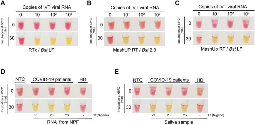
www.nature.com/scientificreports/

                                            Figure 2.  Detection of SARS-CoV-2 in NP samples using RT-LAMP. (A) Comparison of RT-LAMP and
                                            RT-PCR results. The Ct values (RT-PCR results) of 126 COVID-19 positive patients (y-axis) were compared to
                                            the RT-LAMP readout (x-axis) taken after 30 min of incubation at 65 °C (positive, +/yellow; negative, −/pink).
                                            The dotted red line indicates the Ct below, which there is 100% agreement between RT-LAMP and RT-PCR. (B)
                                            Sensitivity of the RT-LAMP assay across different ranges of Ct values (which reflect different viral loads). The
                                            thicker horizontal lines indicate the specificity calculated for the indicated Ct range (according to the data of
                                            panel (A) and Table 1). The vertical lines indicate the corresponding 95% confidence intervals.

                                                                         RT-LAMP
                                             RT-PCR (Ct value range)**   True positives   False negatives   % Sensitivity (95% CI)*
                                             [0–40]                      84               42                67.2 (58.56–74.81)
                                             [0–32]                      68                0                 100 (94.65–100)
                                             [32–35]                     17               20                45.95 (31.04–61.62)
                                             [35–40]                     11               27                28.95 (17–44.76)

                                            Table 1.  RT-LAMP sensitivity across different ranges of Ct values. *Wilson’s binominal confidence interval.
                                            **Ct values rounded to the nearest unit.

                                            Figure 3.  Limit of detection of the saliva RT-LAMP assay. Spike-in experiments of a healthy donor saliva
                                            with (A) tenfold dilutions of in vitro transcribed (IVT) viral RNA (N-gene) and (B) in vitro propagated
                                            SARS-CoV-2 virions. Saliva samples were processed as described in “Materials and methods” and 2 μL were
                                            analyzed by colorimetric RT-LAMP. PFUs plaque forming units. Figures are representative of three independent
                                            experiments.

                                                Saliva and matched NP swab specimens of 49 individuals infected with SARS-CoV-2 (as previously deter-
                                            mined by RT-PCR) were collected and analyzed by RT-LAMP (saliva) and RT-PCR (NP fluid). In addition, 15
                                            saliva samples of healthy donors were tested by RT-LAMP. Saliva samples were self-collected as described above,
                                            and individuals were asked not to eat or drink before testing. A set of 10 of the 49 COVID-19-positive patients
                                            was asked to induce salivation by placing the tongue on the salivary sublingual glands. For this group, we could

          Scientific Reports |   (2021) 11:16430 |                 https://doi.org/10.1038/s41598-021-95799-6                                               4

Vol:.(1234567890)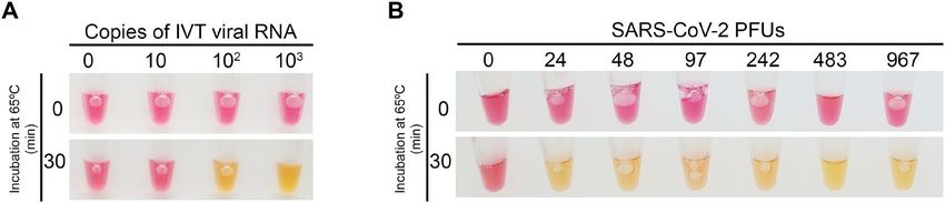
www.nature.com/scientificreports/

                                  Figure 4.  Detection of SARS-CoV-2 in saliva samples using RT-LAMP. RT-LAMP analysis of saliva samples of
                                  confirmed COVID-19 patients who (A) induced or not (B) salivation before sample collection. (C) Comparison
                                  of RT-LAMP and RT-PCR results. The Ct values (RT-PCR results) of 39 COVID-19 positive patients (y-axis)
                                  were compared to the RT-LAMP readout of the matched saliva samples (x-axis), after 30 min of incubation at
                                  65 °C (positive, +/yellow; negative, −/pink). Black circles—direct saliva; orange circles—RNA extracted from
                                  saliva. (D) RNAs from the saliva of false negative samples (as determined by the direct saliva test) were extracted
                                  using plasmid DNA miniprep columns (ZR Plasmid Miniprep-Classic Kit, Zymo Research) and re-analyzed.
                                  NTC no template control. Cts (N gene) obtained for the paired NP samples are indicated.

                                  only detect SARS-CoV-2 sequences in the saliva of one patient using the direct RT-LAMP assay (Fig. 4A).
                                  However, after RNA extraction, 8 out of 10 individuals were identified as being SARS-CoV-2-positive. Taking
                                  into account the Ct values obtained for the paired NP samples (Fig. 4A), we put forward the hypothesis that
                                  by stimulating salivation we were diluting the saliva viral load, which might have accounted for a high number
                                  of false negatives. Corroborating this idea, for all other positive samples where salivation was not induced, we
                                  obtained a good correlation with the RT-PCR results (Fig. 4B,C), as 33 out of 39 samples were identified as posi-
                                  tive samples, with no false positives registered. Therefore, the direct RT-LAMP assay had a sensitivity of 85%

Scientific Reports |   (2021) 11:16430 |               https://doi.org/10.1038/s41598-021-95799-6                                                  5

                                                                                                                                              Vol.:(0123456789)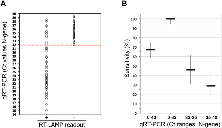
www.nature.com/scientificreports/
Figure 5. Analytical sensitivity of an in-house-made colorimetric RT-LAMP assay. Tenfold dilutions of in vitro
transcribed (IVT) viral RNA (N-gene) were amplified via RT-LAMP and detected using a colorimetric buffer
together with (A) RTx (New England Biolabs) and Bst LF (homemade), (B) MashUP RT (homemade) and Bst
2.0 (New England Biolabs) or (C) MashUP RT (homemade) and Bst LF (homemade). The in-house-made setup
was next used to detect SARS-CoV-2 sequences in (D) RNAs extracted from the NP fluid (NPF) and (E) saliva
samples of COVID-19 positive patients. The reactions were incubated at 65 °C for 30 min. In (D) and (E) the
Cts (N gene) obtained for the paired NP samples are indicated. NTC no template control, HD healthy donor.
Figures are representative of three independent experiments.
(CI 70–93%) for saliva samples with matched NP swabs with Ct ≤ 28 (Fig. 4C). Reaction volumes, but not saliva
amounts, were scaled up to increase the assay sensitivity (Fig. 4B).
All saliva samples that were falsely negative by direct RT-LAMP were positive after RNA extraction (Fig. 4B).
This step increases by 4–9 times the estimated cost of the assay (1€). Inspired by the work of Yaffe et al.37, to keep
RT-LAMP affordable, we tested whether we could use silica columns routinely used in molecular biology labo-
ratories to purify bacterial plasmids (mipreps), to extract viral RNA from saliva samples. As shown in Fig. 4D,
false negative samples were found to be positive after RNA purification using this method, with an estimated
cost per RT-LAMP test of 2€.
Development of an in‑house‑made colorimetric RT‑LAMP. Aiming to establish a colorimetric RT-
LAMP test fully independent of commercial suppliers, we produced the two enzymes needed for the assay and
benchmarked them against commercial alternatives using IVT RNA of SARS-CoV-2.
As for the strand displacement polymerase, the gene encoding the large (Klenow) fragment of Geobacil-
lus stearothermophilus was synthesized, with codon optimized for expression in E. coli, and inserted into the
pET28 + vector. After a simple 2-step purification protocol, we ended up with 250 μL of Bst LF, at a concentration
of 7.6 mg/mL. We next determined the LoD of the assay combining 1 μL of the purified Bst LF, 50-fold diluted
(0.15 μg per 20 μL reaction), the in-house-made colorimetric reaction buffer, and RTx (New England Biolabs).
This semi-commercial assay consistently detected 1–10 copies of the SARS-CoV-2 N gene per reaction (Fig. 5A).
We found that, under our colorimetric conditions, Bst LF outperformed Bst 2.0 (New England Biolabs) (Fig. 1A
and 5A). The amount of the produced Bst LF was enough to perform 12,500 tests at that analytical sensitivity
(1–10 copies).
Alternatives to the commercial RTx were also explored. We started by testing several non-thermostable
reverse transcriptases (from NZYtech and Roche), but it was not possible to detect LAMP products with an
acceptable sensitivity (less than 106 viral IVT RNA copies per reaction, data not shown). We also expressed and
purified the MashUP RT enzyme (clone available at https://pipettejockey.com) that, when combined with Bst
2.0, was able to detect down to 10 IVT viral RNA copies (Fig. 5B), a LoD similar to the one obtained with the
commercial enzyme (Fig. 5A). The MashUP purification consists of a single-step protocol, and sufficient enzyme
was obtained to perform 500 assays (0.5 μL corresponding to 3.4 μg/μL were used directly in the reaction).
Finally, we combined the produced enzymes (Bst LF and MashUP) with the homemade colorimetric reaction
mixture and assessed (i) the LoD of the assay (Fig. 5C) and, as proof of concept, (ii) whether this setup could
identify SARS-CoV-2 N-gene sequences in the RNA extracted from the NP fluid and saliva of COVID-19 patients
(Fig. 5D,E). Our in-house-made assay successfully detected SARS-CoV-2 viral sequences in all the three COVID-
19 patients’ samples (Fig. 5D). Moreover, when using patients’ saliva, processed as described above, instead of NP
RNA, the assay was also capable of identifying SARS-CoV-2 infected patients (Fig. 5E). Corroborating the work
Scientific Reports | (2021) 11:16430 | https://doi.org/10.1038/s41598-021-95799-6 6
Vol:.(1234567890)www.nature.com/scientificreports/
of Alekseenko et al.38, these results clearly indicate that, using simple expression and purification protocols and
home-made buffers, it is possible to establish a colorimetric assay, fully independent of specific supply chains,
that efficiently detects SARS-CoV-2 RNA sequences from clinical specimens.
A new colorimetric method for detection of RT‑LAMP amplification products. The strong and
evident color shift observed with phenol red renders this pH-sensitive dye much preferred for end-point colori-
metric detection of LAMP products. However, when the phenol red method is used with crude samples, interfer-
ence of the sample pH with the assay readout is often observed. Indeed, when establishing the direct RT-LAMP
saliva protocol, we had to discard one sample due to the initial acidification of the reaction, as a strong color shift
to yellow was observed immediately after sample addition into the reaction mixture. Although several colori-
metric indicators are available for detection of LAMP p roducts3,11–15, the pale color shift they produce, which is
difficult to distinguish by the naked eye, has certainly restrained their wide use. To overcome these limitations,
we developed a new colorimetric detection method based on the complexometric indicator, murexide (MX),
which forms a complex with divalent zinc (Zn2+)39. In the absence of Z n2+, MX has a pink color, whereas in the
presence of the divalent cation it turns yellow. Because pyrophosphate (PPi) forms a strong complex with zinc,
we reasoned that the PPi released during DNA polymerization would displace Z n2+ cations from MX, inducing
a color change from yellow to pink. By mimicking the reaction components in a test tube containing the Zn-MX
complex, an evident color shift from yellow to pink was observed immediately after PPi addition (Fig. 6A).
Unfortunately, we found that Zn, but not MX, strongly inhibited the LAMP reaction (data not shown), making
it impossible to use Zn-MX in a one-step colorimetric assay. Therefore, after an incubation period at 65 °C for
30 min, the tubes were opened and MX (0.5 mM) and Z nCl2 (2.5 mM) were added to the reaction. To avoid car-
ryover problems due to the post-amplification opening of the tubes, this step was performed in a separate room.
Using the in-house produced enzymes, we first compared the sensitivity of Zn-MX with that of phenol red
using IVT RNA (Fig. 6B,C). Like phenol red, Zn-MX showed an evident color difference depending on the
presence (pink) or absence (yellow) of LAMP amplification. Moreover, the method enabled the clear detection
of SARS-CoV-2 in crude saliva samples of nine COVID-19 positive patients (Fig. 6D), whereas with phenol red
the viral genetic material was only identified in eight of these samples (Fig. 6E).
Discussion
Widespread testing, preferably based on different supply chains, is required to curtail the ongoing pandemic. To
address that need, we have in this work evaluated a LAMP-based colorimetric test to rapidly detect SARS-CoV-2
in RNAs extracted from patient’s NP fluids, using a single tube protocol. The assay also allows for detection of the
virus directly from patient´s saliva with minimal processing and increased protection of the testing personnel.
We also showed that using simple expression and purification protocols together with homemade buffers, it is
possible to establish an inexpensive colorimetric assay, fully independent of specific supply chains, that efficiently
detects SARS-CoV-2 RNA.
While not as sensitive as the reference diagnostic method for COVID-19, RT-PCR, the simplicity, turnaround
time and low associated costs of our test make it an attractive and efficient tool for infection control. According to
existing literature, the LoD of the test is sufficient to identify individuals with viral titers high enough to transmit
the virus (300–1000 viral copies per μL)27,40,41. This test sensitivity is understood to be adequate for surveillance
and screening of the asymptomatic population. The availability of such a testing solution is therefore of great
importance, as infectiousness peaks occur before or at the symptoms o nset42. Indeed, the rapid evolution of
COVID-19 has been partly attributed to transmissions occurring through people who are presymptomatic or
asymptomatic43; efforts to implement a strategy enabling communities to test asymptomatic individuals require
urgent attention and testing tools to support it.
Several authors have recently shown that the use of different primer sets boosts RT-LAMP sensitivity, pos-
sibly due to better primer efficiency and/or higher target abundance. Also different saliva treatment protocols,
combining certain chemicals and proteinase K, have been shown to improve SARS-CoV-2 detection in saliva
samples16,21,22,28,29,31. Thus, we reason that there is still room to improve the sensitivity of our test.
As expected, RNA extraction greatly improved the saliva test sensitivity, by increasing the concentration
of the viral sequences in the sample. Many other authors have reported similar findings21,22,44,45 and extensive
efforts have been made to establish alternative protocols that enable RNA enrichment using fast and inexpensive
methodologies21,22. Here we showed that RNA extraction using common plasmid DNA extraction columns is
an economical way to concentrate and purify viral RNA from saliva samples.
To eliminate the impact of acidic saliva samples on the test readout, we have developed a new colorimetric
reading, independent of changes in the pH of the LAMP reaction. The method uses a divalent zinc salt (such
as ZnCl2) and the complexometric indicator murexide to form a transient complex (Zn-MX). The presence of
PPi, a by-product of the reaction, is indicated by the indicator displacement method, since Z n2+ forms a more
stable complex with PPi and thus releases murexide. As the presence of zinc inhibits the amplification reaction,
the metal can only be added at the end of the reaction, thus requiring the tubes to be opened post-amplification.
This procedure poses the threat of carryover contaminations, very common in LAMP reactions46,47, which leads
to false positives. We therefore do not anticipate that the Zn-MX method, in its current formulation, can be used
routinely in a molecular diagnostic laboratory. However, the molecular saliva-based tests currently available for
COVID-19, whose workflow already demands opening the LAMP reaction tube, may certainly benefit from our
method48. Additionally, the method can be safely used in closed systems using microfluidic diagnostic cartridges,
similar to the one recently described by Ganguli et al.49.
Scientific Reports | (2021) 11:16430 | https://doi.org/10.1038/s41598-021-95799-6 7
Vol.:(0123456789)www.nature.com/scientificreports/
Figure 6. Alternative colorimetric detection based on the complex murexide-zinc. (A) A strong color change
from yellow to pink is observed when pyrophosphate (PPi) is added to a solution containing Zn-MX. Tenfold
dilutions of in vitro transcribed (IVT) viral IVT RNA (Ngene) were amplified via RT-LAMP and detected
using phenol red (B) or Zn-MX (C). Amplification was confirmed by agarose gel electrophoresis (AGE). Saliva
samples of a healthy donor (HD) and of nine COVID-19 patients were analyzed by RT-LAMP followed by
detection with Zn-MX (D) or phenol red (E) and amplification was confirmed by AGE (Original gel images in
Fig. S1).
Overall, this study, while addressing some of the testing bottlenecks imposed by the current pandemic, rein-
forces RT-LAMP as a powerful method for sensitive and inexpensive molecular diagnosis of COVID-19 that
can be easily deployable in limited resource settings.
Materials and methods
Sample collection, processing and storage. Clinical specimens were collected at Hospital das Forças
Armadas and processed in Laboratório de Bromatologia e Defesa Biológica (Unidade Militar Laboratorial de
Defesa Biológica e Química). Saliva specimens (~ 1 mL) were self-collected into sterile tubes (50 mL or 1.5 mL).
Patients were asked not to eat or drink before testing. NP swab-matched samples were collected in parallel and
placed in 3 mL Universal Viral Transport Media. Tubes containing clinical specimens were decontaminated with
an alcohol-based solution and identified. After collection, samples were kept at 4 °C for 2–4 days or processed
immediately. Samples were inactivated by incubation at 95 °C for 5 min (NP swabs) or 30 min (saliva samples).
Salivas were centrifuged at 5000g for 5 min and 200 μL of the supernatant were diluted in TE 10 × (1 ×, final
concentration) and frozen at − 80 °C until analysis. The saliva pellets were also frozen.
Scientific Reports | (2021) 11:16430 | https://doi.org/10.1038/s41598-021-95799-6 8
Vol:.(1234567890)www.nature.com/scientificreports/
RNA extraction from clinical samples. Total viral RNA was extracted from 140 μL of NP deactivated
samples using Viral RNA Mini Kit (QIAGEN) and eluted in 60 μL of RNAse free water, to ensure the RNA
elution buffer has no impact of pH in RT‐LAMP reactions. As for saliva samples, total RNA (from the pellets)
was isolated using the RNeasy Mini Kit (QIAGEN) following the manufacturer’s instructions or the LogSpin
method37 as described by the authors. Briefly, the pellet was mixed by vortexing with 250 μL a guanidine-based
solution (8 M guanidine-HCl, 20 mM MES hydrate and 20 mM EDTA). The mixture was centrifuged at 16,000g
for 5 min and the supernatant was mixed with 250 μL of 100% ethanol, and loaded into the ZR plasmid miniprep
columns (ZYMO Research). The column was washed twice with 450 μL of 3 M Na-Acetate and 320 μL of 70%
ethanol. RNA was eluted in 30 μL of water.
SARS‑CoV‑2 RNA standard. To prepare the SARS-CoV-2 RNA standard, the N gene was amplified from
the plasmid 2019-nCoV_N_Positive Control (Integrated DNA Technologies) with a T7-promoter-containing
primer (5′-TAATACGACTCACTATAGGatgtctgataatggaccccaaaa-3′) and the reverse primer (5′-ttaggcctgagtt-
gagtcagc-3′), then the product was in vitro transcribed using the HiScribe T7 High Yield RNA Synthesis Kit,
NEB), according to the manufacturer’s instructions. Template DNA was removed using Turbo DNase (Invit-
rogen) and RNA was then purified using the RNeasy Mini Kit (QIAGEN). Standard RNA copy numbers were
calculated from concentration measured using Take3 from Epoch from Biotek and confirmed using a Ultro-
spec2100pro (Amersham Biosciences).
Virus isolation and spike experiments. SARS-Cov-2 isolate, BetaCoV/Portugal/ICV1006/2020, was
obtained at INIAV from a patient confirmed positive for SARS-CoV-2 by RT-PCR. Virus isolation and pro-
duction of the virus stock were accomplished in Vero E6 cells (African green monkey kidney cells, catalog
no.ATCC CRL-1586) maintained in Eagle’s minimum essential medium (MEM) supplemented with 10% fetal
bovine serum (FBS), penicillin (100 U/mL) and streptomycin (100 mg/mL), at 37 °C in a 5% carbon dioxide
atmosphere. The infectivity titer of the viral stock prepared from infected cell culture supernatants was deter-
mined by a standard plaque assay. Aliquots of saliva (500 μL) were spiked with decreasing numbers of plaque
forming units (pfus) of isolate ICV1006 and used to evaluate the limit of detection of the saliva RT-LAMP assay.
RT‑PCR. SARS-CoV-2 N-gene and an internal control (RNase P) were amplified by RT-PCR using the
TaqMan 2019-nCoV Assay Kit v1 (Termofisher) with TaqMan Fast Virus 1-step Master Mix (Termofisher) and
the CFX96 thermocyler (BioRad), according to the manufacturer’s instructions.
RT‑LAMP assays. RT-LAMP reaction was performed in a total volume of 20 μL containing the following
components: 8 U Bst 2.0 (NEB), 7.5 U RTx (NEB) and 1 × colorimetric buffer mix [1.6 μM FIP/BIP primers,
0.4 μM LF/LB primers, 0.2 μM F3/B3 primers Gene N-A24, 10 mM ( NH4)2SO4 (Merck), 50 mM KCl (BDH),
8 mM MgSO4 (BDH), 0.1% Tween 20, 0.2 mM Phenol Red (Sigma), 1.4 mM each dNTP (NZYTech)]. For the
in-house-made assay, we used the same colorimetric buffer mix, 0.5 μL of MashUP RT (6.8 mg/mL) and 1 μL of
Bst LF (7.6 mg/mL) 50 × diluted. WarmStart colorimetric LAMP 2 × master mix (M1800S, NEB) was also used
with the above final primer concentration.
When the complexometric indicator MX-Zn was used, samples were assembled as described above, but
without phenol red. After 30 min, 2 μL of 5 mM Murexide and 1 μL of 50 mM of Z nCl2 were added to the reac-
tion, in a post-LAMP workspace. All reactions were performed in a thermocycler at 65 °C and pictures were
taken at the indicated time points. Figures depicting the readout of the RT-LAMP assays are representative of
three independent experiments.
Zinc‑murexide colorimetric method. All reagents obtained from commercial sources in analytical
grade. Analytical solutions were prepared in ultrapure grade water from a Milli-Q system, as follows: MOPS
buffer pH = 7.4 at 20 mM, magnesium chloride (MgCl2) at 47.5 mM, zinc chloride (ZnCl2) at 47.1 mM, sodium
pyrophosphate (Na4P2O7) at 50 mM, ATP at 25 mM, and murexide (MX) at 0.5 mM. The MX solution was
prepared immediately before use or otherwise kept frozen. Samples (1 mL), simulating the starting conditions
of the RT-LAMP assay, contained 8 mM of magnesium chloride and 1.4 mM of ATP, buffered at pH = 7.4 with
10 mM of MOPS. To these samples were added a few drops of a MX solution to attain a suitable color intensity,
which turned the samples violet, indicating that MX was in the free form. Addition of Z nCl2 at 8 mM to the
samples rendered them orange, indicating a change of the indicator to its complexed form. Finally, titration of
pyrophosphate into the samples caused a color change back to pink from ca. 16 mM, pointing to a release of
the indicator caused by the binding of zinc to pyrophosphate. These color changes demonstrated that MX is a
suitable colorimetric indicator to detect pyrophosphate in presence of magnesium (Supplementary Figure S1).
Expression and purification of Bst1 klenow. The gene encoding the klenow fragment of Bst1 (UniProt
sequence P52026, residue 291–876) was synthesized (codon optimized for expression in E. coli) and inserted into
the pET28 + vector with nucleotides encoding an N-terminal 6HisTag and a TEV cleavage site (Genescript). The
resulting plasmid was used for transformation of E. coli BL21 (DE3) pLysS. Overnight pre-cultures (10 mL) were
grown at 37 °C and used to inoculate 1 L Power Broth (Molecular Dimensions) with 100 µg mL−1 ampicillin and
50 µg mL−1 kanamycin. The culture was grown at 37 °C until OD600 reached 0.7–0.9. At this point, the culture was
moved to 18 °C and expression was induced by adding 0.5 mM isopropyl-β-d-thiogalactopyranoside (IPTG).
After overnight expression, the cells were harvested by centrifugation at 7548g for 30 min at 4 °C, flash frozen
and stored at − 20 °C. Upon protein purification, the cells were resuspended in 20 mL extraction buffer [150 mM
Scientific Reports | (2021) 11:16430 | https://doi.org/10.1038/s41598-021-95799-6 9
Vol.:(0123456789)www.nature.com/scientificreports/
NaCl, 50 mM Tris–HCl pH 7.5, 10 mM M gCl2, 1 mg/mL DNase I, 1 mg/mL lysozyme and one tablet EDTA
free proteinase inhibitor (Roche)] and subjected to multiple freeze/thaw cycles (alternating room temperature
water bath and liquid nitrogen). The lysate was cleared by centrifugation at 48,385g for 30 min at 4 °C and the
supernatant was carefully removed and added to a 5 mL HisTrap HP purification column (Cytiva), previously
equilibrated in buffer A (150 mM NaCl, 50 mM Tris–HCl pH 7.5). The protein was eluted over a 10 CV gradi-
ent from 5 to 100% buffer B (buffer A with 0.5 M Imidazol). Fractions containing Bst1 Klenow were identified
by SDSPAGE, pooled and dialyzed overnight in 2 L buffer A in the presence of TEV (1:20) at 4 °C. The dialyzed
and TEV cleaved protein was thereafter added to a 5 mL HisTrap column and eluted in the Flow Through (due
to the removal of the HisTag). The HisTag free Bst1 Klenow was thereafter desalted through a HiTrap desalting
column (Cytiva) followed by a final purification step on a 5 mL HiTrap Heparin HP column (Cytiva), to remove
eventual residual DNA bound to the protein. The protein was eluted over a 10 CV gradient in buffer B2 (buffer
A and 1 M NaCl). Fractions containing BstKlenow was identified by SDSPAGE, pooled, concentrated to 7.6 mg/
mL by Amicon Ultra-15 concentration filter units (10 kDa cut off, Millipore) and stored at − 80 °C.
Expression and purification of MashUP reverse transcriptase. The MashUp RT plasmid (kindly
provided by https://pipettejockey.com), which encodes a modified Feline Leukemia Virus Reverse Transcriptase
(RT) and plasmid pGTf2 that encodes for a chaperon were co-transformed into E. coli BL21 (DE3) competent
cells and plated on L-Broth (LB) agar (NZYTech) plates containing 50 μg/mL kanamycin and 30 μg/mL chlo-
rophenicol. Overnight cultures were inoculated with fresh transformants and grown at 37 °C, 150 RPM in LB
selective medium. Subsequently, the overnight culture was diluted 100 × in Terrific Broth (TB). The cells were
grown at 37 °C, 150–170 RPM until OD 600 nm reach 0.8–1.0. Then, temperature was lowered to 18 °C and
protein expression induced with 0.5 mM IPTG and 5 ng/mL tetracycline, for the RT and chaperone, respectively,
and grown additionally for 18 h at 18 °C. The cells were harvested by centrifugation at 4500×g for 10 min at
4 °C and resuspended in MashUp-RT lysis buffer (25 mM Tris–HCl pH 8, 300 mM NaCl, 10% glycerol, 40 mM
imidazole, 0.5% Triton X-100), supplemented with one tablet of Complete EDTA-free protease inhibitor cock-
tail (one unit per 1 L). Cells were disrupted by French press and the extract was clarified by centrifugation at
100,000×g, 90 min at 4 °C. The supernatant was loaded into an IMAC column equilibrated with lysis buffer. The
column was washed with the same buffer and the adsorbed proteins were eluted from the column with 25 mM
Tris–HCl pH 8, 300 mM NaCl, 10% glycerol, 500 mM imidazole, 0.5% Triton X-100. Protein was concentrated
in an Ammicon ultrafiltration device with a 30 kDa cutoff. Total protein present in the sample was quantified by
BCA assay (6.8 mg/mL) using albumin as a standard.
Ethics statement. The Director of the Hospital das Forças Armadas (HFA) approved all experimental pro-
cedures, which were carried out following the guidelines of the HFA Ethics Committee. The study was con-
ducted in accordance with the European Statements for Good Clinical Practice and the declaration of Helsinki
of the World Health Medical Association. Informed consent was obtained from all participants.
Received: 28 January 2021; Accepted: 23 July 2021
References
1. Nagamine, K., Hase, T. & Notomi, T. Accelerated reaction by loop-mediated isothermal amplification using loop primers. Mol.
Cell Probes 16, 223–229. https://doi.org/10.1006/mcpr.2002.0415 (2002).
2. Notomi, T. et al. Loop-mediated isothermal amplification of DNA. Nucleic Acids Res. 28, E63. https://doi.org/10.1093/nar/28.12.
e63 (2000).
3. Tomita, N., Mori, Y., Kanda, H. & Notomi, T. Loop-mediated isothermal amplification (LAMP) of gene sequences and simple
visual detection of products. Nat. Protoc. 3, 877–882. https://doi.org/10.1038/nprot.2008.57 (2008).
4. Ahn, S. J. et al. Rapid and simple colorimetric detection of multiple influenza viruses infecting humans using a reverse transcrip-
tional loop-mediated isothermal amplification (RT-LAMP) diagnostic platform. BMC Infect. Dis. 19, 676. https://doi.org/10.1186/
s12879-019-4277-8 (2019).
5. Bhadra, S. et al. Real-time sequence-validated loop-mediated isothermal amplification assays for detection of Middle East respira-
tory syndrome coronavirus (MERS-CoV). PLoS One 10, e0123126. https://doi.org/10.1371/journal.pone.0123126 (2015).
6. Hong, T. C. et al. Development and evaluation of a novel loop-mediated isothermal amplification method for rapid detection of
severe acute respiratory syndrome coronavirus. J. Clin. Microbiol. 42, 1956–1961. https://doi.org/10.1128/jcm.42.5.1956-1961.
2004 (2004).
7. Jayawardena, S. et al. Loop-mediated isothermal amplification for influenza A (H5N1) virus. Emerg. Infect. Dis. 13, 899–901.
https://doi.org/10.3201/eid1306.061572 (2007).
8. Lee, S. H. et al. One-pot reverse transcriptional loop-mediated isothermal amplification (RT-LAMP) for detecting MERS-CoV.
Front. Microbiol. 7, 2166. https://doi.org/10.3389/fmicb.2016.02166 (2016).
9. Thompson, D. & Lei, Y. Recent progress in RT-LAMP enabled COVID-19 detection. Sens. Actuators Rep. https://d oi.o
rg/1 0.1 016/j.
snr.2020.100017 (2020).
10. Quyen, T. L., Ngo, T. A., Bang, D. D., Madsen, M. & Wolff, A. Classification of multiple DNA dyes based on inhibition effects on
real-time loop-mediated isothermal amplification (LAMP): Prospect for point of care setting. Front. Microbiol. 10, 2234. https://
doi.org/10.3389/fmicb.2019.02234 (2019).
11. Goto, M., Honda, E., Ogura, A., Nomoto, A. & Hanaki, K. Colorimetric detection of loop-mediated isothermal amplification
reaction by using hydroxy naphthol blue. Biotechniques 46, 167–172. https://doi.org/10.2144/000113072 (2009).
12. Tanner, N. A., Zhang, Y. & Evans, T. C. Jr. Visual detection of isothermal nucleic acid amplification using pH-sensitive dyes. Bio-
techniques 58, 59–68. https://doi.org/10.2144/000114253 (2015).
13. Fischbach, J., Xander, N. C., Frohme, M. & Glokler, J. F. Shining a light on LAMP assays—A comparison of LAMP visualization
methods including the novel use of berberine. Biotechniques 58, 189–194. https://doi.org/10.2144/000114275 (2015).
Scientific Reports | (2021) 11:16430 | https://doi.org/10.1038/s41598-021-95799-6 10
Vol:.(1234567890)www.nature.com/scientificreports/
14. Lamb, L. E., Bartolone, S. N., Ward, E. & Chancellor, M. B. Rapid detection of novel coronavirus/Severe Acute Respiratory Syn-
drome Coronavirus 2 (SARS-CoV-2) by reverse transcription-loop-mediated isothermal amplification. PLoS One 15, e0234682.
https://doi.org/10.1371/journal.pone.0234682 (2020).
15. Park, G. S. et al. Development of reverse transcription loop-mediated isothermal amplification assays targeting severe acute respira-
tory syndrome coronavirus 2 (SARS-CoV-2). J. Mol. Diagn. 22, 729–735. https://doi.org/10.1016/j.jmoldx.2020.03.006 (2020).
16. Anahtar, M. N. et al. Clinical assessment and validation of a rapid and sensitive SARS-CoV-2 test using reverse-transcription
loop-mediated isothermal amplification. medRxiv https://doi.org/10.1101/2020.05.12.20095638 (2020).
17. Buck, M. D. et al. Standard operating procedures for SARS-CoV-2 detection by a clinical diagnostic RT-LAMP assay. medRxiv
https://doi.org/10.1101/2020.06.29.20142430 (2020).
18. Butler, D. J. et al. Shotgun transcriptome and isothermal profiling of SARS-CoV-2 infection reveals unique host responses, viral
diversification, and drug interactions. bioRxiv https://doi.org/10.1101/2020.04.20.048066 (2020).
19. Dao Thi, V. L. et al. A colorimetric RT-LAMP assay and LAMP-sequencing for detecting SARS-CoV-2 RNA in clinical samples.
Sci. Transl. Med. https://doi.org/10.1126/scitranslmed.abc7075 (2020).
20. Huang, W. E. et al. RT-LAMP for rapid diagnosis of coronavirus SARS-CoV-2. Microb. Biotechnol. 13, 950–961. https://doi.org/
10.1111/1751-7915.13586 (2020).
21. Kellner, M. J. et al. A rapid, highly sensitive and open-access SARS-CoV-2 detection assay for laboratory and home testing. bioRxiv
https://doi.org/10.1101/2020.06.23.166397 (2020).
22. Rabe, B. A. & Cepko, C. SARS-CoV-2 detection using isothermal amplification and a rapid, inexpensive protocol for sample
inactivation and purification. Proc. Natl. Acad. Sci. U.S.A. 117, 24450–24458. https://doi.org/10.1073/pnas.2011221117 (2020).
23. Yu, L. et al. Rapid detection of COVID-19 coronavirus using a reverse transcriptional loop-mediated isothermal amplification
(RT-LAMP) diagnostic platform. Clin. Chem. 66, 975–977. https://doi.org/10.1093/clinchem/hvaa102 (2020).
24. Zhang, Y. et al. Rapid molecular detection of SARS-CoV-2 (COVID-19) virus RNA using colorimetric LAMP. medRxiv https://
doi.org/10.1101/2020.02.26.20028373 (2020).
25. To, K. K. et al. Temporal profiles of viral load in posterior oropharyngeal saliva samples and serum antibody responses during
infection by SARS-CoV-2: An observational cohort study. Lancet Infect. Dis. 20, 565–574. https://doi.org/10.1016/S1473-3099(20)
30196-1 (2020).
26. To, K. K. et al. Consistent detection of 2019 novel coronavirus in saliva. Clin. Infect. Dis. 71, 841–843. https://doi.org/10.1093/cid/
ciaa149 (2020).
27. Wyllie, A. L. et al. Saliva or nasopharyngeal swab specimens for detection of SARS-CoV-2. N. Engl. J. Med. 383, 1283–1286. https://
doi.org/10.1056/NEJMc2016359 (2020).
28. Dudley, D. M. et al. Optimizing direct RT-LAMP to detect transmissible SARS-CoV-2 from primary nasopharyngeal swab and
saliva patient samples. medRxiv https://doi.org/10.1101/2020.08.30.20184796 (2020).
29. Lalli, M. A. et al. Rapid and extraction-free detection of SARS-CoV-2 from saliva with colorimetric LAMP. medRxiv https://doi.
org/10.1101/2020.05.07.20093542 (2020).
30. Ranoa, D. R. E. et al. Saliva-based molecular testing for SARS-CoV-2 that bypasses RNA extraction. bioRxiv https://doi.org/10.
1101/2020.06.18.159434 (2020).
31. Yang, Q. et al. Saliva TwoStep: An RT-LAMP saliva test for SARS-CoV-2 and its assessment in a large population. medRxiv https://
doi.org/10.1101/2020.07.16.20150250 (2020).
32. Kim, D. et al. The architecture of SARS-CoV-2 transcriptome. Cell 181, 914-921 e910. https://doi.org/10.1016/j.cell.2020.04.011
(2020).
33. Lai, M. M. & Cavanagh, D. The molecular biology of coronaviruses. Adv. Virus Res. 48, 1–100 (1997).
34. Li, S. et al. The epitope study on the SARS-CoV nucleocapsid protein. Genomics Proteomics Bioinform. 1, 198–206. https://doi.org/
10.1016/s1672-0229(03)01025-8 (2003).
35. Wu, H. Y. & Brian, D. A. Subgenomic messenger RNA amplification in coronaviruses. Proc. Natl. Acad. Sci. U.S.A. 107, 12257–
12262. https://doi.org/10.1073/pnas.1000378107 (2010).
36. Zhu, J., Guo, J., Xu, Y. & Chen, X. Viral dynamics of SARS-CoV-2 in saliva from infected patients. J. Infect. 81, e48–e50. https://
doi.org/10.1016/j.jinf.2020.06.059 (2020).
37. Yaffe, H. et al. LogSpin: A simple, economical and fast method for RNA isolation from infected or healthy plants and other eukary-
otic tissues. BMC. Res. Notes 5, 45. https://doi.org/10.1186/1756-0500-5-45 (2012).
38. Alekseenko, A. et al. Detection of SARS-CoV-2 using non-commercial RT-LAMP regents and raw samples. medRxiv https://doi.
org/10.1101/2020.08.22.20179507 (2020).
39. Schwarzenbach, G. & Flaschka, W. Complexometric Titrations, 2nd edn 60–62, 260 (1969).
40. Lescure, F.-X. et al. Clinical and virological data of the first cases of COVID-19 in Europe: A case series. Lancet Infect. Dis 20,
697–706. https://doi.org/10.1016/S1473-3099(20)30200-0 (2020).
41. Wolfel, R. et al. Virological assessment of hospitalized patients with COVID-2019. Nature 581, 465–469. https://doi.org/10.1038/
s41586-020-2196-x (2020).
42. He, X. et al. Temporal dynamics in viral shedding and transmissibility of COVID-19. Nat. Med. 26, 672–675. https://doi.org/10.
1038/s41591-020-0869-5 (2020).
43. Moghadas, S. M. et al. The implications of silent transmission for the control of COVID-19 outbreaks. Proc. Natl. Acad. Sci. 117,
17513–17515. https://doi.org/10.1073/pnas.2008373117 (2020).
44. Broughton, J. P. et al. CRISPR–Cas12-based detection of SARS-CoV-2. Nat. Biotechnol. 38, 870–874. https://doi.org/10.1038/
s41587-020-0513-4 (2020).
45. Klein, S. et al. SARS-CoV-2 RNA extraction using magnetic beads for rapid large-scale testing by RT-qPCR and RT-LAMP. Viruses
12, 863. https://doi.org/10.3390/v12080863 (2020).
46. Hsieh, K., Mage, P. L., Csordas, A. T., Eisenstein, M. & Tom Soh, H. Simultaneous elimination of carryover contamination and
detection of DNA with uracil-DNA-glycosylase-supplemented loop-mediated isothermal amplification (UDG-LAMP). Chem.
Commun. 50, 3747–3749. https://doi.org/10.1039/C4CC00540F (2014).
47. Ma, C. et al. A novel method to control carryover contamination in isothermal nucleic acid amplification. Chem. Commun. 53,
10696–10699. https://doi.org/10.1039/C7CC06469A (2017).
48. Helgouach, N. et al. EasyCOV: LAMP based rapid detection of SARS-CoV-2 in saliva. medRxiv https://doi.org/10.1101/2020.05.
30.20117291 (2020).
49. Ganguli, A. et al. Rapid isothermal amplification and portable detection system for SARS-CoV-2. Proc. Natl. Acad. Sci. 117, 22727.
https://doi.org/10.1073/pnas.2014739117 (2020).
Acknowledgements
We thank the members of the COVID-19 task force of ITQB NOVA and Mariana Pinho for helpful discussions
and suggestions. In particular, we thank Claudio M. Soares and Adriano O. Henriques for their unconditional
support and invaluable help in making this work possible. The authors acknowledge the assistance of Paula Chi-
cau, ITQB NOVA, in the development of the saliva test and are grateful to Chuck Farah, São Paulo University, for
Scientific Reports | (2021) 11:16430 | https://doi.org/10.1038/s41598-021-95799-6 11
Vol.:(0123456789)www.nature.com/scientificreports/
the pGTf2 plasmid. This work was supported by (i) Project LISBOA-01-0145-FEDER-007660 (“Microbiologia
Molecular, Estrutural e Celular”) funded by FEDER funds through COMPETE2020—“Programa Operacional
Competitividade e Internacionalização” (POCI), (ii) the European Union’s Horizon 2020 research and innova-
tion programme under grant agreement No 810856 and (iii) “Fundação para a Ciência e a Tecnologia” (FCT)
through programme IF (IF/00124/2015) to C.P, (iv) through the project “DETECT: Development of an Easy, fast-
Track and Economical Colorimetric Test for autonomous national diagnosis of COVID-19” Ref 433_613549914
(20/7/153), attributed to C.P., under the scope of the 2nd edition of the programme RESEARCH4COVID19 and
partially supported by (v) the project “STOP-COVID—Strategies to prevent COVID-19 by early detection of
asymptomatic carriers at increased risk: epidemiological studies and validation of a rapid in-house diagnostic
test”, Ref 072559, funded by FEDER—“Fundo Europeu de Desenvolvimento Regional” from “Programa Opera-
cional Regional Lisboa”.
Author contributions
Conceptualization: C.A. and C.P. Performed the experiments: C.A., W.A., C.S., I.L.G., G.S.A., R.V., M.S., C.P.
Enzyme expression and purification: E.M. and A.G.D. Colorimetric method development: C.A., M.R.V., L.M.P.L.,
C.P. Virus isolation: A.M.H. and M.F. Sample collection: H.S.S.T., M.S.R., M.A.R.S. Writing—original draft: C.P.,
Writing—review and editing: C.A., M.S., W.A., E.M., A.G.D., L.M.P.L., I.L.G., M.F., R.V., C.P. Project administra-
tion: M.S. and C.P. Funding acquisition: C.P.
Competing interests
The authors declare no competing interests.
Additional information
Supplementary Information The online version contains supplementary material available at https://doi.org/
10.1038/s41598-021-95799-6.
Correspondence and requests for materials should be addressed to M.S. or C.P.
Reprints and permissions information is available at www.nature.com/reprints.
Publisher’s note Springer Nature remains neutral with regard to jurisdictional claims in published maps and
institutional affiliations.
Open Access This article is licensed under a Creative Commons Attribution 4.0 International
License, which permits use, sharing, adaptation, distribution and reproduction in any medium or
format, as long as you give appropriate credit to the original author(s) and the source, provide a link to the
Creative Commons licence, and indicate if changes were made. The images or other third party material in this
article are included in the article’s Creative Commons licence, unless indicated otherwise in a credit line to the
material. If material is not included in the article’s Creative Commons licence and your intended use is not
permitted by statutory regulation or exceeds the permitted use, you will need to obtain permission directly from
the copyright holder. To view a copy of this licence, visit http://creativecommons.org/licenses/by/4.0/.
© The Author(s) 2021
Scientific Reports | (2021) 11:16430 | https://doi.org/10.1038/s41598-021-95799-6 12
Vol:.(1234567890)You can also read