Targeting Abnormal Hematopoietic Stem Cells in Chronic Myeloid Leukemia and Philadelphia Chromosome-Negative Classical Myeloproliferative ...
←
→
Page content transcription
If your browser does not render page correctly, please read the page content below
International Journal of
Molecular Sciences
Review
Targeting Abnormal Hematopoietic Stem Cells in Chronic
Myeloid Leukemia and Philadelphia Chromosome-Negative
Classical Myeloproliferative Neoplasms
Yammy Yung, Emily Lee, Hiu-Tung Chu, Pui-Kwan Yip and Harinder Gill *
Division of Haematology, Medical Oncology and Haemopoietic Stem Cell Transplantation,
Department of Medicine, LKS Faculty of Medicine, The University of Hong Kong, Hong Kong, China;
u3558354@hku.hk (Y.Y.); emilylmy@hku.hk (E.L.); u3557654@hku.hk (H.-T.C.); u3557642@hku.hk (P.-K.Y.)
* Correspondence: gillhsh@hku.hk; Tel.: +852-2255-4542; Fax: +852-2816-2863
Abstract: Myeloproliferative neoplasms (MPNs) are unique hematopoietic stem cell disorders sharing
mutations that constitutively activate the signal-transduction pathways involved in haematopoiesis.
They are characterized by stem cell-derived clonal myeloproliferation. The key MPNs comprise
chronic myeloid leukemia (CML), polycythemia vera (PV), essential thrombocythemia (ET), and
primary myelofibrosis (PMF). CML is defined by the presence of the Philadelphia (Ph) chromosome
and BCR-ABL1 fusion gene. Despite effective cytoreductive agents and targeted therapy, complete
CML/MPN stem cell eradication is rarely achieved. In this review article, we discuss the novel agents
and combination therapy that can potentially abnormal hematopoietic stem cells in CML and MPNs
and the CML/MPN stem cell-sustaining bone marrow microenvironment.
Keywords: stem cells; chronic myeloid leukemia; myeloproliferative neoplasm; targeted therapy
Citation: Yung, Y.; Lee, E.; Chu,
H.-T.; Yip, P.-K.; Gill, H. Targeting
Abnormal Hematopoietic Stem Cells 1. Introduction
in Chronic Myeloid Leukemia and Myeloproliferative neoplasms (MPN) are a collection of clonal hematopoietic stem
Philadelphia Chromosome-Negative cell disorders characterized by the proliferation of one of more of the hematopoietic
Classical Myeloproliferative lineages [1–4]. The major clinicopathologic entities comprise chronic myeloid leukemia
Neoplasms. Int. J. Mol. Sci. 2021, 22, (CML), polycythemia vera (PV), essential thrombocythemia (ET), and primary myelofibro-
659. https://doi.org/10.3390/
sis (PMF) [5,6]. CML is defined by the presence of the Philadelphia (Ph) chromosome that
ijms22020659
results from t(9;22) (q34.1;q11.2) and formation of the constitutively expressed oncopro-
tein BCR-ABL1 [7,8]. Philadelphia chromosome-negative myeloproliferative neoplasms
Received: 9 December 2020
(Ph-negative MPNs) arise from a single clonal hematopoietic stem cell (HSC) leading
Accepted: 6 January 2021
to proliferation of more than one cell lineage, with transitional forms from one entity
Published: 11 January 2021
to another. Over 95% of PV, ET, and PMF are associated with mutually exclusive so-
Publisher’s Note: MDPI stays neu-
matic driver mutations JAK2V617F, calreticulin (CALR), and myeloproliferative leukemia
tral with regard to jurisdictional clai-
protein (MPL) [9–13]. Acquisition of somatic driver mutations leads to the development
ms in published maps and institutio- of the MPN stem cells. In addition, disease initiation and progression of Ph-negative
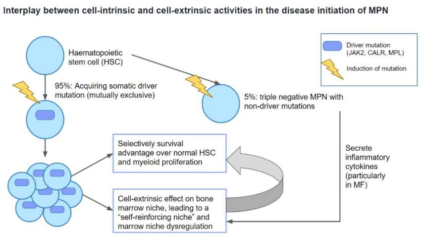
nal affiliations. MPNs involve the interplay between cell-intrinsic and cell-extrinsic activities. This is
characterized by survival advantage of MPN stem cells over normal HSCs that is sus-
tained by a dysregulated bone marrow niche via a positive feedback mechanism [9,14]
(Figure 1). Further acquisition of non-driver mutations then plays a pivotal role in de-
Copyright: © 2021 by the authors. Li- termining disease phenotype and promoting leukemic progression [9,10,13] (Figure 2).
censee MDPI, Basel, Switzerland. At diagnosis, all CML patients harbor abnormal HSCs [8,15,16]. They are characterized
This article is an open access article
by an unlimited potential and unrestricted ability to self-regenerate, remain quiescent,
distributed under the terms and con-
mediate BCR-ABL1-independent tyrosine kinase inhibitor (TKI)-resistance and evade the
ditions of the Creative Commons At-
host immunity, and allowing disease initiation, development, maintenance and progres-
tribution (CC BY) license (https://
sion [8,15,17–22]. The abnormal HSCs in CML are able to survive and thrive through vari-
creativecommons.org/licenses/by/
ous mechanisms such as modulation of downstream signaling pathways (e.g., JAK/STAT,
4.0/).
Int. J. Mol. Sci. 2021, 22, 659. https://doi.org/10.3390/ijms22020659 https://www.mdpi.com/journal/ijms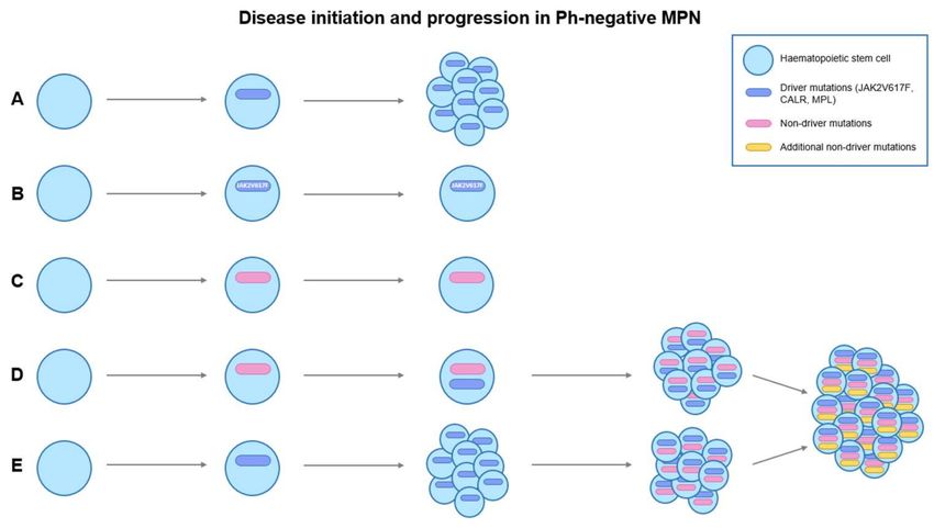
Int. J. Mol. Sci. 2021, 22, 659                                                                                                      2 of 39

                                  PI3K/AKT/mTOR, Wnt/β-catenin, Hedgehog signalling), induction of autophagy, selec-
                                  tive advantage in homing and engraftment in the bone marrow microenvironment (BMM),
                                  and alterations in cellular metabolism [8,15,23–29]. While leukemia stem cells (LCSs) are
                                  generally CD34+ /CD38− , the abnormal HSC populations in CML have extremely het-
                                  erogeneous and unstable cell surface antigens expression, and vary greatly in terms of
                                  their leukemogenic capacity [8,17,21,30,31]. Although TKIs show some efficacy in targeting
                                  abnormal HSCs in CML, they may not be adequate for disease eradication.

                                  Figure 1. Interplay between cell-intrinsic and cell-extrinsic activities in the disease initiation of MPN.
                                  MPN: myeloproliferative neoplasm; MF: myelofibrosis.

                                  Figure 2. Disease initiation and progression in Ph-negative MPNs. (A) shows the acquisition of
                                  somatic driver mutations (JAK2V617F, CALR, MPL) in a haematopoietic stem cell (HSC), leading
                                  to myeloproliferation. (B) shows the acquisition of somatic driver mutation, JAK2V617F, in HSC
                                  without progression to MPN. One of the reasons is the insufficient JAK2V617F allele burden to give
                                  rise to a MPN phenotype. (C) shows the acquisition of non-driver mutations without progression to
                                  MPN, indicating the presence of clonal hematopoiesis of indeterminate potential (CHIP). (D) The
                                  presence of CHIP increases the chance of HSC to acquire driver mutations, leading to myeloprolifera-
                                  tion. (D,E) Acquisition of additional non-driver mutations in MPN stem cells could lead to disease
                                  progression or leukemic transformation to secondary acute myeloid leukemia (sAML).

                                       The persistence of abnormal HSCs in CML and MPN has led to the development of
                                  novel therapies targeting CML or MPN stem cells. In this review, we discuss the current
                                  and emerging therapeutic options that may target CML and MPN stem cells with the aim
                                  of disease modification and eradication. The various pathways involved in the biology
Int. J. Mol. Sci. 2021, 22, 659 3 of 39
of CML and Ph-negative MPNs are depicted in Supplementary Materials Files S2–S7,
highlighting the rationale of their therapeutic targeting.
2. Current Therapeutic Options in CML and Their Effects on CML Stem Cells
TKIs competitively bind to the ATP-binding site of the BCR-ABL1 to reduce abnormal
phosphorylation of the dysregulated tyrosine kinase and inhibit downstream pathways
and leukaemogenesis [7,32,33]. TKIs (Table 1) have modest effects on CML stem cells as
single agents may be rendered ineffective in targeting CML stem cells as a result of BCR-
ABL1-independent mechanisms. They may arise from mutations in epigenetic regulators
(e.g., DNMT3A, EZH2, IDH1/2) [7,34] or the loss of tumour suppressor genes (e.g., TP53,
PTEN, TET1/2) [17,29] and genes that code for anti-oxidant systems (e.g., FoxO, EPAS1) [17].
Quiescence of CML stem cells is another major challenge, and may contribute to TKI
resistance and relapse in CML. Despite the fact that 50% of CML patients achieve treatment-
free remissions without relapse after achieving deep molecular response, most harbour
residual CML stem cells [15,17,35].
Table 1. Tyrosine kinase inhibitors and their effects on CML stem cells.
TKI Observations References
1. Reduction in CD26, a specific CML stem cell marker, in circulation after imatinib treatment
Imatinib 2. In-vitro enhanced mitochondrial oxidative phosphorylation upon imatinib discontinuation, [16,34]
leading to CML stem cell proliferation
1. Lower CD34+ cell proliferation in dasatinib-treated cells compared to imatinib-treated
Dasatinib [34]
group upon TKI discontinuation
1. Induction of apoptosis in CML CD34+ /CD38− stem cells
2. Inhibition of CML stem cell engraftment in murine models
Nilotinib [36,37]
3. More rapid and deeper CD34+ /lin− Ph+ cell clearance may might increase
treatment-free remission
1. Higher potency in inhibiting Src phosphorylation, hence inhibiting CML primitive
progenitor cells compared to imatinib
Bosutinib [31,38]
2. Growth inhibition of CD34+ /CD38− CML stem cells in combination with
gemtuzumab ozogamicin
1. Higher efficacy in eradicating CML-associated LSK cells than imatinib and dasatinib in
Ponatinib [22]
murine models
TKI: Tyrosine kinase inhibitor; CML: Chronic myeloid leukaemia: LSK cells: lin−Sca-1+ c-Kit+ cells.
2.1. First and Second Generation TKIs
Imatinib (IM), a first generation TKI, is highly effective in inducing apoptosis in BCR-
ABL1-positive cells. CML stem cells have dysregulated intracellular calcium signalling,
uncontrolled pro-inflammatory cytokines such as interleukin-6 (IL-6), IL-8 and nuclear
factor kappa beta (NF-κB), and overproduction of activator protein 1 (AP-1), which are
hallmarks for CML stem cell survival [29,39–41]. In-vivo and in vitro studies showed
that these could be reversed by IM antagonizing inositol-triphosphate (IP3)-mediated
calcium mobilization (p < 0.05) and oxidative stress via IP3 receptor inhibition (IP3R) on
the endoplasmic reticulum (ER) [39,40,42]. Herrmann et al. demonstrated a decrease in
CD26+ stem cells after in vitro IM therapy [16], yet Willmann et al. showed otherwise [36].
Moreover, nilotinib induced CML stem cell apoptosis [36], and nilotinib and dasatinib
showed higher potency in IP3R inhibition [42]. IM may also downregulate overexpressed
EZH2 in CML stem cells, with minimal effects in normal HSCs [17,43]. Also, in-vitro
studies showed that post-dasatinib or -IM therapy, programmed death receptor 1 (PD-1,
immune marker for immune-evasion) expression was found to be reduced on CD8+ T cells
and monocytic myeloid-derived suppressor cells (MDSCs), leading to increased cytotoxic T-Int. J. Mol. Sci. 2021, 22, 659 4 of 39
lymphocyte- (CTL) and Natural Killer (NK) cell-mediated cytotoxicity [18,44–46]. However,
contradicting evidence was presented in another in vitro study, which showed that IM
enhanced mRNA and protein expression of autophagy-related 4B cysteine peptidase
(Atg4B), resulting in TKI-induced autophagy and selective survival in CD34+ CML cells
(p ≤ 0.05) [39].
2.2. Ponatinib
Ponatinib, a third generation TKI, is indicated in CML with BCR-ABL1T315I mu-
tations or refractoriness to ≥2 TKIs. Steric hindrance is produced due to replacement
of threonine by isoleucine at the ATP-binding site [47,48]. Presence of a carbon-carbon
triple bond in ponatinib allows it to be 500-fold more potent than IM in overcoming
TKI-resistance [22,47–49]. Other pathways targeted by ponatinib include VEGFR, KIT, SRC,
FGFR, PDGFR, FLT3, and KIT [47]. In-vivo murine models with CML stem cells that
are lin− Sca-1+ c-kit+ showed that ponatinib was effective in CML stem cell eradication
and spleen size reduction. Thirty-percent residual BCR-ABL1 chimerism at 28 days was
achieved compared to >50% in dasatinib and IM [22,47,50].
2.3. Asciminib
Asciminib (ABL001), a recent, FDA-approved, fourth generation TKI, is an allosteric in-
hibitor that binds to the BCR-ABL1 myristoyl-pocket (STAMP) [8,33,49,51,52]. It is effective
against BCR-ABL1 KD-dependent and -independent mutations as monotherapy or in com-
bination with other TKIs to restore TKI-sensitivity in resistant cell lines and produce drug
synergism in reducing CRK-like protein (CRKL) phosphorylation for CML stem cells [49,53].
Initial results in a phase I trial (ClinicalTrials.gov number, NCT03595917) demonstrated
that 82% of patients with TKI-resistance achieved major cytogenetic response (MCyR) by
3 months and 30% of patients reached CCyR at 5 months [51]. In the phase III ASCEBEL
trial, asciminib showed superiority over bosutinib in achieving MMR at 24 weeks [53].
Ongoing trials using asciminib as monotherapy, in combination with other TKIs and/or
corticosteroids are underway (ClinicalTrials.gov identifiers: NCT04216563, NCT03906292,
NCT04360005, NCT03106779, NCT03595917, NCT03578367 and NCT02081378).
2.4. Interferon-α
IFNα was used as first-line treatment before the emergence of TKIs. It induces apop-
tosis of LSCs via Fas-receptors upregulation, FADD/caspase-8 pathway activation, and
cytochrome-c release, leading to mitochondrial disruption and cellular apoptosis indepen-
dent of anti-apoptotic B-cell lymphoma 2 (Bcl-2), cell-cycle arrest and tumour-suppressor
p53 [54–56]. IFNα also restores normal function of the dysregulated BMM through β1-
integrin for cellular differentiation and elimination of the protective barrier established for
LSC quiescence [54,57,58]. IFN-α-mediated increase in expression of major histocompati-
bility complex (MHC) class I molecules and tumour-associated antigens cause reactivation
of CTL and prompt CTL-mediated cytotoxicity against LSCs [54,55]. The 5-year survival
rate of IFNα was 57% as shown in a meta-analysis of 7 data sets of randomized trials
consisting of 1,554 patients [54,59]. In another study using IFNα monotherapy, the 10-year
survival rate was 72%, where 46% remained in CCyR [55,60]. These highlight the potential
re-emergence of IFNα for LSC elimination, where clinical trials using IFNα alone or in com-
bination with other TKIs showed promising results for TFR (ClinicalTrials.gov identifiers:
NCT02001818, NCT01657604, NCT03117816, NCT03831776, NCT04126681, NCT01316250,
NCT02381379, and NCT00452023).
3. Current Therapeutic Options in MPN and Their Effects on MPN Stem Cells
3.1. IFNα
A major significance of Peg-IFNα-2a is its ability to target MPN stem cells and reduce
mutant allele burden in MPN [61–68]. Sustained molecular, haematological response and
regression of BM fibrosis were seen in some patients after discontinuation of Peg-IFNα-2a,Int. J. Mol. Sci. 2021, 22, 659 5 of 39
indicating the eradication of MPN stem cells [65,69] (Table 2). Interestingly, the effect of
Peg-IFNα-2a on JAK2V617F+ stem cells was greater than that on CALR-mutated stem cells,
with no difference in hematological response [70,71]. This is due to the phosphorylation
and activation of JAK1-STAT1 pathway in JAK2V617F cells, but not in CALR-mutated cells,
resulting in JAK2V617F-positive cells priming towards Peg-IFNα-2a [70]. There is a paucity
of data suggesting that Peg-IFNα-2a targeting MPL-mutated clones could be due to the
low frequency of MPL mutations in MPN.
Table 2. Targeting of JAK2V617F and CALR-mutated MPN by IFNα preparations.
IFNα Preparation MPN Subtype Observations References
1. Preferential targeting JAK2V617F HSPC, especially in
CD90+ /CD34+ /CD38− HSC-enriched progenitors, compared to
mature blood cells (p < 0.05).
PV, ET, MF 2. Faster response in homozygous JAK2V617F clones than that of [71]
heterozygous clones.
3. Faster response in targeting JAK2V617F HSPC than
IFNα
CALR-mutated HSPC.
1. Sixty-two percent showed decrease in CALR-mutant allele burden
CALR-mutated (median decline: 29% from baseline).
ET, MF [62,72]
2. Nineteen percent (4 out of 21) showed MR with >50% mutant allele
burden reduction.
1. Reduction of median CALR-mutant allele burden from 41% to 26%.
CALR-mutated 2. Seven percent (2 out of 31) showed complete MR.
Peg-IFNα-2a ET [73]
3. Variation in responses in patients with additional
molecular mutations
1. Preferential inhibition and reduction of JAK2V617F-mutated primary
hematopoietic progenitors with sparing of JAK2-wild type cells.
Ropeginterferon
PV [69]
α-2b 2. Increase in proportion of wild-type to JAK2V617F-mutated colonies
after 12 months of treatment compared with hydroxyurea, reflecting
reduction of malignant progenitors in the BM.
PV: polycythemia vera; ET: essential thrombocythemia; MF: myelofibrosis; HSPC: hematopoietic stem and progenitor cells; HSC: hematopoi-
etic stem cell; MR: molecular response; Peg-IFNα-2a: pegylated interferon-alpha-2a; IFNα: interferon-alpha; BM: bone marrow.
3.2. JAK Inhibitors
Treatment with ruxolitinib showed some reduction in JAK2V617F mutant allele burden
in PV patients [74,75], although its effect on reduction of mutant allele burden in MF
is mild [76]. Additionally, several second generation JAK inhibitors were developed to
improve efficacy and reduce side effects of ruxolitinib [77–81]. However, none of them show
significance in eradicating LSCs [77,79,80,82,83]. Some studies have shown that fedratinib,
a newly FDA approved JAK2/FLT3 inhibitor in 2019, reduced JAK2V617F variant allele
frequency (VAF) [79,84]. Yet, the results were not consistent with other trials [79,83,85].
Therefore, combinations of various novel therapies with ruxolitinib emerge in the hope of
eliminating MPN LSCs [86].
3.3. Allogeneic HSCT
Allo-HSCT is able to overcome high molecular risks (HMR) mutations, and its recipi-
ents usually harbor additional molecular mutations. These additional molecular mutations,
including those conferring a poor prognosis in MPN (ASXL1, EZH2, SRSF2, IDH1/2,
TP53) generally do not affect relapse-free survival (RFS) and OS in patients receiving
allo-HSCT [87]. However, discrepancies are seen in terms of relapse risks. The ASXL1
mutation, which accounts for >90% in intermediate-2 and high-risk MF patients, is found to
be associated with higher relapse risks [88]. In a study assessing the outcome of allo-HSCTInt. J. Mol. Sci. 2021, 22, 659 6 of 39
in MPL-mutated PMF and secondary myelofibrosis (SMF), the only relapsed patient har-
bored ASXL1 and EZH2 mutation [89]. Meanwhile, some post-transplant ASXL1-mutated
patients may die without relapsing [87].
4. Novel Therapy Targeting Signaling Pathways in CML Stem Cells
4.1. Novel Tyrosine Kinase Inhibitors
PF-114 is an orally bioavailable fourth generation TKI that is selectively active against
BCR-ABL1-dependent and -independent mutations, as well as non-mutational TKI-resistant
cell lines [8,90–92]. It acts as a BCR-ABL1 KD antagonist and/or STAMP inhibitor, suppress-
ing the constitutively active PI3K/AKT/ERK1/2 and JAK/STAT3/5 signalling, elevating
p27 levels, leading to G1 cell cycle arrest [91,93]. It induced apoptosis in patient-derived
K562 and KCL-22 cell lines [91]. In K562 nude mouse xenograft, it caused complete erad-
ication of the tumour bulk (p < 0.001) without recurrence [90]. It also showed excellent
toxicity profile as it spared VEGFR, FLT3, EPHRIN, FGFR and B-RAF, implying less car-
diac, pulmonary and vascular complications [90], hence making it a promising agent for
patients refractory and/or resistant to frontline therapies. Phase I/II trials showed that
55% of heavily pretreated patients receiving PF-114 300mg daily achieved MCyR and 36%
achieved MMR [92].
4.2. Targetting microRNAs
MicroRNAs (miR) are short pleiotropic non-coding RNA sequences that cleave or
repress transcription, hence controlling at least 10–40% of human mRNA expression [94,95].
Malfunctioning miR is key to leukemogenesis and TKI-resistance, maintenance and self-
regeneration [17,94–96]. Overt expression of miR-29a leads to the depletion of tumor
suppressor TET2 and antioxidant-coding EPAS1, with upregulation of anti-apoptotic genes
Bcl-2 and Mcl-1 apoptosis regulator (Mcl-1) [94,97–99]. Downregulation of Bcl-2 inhibitor
miR-153-3p is mediated by uncontrolled c-Myc/miR-150 expression in LSC and Ph+ cells,
impairing myeloid differentiation and promoting TKI-resistance [100,101]. Prolonged ex-
posure of TKIs triggers drug resistance in LSCs and K562 cells, and is associated with low
miR-217 and high DNA methyltransferases (DNMTs) [94,102,103]. miR-424 acts as a direct
inhibitor of BCR-ABL1, showing markedly low expression in CD34+ /CD38− and BCR-
ABL1+ cells alongside high levels of oncoprotein Cobll1 [94,97,104,105]. In vitro studies
suggested that miR-217 overexpression might allow restoration of tumour suppressor ef-
fects [94,97,104]. Reduced tumor suppressor miR-142 levels in LSCs and TKI-resistant cells
were found to be associated with excessive oncoproteins Mcl-1, cKIT, and SRI, precipitating
unimpeded PI3K/AKT, JAK/STAT, and RAS/RAF/MEK/ERK downstream signalling
with anti-apoptotic, pro-survival and therapy-resistant effects [94,97,106–109]. Correction
of dysregulated miR levels in preclinical studies exhibited propitious results in reduction
of tumour bulk. This may not only yield a new field of clinical investigation for targeted
therapy, but also as a tool in aiding diagnosis, a prognostic indicator and a predictor of
treatment response [110].
4.3. Targeting BCR-ABL1/Gab2/Grb2 Axis
Dysregulated growth factor receptor-bound protein 2 (Grb2) expression permits direct
binding to the Src homology (SH2) domain of BCR-ABL1, forming Grb2-SOS complexes
and leading to downstream hyperactivation of RAS/MAPK pathway [111–114]. BP1001, a
liposome-incorporated antisense oligodeoxynucleotide targeted against the Grb2 mRNA,
inhibits subsequent protein expression and the RAS/MEK/ERK pathway. Preclinical
studies showed that Grb2 depletion induced LSC knockdown in CD34+ cells, without
affecting normal HSCs and STAT inhibition [111,114]. Combination with TKIs overcomes
resistance and induces drug synergism [113]. A phase I clinical trial showed that BP1001
enhanced the effects of dasatinib by 2–6-fold and reduced phosphorylation of ERK1/2
and Grb2 levels by >25% in 52% and 49% patients respectively [113]. Trametinib, a MEKInt. J. Mol. Sci. 2021, 22, 659 7 of 39
inhibitor in combination with imatinib inhibited the MEK/ERK and NF-κB-mediated LSC
survival, restoring TKI sensitivity in-vitro and in-vivo [18,115,116].
4.4. Targeting MAPK/MNK1/2 Pathway
The MAPK/MNK1/2 pathway is amplified in CML stem cells but not in normal HSCs.
The constitutive phosphorylation of eukaryotic initiation factor 4E (eIF4E, an oncoprotein
essential for LSC proliferation) induces nuclear activation and translocation of β-catenin,
contributing to leukemogenesis and TKI-resistance [117]. Preclinical studies showed that
ETC-1907206, a selective MNK1/2 inhibitor, suppressed eIF4E phosphorylation and β-
catenin signalling [117].
4.5. Targeting mTOR Pathway
mTOR, is a catalytic kinase for the protein complexes mTORC1 and mTORC2 in the
PI3K/AKT/mTORC1 pathway [118,119]. It inhibits mRNA translation through initiation
factor 4E binding protein (4E-BP1) and p70 ribosomal S6 kinase (p70S6K, S6K) [118–120].
Liver kinase B1 (LKB1, tumour suppressor), an upstream kinase of AMP-activated protein
kinase (AMPK) induces AMPK phosphorylation of tuberous sclerosis complex 2 (TSC2) to
suppress Ras homolog enriched in brain (Rheb) and inhibit mTOR. In LSCs, dysregulated
PI3K/AKT/mTOR signalling increases reactive oxygen species (ROS) production, leading
to the loss of negative regulation by LKB1 and AMPK, promoting survival, proliferation,
drug resistance and stemness [118,121]. Preclinical studies showed that metformin, an
AMPK activator inhibited aberrant PI3K/AKT/mTOR hyperactivation to reduce oxidative
phosphorylation of glucose and fatty acids, leading to LSC apoptosis [122,123]. However,
this led to the compensatory upregulation of glucose and glycolysis which could be be
overcome by the addition of 2 deoxy-glucose (2-DG), a hexokinase inhibitor that mimics d-
glucose and inhibits glycolysis to induce cell death [123,124]. Combination with TKIs boosts
effects and induces TKI-mediated apoptosis [123,124]. Resveratrol, a natural antioxidant
found in grapes is also found to stimulate AMPK activation, hence upregulating p38 expres-
sion and JNK-mediated phosphorylation of H2AX, downregulating Bcl-2 and triggering
caspase-3-mediated LSC apoptosis and cell-cycle arrest [120,125,126]. 5-aminoimidazole-4-
carboxamide riboside (AICAR), undergoes phosphorylation and binds to an allosteric site
of AMPK to activate it and inhibits mTOR regardless of TKI sensitivity [127].
4.6. Bcl-2 Targeting
Bcl-2, a key anti-apoptotic gene that regulates mitochondrial-mediated apoptosis
through the JAK/STAT and PI3K/AKT pathways [8,18,128,129], is overwhelmingly ex-
pressed in LSCs. In addition, BCR-ABL1 induces upregulation of Bcl-2 anti-apoptotic
proteins, including Mcl-1 and B-cell lymphoma-extra large (Bcl-xL) [8,18,128]. Venetoclax,
a Bcl-2 inhibitor, binds to the hydrophobic groove of the Bcl-2 homology 3 (BH3) domain
of Bcl-2, releasing its inhibition on Bcl-2-associated X protein (BAX) to drive p53/BAX-
mediated programmed cell death [18,128]. Preclinical studies showed synergism between
venetoclax and TKI in targeting mitochondrial oxidative phosphorylation to eliminate
CD34+ /CD38+ and CD34+ cells [18,128]. A retrospective study using venetoclax in com-
bination with TKIs showed 60% complete remission (CR), 75% ORR, median survival of
10.9 months and median RSF of 3.9 months [130].
4.7. JAK2 Inhibition
JAK2 mediates cytokine-mediated signaling in CML cells. It leads to uncontrolled
STAT3/5 phosphorylation by directly binding to the SH2 domain of BCR-ABL1, which is
stabilized by Abelson helper integration site 1 (AHI-1), an oncogenic adaptor for LSC sur-
vival. [18,30,131,132]. In LSCs, induced expression of MPL enhances JAK/STAT signaling
to trigger ROS formation and subsequent clonal evolution, contributing to stemness and
TKI-resistance [8,18,30,131]. However, LSCs remain sensitive to JAK2 inhibitors such as rux-
olitinib. Preclinical studies showed that combining ruxolitinib with the CML-specific TKIsInt. J. Mol. Sci. 2021, 22, 659 8 of 39
eliminated CD34+ /CD38− stem cells with no effects on normal HSCs in-vitro, and reduced
CD34+ cell engraftment to the BM in-vivo [131]. Sweet et al. showed that 33% of patients
had ≥1 log reduction in BCR-ABL1 transcripts and 44% achieved MR4.5 when co-treated
with nilotinib in a phase I trial [133]. Another phase I trial with nilotinib demonstrated that
40% of patients had molecularly undetectable BCR-ABL1 transcripts over 6 months [134].
A phase II trial using ruxolitinib alone showed 60% ORR, where 33% observed clinical
benefit in one or more categories: platelet count improvement, hemoglobin improvement,
≥50% reduction in spleen size and ≥50% reduction in symptoms [135]. A phase I/II trial
in combining ruxolitinib with CML-specific TKIs showed achievement of CCyR in 87.5%
and MMR in 37.5% of patients [136].
4.8. Targeting PPARγ/STAT5/HIF2α Axis
STAT5 activation leads to the induction of hypoxia inducible factor-2α (HIF-2α)/CITED
pathway for adaptation in low oxygen levels of the BMM to maintain LSC dormancy
and self-renewal potential [137–140]. PPARγ, a negative regulator of the STAT5/HIF-
2α/CITED pathway inhibits adhesion of LSCs to the extracellular matrix and drives apop-
tosis [8,137,140,141]. Preclinical studies demonstrated that thiazolidinediones (PPARγ ago-
nists) upregulate matrix metalloproteinase-9 (MMP-9) and MMP-2 to inhibit LSC invasion
and adhesion to the BMM. They also activate caspase-3 for LSC apoptosis [8,137,140,141].
Other findings include upregulation of PPARα ligands e.g., clofibrate and enhanced ex-
pression of human organic cation transporter 1 (hOCT1) via WY-12643, which increase
cellular uptake of TKIs to promote TKI-mediated apoptosis [139,141]. Preliminary clinical
studies in 3 CML patients showed that combined use of pioglitazone and IM accomplished
sustained complete molecular remission for up to 4.7 years in all patients, even after pi-
oglitazone withdrawal [141]. Phase II ACTIM trial showed that IM and pioglitazone had
no drug interactions, yet their combination achieved MR4.5 at 12 months in 56% of pa-
tients [138]. Novel STAT3 inhibitor BP-5087, derived from SF-1-066, demonstrated 10-fold
greater potency in reducing STAT3 phosphorylation and translocation, inhibiting survival
of TKI-resistant CML cells and LSCs in preclinical studies [18,142]. Combination with
TKIs showed dramatic increase in effects, whereas monotherapy of either was evidently
inferior [142]. However, STAT3/5 inhibition may be less effective than JAK inhibition as
other STATs may compensate for STAT3/5 loss [143].
4.9. Prostaglandin E (PGE) 1 Analogue
PGE2 is a pro-inflammatory prostaglandin upregulated by BCR-ABL1 [17,144]. It
promotes β-catenin nuclear accumulation, stabilization and localization to promote the
β-catenin/Wnt signalling, conferring to LSC stemness, TKI-resistance and disease progres-
sion [17,144]. On the contrary, PGE1 exhibits protective functions against LSCs [17,144].
Preclinical studies showed that misoprostol, a PGE1 analogue acts via EP4 receptor to
inhibit Tcf1/Lef1 and Fos/FosB, hence reducing LSCs by >10-fold [17,144]. Exhibiting
negligible effects on normal HSCs, the activation of PGE1 poses as a promising target for
CML stem cell eradication.
4.10. Activation of Promyelocytic Leukaemia—Nuclear Bodies (PML-NB)
Promyelocytic leukaemia (PML) forms PML-NBs to repair DNA double-strand breaks
(DSBs), maintain telomere homeostasis and maintain normal HSC asymmetric division
through the PML/PPAR/FAO pathway [17,145,146]. Preclinical studies showed that PML
upregulation in mesenchymal stromal cells upregulated inflammatory cytokines (IL-6/IL-
6R and CXCL1/CXCR2), which are crucial for the maintenance in the BMM and TKI-
resistance of CML stem cells [146]. Arsenic trioxide (ATO) was used as a first-line treatment
for CML before the development of TKIs, but preclinical studies showed limited effective-
ness in targeting CML stem cells as a single agent [147–149]. However, combination with
TKIs showed LSC targeting, downregulation of VEGFR and angiogenesis, upregulation
of NKG2D ligands to induce NK-cell mediated cytotoxicity, growth arrest, inhibition ofInt. J. Mol. Sci. 2021, 22, 659 9 of 39
RAS/MAPK and PI3K/AKT pathways, and apoptosis via extrinsic pathways (caspase-
8/-10, TNFR1) and intrinsic (BAX) pathways and the induction of ER stress [147–149].
ATO/IFNα combination therapy demonstrated superior in-vivo and in vitro results com-
pared to ATO/TKIs, where it induced cell-cycling of dormant LSCs and inhibited the
Hh pathway, hence, leading to autophagy-induced cell death [145]. The established abil-
ity of ATO/IFNα to overcome TKI-resistance and abolish CML stem cells in preclinical
studies [145,150] has led to phase I clinical trials [151]. In a cohort of eight patients, de-
crease in BCR-ABL1 fusion transcript was seen in 100% and 87.5% patients after trial and
12 months after trial respectively. MR4.5 or above was achieved in 87.5% and 55.6% patients
immediately after study and 12 months later, respectively [151].
5. Targeting the CML Stem Cell Microenviroment, Survival and Self-Renewal
Normal HSCs interact with endothelial cells, neural cells, osteoclasts, mesenchymal
stromal cells and osteoblasts in the BMM [17,25,152,153]. Selectins, integrins, and CD44
expressions are required for HSC engraftment and adhesion between fibronectin on the
extracellular matrix and CD106 (VCAM-I) on the BM endothelium [23,25,152–154]. HSC
rolling and homing is mediated by interaction between constitutively expressed E- and P-
selectins and VLA4, where SDF1 and its receptor CXCR4 acts as a chemo-attractant through
β1/2− integrins and SDF1 for stable engraftment [23,25,152–156]. BCR-ABL1 impairs
the SDF1/CXCR4 axis in normal HSCs but upregulates it in CML stem cells, conferring
selective homing and survival in the BM niche [17,23,152,153,157,158]. In addition, CML
stem cells have defective β1-integrin levels (VLA4 or VLA5), allowing redistribution and
mobilization into the PB and other organs, e.g., spleen with the potential of uncontrolled
extramedullary myeloproliferation and local LSC reservoirs [152,154,157]. CML stem cells
alter extrinsic factors and upregulate expression of CD44+ and E-selectin to promote
prominent BMM changes such as marrow fibrosis for exclusive stem cell engraftment and
dormancy, offering protection from drug-targeting [25,152,154,158–162].
5.1. Dipeptidyl-Peptidase (DPP-4) Inhibition
DPP-4 (CD26) is an overtly expressed protease on LSC surface, where it cleaves
the SDF1/CXCR4 axis to facilitate LSC mobilization into the PB independent of niche
regulations [16,17]. TKIs decrease CD26+ LSCs but levels dramatically increase following
resistance or relapse [16,17,137]. CD26 is not expressed on normal HSCs, suggesting that
it may be a marker for LSCs as concentrations correlate with white blood cell (WBC)
count [16]. DPP-4 inhibitors (gliptins) normalize the dysregulated SDF1/CXCR4 axis to
restore and promote homing of LSCs [16,36]. Interestingly, Willmann et al. demonstrated
that single agent nilotinib could inhibit engraftment and induce apoptosis of LSCs, while
neither vildagliptin nor imatinib addition exhibited these effects [36]. Combination of
nilotinib with vildagliptin also did not produce cooperative results, suggesting insignificant
effects of co-administration [36]. However, vildagliptin alone reduced disease expansion
through limiting LSC mobilization [16,36]. In samples of two nilotinib-pretreated CML
patients with diabetes mellitus using gliptins for diabetic control, BCR-ABL1 transcripts
were near undetectable or undetectable [16].
5.2. E-Selectin Antagonist
Uproleselan (GMI-1271) is an E-selectin inhibitor which dislocates homed LSC from
the BM niche into PB for cellular differentiation [154,163]. Promising phase III study results
in acute myeloid leukaemia (AML) for LSC eradication [163] has led to preclinical studies
in CML. In vitro studies demonstrated cell cycle progression via upregulated CDK6 (cell
cycle promotor) and downregulated p16 (cell cycle inhibitor), leading to an increase in cells
in G-phase and increase G2 /S/M phase when used as monotherapy or in combination
with IM [152,154]. Reduced CD44+ expression via the Scl/Tal1 pathway, increased CML
stem cell cycling, and restoration of TKI-sensitivity were also noted [152,154]. MurineInt. J. Mol. Sci. 2021, 22, 659 10 of 39
models showed depletion of LSC and BCR-ABL1+ cells, spleen size reduction, impaired
LSC engraftment to the BM and spleen, and improved OS [152,154].
5.3. Targeting SDF1/CXCR4/CXCR7 Axis
Preclinical studies showed that disruption of the SDF1/CXCR4/CXCR7 axis of mes-
enchymal stromal cells reduced EZH2 expression [164], increased self-renewal capacity
in LSCs and the ability to override TKI-resistance with no effect on osteoprogenitor cells,
mesenchymal stromal cells and normal HSCs [164–166]. NOX-A12, a pegylated Spiegelmer,
inhibits SDF1 and antagonizes the SDF1-CXCR4 or -CXCR7 interactions to inhibit LSC
homing and causes TKI-sensitization [167]. In-vitro studies showed enhanced abolishment
of SDF1-mediated migration in BCR-ABL1+ cells and induction of apoptosis when com-
bination with imatinib was used (p < 0.00005) [168]. In-vivo studies showed eradication
of FLT3-ITD+ cells and inhibition of SDF1-mediated migration of FLT3-ITD+ cells [168].
Plerixafor, an allosteric CXCR7 agonist and CXCR4 antagonist/partial agonist, is clini-
cally used for stem cell mobilization in HSCT in multiple myeloma and non-Hodgkin
lymphoma [165,168,169]. Its use in in vitro studies with K562 and KU812 cell lines showed
reduction of drug-resistance, cellular migration and adhesion to BMM and sensitization to
TKI [165]. Plerixafor in in-vivo murine models mobilized LSCs to the PB, potentiating TKI-
induced tumor bulk eradication [165]. However, Agarwal et al. presented contradicting
in-vivo results, which demonstrated that TKI plus plerixafor led to stem cell infiltra-
tion of the central nervous system (CNS) and subsequent development of neurological
deficits [170].
5.4. Hypoxia-Inducible Factor (HIF) Targeting
HIFs interact with HIF-responsive elements (HRE) for gene regulation, depend-
ing on the oxygen concentration of the microenvironment [17,28,171–173]. The hypoxic
BM niche contains high ROS levels which causes upregulation of HIF-1α and HIF-2α,
suppressing BCR-ABL1 oncoprotein yet permitting CML stem cell survival, quiescence,
immune-evasion, TKI-resistance and potential transformation of normal HSC into in-
duced pluripotent stem cells capable of becoming LSCs [17,28,172,173]. This is mediated by
glucose transporter 1 (GLUT1) and tumour M2-pyruvate kinase (PKM2), which lead to in-
creased glycolytic flux [28], p21 upregulation for cellular proliferation [172], suppression of
p53 [28,172,173], increased transcription of antioxidant enzymes (FoxO and Nrf2) [28,173],
overt Oct4 and c-Myc for transformation of other haematopoietic cells into LSCs [173],
and evasion of cellular immunity through B7H1/programmed death ligand 1 (PD-L1)
expression, and soluble factors such as nitric oxide [28]. LSCs have high enough ROS
levels for clonal evolution yet low enough levels to maintain stemness [8,17]. Acriflavine,
a HIF-1 inhibitor prevents dimerization of the HIF complex and reduces LSC formation,
maintenance, survival and stemness through three mechanisms [17,173,174]: depleting
c-Myc at mRNA and protein levels, promoting expression of tumour suppressors (e.g., p57,
p19Arf and p16Ink4a ) and inhibiting genes that favour LSC stemness (e.g., NANOG, Oct4,
Sox9). In vitro studies using K562, KCL22, and LAMA-84 CML cell lines and in-vivo stud-
ies demonstrated cytotoxicity against BCR-ABL1+ cells and LSCs, where adverse effects on
normal HSCs were significantly less-severe in-vivo [174].
5.5. Targeting Hh Pathway
Hh homologues bind to the Patched (PTCH) receptor, activating Smoothened (Smo)
and Gli family of transcription factors to mediate downstream signalling (e.g., Myc, cyclin-
D1, Bcl-2, SOX2) for cellular regeneration and homeostasis [8,18,29,175,176]. Low Shh
levels in mesenchymal stromal cells along with hyperactivation of Shh and Smo in CD34+
and c-kit+ (p < 0.05) LSCs stimulate cyclin-D1-mediated LSC quiescence, maintenance and
uncontrolled expansion through the Wnt/β-integrin pathway [175]. Hh overactivation is
seen in 50% chronic phase (CP)-CML, 70% accelerated phase (AP)-CML and >80% blast-
phase (BP)-CML patients [175]. In vitro and vivo studies showed that LDE225 (sonidegib),Int. J. Mol. Sci. 2021, 22, 659 11 of 39
a highly selective and potent Smo inhibitor, was effective alone and in combination with
TKIs in eradicating Hh-mediated self-renewal capacity of CD34+ and BCR-ABL1+ CML
cells [18,177]. In a phase I trial CA180323, another Smo inhibitor, BMS-833923, was in-
vestigated in combination with dasatinib, showing no drug interactions and undesirable
toxicity profiles and minimal reduction of BCR-ABL1 progenitors [176]. Although preclini-
cal and preliminary clinical studies showed conflicting results, the Hh pathway remains a
significant target worth investigating.
5.6. Targeting Wnt/β-Catenin Signalling
Porcupine (PORCN)-dependent acetylation of Wnt ligands is essential in Wnt/β-
catenin signalling for maintenance of cellular functions [17,178,179]. BCR-ABL1 drives consti-
tutive secretion of Wnt-ligands and overexpression of frizzled-4 (FZD4) receptors to promote
nuclear transduction and stabilization of β-catenin, mediating TKI-resistance [178–180].
Riether et al. proposed that it might be induced by prolonged TKI exposure as TKI-therapy
depleted miR29 and amplified CD70 expression, leading to CD27-mediated Wnt activation
for LSC quiescence and therapy resistance [181]. In an in-vivo study using transgenic
murine models with CD34+ and c-kit cells, potent PORCN inhibitor WNT974 in com-
bination with nilotinib was efficacious in reducing neutrophils, white blood cells and
myeloid cells in PB, with eradication of CML stem cells and other progenitors in the BM
and spleen [18,178,179]. Mice treated with nilotinib monotherapy died after 30 days while
nilotinib plus WNT974-treated mice had prolonged survival with prominent suppression of
c-Myc, cyclin-D1 and Axin-2 expression [18,178]. C82, a β-catenin inhibitor, downregulated
CD44, c-Myc, STAT5, survivin, and CRKL in T315I and E255V mutant cell lines, eliminating
LSCs in-vitro and in-vivo [27].
5.7. Targeting Protein Phosphatase 2A (PP2A)
PP2A is a serine-threonine phosphatase that acts as a tumour suppressor, contributing
to >90% intracellular phosphatase activity alongside PP1 [29,182–185]. PP2A dephosphory-
lates Myc, disrupting Myc/MAX interaction and inhibits gene expression for mitochon-
drial biogenesis. BCR-ABL1 oncoprotein amplifies endogenous expression of potent PP2A
inhibitors protein SET (SET), cancerous inhibitor of PP2A (CIP2A) and PP2A-Aα that inac-
tivate phosphatase activity, hence, resulting in high levels of Myc and uncontrolled DNA
synthesis for LSCs survival and maintenance [29,182,183,186,187]. The Myc/MAX complex
can directly bind BCR-ABL1 to upregulate its mRNA and protein content, establishing a
positive feedback loop for LSCs [182,185]. Myc inhibitor 10058-F4 suppressed CIP2A in 80%
of CD34+ cells (p = 0.04) and 85% of K562 cells (p = 0.01), preventing Myc/MAX interaction
and restoring tumour-suppressor functions in vitro K562 and CD34+ cell lines [29,184].
However, Myc-targeting remains a therapeutic challenge due to the lack of a clear ligand
binding domain [182]. OP449, a SET antagonist reactivated PP2A and significantly reduced
JAK/STAT5 and PI3K/AKT pathways in vitro CD34+ and K562 CML cells, as well as alle-
viated tumour burden in vivo xenografted mice with human CML cells [182,186]. FTY720,
a SET antagonist activates extrinsic and intrinsic apoptotic pathways in a PP2A-dependent
manner [29,184,186,188] or via the activation of caspase-3/-8/-9 and pro-apoptotic BH3-
only proteins (BIM and BID) in-vitro K562, MYL, KBM5 and KCL22 cell lines [184]. It
can also overcome BIM-deletion-mediated, Gal-3-mediated BCR-ABL1 kinase-domain-
mediated TKI resistance, with synergistic activity in combination with imatinib [184].
However, Bcl-2 expression partially hinders FTY720-mediated apoptosis [184]. Combina-
tion of FTY720 or OP449 with TKIs showed drug synergism [182,184,186,188].
6. Targeting CML Stem Cells via Epigenetic, Ribosomal and Transcriptional Regulation
Epigenetic modifications are reversible and heritable changes that regulate DNA ex-
pression while maintaining the same nucleotide sequence [189–191]. High ROS levels
and hypoxic conditions of the BMM lead to DNA damage and ineffective repair, making
LSCs prime candidates to undergo genetic evolution. Pre-leukaemic lesions in epigeneticInt. J. Mol. Sci. 2021, 22, 659 12 of 39
regulators (e.g., DNMT3A, IDH1/2, TET1/2, TP53) result in clonal hematopoiesis of interme-
diate potential (CHIP), a predisposing factor for haematological malignancies [8,192–195].
Despite having a peak incidence of 15–20% in the general healthy population after the
age of 70 [8,196–199], CHIP is not a cause for CML [8,21,193]. However, the concurrent
presence of CHIP and leukaemia drives LSC transformation and survival, and is associated
with an inferior prognosis [8,17,189,195–199].
6.1. Bromodomain and Extra-Terminal (BET) Inhibitor
BET proteins are epigenetic regulators of transcription, inflammatory processes, and
cell-cycle regulation [18,200–202]. Bromodomain-containing protein 4 (BRD4) is the only
ubiquitously expressed member of the family directly bound to P-TEFb to maintain chro-
matin stability and G2 /M phase transition in the cell cycle. BCR-ABL1 and LSCs can acquire
secretory-associated senescent phenotype (SASP) to drive BRD4 activity and upregulation
of Myc, leading to overt release of pro-inflammatory cytokines (e.g., IL-1, IL-6, IL-8, IL-17,
IL-23, BMP2, TNFα, CCL9, NF-κB) to favour LSC senescence. Aberrant BRD4 activity
also induces active transcription and expression of PD-L1 on leukaemic cells, myeloid
dendritic cells and macrophages for immune-evasion. BRD4 inhibitor JQ-1, as well as
BRD4 degraders dBET1 and dBET6 have shown to be promising and potent inhibitors that
downregulate Myc in targeting LSCs and overcoming TKI-resistance [200,202–205]. In vitro
studies showed that JQ-1 increased IL-12β to reduce VEGFR-mediated angiogenesis while
decreasing PD-L1 expression to promote CTL-cytotoxicity [200–202,204] and IL-6-mediated
Jagged1/Notch1 cellular invasion and migration [201]. In vitro studies using K562 and
KU812 cell lines showed superior potency of dBET6 and dBET1 over JQ-1 in suppressing
BRD4 and Myc expression, where dBET6 and dBET1 could eliminate BCR-ABL1+ cells
and CD34+ /CD38− LSCs while JQ-1 failed [202]. In vivo studies demonstrated that dBET6
could override niche cell-induced TKI-resistance in CML LSCs, while JQ-1 was only able
to restore TKI effects completely in KU812 cells and partially in K562 cells [202]. All three
BRD4 inhibitors inhibited IFNγ-induced PD-L1 expression in LSCs [202,205]. Promising
preclinical studies have led to the development of novel BRD4 inhibitor CPI-0610, which is
currently in phase I clinical trials (ClinicalTrials.gov identifier: NCT02158858).
6.2. EZH2 Inhibition
EZH2 is a histone methyltransferase and a component of the polycomb repressive
complex 2 (PRC2) for histone H3 methylation (H3K27me3) and transcription inactiva-
tion [17,206–208]. EZH2 hyperactivity blocks myeloid differentiation to promote LSC
expansion, survival and TKI-resistance [17,43,207–211]. In vitro studies showed that EZH2
inhibitor EPZ-6438 upregulated tumour suppressor p16 to deplete leukaemic cells in K562,
HEL, Kasumi-1, ME-1, Mv4-11 and MOLM13 cell lines [207,210]. Inactivation of EZH2
showed significant reduction in WBC count and LSCs in the PB, prolonged survival, and
absence of splenomegaly and pulmonary hemorrhage compared to the control arm [210].
Another in vitro study showed 20–40% reduction in CD34+ cells and 60–80% reduction in
progenitor granulocyte/erythroid/megakaryocyte/macrophage (GEMM) and total colony
forming cell (CFC) [43]. Combination with TKI increased activation of H3K27me3 targets
(e.g., CDKN2A) and upregulated pro-apoptotic targets of p53 (e.g., NOXA, p53 upregulated
modulator of apoptosis (PUMA), BAX, CDKN2A, TNFRS10B), showing >70% reduction
in CD34+ /CD38− /CD45+ cells, near complete eradication of CD34+ /CD45+ cells and
restoration of TKI-sensitivity [43,211]. Murine xenograft models with CD34+ cells showed
similar findings [43].
6.3. Histone Deacetylase (HDAC) Inhibitor
HDAC packs histones tightly to inhibit transcription. Aberrant activity in CML LSC
halts myeloid differentiation to allow LSC survival [18,212–214]. Panobinostat (LBH589),
a HDAC inhibitor combined with TKIs in preclinical studies showed impairment of
LSC quiescence and engraftment, promoting TKI-mediated apoptosis [18,212–214] in vivoInt. J. Mol. Sci. 2021, 22, 659 13 of 39
CD34+ mice [212,213] and in vitro cell lines CD34+ /CD38− , K562, K562/IM-R1, Ba/F3 and
Fa/F3/T315I [18,212,213]. A phase I clinical trial exhibited no dose-limiting toxicities and
44% patients achieving >1 log reduction in BCR-ABL1/ABL1 transcripts, but was discon-
tinued due to slow accrual [18]. A phase II study also showed no MCyR nor molecular
response. However, in light of encouraging preclinical results, a phase Ib trial is currently
underway (ClinicalTrials.gov identifier: NCT03878524). Chidamide, an orally bioavailable
HDAC inhibitor in vitro increased acetylation of Histone H3, activated caspase-3/-9 and
decreased levels of β-catenin, surviving and Myc in CD34+ cells, inducing apoptosis [215].
It also exhibited limited toxicity to normal HSCs, and produced drug-synergism in combi-
nation with TKIs in overriding BCR-ABL1-dependent and -independent mutations [215].
Another novel pan-HDAC inhibitor MAKV-8 reduced c-Myc expression, activated caspase-
3/-9-mediated apoptosis and triggered ER stress for LSC eradication using K562 and
MEG-01 cell lines in vitro [214]. When used in combination with TKIs, in vivo K562 cells
in zebra-fish were completely abolished [214].
6.4. Protein Arginine Methyltransferase (PRMT5) Inhibitor
PRMT5 catalyzes histone methylation for RNA metabolism, transcriptional regulation,
ribosome biogenesis and cell-cycle regulation [216]. Aberrantly expressed PRMT5 binds to
BCR-ABL1 to form a positive feedback loop, which is associated with worse progression-
free survival in CML patients [216–218]. Preclinical studies showed that PJ-68, a PRMT5
inhibitor suppressed the Wnt/β-catenin pathway and induced negative regulators of LSC
cellular renewal P15INK4B and p27KIP1 for CD34+ CD38− apoptosis [18,218].
7. Targeting CML Stem Cells via P53 Modulation
P53 is crucial in tumour suppression and apoptotic control [29,219,220]. It is dys-
functional in CML as it directly binds to IκBα and BCR-ABL1, leading to dysregulated
p53 and Myc levels [29,219,220]. Preclinical studies showed that depletion of E3 ligase
FBWX7 (SCF) led to Myc-induced p53 upregulation, promoting p53-mediated cell-cycle
regulation and apoptosis in CD34+ and HeLa cell lines [219]. This spared normal HSCs and
achieved near complete elimination of BCR-ABL1+ cells and LSCs [219]. Dasatinib showed
some efficacy in STAT5 inhibition and mutant TP53 reduction. However, it was insuffi-
cient to eliminate LSCs [219,221]. RITA (NSC652287) binds and prevents p53 degradation.
In vitro studies showed degradation of IκBα and downregulation of NF-κB-regulated pro-
liferative (c-Myc) and anti-apoptotic (Bcl-2, XIAP, cIAP1) genes, inhibition of PI3K/AKT
and JAK/STAT5 signalling pathways, increased p53-mediated apoptosis and decreased
c-Myc levels leading to subsequent CD34+ and K562 cellular knockdown [219,222,223].
In-vivo mouse CML models using RITA in combination with CPI-203 (BET family inhibitor)
showed decreased levels of CD11b, CD19, CD33, CD34 and CD45, suggesting reduced
LSC engraftment [219,222]. P53-mediated apoptosis requires phosphorylation at Serine-46
(Ser46), where deficiency leads to drug resistance. In vitro studies showed that, despite the
ability of RITA to override HDM2 inhibition, it remained ineffective against p53-mutant
cells lacking phosphorylated Ser46 [224,225].
7.1. Sirtuin 1 (SIRT1) Inhibition
SIRT1, a NAD+ -dependent deacetylase, is a potent suppressor of tumour suppressor
p53 found to be overly expressed in CML stem cells [18,221,226]. It activates PGC-1α to pro-
mote mitochondrial DNA replication to maintain the bioenergetic demands of LSCs [226].
SIRT1 deletion in vitro and in vivo demonstrated downregulation of mitochondrial genes
and upregulation of p53 acetylation in LSCs and progenitor cells [221,226]. While TKI
treatment did not affect mitochondrial respiration [226], combination with SIRT inhibitors
restored sensitivity to TKIs and subsequent TKI-mediated apoptosis [221,226].Int. J. Mol. Sci. 2021, 22, 659 14 of 39
7.2. Human Double Minute 2 Protein (HDM2) Inhibition
HDM2, another p53 negative regulator, inhibits TP53 transcription via binding to
its transactivation domain [18,227,228]. Hyperactivity of CML LSCs leads to p53 protea-
somal degradation and evasion of apoptosis. DS-5272, an HDM2 antagonist, restored
TKI sensitivity via p53 reactivation and induction of NOXA, leading to silencing of anti-
apoptotic Mcl-1 [229]. Combination with TKIs or BET inhibitors suppressed downstream
Myc-related pathways and upregulated p53, NOXA and BAXA, reducing the threshold for
TKI-mediated apoptosis [229]. In vitro and in vivo results demonstrated high selectivity
and near complete eradication of LSCs [229]. MI-219 directly stabilized and reactivated
p53, reduced CD44+ for LSC homing and engraftment, and depleted important genes for
LSC self-renewal (e.g., JARID2, PRDM16) both in vitro and in vivo [18,227]. MI-219 had
limited effects on normal HSCs, and it upregulated IFNAR1 to drive LSCs into the cell
cycle and exhaust them [227].
8. Targeting Autophagy in CML Stem Cells
Autophagy is the stress-induced formation of autophagosome for recycling and degra-
dation of damaged and/or aged cytoplasmic components to sustain bioenergetic and
nutritional demands [24,29,230–232]. A metabolic shift in LSCs results in increased glu-
cose influx, pyruvate shuttling, glycolysis, anaplerosis, oxidative phosphorylation, and
ROS overload (Warburg effect) for survival and maintenance of stemness [8,29,171,232].
Moreover, the upregulation of Beclin-1 is essential in autophagic flux [29,230,231], acting
as a protective mechanism to avoid oxidative stress and apoptosis for the maintenance of
stemness [24,29,230–232].
8.1. Tigecycline
Tigecycline, a third generation tetracycline, is active against LSCs via three mecha-
nisms with negligible effects on normal HSCs [8,203,232–235]: Atg7 knockdown deplet-
ing glucose levels in LSCs, downregulation of signalling pathways (e.g., Wnt/β-catenin,
PI3K/AKT/mTORC1, p21CIP1 /Warf1, hypoxia-inducible factors (HIF), c-Myc) for au-
tophagosome formation, and binding to 28S subunit of ribosome (homologous to 30S in
bacteria) to activate cytochrome-c/caspase-9/caspase-3 causing defective mitochondrial
translation, oxidative phosphorylation, electron transport chain and oxygen consumption.
In vitro studies using CD34+ /CD38− cells and in vivo murine models demonstrated su-
perior efficacy in the reduction of tumour load in combination with IM than either agent
alone [233].
8.2. Chloroquine (CQ)
CQ becomes protonated and trapped in lysosomes to alkalize acidic hydrolases,
prevents fusion with autophagosomes, increases cellular stress and drives apoptosis [236].
In vitro studies showed that CQ eliminated BCR-ABL1+ cells and sensitized CD34+ /CD38−
to TKI-mediated apoptosis [24,237]. These lead to CHOICES (Chloroquine and Imatinib
Combination to Eliminate Stem cells), a randomized phase II trial in comparing IM alone
versus IM plus hydroxychloroquine (HCQ) [24,238]. MMR was 92% (IM/HCQ) vs. 80%
(IM) at 12 months, and the qPCR level at 24 months with ≥0.5 log reduction was 75%
(IM/HCQ) versus 67% (IM) [238].
9. Immunotherapeutic Targeting of CML Stem Cell
Targeting PD-1/PD-L1 Axis
The PD-1/PD-L1 axis is responsible for self-tolerance [44,239]. CML induces IFNγ-
mediated PD-L1 expression for LSCs to aid evasion of CTL-cytotoxicity and recruitment of
MDSCs and regulatory T cells for immune-evasion. Preclinical studies showed that T-cell
immunotherapy with PD-1 inhibition eliminated LSCs [239]. Nivolumab, a monoclonal
IgG4 antibody (Ab) against PD-1, was used on an 82-year-old man, with the ability to
achieve undetectable BCR-ABL1 transcripts as a single agent [240]. Results of phase IInt. J. Mol. Sci. 2021, 22, 659 15 of 39
clinical trials with dasatinib are pending (ClinicalTrials.gov identifier: NCT02011945).
Avelumab [241], a monoclonal IgG1 Ab against PD-L1, is currently in clinical trials with
various TKIs (ClinicalTrials.gov identifier: NCT02767063).
10. Novel Therapies Targeting MPN Stem Cells via Signaling, Apoptotic and Cell
Cycle Pathways
10.1. Telomerase Inhibition
Telomerase is a ribonuclear protein complex comprised of human telomerase reverse
transcriptase (hTERT), an RNA template (hTR), and specialized proteins (e.g., dyskerin),
that extend the length of telomere [86,242–245]. It maintains replicative potential and
is actively expressed in HSPC [86,242–245]. In MPN, telomerase is overexpressed and
upregulated [86,242,243].
Imetelstat (GRN163L) is a 13-mer oligonucleotide that inhibits hTR in
telomerase [86,242–246], resulting in selective apoptosis of MPN stem cells [242,243,245].
Preclinical studies demonstrated preferential apoptosis of MF CD34+ cells irrespective of
driver mutations [243], and reduced polyploidization and maturation of CD41+ /CD42b+
megakaryocytes [242]. Decreased malignant megakaryocytes led to reduced growth fac-
tors (e.g., platelet-derived growth factor (PDGF), fibroblast growth factor 2 (FGF-2)) and
inflammatory cytokines, hence reduction in BM fibrosis in MPN cultures [242] A pilot
study suggested that additional spliceosome mutations (e.g., U2AF1, SF3B1) might lead
to suboptimal telomerase upregulation and increase patients’ sensitivity to telomerase
inhibition [244] (Table 3).
Table 3. Clinical trials of novel therapies as single agents for targeting Ph-negative MPNs.
Novel
MPN Subtype Observations References
Therapy
1. CR and PR: 21% (7 out of 33)
2. (Median duration of CR: 18 months; median duration of PR:
10 months)
DIPSS-plus 3. Reversal of marrow fibrosis in all 4 CR patients. Three of them
Int-2/high risk MF [244]
demonstrated MR.
4. Thirty-eight percent SF3B1/U2AF1 patients showed complete
response, which was higher than that of patients who did not
harbour SF3B1/U2AF1 mutations (4%) (p = 0.04)
Telomerase
inhibitor
(imetelstat) 1. ≥35% SVR at week 24: 10.2% in 9.4 mg/kg arm; 0% in 4.7 mg/kg arm
2. Median OS: NR in 9.4 mg/kg arm; 19.9 months in 4.7 mg/kg arm
DIPSS Int-2/high
3. No significant difference of OS in all 3 driver mutations (JAK2V617F, [245]
risk MF
CALR, MPL)
4. Higher median OS in triple-negative patients in 9.4 mg/kg arm
1. HR: 100%
ET 2. MR in 88% JAK2V617F patients [246]
3. Reduction by 15–66% in CALR and MPL mutant allele burden
1. ORR after 6 cycles: 58% in idasanutlin monotherapy; 50% when
MDM2 PV/ET (only 1
combined with Peg-IFNα-2a
inhibitor ET patient [247]
2. (Median duration of response: 16.8 months)
(idasanutlin) was enrolled)
3. Median reduction of JAK2V617F VAF: 43%You can also read


