Eyeing up the Future of the Pupillary Light Reflex in Neurodiagnostics - MDPI
←
→
Page content transcription
If your browser does not render page correctly, please read the page content below
diagnostics
Review
Eyeing up the Future of the Pupillary Light Reflex
in Neurodiagnostics
Charlotte A. Hall and Robert P. Chilcott *
 Research Centre for Topical Drug Delivery and Toxicology, University of Hertfordshire, Hatfield SP10 1JX, UK;
 c.hall5@herts.ac.uk
 * Correspondence: tox.publications@herts.ac.uk; Tel.: +44-(0)7718696629

 Received: 22 February 2018; Accepted: 12 March 2018; Published: 13 March 2018

 Abstract: The pupillary light reflex (PLR) describes the constriction and subsequent dilation of the
 pupil in response to light as a result of the antagonistic actions of the iris sphincter and dilator
 muscles. Since these muscles are innervated by the parasympathetic and sympathetic nervous
 systems, respectively, different parameters of the PLR can be used as indicators for either sympathetic
 or parasympathetic modulation. Thus, the PLR provides an important metric of autonomic nervous
 system function that has been exploited for a wide range of clinical applications. Measurement of the
 PLR using dynamic pupillometry is now an established quantitative, non-invasive tool in assessment
 of traumatic head injuries. This review examines the more recent application of dynamic pupillometry
 as a diagnostic tool for a wide range of clinical conditions, varying from neurodegenerative disease to
 exposure to toxic chemicals, as well as its potential in the non-invasive diagnosis of infectious disease.

 Keywords: pupillometry; acetylcholine; cholinergic system; neurodegeneration; trauma; infection;
 recreational drugs; chemicals; toxins; autism

1. Introduction
     The origin of the phrase “the eyes are the window to the soul” is attributed to the Roman Consul
Cicero, but during the past three decades the ability of the eye to act as a window into nervous system
function has been exploited for a wide range of clinical applications, including mental health and
neurodegenerative disorders, as well as exposure to toxic or illicit substances and trauma.
     The pupillary light reflex (PLR) describes the constriction and subsequent dilation of the pupil in
response to light, which not only serves as a major determination of retinal image quality [1,2], but also
provides an important metric of autonomic nervous system function [3]. As such, measurement of the
pupil’s response to light serves as a non-invasive tool for basic neuroscience research and the study of
parasympathetic and sympathetic balance.

2. Pupillary Light Reflex
     The pupil has a large dynamic range, typically from 7.5–8 mm diameter at full mydriasis to
1.5–2 mm diameter at full miosis, and is controlled by the antagonistic actions of the iris sphincter and
dilator muscles [4]. The sphincter and dilator are innervated by the parasympathetic and sympathetic
nervous systems, respectively; thus, different parameters of the PLR can be used as indicators for either
sympathetic or parasympathetic modulation. Factors which affect average pupil diameter include
age, sex, iris colour, retinal and optic nerve health and optical media clarity [5]; however, the most
powerful determinant of pupil size is ambient light level.

Diagnostics 2018, 8, 19; doi:10.3390/diagnostics8010019                     www.mdpi.com/journal/diagnostics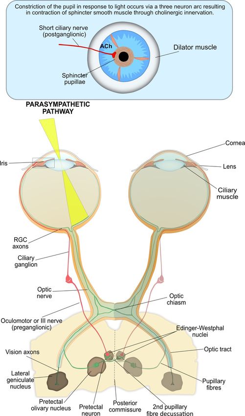
Diagnostics 2018, 8, 19 2 of 20
Diagnostics 2018, 8, x FOR PEER REVIEW 2 of 20
3. Measuring thethe
3. Measuring PLRPLR
TheThe
dynamics
dynamicsof the PLR
of the follow
PLR a general
follow a generalpattern consisting
pattern of 4ofphases:
consisting 4 phases:response
responselatency,
latency,
maximum constriction, pupil escape and recovery (Figure 1), which can be influenced
maximum constriction, pupil escape and recovery (Figure 1), which can be influenced by the by the duration,
intensity andintensity
duration, spectral composition
and spectral of the light. The
composition PLR
of the provides
light. a physiological
The PLR measure of normal
provides a physiological measure
or abnormal nervous system function and the symmetry of the PLR in response to
of normal or abnormal nervous system function and the symmetry of the PLR in response tostimulation of either
eye,stimulation
because ofofpupillary fibre
either eye, decussation,
because provides
of pupillary an opportunity
fibre decussation, to compare
provides the pupillomotor
an opportunity to compare
drive
theinpupillomotor
both eyes [6].drive in both eyes [6].
Figure
Figure 1. Schematic
1. Schematic of the
of the pupillogram
pupillogram (blue
(blue line)
line) andand associated
associated PLRPLR parameters.
parameters. TheThe
lightlight stimulus
stimulus
at time zero results in a rapid reduction in pupil diameter. Latency (t ) is calculated
at time zero results in a rapid reduction in pupil diameter. Latency (tL ) is calculated as the elapsed
L as the elapsed
timetime between
between light
light onset
onset andand the the
startstart of constriction.
of constriction. TheThe
pupilpupil
thenthen rapidly
rapidly constricts
constricts (maximal
(maximal
constriction
constriction velocity;
velocity; MCV)MCV) fromfrom
the the baseline
baseline 0) pupil
(D0 )(Dpupil diameter
diameter to the
to the minimum
minimum (Dmin(D)min ) pupil
pupil
diameter; the constriction time (t ) and maximum constriction amplitude (MCA)
diameter; the constriction time (tC ) and maximum constriction amplitude (MCA) are calculated as the
C are calculated as the
time interval and size difference between these two values, respectively. At offset of light stimulus or
time interval and size difference between these two values, respectively. At offset of light stimulus
during sustained
or during sustained light
lightstimulation
stimulationthe thepupil
pupilundergoes
undergoesa period
a periodof of rapid
rapid redilation
redilation or pupillary
or pupillary
“escape”
“escape” to a partially
to a partially constricted
constricted state. Subsequently
state. Subsequently the pupilthe pupil
slowly slowly
returns returns
to the to diameter.
baseline the baseline
diameter.
Response latency describes the delay in pupil constriction following the onset of a light stimulus,
Response latency describes the delay in pupil constriction following the onset of a light stimulus,
with the latency shortening as light intensity increases, to a minimum of 180–230 ms [7,8]. The latency
with the latency shortening as light intensity increases, to a minimum of 180–230 ms [7,8]. The latency
period is due to the delay in iris smooth muscle contraction and to a lesser extent the temporal
period is due to the delay in iris smooth muscle contraction and to a lesser extent the temporal
dynamics of retinal output and innervation pathways [7,8].
dynamics of retinal output and innervation pathways [7,8].
The latency period is followed by a period of rapid constriction of the pupil until it reaches
The latency period is followed by a period of rapid constriction of the pupil until it reaches the
the maximum constriction velocity (MCV), after which constriction slows until the minimum pupil
maximum constriction velocity (MCV), after which constriction slows until the minimum pupil
diameter is reached. The onset of pupil contraction can be determined using velocity and acceleration
diameter is reached. The onset of pupil contraction can be determined using velocity and acceleration
analysis [9]. The maximum constriction velocity varies with light stimulus intensity, duration, spectral
analysis [9]. The maximum constriction velocity varies with light stimulus intensity, duration,
composition, retinal size and location [10]. The maximum constriction amplitude (MCA) represents the
spectral composition, retinal size and location [10]. The maximum constriction amplitude (MCA)
difference between the baseline and minimum pupil diameter. However, the baseline pupil diameter
represents the difference between the baseline and minimum pupil diameter. However, the baseline
can be affected by a number of factors and can influence the MCA (a smaller MCA is observed with a
pupil diameter can be affected by a number of factors and can influence the MCA (a smaller MCA is
smaller baseline pupil diameter). Therefore, the MCA should be normalised to baseline pupil diameter
observed with a smaller baseline pupil diameter). Therefore, the MCA should be normalised to
to account for this effect [6]. After this peak constriction, the pupil quickly redilates or “escapes” to a
baseline pupil diameter to account for this effect [6]. After this peak constriction, the pupil quickly
partially constricted state during a prolonged light stimulus lasting from 1–2 up to 100 s, before slowly
redilates or “escapes” to a partially constricted state during a prolonged light stimulus lasting from
redilating to the initial size [11].
1–2 up to 100 s, before slowly redilating to the initial size [11].
In addition to the dynamic phases of the PLR during light stimulation, there is also a sustained
component [12,13]. Both outer and inner photoreceptors contribute to the early sustained post-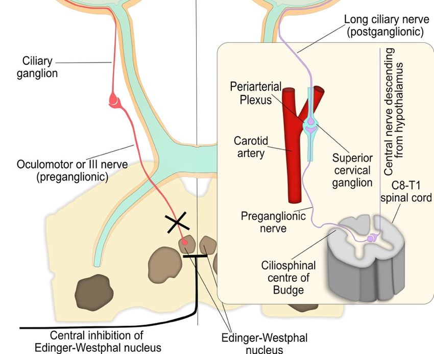
Diagnostics 2018, 8, 19                                                                               3 of 20

      In addition to the dynamic phases of the PLR during light stimulation, there is also a
sustained component [12,13]. Both outer and inner photoreceptors contribute to the early sustained
post-illumination pupil response (PIPR;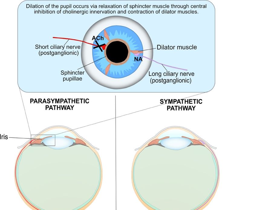
Diagnostics 2018, 8, 19 4 of 20
Diagnostics 2018, 8, x FOR PEER REVIEW 4 of 20
as well as in the PLR [31–33]. The ipRGCs regulate pupil size through the integration of extrinsic signals
from rods and cones but also through intrinsic (melanopsin) phototransduction [12]. Melanopsin is
protein-coupled photopigment, which is maximally sensitive to 482 nm wavelength light and, unlike
a G protein-coupled photopigment, which is maximally sensitive to 482 nm wavelength light and,
rods and cones, depolarizes in response to light following activation of a phototransduction cascade
unlike rods and cones, depolarizes in response to light following activation of a phototransduction
involving Gq/11 and phospholipase C [34–37]. Unlike rods and cones, which have their
cascade involving Gq/11 and phospholipase C [34–37]. Unlike rods and cones, which have
photopigment concentrated in specialised light-absorbing cellular domains (outer segment),
their photopigment concentrated in specialised light-absorbing cellular domains (outer segment),
melanopsin
melanopsin is distributedthroughout
is distributed throughout the
the plasma membraneof
plasma membrane ofipRGCs
ipRGCs[28].
[28].The
TheipRGCs
ipRGCsalso
alsodirectly
directly
contribute
contributetotothe
thePIPR
PIPRasasaasustained
sustained constriction
constriction of
of the PLR (>30
the PLR (>30 s)
s) in
in response
response to
to high
highintensity,
intensity,
short wavelength light [14,15,28,38–40].
short wavelength light [14,15,28,38–40].
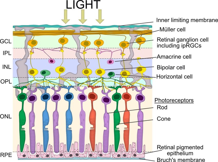
Figure2. 2.
Figure Simplified
Simplified schematic
schematic viewview of retinal
of retinal layerslayers
involvedinvolved
in the in the pupillary
pupillary light Vertical
light reflex. reflex.
Vertical signalling pathways in the retina are composed of the photoreceptors (rod
signalling pathways in the retina are composed of the photoreceptors (rod and cone cells), bipolar and cone cells),
bipolar
cells andcells andganglion
retinal retinal ganglion cells (RGC),
cells (RGC), including
including intrinsically
intrinsically photosensitive
photosensitive retinal
retinal ganglion
ganglion cells
cells (ipRGCs). There are also two lateral pathways comprised of horizontal cells in
(ipRGCs). There are also two lateral pathways comprised of horizontal cells in the outer plexiform the outer plexiform
layer(OPL)
layer (OPL)and
andthe
theamacrine
amacrinecells
cellsin
inthe
theinner
inner plexiform
plexiform layer
layer (IPL).
(IPL). These
Thesecells
cellsmodulate
modulatethe theactivity
activity
of other retinal cells in the vertical pathway. The somata of the neurons are in three cellular layers.
of other retinal cells in the vertical pathway. The somata of the neurons are in three cellular layers.
The rod and cone cells are located in the outer nuclear layer (ONL), which is adjacent to the retinal
The rod and cone cells are located in the outer nuclear layer (ONL), which is adjacent to the retinal
pigment epithelium (RPE). The horizontal cell, bipolar cell and amacrine cell somas are located in
pigment epithelium (RPE). The horizontal cell, bipolar cell and amacrine cell somas are located in the
the inner nuclear layer (INL), whilst the ganglion cell somata are located in the ganglion cell layer
inner nuclear layer (INL), whilst the ganglion cell somata are located in the ganglion cell layer (GCL).
(GCL). The axon terminals of the bipolar cells stratify at different depths of the inner plexiform layer,
The axon terminals of the bipolar cells stratify at different depths of the inner plexiform layer, which
which is subdivided into the OFF outer sublamina (where OFF bipolar cells terminate) and the ON
is subdivided into the OFF outer sublamina (where OFF bipolar cells terminate) and the ON inner
inner sublamina (where ON bipolar cells terminate). There are also ON and OFF bands of melanopsin
sublamina (where ON bipolar cells terminate). There are also ON and OFF bands of melanopsin
dendrites from the ipRGCs, but both lie outside of the ON and OFF cholinergic bands within the IPL.
dendrites from
The bipolar theare
cells ipRGCs, but bothspecific
photoreceptor lie outside of the
and the ON and
bipolar OFF cholinergic
dendrites bands within
synapse exclusively withthe IPL.
either
The bipolar cells
rod or cone cells. are photoreceptor specific and the bipolar dendrites synapse exclusively with either
rod or cone cells.
4.3. The Interneuron and Efferent Arms of Pupil Constriction
The interneuron and efferent arms of pupil constriction are summarised in Figure 3. The RGC
axons form the first interneuron arm of the PLR arc and carry the neuronal signal from the
photoreceptors [2].Diagnostics 2018, 8, 19 5 of 20
4.3. The Interneuron and Efferent Arms of Pupil Constriction
The interneuron and efferent arms of pupil constriction are summarised in Figure 3. The RGC
axons form the first interneuron arm of the PLR arc and carry the neuronal signal from the
photoreceptors [2].
Diagnostics 2018, 8, x FOR PEER REVIEW 5 of 20
Figure 3. The parasympathetic nervous system is the main system responsible for pupil constriction
Figure 3. The parasympathetic nervous system is the main system responsible for pupil constriction in
in response to light. The integrated afferent input is transmitted along the axons of the retinal ganglion
response to light. The integrated afferent input is transmitted along the axons of the retinal ganglion
cells (RGC), which contribute to the optic nerve. At the optic chiasm, nerves from the nasal retina
cells (RGC), which contribute to the optic nerve. At the optic chiasm, nerves from the nasal retina cross
cross to the contralateral side, whilst nerves from the temporal retina continue ipsilaterally. The
to the contralateral
pupillary side, whilst
RGC axons exit thenerves from and
optic tract the temporal
synapse atretina continueolivary
the pretectal ipsilaterally.
nucleus.ThePretectal
pupillary
RGC neurons are projected either ipsilaterally or contralaterally, across the posterior commissure, to theare
axons exit the optic tract and synapse at the pretectal olivary nucleus. Pretectal neurons
projected either ipsilaterally
Edinger-Westphal or From
nucleus. contralaterally,
there, theacross the posterior
pre-ganglionic commissure, fibres
parasympathetic to the Edinger-Westphal
travel with the
nucleus. From there, the pre-ganglionic parasympathetic fibres
oculomotor, or III cranial nerve, and synapse at the ciliary ganglion. Thetravel with the oculomotor,
post-ganglionicor III
cranial nerve, and synapse at the ciliary ganglion. The post-ganglionic parasympathetic
parasympathetic neurons (short ciliary nerves) travel to and innervate the contraction of the iris neurons
(short ciliary muscle
sphincter nerves)viatravel
the to and innervate
release the contraction
of acetylcholine of the iris sphincter
at the neuromuscular junction,muscle via in
resulting thepupil
release
of acetylcholine
constriction. at the neuromuscular junction, resulting in pupil constriction.
At the optic chiasma, approximately half the RGCs from the nasal plane in each eye decussate
to the opposite optic tract [41]. At the terminus of the optic tract, the axons of RGCs responsible for
the PLR separate from the visual axons and carry the afferent pupillomotor signal through theDiagnostics 2018, 8, 19 6 of 20
At the optic chiasma, approximately half the RGCs from the nasal plane in each eye decussate to
the opposite optic tract [41]. At the terminus of the optic tract, the axons of RGCs responsible for the
PLR separate from the visual axons and carry the afferent pupillomotor signal through the brachium
of the superior colliculus to synapse at the pretectal olivary nucleus in the dorsal midbrain [8].
The pretectal neurons integrate the input signals (retinal, supranuclear and infranuclear),
that modulate the PLR and form the second interneuron of the reflex arc. These pretectal nuclei
project to either the ipsilateral or contralateral Edinger–Westphal (EW) nucleus within the oculomotor
nuclear complex, which contain the pre-ganglionic parasympathetic neurons that control the iris
sphincter [42]. The bilateral neuron projection results in a double decussation of pupillary fibres, first at
the optic chiasm and then within the pretectal area, and ensures each EW nucleus receives information
about the level of incoming light from each eye. Therefore, unilateral light stimulation causes an equal
direct and consensual pupillary constriction. However, contraction anisocoria, whereby the direct
pupillary constriction is slightly stronger than the consensual reaction, may present if asymmetry
occurs during crossing of fibres at the chiasm or pretectal olivary nucleus; this is normally clinically
insignificant [8].
From the EW nuclei, the efferent pre-ganglionic axons pass into the right and left fascicles of
the oculomotor nerve (third nerve) to join the motor axons destined for the eye muscles (Figure 3).
The oculomotor nerve bifurcates into a superior and inferior division near the anterior cavernous sinus.
The parasympathetic fibres travel with the inferior division through the superior orbital fissure toward
the orbital apex and synapse at the ciliary ganglion (CG).
The short ciliary (post-ganglionic) nerves pierce the globe around the optic nerve and pass
between the choroid and sclera toward the iris. These nerves innervate the contraction of the iris
sphincter muscle via the neurotransmitter acetylcholine (ACh), resulting in constriction of the pupil.
4.4. Pupil Reflex Dilation: Central and Peripheral Nervous System Integration
Dilation of the pupil following a light stimulus occurs through two integrated processes driven
by the sympathetic neurons and corresponds to the recovery phase of the PLR and is summarised in
Figure 4. Firstly, the parasympathetic innervation of the pupil sphincter is suppressed by supranuclear
inhibition via central sympathetic neurons, resulting in relaxation of the muscle and pupil dilation.
These sympathetic neurons primarily originate in the reticular activating formation in the brainstem
and inhibit the pre-ganglionic parasympathetic neurons at the EW nucleus via α2 -adrenergic receptor
activation. Secondly, the iris dilator muscle contracts via excitation of the α1 -adrenergic sympathetic
pathway. This peripheral sympathetic nerve activation greatly enhances the dynamics of pupil dilation
in terms of speed and maximal pupil diameter attained.
The sympathetic influence on the iris dilator muscle consists of a paired, three-neuron arc on
both the right and left side of the central and peripheral nervous system, without decussations,
extending from the hypothalamus to the iris dilator muscle (Figure 4) [43–45]. In this three-neuron
arc, the synaptic transmission is mediated by ACh at the first two junctions, while the post-ganglionic
fibres innervate the dilator muscle via noradrenaline.
4.5. Other Inputs to the Iris
Nerves within the ophthalmic division of the trigeminal nerve provide sensory innervation to
the iris and may play an additional role in modulating pupil diameter [46]. Mechanical and chemical
irritation of the eye can cause a strong miotic response that is non-cholinergic and fails to reverse
with autonomic-acting drugs. In addition to the neuronal mechanisms involved in pupil size control,
circulating catecholamines and peptide hormones may act on the iris dilator or sphincter muscles,
either directly through the bloodstream or potentially indirectly through the tears [47,48].Diagnostics 2018, 8, 19 7 of 20
Diagnostics 2018, 8, x FOR PEER REVIEW 7 of 20
Figure 4. Both parasympathetic and sympathetic nervous systems are required for pupil dilation as
Figure 4. Both parasympathetic and sympathetic nervous systems are required for pupil dilation
part of the PLR. The parasympathetic innervation of the pupil sphincter is inhibited by central
as part of the PLR. The parasympathetic innervation of the pupil sphincter is inhibited by central
supranuclear
supranuclear inhibition
inhibition ofof Edinger–Westphal
Edinger–Westphalnuclei nucleivia 2-adrenergic receptor activation, resulting
viaαα-adrenergic receptor activation, resulting in
2
in
relaxation of the pupil sphincter muscle. The sympathetic influence on theon
relaxation of the pupil sphincter muscle. The sympathetic influence iristhe iris muscle
dilator dilator consists
muscle
consists of a paired three-neuron arc on both the right and left sides of the
of a paired three-neuron arc on both the right and left sides of the central and peripheral nervouscentral and peripheral
nervous system without
system without decussations.
decussations. The first-order
The first-order (central)(central)
neuron neuron originates
originates in theinhypothalamus
the hypothalamus and
and descends to synapse with the pre-ganglionic in the ciliospinal
descends to synapse with the pre-ganglionic in the ciliospinal centre of Budge at C8-T1centre of Budge at C8-T1
of the of the
spinal
spinal
cord. cord. The pre-ganglionic
The pre-ganglionic neuron neuron
ascendsascends
from from the ciliospinal
the ciliospinal centre centre of Budge
of Budge to synapse
to synapse withwith
the
the post-ganglionic neuron at the superior cervical ganglion, which is located at the
post-ganglionic neuron at the superior cervical ganglion, which is located at the periarterial plexus periarterial plexus
near the carotid
near the carotidartery
artery bifurcation.
bifurcation. Finally,
Finally, long long
ciliaryciliary (post-ganglionic)
(post-ganglionic) nerves nerves
travel totravel to and
and innervate
innervate the contraction of the iris dilator muscles, via a release of noradrenaline
the contraction of the iris dilator muscles, via a release of noradrenaline (NA) at the neuromuscular (NA) at the
neuromuscular
junction, resultingjunction, resulting
in pupil in pupil
dilation. dilation. The
The synaptic synaptic transmission
transmission at the other
at the other junctions is junctions
mediated
is
bymediated by acetylcholine.
acetylcholine.Diagnostics 2018, 8, 19 8 of 20
5. Clinical Applications of Pupillometry
Conditions that influence the integration of parasympathetic stimulation and inhibition,
sympathetic stimulation and humoural release of neurotransmitters may each affect the dynamics
of the PLR and may be clinically diagnostic. Pupil function abnormalities have been reported for
a wide range of disorders, including alcoholism [49,50], mental health disorders such as seasonal
affective disorders [51], schizophrenia [52] and generalised anxiety disorder [53], Alzheimer’s [54–56]
and Parkinson’s [57–60] diseases, autism spectrum disorders [61,62], as well as glaucoma [14,63–65]
and autonomic neuropathies associated with diabetes [66–70]. Additionally, pupillometry has been
applied to other clinical fields, such as monitoring of central states in anaesthesiology and analgesia,
as well as monitoring and prognosis following head injuries, cardiac arrest and drug overdose [71].
5.1. Neurodegenerative Disorders
There is an established body of evidence to indicate that cholinergic hypofunction is a significant
component of neurodegenerative diseases, such as Alzheimer’s and Parkinson’s diseases, which are
due to ACh and dopamine deficiencies, respectively. Moreover, the majority of studies that analysed
ACh-dependent PLR parameters have revealed significant differences between normal age-matched
patients and those with Alzheimer’s [55–57,72–74] or Parkinson’s disease [58,59,75]. Studies typically
used light stimuli wavelength of 820 nm with a light intensity of 24.6 cd/m2 . Specifically, patients with
Alzheimer’s [57] or Parkinson’s disease, with or without any detectable cognitive deficits or psychiatric
disorders [55,57,75], had significantly lower MCV and MCA values. Ferrario et al. [76] did not observe
a significant difference in MCA between Alzheimer’s patients and controls; however, this may be
attributed to the longer light stimulus used (1 s) compared to the other studies (20–150 ms) [77].
The MCV and MCA parameters correspond to the first portion of the characteristic V-shaped
pupillometric response (Figure 4) and are considered the most sensitive markers of cholinergic
activity [78]. Therefore, the MCA and secondarily the MCV are the best PLR predictors to discriminate
between healthy individuals and patients with Alzheimer’s or Parkinson’s disease [57,72]. A significant
increase in latency has been observed for subjects with Parkinson’s disease [57–59]; however, this was
not observed consistently in those with Alzheimer’s disease [54,56,72]. Additionally, Bittner et al.
examined the pupil’s response under repetitive light stimulation, which is systematically unstable in
normal patients [54]. However, these changes were not observed in patients with Alzheimer’s disease.
A number of possible pathophysiological mechanisms have been proposed for the observed
changes in pupillary response seen in Alzheimer’s and Parkinson’s patients [60], but Fotiou et al. [57]
and others have concluded that the most significant factor behind these findings was likely to be
the involvement of a central cholinergic deficit. Pupillometry, including repeated light stimulation
pupillometry, may serve as a useful diagnostic tool even at early, subclinical stages of autonomic
nervous system dysfunction [57,58].
5.2. Trauma
The PLR is a well-established measurement in the management and prognosis of patients with
acute brain injuries, in conjunction with other clinical parameters such as age, mode of injury and
Glasgow Coma Scale [79–81]. Typical light stimuli parameters are a 465 nm wavelength with a duration
of 1 s and low luminance (0.001 cd/m2 ) used to stimulate rod cells or high luminance (450 cd/m2 ) used
to stimulate ipRGCs. In particular, the location of the pupillomotor nuclei within the dorsal midbrain
and efferent oculomotor nerve are important in the determination of brainstem compression and the
onset of transtentorial herniation [82]. Morris et al. reported that loss of the PLR or development of
anisocoria or pupil asymmetry >2 mm in patients who sustained traumatic brain injuries was correlated
with increased morbidity and mortality rates [83]. However, manual examination using a penlight
is subject to large inter-examiner discrepancies that can be as high as 40%, particularly when pupils
are constricted [84], and these may be further confounded by a variety of factors, including alcohol,Diagnostics 2018, 8, 19 9 of 20
narcotics or hypothermia, which are common to many trauma patients [85]. Furthermore, Couret et al.
observed an error rate of approximately 20% even for intermediate-sized pupils (2–4 mm), with a 50%
failure rate in the detection of anisocoria [86]. Additionally, Larson and Muhiudeen found complete
failure in the detection of the PLR by manual examination when the reflex amplitude was less
than 0.3 mm [87]. Automated pupillometry using a pupillometer is a more sensitive technique that
has smaller inter-examiner discrepancies compared to manual examination [86,88]. The ability of
pupillometry to detect subtle changes in pupillary reaction even when pupils are constricted has
potential clinical significance and may provide a useful tool in the early detection, monitoring and
management of brain injuries [89]. In support of this, several groups have demonstrated that use of a
pupillometer was superior to manual assessment in predicting the 90-day outcome following cardiac
arrest [90,91].
5.3. Autism
The cholinergic system is key to normal pre- and postnatal neurodevelopment, and numerous
studies, ranging from neuroimaging data [92], to post mortem histopathological analysis of brain
tissue [93,94], animal models [95,96] and molecular genetic studies [97], have suggested that alterations
in the cholinergic system may be a contributing factor to the aetiology of autism spectrum disorder
(ASD). Moreover, an atypical PLR is reported in both children and adults with ASD [60,98–100].
Common measurement parameters used include a light stimulus wavelength of 530 nm and a
duration of 100 ms with a light intensity of 63.1 cd/m2 . Typically, these differences are characterised by
longer latency, reduced constriction amplitude [61,99] and reduced constriction velocity [61] compared
to children without ASD. However, Nyström et al. reported the opposite for high ASD-risk infants,
defined as those who had a sibling with ASD. The difference in age among the studies’ subjects,
who ranged from 10-month-old infants [100] up to children >5 years of age [61,98,99], may provide an
explanation for the contrasting data.
Children without ASD exhibit an age-dependent decrease in PLR latency before reaching a
plateau at >8 years of age [98,99]; this correlates with similar trends in white-matter maturation
rates as determined by flash visual evoked potential studies [101]. However, in children with ASD
there was no age-dependent decrease in PLR latency [99]. Moreover, brain-imaging studies have
shown that the neurodevelopment trajectory of brain maturation is atypical in children with ASD.
Initially, young children (Diagnostics 2018, 8, 19 10 of 20
a measurement of drug metabolites, which may be present in the urine for an extended period even
though any drug-induced physiological effects or impairment have ceased.
5.4.1. Alcohol
Pupillometry may have a role in both the detection of alcohol intoxication and treatment
management during alcohol withdrawal. Data from chromatic pupillometry studies demonstrated
a significant increase in both baseline pupil diameter and peak constriction amplitude following a
600 nm wavelength light stimulus at exhaled breath alcohol concentrations of ≥0.25 mg/L [115].
However, following a high dose of alcohol (1 g/kg body weight) the opposite was observed,
with significant decreases in pupil diameter, constriction amplitude and velocity compared to
control groups, suggesting inhibition of parasympathetic nerve activity [116,117]. These apparently
contradictory results are likely to reflect the acute, dose-dependent inhibition of the parasympathetic
nervous system, which results in the predominance of sympathetic nerve activity [118].
Pupillometry may also have a role in the development of clinical management tools to prevent
severe autonomic dysfunction during alcohol withdrawal [119]. Specifically, prolonged latency
and decreased constriction velocity parameters were described for participants undergoing alcohol
withdrawal. The reduction in parasympathetic innervation of the pupil is likely to be due to increased
activation of the locus coeruleus, as previously described during alcohol withdrawal [120,121].
5.4.2. Recreational Drugs
The pupillary response to 3,4 methylenedioxymethamphetamine (MDMA) and tetrahydrocannabinol
(cannabis) is characterised by an indirect central parasympathetic inhibition, resulting in
significantly increased latency and decreased constriction amplitude and velocity [113,122,123].
Additionally, increased sympathomimetic activity due to increased noradrenaline and serotonin
signalling was reported following MDMA intoxication, resulting in mydriasis and a reduction in
the PLR recovery time [123].
However, for cannabis intoxication there are conflicting reports regarding the effect of the drug
on baseline pupil diameter. Hartman et al. observed a significantly increased pupil size compared
to control participants, under both scotopic and photopic light conditions as well as following direct
light stimulation, suggesting increased sympathetic nervous system activity [112]. This is in contrast to
data reported by Fant et al. and others, which demonstrated a significant cannabis-induced effect on
the PLR but either a small (0.5 mm) decrease [116,124] or no change [122] in baseline pupil diameter.
These differences may be attributed to study design, as the studies that observed little or no effect
on pupil diameter used a defined, high dose (27 mg ∆9 THC) and a specified time duration between
drug administration and pupil measurements, whereas Hartman et al. used Drug Recognition Expert
examination data; thus, the exact dose and timings are undefined [113,116,122].
5.5. Exposure to Toxins and Toxic Chemicals
Changes in pupil size and response to light have been reported following exposure to toxic
chemicals such as organophosphates, as well as bacterial toxins including botulinum toxin.
Ophthalmic manifestations are early and persistent signs of botulism. Botulinum toxins (BTx)
block the release of ACh at neuromuscular junctions, post-ganglionic parasympathetic nerve endings,
and post-ganglionic sympathetic nerve endings that release Ach, resulting in paralysis of the
sympathetic and parasympathetic innervation of the iris [125,126]. This may result in transient
pupil dilation and attenuation of the PLR by uptake of BTx into the parasympathetic ciliary ganglion
or the parasympathetic neuromuscular junctions at the iris sphincter muscle [127]. There are a number
of reports in the literature describing mydriasis with an attenuated PLR as a consequence of ingesting
contaminated food [128], or following injection of BTx [129,130].
Organophosphates, a family of chemicals that includes nerve agents and pesticides,
inhibit cholinesterase activity, resulting in increased levels of ACh at the nerve synapses; they thus actDiagnostics 2018, 8, 19 11 of 20
as an indirect cholinergic agonist. Published studies by Dabisch et al. and others suggest that
the majority of cholinesterase inhibition observed within the eye is a result of the nerve agent
vapour acting directly on the ocular tissues, rather than distributing to the eye as a result of systemic
absorption [131–133]. The localised increase in ACh leads to contraction of the pupillary sphincter
muscle, resulting in dose-dependent miosis [132,134–136]. Miosis is a highly sensitive index of
exposure and can occur at exposure levels below those that cause systemic effects [137,138]. In relevant
animal models, the amounts of sarin and cyclosarin required to produce miosis were up to 30- and
135-fold lower, respectively, than the amounts required for lethality [139].
The PLR is also reduced following organophosphate exposure, as a result of developing tolerance
to cholinergic agonists and desensitization of muscarinic ACh receptors within retinal tissue following
prolonged exposure [135,140,141]. The threshold dose required to attenuate the PLR is similar to that
required to produce miosis, but the duration of the response is very different [140].
Dabisch et al. [140] observed rapid miosis and attenuation of the PLR in a rodent model following
a single low-dose soman vapour exposure, but while pupil size returned to normal after 48 h,
the PLR took up to 10 days to fully recover. Similarly, exposure to dichlorvos vapour resulted in a
dose-dependent transient miotic response in the guinea pig; however, a persistent enhanced pupillary
response to light was observed [136]. The recovery of pupil size is attributed to desensitisation of
the muscarinic receptors rather than reactivation of cholinesterases within the eye, which may take
up to 6 days to recover. Consequently, the PLR is attenuated until muscarinic receptor function is
regained [142]. In support of this, oximes, which reactivate acetylcholinesterase, had no effect on
sarin-induced miosis in animal models. Moreover, tropicamide—a muscarinic receptor antagonist
that competes with ACh for binding sites, preventing receptor desensitisation—rapidly increased
pupil size and restored PLR [143]. However, organophosphates inactivate cholinesterases at both
muscarinic and nicotinic receptor sites and paradoxical pupil dilation or mydriasis may occur in certain
circumstances due to dominant nicotinic effects at the pre-ganglionic fibres of the sympathetic nervous
system, resulting in increased innervation of the dilator muscle [144,145].
5.6. Response to Infection
An area of interest that currently remains unexplored is the response of the PLR to infection and the
potential diagnostic value of pupillometry. The brain monitors and modulates immune status through
both humoural and neural pathways [146,147]. Neuroendocrine responses control inflammation at
the systemic level through the hypothalamic-pituitary-adrenal axis [148]. The first branch of this
pathway, the vagus nerve, is activated either directly by cytokines (released from innate immune cells)
or indirectly through the chemoreceptive cells located in the vagal paraganglia [149]. The release
of pro-inflammatory cytokines needs to be carefully controlled, as excessive or uncontrolled release
(also known as a “cytokine storm”) may contribute to the pathogenesis of infections, including the
novel coronaviruses SARS and MERS [150–152], influenza [153], and Ebola [154], as well as potential
bacterial biothreat agents such as Burkholderia pseudomallei [155] and Yersinia pestis [156].
Signals from the vagus afferent fibres eventually project to the locus coeruleus region of the brain.
The locus coeruleus exerts a dual influence on the PLR, ultimately leading to pupil dilation [157,158].
Firstly, it contributes to the sympathetic outflow that innervates the pupillary dilator muscle.
Secondly, it attenuates the parasympathetic outflow via inhibition of the EW nucleus. Furthermore,
experimental models of infection—including sepsis [159–161], pneumococcal pneumonia [162],
endotoxaemia [163,164], leptospirosis [165,166] and influenza A [167,168]—have also highlighted the
significance of the cholinergic signalling pathway during infection. Changes to cholinergic signalling
are likely to influence the PLR both directly, through ACh receptors located on iris sphincter muscles,
and indirectly, through altered parasympathetic nervous system function. Therefore, measurement
of the PLR using dynamic pupillometry may offer the potential to detect systemic changes in
parasympathetic and sympathetic system function in response to infection and inflammation.Diagnostics 2018, 8, 19 12 of 20
6. Limitations
Pupillometry shows promise as a non-invasive diagnostic technology for a wide range of
conditions. However, there are a number of limitations that require consideration and further research
is needed to enable translation into clinical settings. Firstly, PLR measurements can be influenced by the
light stimuli used [6,169], sex [170], age [171,172] and iris colour [173]. Changes in pupil size are also
observed in response to other stimuli, including spatial structure patterns [174,175], object nearness or
accommodation reflex [10], and a variety of emotional and cognitive stressors [176,177]. Therefore, it is
critical that standardised protocols be developed to enable the use of pupillometry as a diagnostic tool
and limiting factors should be considered as covariates or exclusion criteria in PLR studies to enable
inter-study comparisons [23].
Further research is also required to establish whether the observed changes in PLR associated
with different disorders are sufficient in terms of specificity and sensitivity to be used diagnostically.
However, the ease of use, non-invasive nature and low cost mean pupillometry is well-placed for
inclusion in early diagnostic screening assessments and could complement other sources of information
to identify individuals at risk who warrant further clinic investigations.
Furthermore, there are approaches that have been exploited in particular fields—such as the
use of chromatic pupillometry to measure the response of different sub-types of photoreceptor,
specifically ipRGCs, in diabetes [66] and glaucoma [14,64,65]—which could be applied to other
conditions including neurodegenerative disorders.
7. Conclusions
The pupillary light reflex serves as a valuable indicator of autonomic nervous system
function. Moreover, measurement of the reflex using dynamic pupillometry provides a quantitative,
non-invasive tool, which may aid the diagnosis and clinical management of a wide range of clinical
conditions, varying from neurodegenerative disease to exposure to toxic chemicals.
Acknowledgments: The authors would like to thank Philip Lees for his assistance in finalizing this manuscript.
Conflicts of Interest: The authors declare no conflict of interest.
References
1. Hirata, Y.; Yamaji, K.; Sakai, H.; Usui, S. Function of the pupil in vision and information capacity of retinal
image. Syst. Comput. Jpn. 2003, 34, 48–57. [CrossRef]
2. McDougal, D.H.; Gamlin, P.D. Autonomic control of the eye. Compr. Physiol. 2015, 5, 439–473. [CrossRef]
[PubMed]
3. Girkin, C. Evaluation of the pupillary light response as an objective measure of visual function. Ophthalmol. Clin.
N. Am. 2003, 16, 143–153. [CrossRef]
4. Loewenfeld, I.E. The Pupil: Anatomy, Physiology, and Clinical Applications, 2nd ed.; Butterworth-Heinemann:
Boston, MA, USA, 1999.
5. Winn, B.; Whitaker, D.; Elliott, D.B.; Phillips, N.J. Factors affecting light-adapted pupil size in normal human
subjects. Investig. Ophthalmol. Vis. Sci. 1994, 35, 1132–1137.
6. Adhikari, P.; Pearson, C.A.; Anderson, A.M.; Zele, A.J.; Feigl, B. Effect of age and refractive error on the
melanopsin mediated post-illumination pupil response (PIPR). Sci. Rep. 2015, 5, 17610. [CrossRef] [PubMed]
7. Ellis, C.J. The pupillary light reflex in normal subjects. Br. J. Ophthalmol. 1981, 65, 754–759. [CrossRef]
[PubMed]
8. Lowenstein, O.; Loewenfeld, I.E. The sleep-waking cycle and pupillary activity. Ann. N. Y. Acad. Sci. 1964,
117, 142–156. [CrossRef] [PubMed]
9. Bergamin, O.; Kardon, R.H. Latency of the pupil light reflex: Sample rate, stimulus intensity, and variation
in normal subjects. Investig. Ophthalmol. Vis. Sci. 2003, 44, 1546–1554. [CrossRef]
10. Barbur, J. Learning from the pupil: Studies of basic mechanisms and clinical applications. In The Visual
Neurosciences; MIT: Cambridge, MA, USA, 2004; pp. 641–656.Diagnostics 2018, 8, 19 13 of 20
11. Kawasaki, A.; Kardon, R.H. Intrinsically photosensitive retinal ganglion cells. J. Neuroophthalmol. 2007, 27,
195–204. [CrossRef] [PubMed]
12. Joyce, D.S.; Feigl, B.; Zele, A.J. Melanopsin-mediated post-illumination pupil response in the peripheral
retina. J. Vis. 2016, 16, 5. [CrossRef] [PubMed]
13. Kankipati, L.; Girkin, C.A.; Gamlin, P.D. Post-illumination pupil response in subjects without ocular disease.
Investig. Ophthalmol. Vis. Sci. 2010, 51, 2764–2769. [CrossRef] [PubMed]
14. Adhikari, P.; Feigl, B.; Zele, A.J. Rhodopsin and melanopsin contributions to the early redilation phase of the
post-illumination pupil response (PIPR). PLoS ONE 2016, 11, e0161175. [CrossRef] [PubMed]
15. Gamlin, P.D.; McDougal, D.H.; Pokorny, J.; Smith, V.C.; Yau, K.W.; Dacey, D.M. Human and macaque pupil
responses driven by melanopsin-containing retinal ganglion cells. Vis. Res. 2007, 47, 946–954. [CrossRef]
[PubMed]
16. Rosen, E.S.; Gore, C.L.; Taylor, D.; Chitkara, D.; Howes, F.; Kowalewski, E. Use of a digital infrared
pupillometer to assess patient suitability for refractive surgery. J. Cataract Refract. Surg. 2002, 28, 1433–1438.
[CrossRef]
17. Smith, G.T. Repeatability of the procyon p3000 pupillometer. J. Refract. Surg. 2011, 27, 11. [CrossRef]
[PubMed]
18. Chen, J.W.; Vakil-Gilani, K.; Williamson, K.L.; Cecil, S. Infrared pupillometry, the Neurological Pupil
index and unilateral pupillary dilation after traumatic brain injury: Implications for treatment paradigms.
Springerplus 2014, 3, 548. [CrossRef] [PubMed]
19. De Souza, J.K.; Pinto, M.A.; Vieira, P.G.; Baron, J.; Tierra-Criollo, C.J. An open-source, FireWire camera-based,
Labview-controlled image acquisition system for automated, dynamic pupillometry and blink detection.
Comput. Methods Programs Biomed. 2013, 112, 607–623. [CrossRef] [PubMed]
20. Bremner, F.D. Pupillometric evaluation of the dynamics of the pupillary response to a brief light stimulus in
healthy subjects. Investig. Ophthalmol. Vis. Sci. 2012, 53, 7343–7347. [CrossRef] [PubMed]
21. Nyström, P.; Falck-Ytter, T.; Gredebäck, G. The TimeStudio Project: An open source scientific workflow
system for the behavioral and brain sciences. Behav. Res. Methods 2016, 48, 542–552. [CrossRef] [PubMed]
22. Keivanidou, A.; Fotiou, D.; Arnaoutoglou, C.; Arnaoutoglou, M.; Fotiou, F.; Karlovasitou, A. Evaluation of
autonomic imbalance in patients with heart failure: A preliminary study of pupillomotor function. Cardiol. J.
2010, 17, 65–72. [PubMed]
23. Wang, Y.; Zekveld, A.A.; Naylor, G.; Ohlenforst, B.; Jansma, E.P.; Lorens, A.; Lunner, T.; Kramer, S.E.
Parasympathetic nervous system dysfunction, as identified by pupil light reflex, and its possible connection
to hearing impairment. PLoS ONE 2016, 11, e0153566. [CrossRef] [PubMed]
24. Cepko, C.L. The Determination of Rod and Cone Photoreceptor Fate. Annu. Rev. Vis. Sci. 2015, 1, 211–234.
[CrossRef] [PubMed]
25. Lamb, T.D. Why rods and cones? Eye 2016, 30, 179–185. [CrossRef] [PubMed]
26. Kawamura, S.; Tachibanaki, S. Explaining the functional differences of rods versus cones. Wiley Interdiscip.
Rev. Membr. Transp. Signal 2012, 1, 675–683. [CrossRef]
27. Diamond, J.S. Inhibitory Interneurons in the Retina: Types, Circuitry, and Function. Annu. Rev. Vis. Sci. 2017,
3, 1–24. [CrossRef] [PubMed]
28. Dacey, D.M.; Liao, H.W.; Peterson, B.B.; Robinson, F.R.; Smith, V.C.; Pokorny, J.; Yau, K.W.; Gamlin, P.D.
Melanopsin-expressing ganglion cells in primate retina signal colour and irradiance and project to the LGN.
Nature 2005, 433, 749–754. [CrossRef] [PubMed]
29. Liao, H.-W.; Ren, X.; Peterson, B.B.; Marshak, D.W.; Yau, K.-W.; Gamlin, P.D.; Dacey, D.M.
Melanopsin-expressing ganglion cells on macaque and human retinas form two morphologically distinct
populations. J. Comp. Neurol. 2016, 524, 2845–2872. [CrossRef] [PubMed]
30. Nasir-Ahmad, S.; Lee, S.C.S.; Martin, P.R.; Grünert, U. Melanopsin-expressing ganglion cells in human retina:
Morphology, distribution, and synaptic connections. J. Comp. Neurol. 2017, 10. [CrossRef] [PubMed]
31. Berson, D.M.; Dunn, F.A.; Takao, M. Phototransduction by retinal ganglion cells that set the circadian clock.
Science 2002, 295, 1070–1073. [CrossRef] [PubMed]
32. Barnard, A.R.; Hattar, S.; Hankins, M.W.; Lucas, R.J. Melanopsin regulates visual processing in the mouse
retina. Curr. Biol. 2006, 16, 389–395. [CrossRef] [PubMed]
33. Hattar, S.; Liao, H.W.; Takao, M.; Berson, D.M.; Yau, K.W. Melanopsin-containing retinal ganglion cells:
Architecture, projections, and intrinsic photosensitivity. Science 2002, 295, 1065–1070. [CrossRef] [PubMed]Diagnostics 2018, 8, 19 14 of 20
34. Graham, D.M.; Wong, K.Y.; Shapiro, P.; Frederick, C.; Pattabiraman, K.; Berson, D.M. Melanopsin ganglion
cells use a membrane-associated rhabdomeric phototransduction cascade. J. Neurophysiol. 2008, 99, 2522–2532.
[CrossRef] [PubMed]
35. Hartwick, A.T.; Bramley, J.R.; Yu, J.; Stevens, K.T.; Allen, C.N.; Baldridge, W.H.; Sollars, P.J.; Pickard, G.E.
Light-evoked calcium responses of isolated melanopsin-expressing retinal ganglion cells. J. Neurosci. 2007,
27, 13468–13480. [CrossRef] [PubMed]
36. Sekaran, S.; Lall, G.S.; Ralphs, K.L.; Wolstenholme, A.J.; Lucas, R.J.; Foster, R.G.; Hankins, M.W.
2-Aminoethoxydiphenylborane is an acute inhibitor of directly photosensitive retinal ganglion cell activity
in vitro and in vivo. J. Neurosci. 2007, 27, 3981–3986. [CrossRef] [PubMed]
37. Warren, E.J.; Allen, C.N.; Brown, R.L.; Robinson, D.W. The light-activated signaling pathway in
SCN-projecting rat retinal ganglion cells. Eur. J. Neurosci. 2006, 23, 2477–2487. [CrossRef] [PubMed]
38. Markwell, E.L.; Feigl, B.; Zele, A.J. Intrinsically photosensitive melanopsin retinal ganglion cell contributions
to the pupillary light reflex and circadian rhythm. Clin. Exp. Optom. 2010, 93, 137–149. [CrossRef] [PubMed]
39. Adhikari, P.; Zele, A.J.; Feigl, B. The post-illumination pupil response (PIPR). Investig. Ophthalmol. Vis. Sci.
2015, 56, 3838–3849. [CrossRef] [PubMed]
40. Fu, Y.; Yau, K.W. Phototransduction in mouse rods and cones. Pflugers Arch. Eur. J. Physiol. 2007, 454,
805–819. [CrossRef] [PubMed]
41. Hoyt, W.F.; Luis, O. The primate chiasm. Details of visual fiber organization studied by silver impregnation
techniques. Arch. Ophthalmol. 1963, 70, 69–85. [CrossRef] [PubMed]
42. Kozicz, T.; Bittencourt, J.C.; May, P.J.; Reiner, A.; Gamlin, P.D.; Palkovits, M.; Horn, A.K.; Toledo, C.A.;
Ryabinin, A.E. The Edinger-Westphal nucleus: A historical, structural, and functional perspective on a
dichotomous terminology. J. Comp. Neurol. 2011, 519, 1413–1434. [CrossRef] [PubMed]
43. Remington, L.A. Clinical Anatomy of the Visual System; Elsevier Health Sciences: Amsterdam, The Netherlands,
2011; 303p.
44. Kardon, R.; Anderson, S.C.; Damarjian, T.G.; Grace, E.M.; Stone, E.; Kawasaki, A. Chromatic pupillometry in
patients with retinitis pigmentosa. Ophthalmology 2011, 118, 376–381. [CrossRef] [PubMed]
45. Johnson, L.N.; Hill, R.A.; Bartholomew, M.J. Correlation of afferent pupillary defect with visual field loss on
automated perimetry. Ophthalmology 1988, 95, 1649–1655. [CrossRef]
46. Saari, M.; Koskela, P.; Masar, S.E. Effect of vehicle on pilocarpine-induced miosis. Acta Ophthalmol. 1978, 56,
496–503. [CrossRef]
47. Liu, J.H.; Dacus, A.C. Central cholinergic stimulation affects ocular functions through sympathetic pathways.
Investig. Ophthalmol. Vis. Sci. 1990, 31, 1332–1338.
48. Hansen, M.S.; Sander, B.; Kawasaki, A.; Brøndsted, A.E.; Nissen, C. Prior light exposure enhances the pupil
response to subsequent short wavelength (blue) light. J. Clin. Exp. Ophthalmol. 2011, 2, 1000152. [CrossRef]
49. Rubin, L.S. Pupillometric studies of alcoholism. Int. J. Neurosci. 1980, 11, 301–308. [CrossRef] [PubMed]
50. Rubin, L.S.; Gottheil, E.; Roberts, A.; Alterman, A.; Holstine, J. Effects of alcohol on autonomic reactivity in
alcoholics. Pupillometric studies. III. J. Stud. Alcohol 1980, 41, 611–622. [CrossRef] [PubMed]
51. Roecklein, K.; Wong, P.; Ernecoff, N.; Miller, M.; Donofry, S.; Kamarck, M.; Wood-Vasey, W.M.; Franzen, P.
The post illumination pupil response is reduced in seasonal affective disorder. Psychiatry Res. 2013, 210,
150–158. [CrossRef] [PubMed]
52. Bär, K.J.; Boettger, M.K.; Schulz, S.; Harzendorf, C.; Agelink, M.W.; Yeragani, V.K.; Chokka, P.; Voss, A.
The interaction between pupil function and cardiovascular regulation in patients with acute schizophrenia.
Clin. Neurophysiol. 2008, 119, 2209–2213. [CrossRef] [PubMed]
53. Bakes, A.; Bradshaw, C.M.; Szabadi, E. Attenuation of the pupillary light reflex in anxious patients. Br. J.
Clin. Pharmacol. 1990, 30, 377–381. [CrossRef] [PubMed]
54. Bittner, D.M.; Wieseler, I.; Wilhelm, H.; Riepe, M.W.; Müller, N.G. Repetitive pupil light reflex:
Potential marker in Alzheimer’s disease? J. Alzheimer Dis. 2014, 42, 1469–1477.
55. Fotiou, F.; Fountoulakis, K.N.; Tsolaki, M.; Goulas, A.; Palikaras, A. Changes in pupil reaction to light in
Alzheimer’s disease patients: A preliminary report. Int. J. Psychophysiol. 2000, 37, 111–120. [CrossRef]
56. Tales, A.; Troscianko, T.; Lush, D.; Haworth, J.; Wilcock, G.K.; Butler, S.R. The pupillary light reflex in aging
and Alzheimer’s disease. Aging 2001, 13, 473–478. [PubMed]Diagnostics 2018, 8, 19 15 of 20
57. Fotiou, D.F.; Stergiou, V.; Tsiptsios, D.; Lithari, C.; Nakou, M.; Karlovasitou, A. Cholinergic deficiency in
Alzheimer’s and Parkinson’s disease: Evaluation with pupillometry. Int. J. Psychophysiol. 2009, 73, 143–149.
[CrossRef] [PubMed]
58. Giza, E.; Fotiou, D.; Bostantjopoulou, S.; Katsarou, Z.; Karlovasitou, A. Pupil light reflex in Parkinson’s
disease: Evaluation with pupillometry. Int. J. Neurosci. 2011, 121, 37–43. [CrossRef] [PubMed]
59. Micieli, G.; Tassorelli, C.; Martignoni, E.; Pacchetti, C.; Bruggi, P.; Magri, M.; Nappi, G. Disordered pupil
reactivity in Parkinson’s disease. Clin. Auton. Res. 1991, 1, 55–58. [CrossRef] [PubMed]
60. Stergiou, V.; Fotiou, D.; Tsiptsios, D.; Haidich, B.; Nakou, M.; Giantselidis, C.; Karlovasitou, A.
Pupillometric findings in patients with Parkinson’s disease and cognitive disorder. Int. J. Psychophysiol. 2009,
72, 97–101. [CrossRef] [PubMed]
61. Fan, X.; Miles, J.H.; Takahashi, N.; Yao, G. Abnormal transient pupillary light reflex in individuals with
autism spectrum disorders. J. Autism Dev. Disord. 2009, 39, 1499–1508. [CrossRef] [PubMed]
62. Rubin, L.S. Patterns of pupillary dilatation and constriction in psychotic adults and autistic children. J. Nerv.
Ment. Dis. 1961, 133, 130–142. [CrossRef] [PubMed]
63. Kankipati, L.; Girkin, C.A.; Gamlin, P.D. The post-illumination pupil response is reduced in glaucoma
patients. Investig. Ophthalmol. Vis. Sci. 2011, 52, 2287–2292. [CrossRef] [PubMed]
64. Feigl, B.; Mattes, D.; Thomas, R.; Zele, A.J. Intrinsically photosensitive (melanopsin) retinal ganglion cell
function in glaucoma. Investig. Ophthalmol. Vis. Sci. 2011, 52, 4362–4367. [CrossRef] [PubMed]
65. Nissen, C.; Sander, B.; Milea, D.; Kolko, M.; Herbst, K.; Hamard, P.; Lund-Andersen, H. Monochromatic
pupillometry in unilateral glaucoma discloses no adaptive changes subserved by the ipRGCs. Front. Neurol.
2014, 5, 15. [CrossRef] [PubMed]
66. Feigl, B.; Zele, A.J.; Fader, S.M.; Howes, A.N.; Hughes, C.E.; Jones, K.A.; Jones, R. The post-illumination
pupil response of melanopsin-expressing intrinsically photosensitive retinal ganglion cells in diabetes.
Acta Ophthalmol. 2012, 90, e230–e234. [CrossRef] [PubMed]
67. Dütsch, M.; Marthol, H.; Michelson, G.; Neundörfer, B.; Hilz, M.J. Pupillography refines the diagnosis of
diabetic autonomic neuropathy. J. Neurol. Sci. 2004, 222, 75–81. [CrossRef] [PubMed]
68. Ferrari, G.L.; Marques, J.L.; Gandhi, R.A.; Heller, S.R.; Schneider, F.K.; Tesfaye, S.; Gamba, H.R.
Using dynamic pupillometry as a simple screening tool to detect autonomic neuropathy in patients with
diabetes: A pilot study. Biomed. Eng. Online 2010, 9, 26. [CrossRef] [PubMed]
69. Kuroda, N.; Taniguchi, H.; Baba, S.; Yamamoto, M. The pupillary light reflex in borderline diabetics. J. Int.
Med. Res. 1989, 17, 205–211. [CrossRef] [PubMed]
70. Yuan, D.; Spaeth, E.B.; Vernino, S.; Muppidi, S. Disproportionate pupillary involvement in diabetic autonomic
neuropathy. Clin. Auton. Res. 2014, 24, 305–309. [CrossRef] [PubMed]
71. Dhakal, L.P.; Sen, A.; Stanko, C.M.; Rawal, B.; Heckman, M.G.; Hoyne, J.B.; Dimberg, E.L.; Freeman, M.L.;
Ng, L.K.; Rabinstein, A.A.; et al. Early Absent Pupillary Light Reflexes After Cardiac Arrest in Patients
Treated with Therapeutic Hypothermia. Ther. Hypothermia Temp. Manag. 2016, 6, 116–121. [CrossRef]
[PubMed]
72. Fotiou, D.F.; Brozou, C.G.; Haidich, A.B.; Tsiptsios, D.; Nakou, M.; Kabitsi, A.; Giantselidis, C.; Fotiou, F.
Pupil reaction to light in Alzheimer’s disease: Evaluation of pupil size changes and mobility. Aging Clin.
Exp. Res. 2007, 19, 364–371. [CrossRef] [PubMed]
73. Granholm, E.; Morris, S.; Galasko, D.; Shults, C.; Rogers, E.; Vukov, B. Tropicamide effects on pupil size
and pupillary light reflexes in Alzheimer’s and Parkinson’s disease. Int. J. Psychophysiol. 2003, 47, 95–115.
[CrossRef]
74. Prettyman, R.; Bitsios, P.; Szabadi, E. Altered pupillary size and darkness and light reflexes in Alzheimer’s
disease. J. Neurol. Neurosurg. Psychiatry 1997, 62, 665–668. [CrossRef] [PubMed]
75. Giza, E.; Fotiou, D.; Bostantjopoulou, S.; Katsarou, Z.; Gerasimou, G.; Gotzamani-Psarrakou, A.;
Karlovasitou, A. Pupillometry and 123I-DaTSCAN imaging in Parkinson’s disease: A comparison study.
Int. J. Neurosci. 2012, 122, 26–34. [CrossRef] [PubMed]
76. Ferrario, E.; Molaschi, M.; Villa, L.; Varetto, O.; Bogetto, C.; Nuzzi, R. Is videopupillography useful in the
diagnosis of Alzheimer’s disease? Neurology 1998, 50, 642–644. [CrossRef] [PubMed]
77. Park, J.C.; McAnany, J.J. Effect of stimulus size and luminance on the rod-, cone-, and melanopsin-mediated
pupillary light reflex. J. Vis. 2015, 15, 13. [CrossRef] [PubMed]You can also read