Rho GTPase Regulators and Effectors in Autism Spectrum Disorders: Animal Models and Insights for Therapeutics - MDPI
←
→
Page content transcription
If your browser does not render page correctly, please read the page content below
cells
Review
Rho GTPase Regulators and Effectors in Autism
Spectrum Disorders: Animal Models and
Insights for Therapeutics
Daji Guo , Xiaoman Yang and Lei Shi *
 JNU-HKUST Joint Laboratory for Neuroscience and Innovative Drug Research, College of Pharmacy, Jinan
 University, Guangzhou 510632, China; daji0321@stu2017.jnu.edu.cn (D.G.); yxm3644@stu2018.jnu.edu.cn (X.Y.)
 * Correspondence: t_shilei@jnu.edu.cn or sophielshi80@gmail.com; Tel.: +86-020-85222120

 Received: 26 February 2020; Accepted: 26 March 2020; Published: 31 March 2020

 Abstract: The Rho family GTPases are small G proteins that act as molecular switches shuttling
 between active and inactive forms. Rho GTPases are regulated by two classes of regulatory proteins,
 guanine nucleotide exchange factors (GEFs) and GTPase-activating proteins (GAPs). Rho GTPases
 transduce the upstream signals to downstream effectors, thus regulating diverse cellular processes,
 such as growth, migration, adhesion, and differentiation. In particular, Rho GTPases play essential
 roles in regulating neuronal morphology and function. Recent evidence suggests that dysfunction
 of Rho GTPase signaling contributes substantially to the pathogenesis of autism spectrum disorder
 (ASD). It has been found that 20 genes encoding Rho GTPase regulators and effectors are listed as
 ASD risk genes by Simons foundation autism research initiative (SFARI). This review summarizes
 the clinical evidence, protein structure, and protein expression pattern of these 20 genes. Moreover,
 ASD-related behavioral phenotypes in animal models of these genes are reviewed, and the therapeutic
 approaches that show successful treatment effects in these animal models are discussed.

 Keywords: Rho GTPase; autism spectrum disorder;                   guanine nuclear exchange factor;
 GTPase-activating protein; animal model; behavior

1. Introduction
     Autism spectrum disorder (ASD) is a neurodevelopmental disorder characterized by two core
symptoms: (1) impaired social interaction and communication, and (2) repetitive or restricted interest
and behaviors. The average global prevalence of ASD is ~0.62% [1], and studies in Europe and Asia
have identified individuals with ASD with an average prevalence between 1% and 2% [2–6]. Statistics
from CDC (Centers for Disease Control and Prevention)’s Autism and Developmental Disabilities
Monitoring (ADDM) Network revealed that one in 59 children were diagnosed with ASD in United
States in 2018 [7]. In addition, there is a high rate of co-occurring mental health disorders in ASD
patients [8]. Meta-analysis of twin studies show that monozygotic twins have significantly higher
concordance rate of ASD than dizygotic twins [9,10], thus the aetiology of ASD is closely related to
genetic component. However, the genetic causes of ASD are very complex as a huge number of genes
contribute to the pathogenesis of ASD. Therefore, databases of ASD-related genes, such as SFARI
(Simons foundation autism research initiative) Gene [11,12] and AutDB (the autism gene database) [13],
have been established. With the development of genome sequencing, increasing genes related to
ASD have been identified. As of November 2019, more than 800 genes have been included in SFARI
Gene [14] and more than 1000 genes have been listed in AutDB [15]. Among these ASD susceptibility
genes, many converge on synapse regulation such as the regulation of development and maturation of
synaptic contacts and synaptic transmission [16–18].

Cells 2020, 9, 835; doi:10.3390/cells9040835                                      www.mdpi.com/journal/cells
Cells 2020, 9, 835 2 of 38
Cells 2020, 9, x FOR PEER REVIEW 2 of 37
In the nervous
In the nervoussystem,
system,precise
preciseneuronal
neuronalconnectivity
connectivity depends
depends onon synapses.
synapses. It isItwell
is well
knownknown
that
that dendritic spines, which are enriched with filamentous actin, are dynamic
dendritic spines, which are enriched with filamentous actin, are dynamic structures important for structures important
for synapse
synapse formation,
formation, function
function and and plasticity
plasticity [19].[19]. Rho family
Rho family GTPases
GTPases are keyareregulators
key regulators of theofactin
the
actin cytoskeleton that play critical roles in axonal outgrowth, dendritic
cytoskeleton that play critical roles in axonal outgrowth, dendritic spine morphogenesis, and synapsespine morphogenesis, and
synapse
formation formation
[20]. The[20].Rho ThefamilyRho family GTPases,
GTPases, which belongwhichtobelong
the Rastosuperfamily,
the Ras superfamily,
are smallare small G
G proteins
proteins
sized ~20sizedKDa.~20 KDa. Human
Human Rho family RhoGTPases
family GTPases
includeinclude
20 members20 members
that can that
becan be classified
classified into
into eight
eight groups [21,22]. By cycling between GTP-bound active forms
groups [21,22]. By cycling between GTP-bound active forms and GDP-bound inactive forms, Rho and GDP-bound inactive forms,
Rho GTPases
GTPases regulateregulate a diverse
a diverse arrayarray
of of cellular
cellular events,
events, includingthe
including thecontrol
controlof of growth,
growth, migration,
migration,
adhesion, and differentiation. Rho GTPase activity is regulated by two different
adhesion, and differentiation. Rho GTPase activity is regulated by two different kinds of regulatory kinds of regulatory
protein: guanine nucleotide
protein: guanine nucleotide exchange
exchange factors
factors (GEFs),
(GEFs), which
which catalyze
catalyze the
the replacement
replacement of of GDP
GDP bybyGTP,
GTP,
enabling
enabling thethe GTPases
GTPases to to recognize
recognize downstream
downstream effectors,
effectors, and
and GTP-activating
GTP-activating proteins
proteins (GAPs),
(GAPs), which
which
negatively
negatively regulate GTPase activity by favoring the GDP-bound forms [19,23]. Rho GTPase activity
regulate GTPase activity by favoring the GDP-bound forms [19,23]. Rho GTPase activity
regulation
regulation is is aa complex
complex process
process as as 82
82 GEFs
GEFs [24,25]
[24,25] and
and 66
66 GAPs
GAPs (of(of which
which 57 57 have
have aa common
common GAP GAP
domain)
domain) [26][26] have
have been
been identified
identified so so far
far in
in humans.
humans. The The complexity
complexity of of Rho
Rho GTPase
GTPase signaling
signaling isis also
also
contributed
contributed by by their downstream effectors,
their downstream effectors, as
as there
there are
are over
over 7070 downstream
downstream effectors
effectors identified
identified toto be
be
capable of transducing signals from Rho
capable of transducing signals from Rho GTPases [27]. GTPases [27].
A
A number
number of of extensive
extensive review
review articles
articles have
have summarized
summarized the the impact
impact of of Rho
Rho family
family GTPases
GTPases in in
neural
neural development and diseases [23,28–33]. Moreover, multiple lines of evidence have suggested
development and diseases [23,28–33]. Moreover, multiple lines of evidence have suggested
that
that Rho
Rho GTPase
GTPase signaling
signalingmay maycontribute
contributetotothe thepathogenesis
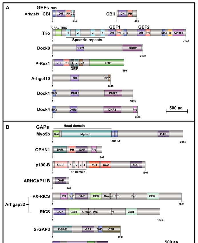
pathogenesisofofASD. ASD.ByBysearching
searching SFARI
SFARI Gene,
Gene, it is
it
notable to find that 8.53% (seven in 82) of RhoGEFs, 12.28% (seven in 57)
is notable to find that 8.53% (seven in 82) of RhoGEFs, 12.28% (seven in 57) of RhoGAPs and 8.21% of RhoGAPs and 8.21% (six in
73)
(sixRho effectors
in 73) are categorized
Rho effectors as ASD-risk
are categorized genes (Figure
as ASD-risk 1); 2.40%1);(20
genes (Figure in 831)
2.40% (20ASD-risk genes directly
in 831) ASD-risk genes
participate
directly participate in Rho GTPase signaling. We also find that all these genes encode proteins
in Rho GTPase signaling. We also find that all these genes encode regulatory regulatory or
effectors of three most-well studied Rho GTPases, Ras homolog family member
proteins or effectors of three most-well studied Rho GTPases, Ras homolog family member A (RhoA), A (RhoA), cell division
cycle 42 (Cdc42),
cell division cycleand Ras-related
42 (Cdc42), andC3 Botulinum
Ras-related C3Toxin Substrate
Botulinum Toxin1 (Rac1)
Substrate(Table 1). In(Table
1 (Rac1) this review,
1). In
the clinical evidence and animal models of these 20 genes are summarized.
this review, the clinical evidence and animal models of these 20 genes are summarized. Moreover, Moreover, therapeutic
approaches that are capable
therapeutic approaches of correcting
that are capable ofthe abnormalities
correcting caused by dysfunctions
the abnormalities of these Rho
caused by dysfunctions of
GTPase regulators and effectors are discussed.
these Rho GTPase regulators and effectors are discussed.
Figure 1. Overlap of human gene sets of RhoGEFs, RhoGAPs, and Rho effectors with autism spectrum
Figure 1. Overlap of human gene sets of RhoGEFs, RhoGAPs, and Rho effectors with autism spectrum
disorder (ASD)
disorder (ASD) risk
risk genes
genes in
in Simons
Simons foundation
foundation autism
autism research
research initiative
initiative(SFARI).
(SFARI).
Table 1. Rho family GTPases involved in ASD.
2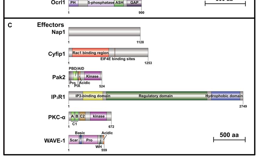
Cells 2020, 9, 835 3 of 38
Table 1. Rho family GTPases involved in ASD.
Upstream/DOWNSTREAM Rho
ASD Candidate Gene Gene Name Chromosome Location Genetic Category SFARI Gene Score
GTPase(s)
Rho GTPase GEF
Cdc42 guanine nucleotide exchange factor Rare Single Gene Category 1 (High
ARHGEF9 Xq11.1-q11.2 CDC42
(GEF) 9 Mutation, Syndromic Confidence)
Rare Single Gene Category 1 (High
TRIO Trio Rho guanine nucleotide exchange factor 5p15.2 RHOA, RAC1
Mutation, Syndromic Confidence)
Rare Single Gene Category 2 (Strong
DOCK8 Dedicator of cytokinesis 8 9p24.3 CDC42
Mutation Candidate)
Phosphatidylinositol-3,4,5-trisphosphate-dependent Category 2 (Strong
PREX1 20q13.13 Genetic Association RAC1
Rac exchange factor 1 Candidate)
Rare Single Gene Category 3 (Suggestive
ARHGEF10 Rho guanine nucleotide exchange factor 10 8p23.3 RHOA
Mutation, Functional Evidence)
Rare Single Gene Category 3 (Suggestive
DOCK1 Dedicator of cytokinesis 1 10q26.2 RAC1
Mutation Evidence)
Rare Single Gene
Category 3 (Suggestive
DOCK4 Dedicator of cytokinesis 4 7q31.1 Mutation, Genetic RAC1
Evidence)
Association, functional
Rho GTPase GAP
Rare Single Gene Category 2 (Strong
MYO9B Myosin IXB 19p13.11 RHOA
Mutation Candidate)
Rare Single Gene Category 2 (Strong
OPHN1 Oligophrenin 1 Xq12 RHOA, RAC1, CDC42
Mutation, Syndromic Candidate)
Rare Single Gene Category 3 (Suggestive
ARHGAP5 Rho GTPase activating protein 5 14q12 RHOA, RAC1, CDC42
Mutation Evidence)
Rare Single Gene Category 3 (Suggestive
ARHGAP11B Rho GTPase activating protein 11B 15q13.2 Unknown
Mutation Evidence)
Rare Single Gene Category 3 (Suggestive
ARHGAP32 Rho GTPase activating protein 32 11q24.3 RHOA, RAC1, CDC42
Mutation, Functional Evidence)
Rare Single Gene Category 3 (Suggestive
SRGAP3 SLIT-ROBO Rho GTPase activating protein 3 3p25.3 CDC42, RAC1
Mutation Evidence)
Rare Single Gene
OCRL Oculocerebrorenal syndrome of Lowe Xq26.1 Syndromic CDC42, RAC1
Mutation, SyndromicCells 2020, 9, 835 4 of 38
Table 1. Cont.
Upstream/DOWNSTREAM Rho
ASD Candidate Gene Gene Name Chromosome Location Genetic Category SFARI Gene Score
GTPase(s)
Rho GTPase Effector
Rare Single Gene Category 1 (High
NCKAP1 NCK-associated protein 1 2q32.1 RAC1
Mutation Confidence)
Rare Single Gene
Category 2 (Strong
CYFIP1 Cytoplasmic FMR1 interacting protein 1 15q11.2 Mutation, Genetic RAC1
Candidate)
Association, Functional
Rare Single Gene Category 2 (Strong
PAK2 p21 (RAC1) activated kinase 2 3q29 CDC42, RAC1
Mutation Candidate)
Rare Single Gene Category 3 (Suggestive
ITPR1 Inositol 1,4,5-trisphosphate receptor type 1 3p26.1 RHOA
Mutation Evidence)
Rare Single Gene Category 3 (Suggestive
PRKCA Protein kinase C alpha 17q24.2 RHOA, RAC1, CDC42
Mutation Evidence)
WASF1 WAS protein family member 1 6q21 Syndromic Syndromic RAC1Cells 2020, 9, 835 5 of 38
2. Rho Family GTPases and ASD
Rho GTPases themselves have been rarely reported as risk genes of ASD. The only evidence
so far is the linkage of RAC1 with ASD. Rac1 is an important Rho GTPase family member
which regulates actin polymerization and spine remodeling through multiple signaling pathways,
including PAKs (p21-activated kinases)-LIMK (LIM-domain-containing protein kinase)-cofilin [34],
IRSp5 (insulin receptor substrate p53)-WAVE (Wiskott–Aldrich syndrome protein (WASP) family
verprolin-homologous protein)-Arp2/3 [35,36], and PKA (protein kinase A) [37]. RAC1, encoding RAC1
and RAC1B, is a candidate gene for ASD listed in AutDB. Seven individuals with de novo mutations of
RAC1 were identified in patients of developmental disorders with divergent phenotypes [38]. One of
these individuals displayed hyperactive behavior, two presented stereotypic movements, and one was
diagnosed with autism [38]. Rac1 is highly expressed in embryonic cortex [39], and is ubiquitously
expressed in the hippocampus, neocortex, thalamus, and cerebellum [40,41]. Rac1 is essential for the
formation of three germ layers during gastrulation [42], and lack of which leads to embryonic lethality
in Rac1 knockout (KO) mice. To understand the brain function of Rac1, several Rac1 conditional KO
(cKO) mouse models have been constructed and studied. For instance, Foxg1-Cre mediated deletion of
Rac1 in the ventricular zone (VZ) of telencephalon [43–45], Nkx2.1-Cre-mediated deletion in medial
ganglionic eminence (MGE) [46], and Nestin-Cre-mediated deletion in precursors of neurons and glia
during early embryonic stage [35] were used to study the important roles of Rac1 for brain development.
Moreover, three studies investigated the behavioral changes in Rac1 cKO mouse models. Haditsch
and colleagues generated a mouse model in which Rac1 is deleted in pyramidal neurons by Cre under
CamKIIα promoter to study the role of Rac1 in memory [47,48]. They demonstrated that loss of Rac1
in the hippocampus impairs long-term potentiation (LTP), and Rac1-deficient mice have impaired
spatial memory and working or episodic-like memory [47]. They also found that the impaired working
memory in these mice is due to prolonged memory retention or perseveration of the previously learned
location [48]. Pennucci and colleagues generated a mouse model named Rac1N mice in which Rac1
is deleted in postmitotic neurons by Synapsin-I-Cre [49]. Rac1N mice show hyperactivity in several
exploration tasks, impairment in spatial and working memory, and defects in retaining the context
memory [49]. This study also reported failed synchronization of cortical networks in Rac1N mice by
quantitative electroencephalogram (EEG). Moreover, spontaneous inhibitory synaptic currents (sIPSCs)
are decreased in CA1 glutamatergic pyramidal cells in these mice [49]. However, these findings only
focus on memory-related behaviors, but not the typical ASD-related ones such as social behaviors.
3. RhoGEF Family and ASD
There are 82 members of the human RhoGEF family, which are divided into two different subtypes:
the classical Dbl family and the atypical Dock family [24]. So far, there are 71 members identified in Dbl
RhoGEF family, which is characterized by a Dbl Homology (DH) domain, the catalytic GEF domain,
and a Pleckstrin-Homology (PH) domain. The DH domain specifically catalyzes the exchange of GDP
for GTP, whereas the role of PH domain varies considerably between different members, but is believed
to facilitate the activation and localization of all Rho GTPases [25,50,51]. The Dock family, which
contains 11 members, shows completely different structural features from the Dbl family. Dock family
proteins have two main domains, the Dock homology region (DHR) 1 domain, which is responsible for
phospholipid binding, and the DHR2 domain, which possesses the GEF activity [52]. Dock proteins
are closely related to neurological disease [28,53]. By examining the overlap of RhoGEF genes and
SFARI Gene, we find the following seven RHOGEFs as ASD-risk genes: ARHGEF9, TRIO, DOCK8,
PREX1, ARHGEF10, DOCK1, and DOCK4 (see Appendix A) (Table 1).
3.1. ARHGEF9 (SFARI Gene Score: 1, High Confidence)
Rho guanine nucleotide exchange factor 9 (Arhgef9), also known as collybistin (CB), is a Dbl
family GEF for Cdc42. ARHGEF9 is located on chromosome Xq11.1-q11.2. The first report on theCells 2020, 9, 835 6 of 38 linkage of ARHGEF9 with ASD identified a de novo microdeletion of Xq11.1 including entire ARHGEF9 in a male patient, who presented with severe intellectual disability (ID), epilepsy, and mild to moderate autism [54]. A second de novo mutation of ARHGEF9 was identified in a female patient diagnosed with ASD, ID and speech delay [55]. Subsequently, more de novo deletions of ARHGEF9 were found in patients with ASD co-occurring with developmental delay (DD) or other mental disorders [56–58], suggesting that ARHGEF9 is a strong candidate for ASD. Alternative splicing of Arhgef9 transcripts creates two CB variants, I and II [59]. CB I has typical domains of Dbl family that include an Src homology 3 domain (SH3), a PH and a DH domain followed by a predicted coiled-coil (CC) domain, whereas CB II lacks SH3 and CC domains [59,60] (Figure 2A). CB is expressed predominantly in the brain, with enrichment in the gray matter, cerebral cortex, hippocampus, and cerebellum [59,61,62]. It was found that the CB1 level is high during early brain developmental stage, whereas CB2 expression maintains high and constant levels throughout brain development [60]. CB KO mice exhibited elevated anxiety levels and impaired spatial memory (Table 2; Supplementary Table S1), and showed reduced GABAergic transmission, increased LTP, and decreased long-term depression (LTD) in hippocampal CA1 region [63]. Two studies using this mouse line investigated electrophysiological characteristics in hippocampal dentate gyrus (DG) region, demonstrating that CB plays an important role in maintaining normal granule cell excitability, GABAergic network inhibition, and synaptic plasticity [64,65].
Cells 2020, 9, 835 7 of 38
Table 2. Summary of ASD-related behavior tests in Rho guanine nucleotide exchange factor (GEF), GTPase-activating protein (GAP), and effector mouse models.
Core Symptoms Comorbidities
Gene Mouse Model Basic locomotion Reference
Social Related Language Anxiety and Schizophrenia
Repetitive Behavior Learning and Memory and Motor
Behavior Communication Depression and Epilepsy
Coordination
Summary of ASD-related behavior tests in Rho GEF mouse models
Spatial learning and
ARHGEF9 Arhgef9 KO mice * NT 1 NT NT Anxiety ↑ Activity − NT [63]
memory ↓
Spatial learning and
Emx1-Trio−/− mice # NT NT NT NT memory ↓ NT NT [66]
Fear memory ↓
Activity ↓
Nestlet shredding (M2 , Anxiety ↑ Object recognition Prepulse
TRIO NEX-Trio+/− mice & Social preference ↓ NT Motor
↑; F3 , −) Depression − memory − inhibition −
coordination ↓ [67]
Anxiety (M, ↑; F, Activity ↑ Prepulse
Nestlet shredding (M, ↑; Object recognition
NEX-Trio−/− mice & Social preference ↓ NT −) Motor inhibition (M,
F, −) memory −
Depression ↑ coordination ↓ ↓; F, −)
Social preference ↓
Reversal learning ↓
Social learning and Ultrasonic Activity −
Fear memory ↓ Prepulse
PREX1 Prex1−/− mice * memory ↓ vocalizations Grooming ↑ Anxiety − Motor [68]
Object recognition inhibition −
Olfactory function (pup) ↓ coordination −
memory −
−
Sociability and
Anxiety ↓ Spatial learning and Prepulse
ARHGEF10 Arhgef10 KO mice * social novelty NT NT Activity ↑ [69]
Depression ↓ memory − inhibition −
preference ↓
Object recognition
Stereotyped circling
memory (F, ↓; M, −)
Ultrasonic (~9% F; M, −)
Social novelty Spatial memory (M, ↓; Activity (~9% F, ↑;
Dock4 KO mice & vocalizations Marble burying (M, −; Anxiety ↑ NT
preference ↓ F, −) M, −)
(pup) ↓ F, NT)
Working memory (M, ↓; [70]
DOCK4 Grooming (M, −; F, NT)
F, −)
Stereotyped circling Object recognition
Social novelty Ultrasonic (~1.7% F; M, −) memory −
Activity (~1.7%
Dock4 HET mice & preference vocalizations Marble burying (M, −; Anxiety − Spatial memory (F, ↓; NT
F,↑; M, −)
(F,↓; M, −) (pup) − F, NT) M, −)
Grooming (M, −; F, NT) Working memory −Cells 2020, 9, 835 8 of 38
Table 2. Cont.
Core Symptoms Comorbidities
Gene Mouse Model Basic locomotion Reference
Social Related Language Anxiety and Schizophrenia
Repetitive Behavior Learning and Memory and Motor
Behavior Communication Depression and Epilepsy
Coordination
Summary of ASD-related behavior tests in Rho GAP mouse models
Working, object
recognition, and spatial Activity ↑
Aggressivity ↓ learning and memory ↓ Motor
OPHN1 Ophn1-/y mice * Social memory − NT NT Anxiety − Fear memory extinction coordination − NT [71–75]
Olfactory function ↓ ↓ Behavioral
Vicarious trial and error lateralization ↓
(VTE) behavior ↓
PX-RICS−/− mice
Epilepsy
(M were used in Social novelty Ultrasonic
Grooming ↑ Reversal learning ↓ Motor (Severe
most behavior tests preference ↓ vocalizations (M NT [76,77]
Marble burying ↑ Fear memory ↓ coordination ↓ progressive
unless otherwise social interaction ↓ and F,↓)
seizures)
ARHGAP32 stated)
PX-RICS+/− mice
(M were used in Social novelty Ultrasonic
Grooming − Motor
most behavior tests preference ↓ vocalizations (M NT Reversal learning − NT [76]
Marble burying ↑ coordination −
unless otherwise social interaction ↓ and F, −)
stated)
Object recognition and
long-term memory ↓ Activity −
WRP−/− mice & NT NT NT Anxiety − Spatial and reversal Motor NT
learning ↓ coordination −
Working memory − [78]
SRGAP3 Object recognition and
long-term memory ↓ Activity −
WRP+/− mice & NT NT NT Anxiety − Spatial and reversal Motor NT
learning ↓ coordination −
Working memory −
Working memory ↓
Prepulse
Marble burying (M, −; Spatial and object Activity (M,↓; F,
SrGAP3-/- mice & Social interaction ↓ NT Anxiety − inhibition (F,↓; [79,80]
F, NT) recognition memory − −)
M, −)
Fear memory ↑Cells 2020, 9, 835 9 of 38
Table 2. Cont.
Core Symptoms Comorbidities
Gene Mouse Model Basic locomotion Reference
Social Related Language Anxiety and Schizophrenia
Repetitive Behavior Learning and Memory and Motor
Behavior Communication Depression and Epilepsy
Coordination
Activity −
Passive avoidance
Ocrl1−/y mice * NT NT NT NT Motor NT [81]
preference −
coordination ↓
OCRL
Ocrl1−/y mice *
(Inpp5b deleted but Social preference − Spatial learning and
NT NT NT Activity ↓ NT [82]
human INPP5B Social novelty − memory −
overexpressed)
Summary of ASD-related behavior tests in Rho effector mouse models
Hippocampus-dependent
memory ↓ Prepulse
Cyfip1 HET mice * Social interaction − NT NT Anxiety − Activity − [83]
Working, spatial, and inhibition −
fearing memory −
Activity −
Ultrasonic
Cyfip1HET mice # Social interest ↓ Marble burying − NT NT Motor NT [84]
vocalizations −
coordination ↓
Cyfip1 m+/p−
(Paternal origin)
Anxiety-like Fear memory (m+/p−, Activity (m+/p−,
and Cyfip1 m−/p+ NT NT NT NT [85]
behavior ↑; m−/p+, −) −; m−/p+, ↓)
(maternal origin)
CYFIP1 mice #
Spatial memory and
flexibility − Activity −
Self-grooming − Prepulse
Cyfip1+/− mice * NT NT NT Object recognition Motor [86]
Marble burying − inhibition ↓
memory ↓ coordination ↓
Working memory −
Cyfip1+/− rat * NT NT NT NT Behavioral flexibility ↓ NT NT [87]
Fear memory (Line 1
and line 2, ↑)
Human CYFIP1
Spatial learning
overexpressing Ultrasonic Grooming − Prepulse
Social preference − Anxiety − memory (Line 2, ↓; line Activity − [88]
mice (Tg line 1 and vocalizations − Digging − inhibition −
1, −)
Tg line 2) &
Working memory (M
and F of both lines, −)Cells 2020, 9, 835 10 of 38
Table 2. Cont.
Core Symptoms Comorbidities
Gene Mouse Model Basic locomotion Reference
Social Related Language Anxiety and Schizophrenia
Repetitive Behavior Learning and Memory and Motor
Behavior Communication Depression and Epilepsy
Coordination
Prepulse
inhibition −
Social preference ↓ Ultrasonic Marble burying ↑ Spatial learning and
PAK2 PAK2+/− mice * Anxiety − Activity − Acoustic [89]
Social memory ↓ vocalizations − Grooming ↑ memory −
startle
response −
Activity −
IP3R1+/− mice& NT NT NT NT NT Motor NT [90]
coordination ↓
ITPR1
L7-Cre; Itpr1flox/flox Motor
NT NT NT NT NT NT [91]
mice # coordination ↓
Wnt1-Cre; Motor
NT NT NT NT NT NT [92]
Itpr1flox/flox mice # coordination ↓
Spatial learning and
memory ↓ Activity ↓
WAVE-1 KO mice # NT NT NT Anxiety ↓ Object recognition Motor NT
memory ↓ coordination ↓ [93]
WASF1
Passive avoidance −
Activity ↓
Learning and memory
WAVE-1 HET mice # NT NT NT Anxiety − Motor NT
−
coordination ↓
*: Only male were used in behavior tests; & : Both male and female mice were used in behavior tests; # : Mice gender was not mentioned; 1 NT: not tested; 2 M: male mice; 3 F: female mice;↑
is increased and ↓ is decreased; − is no change; More detailed information is shown in Supplementary Table S1.Cells 2020, 9, 835 11 of 38
Cells 2020, 9, x FOR PEER REVIEW 10 of 37
Figure 2. Schematics of protein domain structures10 of 20 ASD-related RhoGEFs, RhoGAPs, and Rho
effectors. (A) Seven ASD-related RhoGEF protein domain architectures. Arhgef9 (which has two variants,Cells 2020, 9, 835 12 of 38
CB I and CB II), Trio, P-Rex1, and Arhgef10 belong to Dbl family, which is characterized by a DH
domain (dark violet) and a PH domain (light pink). Dock8, Dock1, and Dock4 belong to Dock family,
which contains two main domains, DHR1 domain (dark orchid) and DHR2 domain (dark magenta).
(B) Seven ASD-related RhoGAP protein domain architectures. In addition to a common catalytic GAP
domain (purple), most RhoGAPs have multiple other functional domains. (C) Six ASD-related Rho
effector protein domain architectures. All protein structures are generated using DOG 2.0 (Domain
Graph, version 2.0) [94] based on corresponding mouse protein sequences except ARHGAP11B, for
which human protein structure is shown as no homologs exist in rodents. Scales represent amino acid
numbers of 500. AID, Autoinhibitory Domain; ASH, ASPM/SPD2/Hydin; BAR, Bin/Amphiphysin/Rvs;
C1, binding site of diacylglycerol (DAG); C2, binding site of Ca2+ ; CBR, β-catenin-binding region;
CC, coiled-coil; CRAL-TRIO, cellular retinaldehyde-binding protein and TRIO guanine exchange
factor; CTR, C-terminal region with proline-rich; DEP, Disheveled, EGL-10, Pleckstrin; DH, Dbl
Homology; DHR1, Dock homology region 1; DHR2, Dock homology region 2; F-BAR, Fes-Cip4
homology Bin/Amphiphysin/Rvs; FF, domain with two conserved phenylalanine residues; GAP,
GTPase-activating proteins; GBD, guanosine triphosphate (GTP)-binding; GBR, GABARAP-binding
region; Ig, immunoglobulin; IP4P, inositol polyphosphate 4-phosphatase; IQ, short calmodulin-binding
motif containing conserved isoleucine and glutamine residues; PBD, p21-binding domain; PDZ,
PSD95/SAP90, DlgA, ZO-1; pG1, pseudoGTPase domain 1; pG2, pseudoGTPase domain 2; PH,
Pleckstrin-Homology; PIX, Pak-interacting exchange factor; Pro, proline-rich; PX, phox homology; Scar,
Scar homology; SH3, Src homology 3; WH, WASF homology.
3.2. TRIO (SFARI Gene Score: 1, High Confidence)
Trio Rho guanine nucleotide exchange factor (Trio), a large protein of the mammalian Dbl
family, activates both Rac1 and RhoA. TRIO, located on chromosome 5p15.2, is a strong candidate
gene for ASD. By using whole-exome sequencing (WES) and transmission and de novo association
(TADA) analysis of rare coding variations, a study identified TRIO as a gene strongly enriched for
variants likely to affect autism risk from 3871 autism cases [95]. Subsequently, several whole-exome
and genome sequencing studies identified several TRIO variations in subjects with autism [96,97]
or ID co-occurring with autism [98,99]. Moreover, a large number of TRIO mutations leading to
either reduced or excessive TIRO activity were found in ASD and neurodevelopmental disorder
(NDD)/ASD/ID patients [100,101]. Trio protein contains two SH3 domains, a CRAL-TRIO (cellular
retinaldehyde-binding protein and TRIO guanine exchange factor) domain, an immunoglobulin
(Ig) domain, several spectrin-like repeats, two RhoGEF domains and a serine kinase domain [102]
(Figure 2A). The GEF domain 1 activates Rac1 and RhoG, whereas the GEF domain 2 acts as an
exchange factor for RhoA [103]. Because of the alternative splicing in Trio, Trio family is consisted
of several isoforms, namely TrioA-E and Tgat [102,104,105]. Trio is enriched in the nervous system,
and different isoforms are highly expressed in the cortex, hippocampus, striatum, and cerebellum,
except for TrioC, which is highly expressed in the cerebellum [105]. Trio has important roles for
embryonic development, as Trio-/- mice die between embryonic day (E) 15.5 and birth [106]. Trio is
also essential for brain development, because TrioNKO mice, in which the Trio is deleted in neuronal
and glia progenitors by Nestin-Cre, have high death rate after birth; hypoplasia was found in the
residual survival individuals with disruptive development of the cerebellum [107]. Two recent studies
on deletion of Trio in the forebrain showed that these cKO mice can survive to adulthood. In one study,
Emx1-Trio-/- mice in which Trio deletion is restricted to the cerebral cortex and hippocampus show
deficient spatial learning [66] (Table 2; Supplementary Table S1). In another study, Trio was deleted by
NEX-Cre in neocortex and hippocampus starting from E11.5 [108]. Both NEX-Trio+/- and NEX-Trio-/-
mice displayed impaired social preference and impaired motor coordination, and anxious behaviors
were observed in all Trio mutant mice except female NEX-Trio-/- mice [67] (Table 2; Supplementary
Table S1). Moreover, partial or full loss of Trio expression in motor cortex Layer 5 pyramidal
neurons lead to disruption in presynaptic release probability, postsynaptic currents, and LTP [67].
However, the change in the ratio of NMDA ((N-methyl-D-aspartate)-type glutamate) receptor/AMPACells 2020, 9, 835 13 of 38
(α-amino-3-hydroxy-5-methyl-4-isoxazolepropionic acid) receptor-mediated excitatory postsynaptic
currents (EPSCs) was opposite in these two mouse lines [67]. These findings show comprehensive
phenotypic changes on behaviors and neural function by Trio deficiency. It was also found in vitro
that gain-of-function forms of TRIO variants lead to increased Rac1 activity and synaptic AMPA
receptor function, whereas loss-of-function forms lead to decreased Rac1 activity and AMPA receptor
function [100,101]. However, gain-of-function Trio mouse models have not been generated and
investigated for ASD pathology thus far.
Katrancha and colleagues found decreased levels of PDE4A5, a negative regulator of PKA
signaling, and corresponding increased PKA signaling in NEX-Trio+/- and NEX-Trio-/- motor cortex [67].
To examine approaches for correcting the abnormalities caused by Trio deficiency, they transfected
Trio+/flox neurons with GFP-P2A-Cre to establish Trio haploinsufficiency neurons as an in vitro model.
These neurons showed increased spine density and decreased dendritic branch number, mimicking
the phenotypes observed in NEX-Trio+/- mouse brain [67]. Moreover, they treated the Trio deficient
neurons with Rp-cAMPS, a competitive PKA antagonist, which reversed the increased dendritic spine
density [67] (Table 3). For a non-pharmacological approach, PDE4A5 overexpression was capable
of correcting the increased spine density but not branching deficits in Trio+/- neurons [67] (Table 3).
However, whether these two therapeutic approaches could rescue abnormal behaviors in NEX-Trio+/-
mice has not been investigated.
Table 3. Treatments for Rho GTPase mouse models.
Mouse/Cellular
Gene Therapeutic Type Therapeutic Strategy Result Reference
Model
Rp-cAMPS treatment (100
Pharmacological Increased dendritic spine
TRIO Trio deficient neurons µM) [67]
density reversed
Non-pharmacological PDE4A5 overexpression
D-serine (for
NMDAR-LTD restored;
electrophysiology: 20 µM;
Pharmacological disruptive social novelty
P-REX1 Prex1-/- mice for mouse: 0.8 g/kg
corrected
[68]
i.p.(intraperitoneal))
NMDAR-LTD restored;
WT P-Rex1 or WT Rac1
disruptive social novelty
Non-pharmacological overexpression (in CA1
and reversal learning
pyramidal neurons)
corrected
D-cycloserine (DCS, 20
Pharmacological Social novelty restored
Dock4 KO mice mg/kg i.p.)
Social novelty and [70]
DOCK4 WT Rac1 overexpression synapatic transmission
Non-pharmacological
(in CA1 region) (mEPSC and LTP)
restored
Dock4 knockdown Decreased dendritic
Non-pharmacological WT Rac1 overexpression [109]
neurons spine density reversed
Rp-cAMPS (bilaterally
Cognitive dysfunction in
infused into PFC; 10 [75]
Y-maze ameliorated
µg/µL; 300–400 nl)
Spine morphology in
olfactory bulbs,
frequency and
Fasudil (dissolved in daily amplitude of mIPSC in [73]
OPHN1 Ophn1-/y mice Pharmacological drinking water at 0.65 olfactory neurons, and
mg/mL for 3 weeks) olfactory behaviors
rescued
Fear memory extinction
[74]
restored
Locomotor activity and
object recognition
Fasudil (orally a daily
memory restored; [72]
dose of 3 mg for 3 months)
abnormal brain
morphology amelioratedCells 2020, 9, 835 14 of 38
Table 3. Cont.
Mouse/Cellular
Gene Therapeutic Type Therapeutic Strategy Result Reference
Model
Deficits of social
preference, reversal
Clonazepam (CZP, 0.03
ARHGAP32 PX-RICS-/- mice Pharmacological learning, and cued fear [76,77]
mg/kg i.p.)
learning memory
reversed
LY367385 (100 µM) and
MPEP
Cyfip1 HET mice mGluR-LTD normalized
Pharmacological (2-Methyl-6-phenylethynyl [83]
hippocampal slices to control levels
pyridine), (10 µM)
CYFIP1
(Incubated in slices)
Motor training (at
Cyfip1HET mice Non-pharmacological postnatal days 40, 50, and Motor deficits alleviated [84]
51)
p-cofilin peptide (15 Social behaviors
PAK2 Pak2+/- mice Non-pharmacological [89]
pmol/g i.v. (intravenous)) moderately improved
CNQX (5 mM; infused into
Wnt1-Cre;Itpr1flox/flox Pharmacological the cerebellum; 0.5 µL/min Dyskinesia ameliorated
ITPR1 mice for 20 min) [92]
Mating with Lurcher mice Dystonic movements
Non-pharmacological
(GluD2LC/+ ) eliminated
3.3. DOCK8 (SFARI Gene Score: 2, Strong Candidate)
Dedicator of cytokinesis 8 (Dock8) belongs to Dock-C subfamily, which lacks recognizable
domains besides the DHR1-DHR2 module [28,110] (Figure 2A). Dock8 displays Cdc42-specific
GEF activity [111,112]. DOCK8 is located on chromosome 9p24.3, which is identified as a linkage
region in large autism extended pedigrees [113,114]. Moreover, multiple variants of DOCK8 are
found in several genome sequencing studies on ASD patients [95,115–119]. Dock8 is primarily
expressed in hematopoietic tissues, and Dock8 deficiency causes a combined immunodeficiency
syndrome [110,112,120]. Recently, a study examined the expression of Dock8 in various cell types in
the central nervous system (CNS), which reported that Dock8 is specifically expressed in microglia,
but not neurons, astroglia, and retinal Müller glia [121]. A Dock8 KO mouse line has been generated,
which showed abnormal microglial activity in retina [121]. However, the role of Dock8 in regulating
neural behaviors has not been explored yet.
3.4. PREX1 (SFARI Gene Score: 2, Strong Candidate)
Phosphatidylinositol-3,4,5-trisphosphate-dependent Rac exchange factor 1 (P-Rex1) is a member
of Dbl family. PREX1 is located on chromosome 20q13.13. A study on the Chinese Han population
revealed that common variations in PREX1 are found in autistic individuals, and PREX1 mRNA
levels are lower in the peripheral blood cells of autism subjects [68]. The main domain structures of
P-Rex1 include a DH domain which has GEF activity, a PH domain which binds to PIP3 (phosphatidyl
inositol (3,4,5) trisphosphate), two DEP (Disheveled, EGL-10, Pleckstrin) and two PDZ (PSD95/SAP90,
DlgA, ZO-1) protein interaction domains, and a C-terminal domain which is similar to IP4P (inositol
polyphosphate 4-phosphatase) [122] (Figure 2A). P-Rex1 activates several RhoGEF family members
in vitro, but it only activates the Rac family and RhoG in vivo [123]. A study showed that P-Rex1 is
expressed mainly in peripheral blood leukocytes and the brain in human [124]. In the developing
mouse brain, the expression of P-Rex1 is present in the cerebral cortex, hippocampus, olfactory bulbs,
and the cerebellum [125], and P-Rex1 is expressed in different types of cell, including neurons, neural
precursor cells, and glial cells [125]. Thus, P-Rex1 may have multiple roles in the nervous system.
A study reported several motor behavioral deficits in Prex1-/- , Prex2-/- double-knockout mice [126].
A more recent study using a Prex1 KO mouse line investigated the behavioral, electrophysiological
and biochemical changes after P-Rex1 is absent. These Prex1-/- mice display core ASD-like features,
including impaired social novelty and social memory, decreased ultrasonic calls in pups, increased timeCells 2020, 9, 835 15 of 38
in grooming, and disrupted behavioral inflexibility [68] (Table 2; Supplementary Table S1). Moreover,
mice with specific knockdown P-Rex1 in hippocampal CA1 region recapitulate the social defects and
disrupted behavioral inflexibility in Prex1-/- mice [68]. The Prex1-/- mice exhibit impairment in NMDA
receptors-dependent LTD in hippocampal neurons [68].
To examine therapeutic approaches to rescue abnormal social behaviors, D-serine, a selective full
agonist of the glycine modulatory sites on the NMDA receptors, was examined [68]. Prex1-/- mice
treated with D-serine restore normal behavior of social novelty in the Three-chamber test. Moreover,
NMDA receptor-dependent LTD impairment in Prex1-/- mice is restored after D-serine treatment [68]
(Table 3). As Prex1-/- mice show reduced Rac1 activity in the hippocampus [68], overexpression of
Rac1 in the hippocampus of Prex1-/- mice is able to correct the failure of social preference and reversal
learning, and the disruption of NMDA receptors function [68] (Table 3). Furthermore, re-expressing
P-Rex1 in Prex1-/- mice also ameliorates impaired NMDA receptor-dependent LTD, deficient social
behavior, and disruptive reversal learning [68] (Table 3).
3.5. ARHGEF10 (SFARI Gene Score: 3, Suggestive Evidence)
Rho guanine nucleotide exchange factor 10 (Arhgef10) belongs to the Dbl family, with a DH
domain functioning as the GEF for RhoA and a putative PDZ-binding motif [127–129] (Figure 2A).
ARHGEF10 is located on chromosome 8p23.3. Several de novo and inherited missense variants in
ARHGEF10 have been identified in ASD patients [95,119,130]. Arhgef10 is ubiquitously expressed
in the central and peripheral nervous system during embryonic development [131] and is widely
expressed in the frontal cortex, striatum, hippocampus, and amygdala in adulthood [69]. An Arhgef10
KO mouse line has been generated, and the mice display defective sociability and social novelty,
hyperactivity, and reduced levels of anxiety and depression [69] (Table 2; Supplementary Table S1).
3.6. DOCK1 (SFARI Gene Score: 3, Suggestive Evidence)
Dedicator of cytokinesis 1 (Dock1), a member of the Dock-A family, activates Rac1 by its DHR2
domain [132]. DOCK1 is located on chromosome 10q26.2. A genome-wide association study reported
a loss-of-function variant of DOCK1 in the affected proband as well as the ASD-affected mother, but
not in the unaffected sibling [133]. A recent study reported that two autistic siblings have unbalanced
translocation on chromosome 10 which leads to DOCK1 deletion [134]. Dock1 consists of an SH3
domain, a DHR1 domain, and a DHR2 domain [132] (Figure 2A). The levels of Dock1 protein are
downregulated during developmental stages in hippocampal neurons [135]. Dock1 has a critical role
for embryonic development, and it has been shown that whole-body Dock1 KO mice are perinatal or
neonatal lethal [136]. There have been no Dock1 cKO mouse models so far for the investigation of
neuronal function and behavioral changes related to ASD.
3.7. DOCK4 (SFARI Gene Score: 3, Suggestive Evidence)
Dedicator of cytokinesis 4 (Dock4), a member of the Dock-B family, is an atypical Rac1 GEF. DOCK4
is located on chromosome 7q31.1 which belongs to AUTS1 (designated as autism susceptibility locus
1), in which several ASD-associated genes reside. A comprehensive single nucleotide polymorphism
(SNP) genotyping, association and copy number variation study in Caucasian autism families identified
the linkage between DOCK4 and ASD [137]. Several subsequent studies using SNP analysis reported
multiple SNPs and chromosome microdeletions or duplications of DOCK4 in autism and/or dyslexia
patients [138,139]. A summary of DOCK4 variations associated with ASD was provided in our previous
study [70]. Dock4 contains an SH3 domain followed by a DHR1-DHR2 module, of which DHR2 is
responsible for its GEF activity, and a proline-rich region [28] (Figure 2A). Dock4 is expressed at the
highest level in the hippocampus, cortex, and cerebellum in adult rat brain [140], and the expression
of Dock4 is upregulated along development in hippocampus in vivo and in hippocampal neurons
cultured in vitro [140]. Our recent study used a Dock4 whole-body KO mouse line to investigate the
phenotypes in behaviors, synapse transmission, and molecular alterations. We found that Dock4 KOCells 2020, 9, 835 16 of 38
mice display impaired social novelty preference, increased vocalizations, elevated anxiety levels, and
disrupted spatial and working memory [70] (Table 2; Supplementary Table S1). Heterozygous (Dock4
HET) mice also show defective social novelty preference and disrupted spatial memory in Y-maze [70]
(Table 2; Supplementary Table S1). Both male and female mice were studied in this study, and the Dock4
deficient mice show sex-dependent differences in anxiety levels and learning and memory. Notably,
a small population of female Dock4 KO and HET mice exhibit repetitive circling behaviors in home
cage and open field arena [70] (Table 2; Supplementary Table S1). Moreover, mice with specific KO
of Dock4 in hippocampal CA1 region also exhibit defective social preference [70]. The hippocampal
CA1 neurons of Dock4 KO mice show impaired excitatory synaptic transmission especially NMDA
receptor-dependent transmission, and decreased LTP [70].
As NMDA receptor impairment appeared to be responsible for the synaptic dysfunction in
Dock4 KO hippocampus, a widely used NMDA receptor agonist D-cycloserine (DCS) was used as a
pharmacological therapeutic strategy. Indeed, social novelty in the Three-chamber test was restored
in Dock4 KO mice treated with DCS [70] (Table 3). For non-pharmacological therapeutic approaches,
overexpressing Rac1 in hippocampus of Dock4 KO mouse corrects defective social preference and
disruptive NMDA receptor function [70] (Table 3). Moreover, overexpressing Rac1 in cultured
Dock4-knockdown hippocampal neurons also reverses the decreased spine density [109] (Table 3).
4. RhoGAP Family and ASD
To date, 66 RhoGAPs have been identified, most of which contain a common RhoGAP domain
that has the catalytic GAP activity [141]. In addition, almost all RhoGAPs have at least two to three
additional domains, which may interact with different proteins and are thus engaging the RhoGAPs
in different signaling pathways [30,141]. RhoGAPs play irreplaceable roles in axonal and dendritic
development, and synaptic plasticity [19,30,31], disruption of which may contribute to the pathological
mechanism of ASD. By overlapping 57 GAP domain-containing RhoGAP genes [26] with SFARI Gene,
we find eight RHOGAPs as ASD-risk genes: MYO9B, OPHN1, ARHGAP5, ARHGAP11B, ARHGAP32,
SRGAP3, and OCRL (Table 1).
4.1. MYO9B (SFARI Gene Score: 2, Strong Candidate)
Myosin IXB (Myo9b), a unique member of myosin family, contains a RhoGAP domain in its
C-terminal tail, which stimulates the GTP hydrolysis of RhoA but not Cdc42 or Rac1 in vitro [142,143].
MYO9B is located on chromosome 19p13.11. Using WES and TADA analysis of rare coding variations
of autism patients, MYO9B was identified as a gene strongly enriched for variants likely to affect autism
risk [95]. Myo9b has a three-part structure: a head domain, four calmodulin-binding motifs containing
conserved isoleucine and glutamine residues (IQ motifs) in the neck, and a RhoGAP tail [142,144]
(Figure 2B). Human MYO9B is highly expressed in the immune system, and has minor levels in the
respiratory system, digestive system, reproductive system, and nervous system [145]. As MYO9B is
related to inflammatory bowel diseases [146] and celiac disease [147], the use of Myo9b KO mice has
been mostly limited in studies of immune cells [148,149]. The role of Myo9b in the nervous system has
been so far investigated in one study, which reported that Myo9b expression in the cerebral cortex
reaches peak at around E18, and is decreased during development [150]. Knockdown of Myo9b in
cultured cortical neurons or in developing cortex results in decreased dendrite length and number [150].
Nonetheless, the function of Moy9b in regulating neural behaviors has not been explored.
4.2. OPHN1 (SFARI Gene Score: 2, Strong Candidate)
Oligophrenin 1 (OPHN1) is a RhoGAP family member that is capable of inhibiting RhoA,
Rac1, and Cdc42 in vitro without any specificity [151]. OPHN1 is located on chromosome Xq12
and is closely related to mental retardation (MR) and cerebellar hypoplasia [152]. Rare missense
variants and rare hemizygous deletions in OPHN1 have been identified in ASD patients in different
studies [153,154]. In recent exome sequencing studies, several de novo and maternally inheritedCells 2020, 9, 835 17 of 38
variants of OPHN1 have been found in ASD patients with other mental disorders [99,155]. OPHN1
possesses a Bin/Amphiphysin/Rvs (BAR) dimerization domain and a PH domain at the N-terminus,
followed by the GAP domain and the proline-rich region at the C-terminus [156,157] (Figure 2B).
OPHN1 is ubiquitously expressed with highest levels in various brain regions, including the olfactory
bulb, frontal lobes, sensory cortex, hippocampus, thalamus, and cerebellum [151,157], and its brain
expression remains high throughout development [150,151,157]. Due to the central role of OPHN1 for
maintaining dendritic spines, Ophn1 KO mice (Ophn1-/y ) have been generated for studying its in vivo
function. Ophn1-/y mice show hyperactivity, decreased behavioral lateralization in paw preference test,
altered spatial memory [71], and impaired object recognition memory [72] (Table 2; Supplementary
Table S1). Furthermore, a study showed that Ophn1-/y mice are impaired in olfactory behavior [73], but
another study reported that olfactory function is normal in the mice during social memory test [71]
(Table 2; Supplementary Table S1). Lack of Ophn1 leads to decreased paired-pulse facilitation (PPF)
in hippocampal CA1 neurons [71] and increased miniature inhibitory postsynaptic current (mIPSC)
amplitude and frequency in olfactory neurons [73]. A further study revealed that Ophn1-/y mice
exhibit disruptive presynaptic plasticity at cortico-lateral amygdala and hippocampal synapses in
a PKA dependent manner [74]. The abnormal presynaptic function leads to deficient fear memory
extinction in Ophn1-/y mice [74] (Table 2; Supplementary Table S1). Another recent study demonstrated
that Ophn1-/y mice display cognitive impairment in Y-maze spatial working memory test with high
occurrence of perseverative behaviors [75] (Table 2; Supplementary Table S1), which suggests poor
behavioral flexibility in Ophn1-/y mice. This study also found that Ophn1-/y mice show decreased
vicarious trial and error (VTE) behavior [75] (Table 2; Supplementary Table S1), which is a pause and
look back and forth behavior reflecting a deliberation process during decision making in rodents [158].
Interestingly, the abnormal behaviors observed in Ophn1-/y mice are found to be resulted from
PKA-dependent dysfunction of medial prefrontal cortex (mPFC) neuronal networks [75]. Consistently,
increasing PKA activity in mPFC of WT mice causes similar impairments in Y-maze as observed in
Ophn1-/y mice [75]. Hence, local infusion of Rp-cAMPS, a competitive PKA antagonist, into mPFC of
Ophn1-/y ameliorates the spatial working memory deficits [75] (Table 3). Moreover, RhoA and its effector
Rho kinase (ROCK) are well-studied downstream mediators of OPHN1 in vivo. As over-activation of
the RhoA-ROCK pathway was observed in Ophn1-/y mice [73], fasudil, a clinically approved inhibitor
of ROCK as well as PKA, has been examined. Chronic fasudil treatment in Ophn1-/y mice is able to
reverse the alterations of spine morphology and mIPSC in olfactory neurons, and restore olfactory
behaviors [73] (Table 3). Furthermore, chronic fasudil treatment restores fear memory extinction,
locomotor activity, and object recognition memory in Ophn1-/y mice (Table 3), but it does not correct
the abnormal working and spatial memory in these mice [72,74]. The abnormal brain morphology in
Ophn1-/y mice, including the enlargement of brain lateral ventricles and the increase in hippocampal
mushroom-shaped spines, are all ameliorated by fasudil treatment [72] (Table 3).
4.3. ARHGAP5 (SFARI Gene Score: 3, Suggestive Evidence)
Rho GTPase activating protein 5 (Arhgap5), also known as p190-B, is a member of the RhoGAP
family that acts on inhibiting RhoA, Cdc42 and Rac1 [159,160]. ARHGAP5 is located on chromosome
14q12. Several de novo mutations in ARHGAP5, which lead to loss-of-function or missense variants,
are found in genome sequencing studies [95,96,161]. From N- to C-terminus, p190-B is composed of
a guanosine triphosphate (GTP)-binding domain (GBD), four FF domains, which characterize two
conserved phenylalanine residues in each domain, two pseudoGTPase domains (pG1 and pG2), and a
GAP domain [162,163] (Figure 2B). p190-B is highly expressed in the brain, stomach, and thymus [164].
A study on the expression pattern of RhoA GAPs shows that the protein level of p190-B is increased
in the cerebral cortex during postnatal development [150]. p190-B plays an important role during
embryonic development in the CNS [165,166] and hematopoietic system [167,168], and homozygous
deletion of p190-B results in perinatal lethality [164]. However, mouse models in which p190-BCells 2020, 9, 835 18 of 38
is deficient in CNS are still missing for investigation of this protein on regulating neural function
and behaviors.
4.4. ARHGAP11B (SFARI Gene Score: 3, Suggestive Evidence)
Rho GTPase activating protein 11B (ARHGAP11B) is a truncated version of ARHGAP11A, which
contains 267 amino acids (aa) mostly comprised of a truncated GAP-domain and a unique C-terminal
sequence [169] (Figure 2B). ARHGAP11B is located on chromosome 15q13.2. Copy number variations
of ARHGAP11B are found in patients with autism, ID [170], and schizophrenia (SCZ) [171]. Besides,
a rare deletion overlapping ARHGAP11B is identified in monozygotic twins with SCZ [172]. Another
study on 1257 autistic patients reported that loss of ARHGAP11B is detected in eight patients, and
two of them carry de novo deletions of SHANK2, a high risk gene of ASD [173]. ARHGAP11B is a
human-specific gene with an important role in human neocortex expansion [174,175], especially for
the amplification of basal radial glial cells. However, ARHGAP11B does not exhibit RhoGAP activity
in vivo [174,176], thus it may not truly belong to RhoGAP family. As there is no homolog gene of
ARHGAP11B in rodents, it is not possible to investigate its function using rodent models.
4.5. ARHGAP32 (SFARI Gene Score: 3, Suggestive Evidence)
Rho GTPase activating protein 32 (Arhgap32), also designated as RICS, Grit, p200RhoGAP,
p250RhoGAP, or GC-GAP, is a member of RhoGAP family. ARHGAP32 is located on chromosome
11q24.3. In a study of 17 patients with Jacobsen syndrome, also called 11q terminal deletion disorder,
eight patients with autistic behaviors have 8.7–14.6 Mb deletions of chromosome 11q, affecting
four genes including ARHGAP32 [177]. Another study on 1543 Chinese ASD probands discovered
the first de novo LGD (likely gene-disrupting) mutation in ARHGAP32 [115]. Recently, a study
using single-molecule molecular inversion probes on ASD patients found an inherited mutation of
ARHGAP32 [118]. Some studies showed that RICS possesses GAP activity toward Cdc42, Rac1, and
RhoA equally [178,179], while others reported that RICS prefers RhoA and Cdc42 [180,181] or even only
RhoA [182] as its substrate in vitro. RICS contains five domains, a GAP domain, a GABARAP-binding
region (GBR), a granin motif (Granin), a polyproline stretch (Pro-rich), and a β-catenin-binding
region (CBR) [183] (Figure 2B). RICS is abundant in the nervous system [178–182], especially in the
cerebral11111 cortex, amygdala, thalamus, and hippocampus [181,184]. The expression of RICS in
mouse brain reaches to peak at about postnatal day (P) 12 during development, and is downregulated
afterwards [185]. Similar expression pattern of RICS is also observed in cultured hippocampal
neurons [185]. Immunofluorescent staining of cultured hippocampal neurons and immunoblotting
of subcellular fractionations reveal that RICS is concentrated in the postsynaptic density [179,181].
Another longer spliced isoform of RICS, namely PX-RICS, has been reported, which has an additional
phox homology (PX) domain and an SH3 domain in its N-terminal region [186] (Figure 2B). PX-RICS
protein is also predominantly expressed in the nervous system [186], and at relatively low levels in the
lung, kidney and spleen [186]. A RICS KO (PX-RICS-/- ) mouse line has been generated, in which both
RICS and PX-RICS protein are absent [184]. PX-RICS-/- mice exhibit defective social novelty preference,
reduced passive social interaction, decreased ultrasonic calls in pups, increased repetitive behaviors,
poor behavioral flexibility, impaired motor coordination, a seizure-prone phenotype, and abnormal
cued fear learning memory [76,77] (Table 2; Supplementary Table S1). Moreover, PX-RICS+/− mice
also exhibit moderate defects in social and repetitive behavior [76] (Table 2; Supplementary Table S1).
Notably, PX-RICS-deficient hippocampal CA1 neurons show decreased mIPSC amplitude, suggesting
impaired GABAA R-mediated synaptic transmission in PX-RICS-/- mice [76]. Hence, clonazepam (CZP),
a benzodiazepine agonist of GABAA R, was examined as a treatment for PX-RICS-/- mice. Indeed, CZP
administration leads to restoration of normal social preference and improved reversal learning and
cued fear learning memory [76,77] (Table 3).Cells 2020, 9, 835 19 of 38
4.6. SRGAP3 (SFARI Gene Score: 3, Suggestive Evidence)
SLIT-ROBO Rho GTPase activating protein 3 (SrGAP3), also called mental disorder-associated GAP
protein (MEGAP) and WAVE-associated RacGAP protein (WRP), is a member of RhoGAP family for Rac1
and Cdc42 but not RhoA [187,188]. SRGAP3 is located on chromosome 3p25.3. Two de novo missense
variants in SRGAP3 are identified in ASD probands from a genome sequencing study [161]. SRGAP3 is
also listed as a neurodevelopmental-disorder risk gene in ID patients co-occurring with autism [98].
SrGAP3 has four main domains including an N-terminal Fes-Cip4 homology Bin/Amphiphysin/Rvs
(F-BAR) domain, a central GAP domain for its Rac1-GAP activity, a C-terminal SH3 domain, and a
C-terminal region (CTR) with proline-rich motif [189,190] (Figure 2B). Both human and mouse SrGAP3
is widely expressed in the whole CNS during embryonic development [187,191,192], and highly
expressed in the hippocampus, amygdala, thalamus, cortex, and cerebellum of the adult brain [187,193].
So far, two SrGAP3-deficient mouse models have been studied. First, a conditional SrGAP3 KO mouse
model named WRP KO mice was generated, in which SrGAP3 is deleted by Nestin-Cre [78]. The WRP
HET and KO mice both show impaired long-term memory and spatial memory in several behavior
tests, and also display abnormal reversal learning in Morris water maze [78] (Table 2; Supplementary
Table S1). Second, a SrGAP3-deficient mouse (SrGAP3-/- mice) was generated, in which an N-terminal
141 aa of SrGAP3 protein instead of the full length protein was expressed, mimicking the SRGAP3
deletion of a patient with severe ID [79]. The SrGAP3-/- male mice display hypoactivity, abnormal
social exploration, and impaired working memory, whereas female mice show normal locomotion
but severely impaired social behaviors (Table 2; Supplementary Table S1) and decreased prepulse
inhibition (PPI) [79]. However, another study using this mouse model reported normal locomotion but
reduced marble burying in SrGAP3-/- male mice [80] (Table 2; Supplementary Table S1).
4.7. OCRL (SFARI Gene Score: S, Syndromic)
Inositol polyphosphate 5-phosphatase OCRL (Ocrl1), which encodes a type II phosphatidylinositol
bisphosphate (PtdIns4,5P2 ) 5-phosphatase, is a RhoGAP family member for Rac1 and Cdc42 [194]. OCRL
is located on chromosome Xq26.1. Mutations in OCRL are related to Lowe syndrome, a multisystem
disorders affecting eyes, the nervous system, and kidney [195]. An assessment of 52 male patients with
Lowe syndrome using the Autism Screening Questionnaire found that 71.2% of patients met the cut-off
score for ASD [196]. Moreover, a study has identified a full gene duplication of OCRL in a male ASD
patient [197]. Ocrl1 has four main domains including a PH domain, a central 5-phosphatase domain, an
ASPM/SPD2/Hydin (ASH) domain, and a C-terminal RhoGAP domain [198,199] (Figure 2B). Human
OCRL1 is widely expressed in different tissues, with the highest levels observed in the brain, liver, and
kidney [81]. A study showed that mice deficient with Ocrl1 alone fail to recapitulate the abnormalities
observed in human [81] (Table 2; Supplementary Table S1). However, another mouse model with
deletion of both Ocrl1 and another type II PtdIns4,5P2 5-phosphatase Inpp5b, but overexpression
of human INPP5B display disorders related to Lowe syndrome [200]. Moreover, these mice show
dysfunctional locomotor activity caused by muscular defects but normal sociability and learning
memory [82] (Table 2; Supplementary Table S1), suggesting that this mouse model may not be used as
an ASD model.
5. Rho GTPase Effectors and ASD
It is well known that Rho GTPases act as molecular switches that transduce upstream signals to
downstream effectors to engage specific signaling cascades. Once in the GTP-bound active forms, the
conformations of effector-binding regions of Rho GTPases are changed to allow interaction with the
effectors [201]. This interaction regulates the function of effectors, resulting in a series of cell responses
to the initial stimuli. There are a large number of molecules involved in Rho GTPase signaling, and
more than 70 proteins have been identified as potential effectors of RhoA, Rac1, and Cdc42 [27].You can also read