Regulation of translation via mRNA structure in prokaryotes and eukaryotes
←
→
Page content transcription
If your browser does not render page correctly, please read the page content below
Gene 361 (2005) 13 – 37
                                                                                                                          www.elsevier.com/locate/gene

                                                                           Review
Regulation of translation via mRNA structure in prokaryotes and eukaryotes
                                                                   Marilyn Kozak ⁎
                       Department of Biochemistry, Robert Wood Johnson Medical School, 675 Hoes Lane, Piscataway, NJ 08854, USA
                                    Received 25 April 2005; received in revised form 31 May 2005; accepted 27 June 2005
                                                              Available online 5 October 2005
                                                                Received by A.J. van Wijnen

Abstract

    The mechanism of initiation of translation differs between prokaryotes and eukaryotes, and the strategies used for regulation differ accordingly.
Translation in prokaryotes is usually regulated by blocking access to the initiation site. This is accomplished via base-paired structures (within the
mRNA itself, or between the mRNA and a small trans-acting RNA) or via mRNA-binding proteins. Classic examples of each mechanism are
described. The polycistronic structure of mRNAs is an important aspect of translational control in prokaryotes, but polycistronic mRNAs are not
usable (and usually not produced) in eukaryotes. Four structural elements in eukaryotic mRNAs are important for regulating translation: (i) the
m7G cap; (ii) sequences flanking the AUG start codon; (iii) the position of the AUG codon relative to the 5′ end of the mRNA; and (iv) secondary
structure within the mRNA leader sequence. The scanning model provides a framework for understanding these effects. The scanning mechanism
also explains how small open reading frames near the 5′ end of the mRNA can down-regulate translation. This constraint is sometimes abrogated
by changing the structure of the mRNA, sometimes with clinical consequences. Examples are described. Some mistaken ideas about regulation of
translation that have found their way into textbooks are pointed out and corrected.
© 2005 Elsevier B.V. All rights reserved.

Keywords: Ribosome binding site; Protein synthesis; Scanning model; AUG; Repressor protein; Reinitiation

1. Introduction                                                                  functions of these proteins (Dever, 2002; Kapp and Lorsch,
                                                                                 2004; Laursen et al., 2005; von der Haar et al., 2004).
    This review focuses on the initiation phase of protein                           Other reviews might be consulted also regarding some
synthesis—in particular, on regulatory mechanisms built into                     important emerging stories, such as temporal control of
the structure of the mRNA.                                                       translation during embryonic development (Kuersten and
    Initiation of translation in prokaryotes is mediated by three                Goodwin, 2003), translation-linked degradation of defective
protein factors, designated IF1, IF2 and IF3. Eukaryotic                         mRNAs (Baker and Parker, 2004), and regulation of translation
initiation factors are more numerous (eIF1, eIF1A, eIF2, eIF2B,                  by microRNAs (Bartel, 2004; Yang et al., 2005). Here, I have
eIF3, eIF4A, eIF4E, eIF4G, eIF5, eIF5B) and some of these                        focused on mechanisms that are more fully defined.
play important regulatory roles (Harding et al., 2001; van der                       Regulation of translation is not limited to the initiation step,
Knaap et al., 2002). One essay cannot cover everything,                          of course. At the level of elongation, the most common
however, and the initiation factors will be discussed herein only                regulatory device involves frameshifting (Márquez et al., 2004;
incidentally. Other reviews do an adequate job of explaining the                 Matsufuji et al., 1995; Namy et al., 2004). Other interesting
                                                                                 regulatory mechanisms are built around the pausing of
    Abbreviations: Csr, carbon storage regulator; IF, initiation factor; eIF,
                                                                                 ribosomes at a particular point in elongation (Chartrand et al.,
eukaryotic IF; IRE, iron-response element; IRES, internal ribosome entry site;   2002; Mason et al., 2000; Murakami et al., 2004; Rüegsegger et
IRP, iron-response protein; LEF-1, lymphoid enhancer factor-1; ORF, open         al., 2001; Snyder et al., 2003).
reading frame; RBS, ribosome binding site; SD, Shine–Dalgarno sequence;              With those acknowledgments concerning what the review
TMV, tobacco mosaic virus; TPO, thrombopoietin; TRAP, trp RNA-binding            omits, here is a preview of what it includes. Section 2 discusses
attenuation protein; upORF, upstream regulatory ORF; UTR, untranslated
region.
                                                                                 aspects of prokaryotic mRNA structure that are important for
 ⁎ Tel.: +1 732 235 5355; fax: +1 732 235 5356.                                  initiation in general. Section 3 describes specific examples of
    E-mail address: kozakma@umdnj.edu.                                           translational regulation in bacteria and bacteriophage. The unit
0378-1119/$ - see front matter © 2005 Elsevier B.V. All rights reserved.
doi:10.1016/j.gene.2005.06.037
14                                                     M. Kozak / Gene 361 (2005) 13–37

on eukaryotes begins with an overview of mRNA structures                 Authentic start codons are preceded by a purine-rich sequence
relevant to initiation (Section 4), followed by examples of              which is complementary to, and base pairs with, a sequence
regulation via reinitiation (Section 5) and mRNA binding                 near the 3′ end of 16S rRNA (Jacob et al., 1987; Steitz and
proteins (Section 6). Section 7 addresses some common questions          Jakes, 1975). This so-called Shine–Dalgarno (SD) sequence in
and misunderstandings about initiation of translation in eu-             mRNA is typically 4 or 5 nt in length. (It can be as long as 8 nt
karyotes. Section 8 traces some of the misunderstandings to              or as short as 3 nt, if two of the three base pairs are G–C. The
recurrent problems in the design and execution of experiments.           mRNA/rRNA complementarity must not be interrupted by
                                                                         unpaired bases.) An exhaustive analysis of E. coli genes
2. Structural elements in prokaryotic mRNAs that control                 documents the existence of an SD sequence in all but a few
initiation of translation                                                exceptional cases (Shultzaberger et al., 2001).
                                                                             The SD is usually positioned some 5–8 nt upstream from the
   mRNA sequences are numbered by designating the A of the               start codon.1 The optimal spacing depends on exactly which
AUG codon as +1. The preceding base is position − 1 and                  bases at the 3′ end of 16S rRNA (3′-AUUCCUCCAC…5′)
negative numbering proceeds upstream.                                    participate in the interaction (Chen et al., 1994a). Spacing is
                                                                         clearly important, as evidenced by cases in which unused AUG
2.1. AUG (or other) start codon                                          codons occur between the SD and the actual start codon. The
                                                                         spacing requirement can be rationalized by structural models of
    Selection of the AUG or alternative start codon by the 30S           the ribosome which show the P site, where AUG binds, on the
ribosomal subunit sets the reading frame for the rest of the             interface side of the 30S subunit while the “anti-SD sequence”
translation process. AUG is recognized via pairing with the              in 16S rRNA is around the corner, on the solvent side
anticodon (3′-UAC-5′) in fMet-tRNA (Mayer et al., 2003).                 (Yusupova et al., 2001).
Structural analyses of initiation complexes help to explain                  In most mRNAs, the standard 4 or 5 base pair SD interaction
why other tRNAs cannot be used in this step (Allen et al.,               is strong enough to mediate efficient translation. Thus,
2005).                                                                   experimentally lengthening the SD sometimes produces no
    Weaker pairing (two rather than three base pairs) with fMet-         increase in translation (Munson et al., 1984) or only a modest
tRNA is part of the reason that translation is less efficient when       increase (Chen et al., 1994b, construct IF6) or even
an alternative start codon replaces AUG. In one study,                   a diminishment (de Smit and van Duin, 1994a; Komarova et al.,
translation was reduced ∼ 8-fold when AUG was replaced by                2002). A stronger-than-normal SD interaction does help,
GUG or UUG (Sussman et al., 1996, Table 3). Notwithstanding              however, when the start codon is not AUG (Weyens et al., 1988)
this reduction in efficiency, 14% of Escherichia coli genes use          or when the initiation site is masked by secondary structure (de
GUG as the start codon and another 3% use UUG (Blattner et               Smit and van Duin, 1994a; Munson et al., 1984). On the latter
al., 1997). Use of UUG as a start codon is more common in                point, the clearest evidence comes from an evolutionary study
Gram-positive bacteria and some bacteriophage (Kunst et al.,             with coliphage MS2 in which expansion or abbreviation of the
1997;Łobocka et al., 2004).                                              SD provoked compensatory changes in the strength of a hairpin
    AUU functions even less efficiently than UUG in                      structure that encompasses the ribosome binding site (Olsthoorn
experimental tests (Sussman et al., 1996), and AUU is the                et al., 1995).
natural start codon in only two E. coli genes. One of these                  Whereas the presence of secondary structure within the
encodes a potentially toxic protein, which explains why                  initiation region can be offset by a stronger-than-normal SD
translation must be restrained (Binns and Masters, 2002). The            sequence, an A/U-rich initiation site that forms no stable
other encodes initiation factor IF3. This factor has the                 secondary structure might require no SD interaction at all
interesting function of proofreading initiation complexes; i.e.          (Fargo et al., 1998). Thus, an A-rich, G-poor leader sequence
IF3 disfavors initiation at nonstandard start codons, as                 derived from tobacco mosaic virus (TMV) which augments
evidenced by increased initiation at AUU, AUA and CUG                    initiation when transposed to bacterial mRNAs (Gallie and
codons when IF3 is mutated (O'Connor et al., 2001; Sussman et            Kado, 1989) might do nothing more than preclude secondary
al., 1996). This leads to the prediction that IF3 mRNA should be         structure. The unusually weak SD in ribosomal protein S1
translated better when IF3 protein levels are low, which is
                                                                           1
indeed the case (Butler et al., 1987). Evolutionary conservation             Feltens et al. (2003) describe an unusual case in which a single SD
of this autoregulatory mechanism underscores its importance              (GGAGG) precedes two functional AUG codons. The sequence is cagG-
(Hu et al., 1993). In addition to functioning as a fidelity factor       GAGGgagAUGgAUG, wherein the first AUG initiates RNase P and the second
                                                                         AUG initiates ribosomal protein L34. The postulated dual use of an SD is not
for selection of the start codon, IF3 also promotes dissociation         certain, however, as an upstream AGG sequence (underlined) is better
of 70S ribosomes, generating the pool of free 30S subunits               positioned to function as the SD for the first AUG. Thus, the hypothesis
required for initiation.                                                 requires testing. Another deviation-from-the-norm was postulated for ribosomal
                                                                         protein S1 mRNA (Boni et al., 2001). Here, an SD located far upstream is
2.2. SD element and nearby sequences                                     supposedly brought close to the AUG codon by an array of hairpin structures.
                                                                         The authors invoke phylogenetic conservation as evidence for the model, but in
                                                                         some species the predicted hairpins are very weak (mostly A–U and G–U base
   The RNA component (16S rRNA) in the 30S ribosomal                     pairs). The model was actually tested only with E. coli S1 mRNA, where some
subunit plays a major role in selecting the translational start site.    but not all mutations produced the expected effects.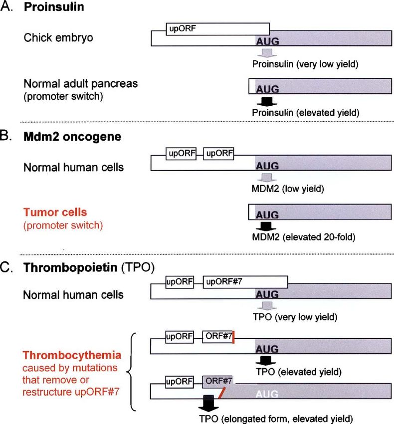
M. Kozak / Gene 361 (2005) 13–37                                                              15

mRNA might suffice because the A/U-richness of the initiation                         In rare cases, coupling allows a downstream cistron to be
site allows only weak secondary structures to form. (A far-                       translated even when it lacks a competent RBS. Ribosomes are
upstream hairpin in S1 mRNA which appears to augment                              delivered to the downstream cistron upon completing translation
translation might do so simply by directing secondary structure                   of the preceding cistron. This inefficient reinitiation mechanism
away from the initiation site, rather than via the complicated                    ensures that certain bacteriophage proteins, needed in only trace
mechanism postulated by Boni et al. (2001).)1                                     amounts, are translated at appropriately low levels (Adhin and van
    The ribosome binding site (RBS), defined as the segment of                    Duin, 1989; Inokuchi et al., 2000; Ivey-Hoyle and Steege, 1992).
mRNA protected against RNase digestion, consists of ∼ 15 nt                       The efficiency of translation increases when the downstream
on each side of the AUG codon (Steitz and Jakes, 1975). The                       cistron has an SD, and genes thus configured enabled testing of the
RBS thus extends slightly 5′ of the SD sequence, but no                           hypothesis that reinitiation indeed involves retention and reuse of
required sequence has been defined upstream of the SD. The                        ribosomes. This was done by mutating the SD sequences of the
stimulatory effect of upstream U-rich or A-rich or A/U-rich                       upstream and downstream cistrons and measuring translation in
sequences (Olins and Rangwala, 1989; Zhang and Deutscher,                         the presence of ribosomes that carry appropriate compensatory
1992; Zhelyabovskaya et al., 2004) might be attributed simply                     mutations (Rex et al., 1994).
to minimizing secondary structure.2                                                   The bacterial genome does not waste space: the terminator
    The sequence between the SD and AUG codon also plays no                       codon of one gene often overlaps the start codon of the next
defined role. Although mutations in this domain sometimes                         (e.g. UGAUG ) and this proximity facilitates reinitiation of
reduce translation of one or another mRNA (Gross et al., 1990),                   translation (Sprengel et al., 1985).3 The expression of foreign
efficient translation can be restored by a variety of sequences.                  genes in E. coli can be augmented by copying this arrangement
Again, the primary requirement might be exclusion of secondary                    (Ishida and Oshima, 2002; Schoner et al., 1986).
structure (Schauder and McCarthy, 1989), which could explain                          Coupled translation is used sometimes to coordinate gene
the general A-richness of E. coli mRNAs in positions − 1 to − 6.                  expression–e.g. allowing production of several ribosomal
    Downstream from the initiation codon, A-rich or A/U-rich                      proteins to be turned on or off via a single control point in the
sequences probably stimulate translation by precluding                            mRNA (Section 3.2)–but coupling does not necessarily ensure
secondary structure (Chen et al., 1994b; Qing et al., 2003). The                  equimolar protein yields. On the contrary, the efficiency of
inhibition of translation by certain G-rich codons (e.g. AGG,                     translation of downstream cistrons varies widely and is
CGG), however, cannot be attributed simply to augmentation of                     sometimes controlled in unusual ways by the upstream cistron
secondary structure. The inhibition is relieved upon inserting or                 (Praszkier and Pittard, 2002; Yu et al., 2001). In one case,
deleting one base in a way that shifts the reading frame,                         coupled translation has the unexpected advantage of enhancing
demonstrating that translation of these particular codons in the                  folding of the protein derived from the downstream cistron
vicinity of the start site is deleterious, for reasons unknown                    (Basu et al., 2004).
(Gonzalez de Valdivia and Isaksson, 2004).
                                                                                  2.4. Unusual mRNA structures and alternative initiation
                                                                                  mechanisms
2.3. Polycistronic mRNA structure: coupled translation
                                                                                     Leaderless mRNAs, albeit rare, are interesting because the
   The expression of prokaryotic genes via polycistronic
                                                                                  SD interaction is clearly precluded when the mRNA begins
transcripts makes possible a type of regulation in which
                                                                                  directly with the AUG codon. (AUG is the only start codon able
translation of a downstream cistron is coupled to that of the
                                                                                  to function in vivo in the absence of a leader sequence; Van
preceding cistron. This is achieved via a conformational
                                                                                  Etten and Janssen, 1998.) Unlike initiation sites in the interior of
constraint which is relieved as ribosomes translate the upstream
                                                                                  the mRNA, an AUG codon positioned exactly at the 5′ end
cistron. In the simplest cases, movement of ribosomes through
                                                                                  might be able to thread into the groove between the 30S and 50S
the upstream cistron (e.g. mok in Fig. 1B) is sufficient to disrupt
                                                                                  subunits, rationalizing the observation that a leaderless mRNA
the base pairing that constrains translation of the next cistron
                                                                                  binds more stably to 70S ribosomes than to the 30S subunit
(hok). (This example will be explained below.) A more
                                                                                  (O'Donnell and Janssen, 2002).
sophisticated control mechanism requires that ribosomes pause
                                                                                     A growing body of evidence indeed supports the idea that
at a particular point during translation of the upstream cistron
                                                                                  leaderless mRNAs are translated via a novel pathway which
(Butkus et al., 2003; Chen and Yanofsky, 2004; Gu et al., 1994;
                                                                                  begins with the 70S ribosome rather than with a free 30S subunit.
Mayford and Weisblum, 1989). In these examples, as in most
                                                                                  This was demonstrated in vitro, using chemically crosslinked 70S
cases of coupled translation, the downstream cistron has
                                                                                  ribosomes, and in vivo via a temperature-sensitive termination
a usable RBS which is temporarily obscured by secondary
                                                                                  factor which promotes accumulation of 70S ribosomes (Moll et
structure.
                                                                                    3
                                                                                      Early studies with the E. coli lacI gene appeared to show reinitiation
 2
    An alternative hypothesis is that ribosomal protein S1 interacts with these   occurring far downstream from a stop codon, but the interpretation of those
so-called enhancer elements, thereby promoting initiation. Although S1            experiments was later revised (Matteson et al., 1991). A rare case involving
occupies an appropriate position on the ribosome (Sengupta et al., 2001),         extensively overlapping genes, wherein the start codon of the second cistron
other evidence undermines the hypothesis: stimulation of translation by S1 is     lies far upstream from the stop codon of the first cistron, raises interesting
not mRNA-specific (Sørensen et al., 1998), and the isolated S1 protein binds      mechanistic questions which require further testing. This very unusual restart
promiscuously to RNAs (McGinness and Sauer, 2004).                                site functions very inefficiently (Adhin and van Duin, 1990).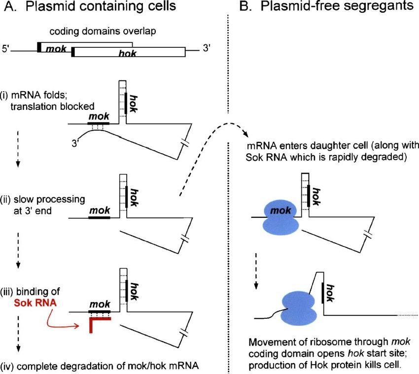
16                                                              M. Kozak / Gene 361 (2005) 13–37

Fig. 1. Translational control of plasmid R1 hok gene. Retention of the R1 plasmid in a bacterial population is ensured by killing plasmid-free segregants. The mRNA
that encodes the host-killing protein (Hok) is silent in plasmid-containing cells; its translation gets turned on only when the mRNA enters a daughter cell
unaccompanied by plasmid DNA. (A) Translation of hok is prevented in plasmid-containing cells via coupling to an upstream, overlapping cistron (mok). (i) mok is
untranslatable because its SD sequence (black bar) is blocked by base-pairing to a sequence near the 3′ end of the mRNA. (ii) Slow constitutive processing of the
mRNA by 3′ exonuclease exposes the mok start site which is quickly blocked by a small, plasmid-encoded antisense RNA (Sok). Sok RNA is very abundant in
plasmid-containing cells (despite its short half life) because it is transcribed from a very strong promoter. (iii) Binding of Sok RNA creates a base-paired structure
recognized by RNase III. The result is degradation of the entire mRNA. (B) In plasmid-free daughter cells, rapid decay of Sok RNA allows translation of mok. As
ribosomes advance through the mok coding domain, the base-paired structure which had prevented translation of hok is disrupted. Production of the toxic Hok protein
kills the cell. The RNA structures here depicted diagrammatically are presented in full by Franch et al. (1997) and evidence for the overall scheme is reviewed by
Gerdes et al. (1997). Recent studies explain how translation of mok/hok is prevented during transcription of the mRNA, i.e. during the interval before the
complementary element near the 3′ end of the mRNA is synthesized: the 5′ end of the nascent transcript forms a different base-paired structure which also blocks the
mok initiation region. (Møller-Jensen et al., 2001).

al., 2004). (Control transcripts containing internal initiation sites                Rangwala, 1989) or a sequence located downstream from the
were not translated by 70S ribosomes in these experiments,                           AUG codon (Sprengart et al., 1996)–but these ideas were
consistent with classic experiments wherein the free 30S subunit                     ruled out by the lack of an effect when the complementary
was shown to be required for initiation; Guthrie and Nomura,                         rRNA sequences were mutated (O'Connor and Dahlberg,
1968). The postulated mechanism rationalizes the effects of                          2001; O'Connor et al., 1999).
initiation factors: translation of leaderless mRNAs is inhibited by
IF3, which promotes dissociation of 70S ribosomes, and                               3. Examples of translational regulation in prokaryotes
augmented by IF2, which stabilizes binding of fMet-tRNA (Grill
et al., 2000; Tedin et al., 1999).                                                      In the upcoming examples, a key point is the ease whereby
    This 70S ribosome-mediated pathway, limited to leaderless                        the initiation step of translation can be blocked by base-paired
mRNAs, is the only credible alternative to the standard SD-                          structures in the mRNA. Once ribosomes enter the elongation
mediated initiation mechanism. Other pairings between                                phase, in contrast, they have a remarkable ability to disrupt
mRNA and 16S rRNA have been postulated–e.g. to explain                               base-pairing (Takyar et al., 2005). Structured elements in the
the stimulatory effect of an mRNA sequence located upstream                          coding domain of an mRNA might transiently slow elongation,
from the SD (the so-called “epsilon” sequence; Olins and                             but ribosomes eventually get through. This is the key to
M. Kozak / Gene 361 (2005) 13–37                                                  17

understanding the aforementioned coupled translation. It also                 RNAs to bind their mRNA targets (Lease and Woodson, 2004;
explains why biotechnologists, looking for ways to improve the                Mikulecky et al., 2004; Valentin-Hansen et al., 2004).
expression of foreign genes in E. coli, sometimes use the simple                 Other small RNAs control translation indirectly: rather
trick of diverting a base-paired structure from the RBS to the                than binding to an mRNA, the small RNA binds to and
coding domain (Paulus et al., 2004; Satchidanandam and                        sequesters a regulatory protein. It is the protein that binds to
Shivashankar, 1997).                                                          mRNA and directly blocks translation. Carbon storage
                                                                              regulator A (CsrA) is an example of this rather unusual type
                                                                              of repressor protein (Baker et al., 2002). Most repressor
3.1. Conformational masking of the initiation site                            proteins function without involvement of small RNAs,
                                                                              however, as described next.
    Translation is often regulated by base-paired structures in the
mRNA which undergo rearrangement, alternately sequestering                    3.2. Repressor proteins (and more about conformational
and exposing the RBS. Many classic examples are described in                  constraints)
an earlier review (de Smit and van Duin, 1990). In some
recently discovered cases, a conformational change in the                         mRNA-specific repressor proteins usually inhibit translation
mRNA is induced by small metabolites or a change in                           by competing with ribosomes for binding to mRNA. In most
temperature (Chowdhury et al., 2003; Johansson et al., 2002;                  cases, the protein binds directly across the RBS (Table 1) or
Nahvi et al., 2004; Nou and Kadner, 2000; Ravnum and                          close enough to it to sterically impede ribosome entry (Jenner et
Andersson, 2001; Winkler et al., 2002).4 The examples                         al., 2005). The mechanism is more complicated, but still
discussed in the next few paragraphs illustrate the basic point–              understandable, when the protein binds far upstream from the
that translation can be turned on and off by refolding of the                 RBS in a way that causes the mRNA to refold: the refolded
mRNA–and show how refolding is brought about by mRNA-                         conformation sequesters the SD and blocks ribosome entry (Du
binding proteins, small trans-acting RNAs, or movement of the                 and Babitzke, 1998). There are a few cases in which, despite
ribosome itself.                                                              binding of a repressor protein near the RBS, the ribosome can
    The first example (Fig. 1) involves short-range in-                       still bind to the mRNA, but only in a nonproductive way that
tramolecular base pairing (between hok and mok cistrons), long-               does not allow fMet-tRNA to pair with the AUG codon
range intramolecular base pairing (between the mok initiation                 (Philippe et al., 1993; Schlax et al., 2001).
site and the 3′ end of the mRNA), and intermolecular pairing                      Most of the proteins listed in Table 1 have as their primary
(between mok and a small antisense RNA called Sok). All these                 function something other than regulating translation. This is
constraints on translation are required because the protein                   important because regulation of translation requires controlled
encoded by the hok gene is so toxic that it must be produced                  binding of the repressor protein, and control is sometimes
only in plasmid-free segregants. The mechanism of inhibition is               achieved via competition between the mRNA and another
unambiguous here and in other cases where an antisense RNA                    substrate, such as tRNA or rRNA.
binds directly across the RBS (Ma and Simons, 1990). A small                      This idea underlies the remarkable feedback mechanism
RNA that binds just upstream from the SD can also inhibit                     whereby the production of ribosomal proteins is coordinated
translation (Malmgren et al., 1996).                                          with the availability of rRNA (Nomura et al., 1984; Zengel and
    Whereas small, plasmid-encoded antisense RNAs have                        Lindahl, 1994). When rRNA is saturated, certain ribosomal
a single mRNA target (e.g. Sok regulates only mok/hok), some                  proteins bind to their own mRNAs and shut off further
small RNAs encoded on the bacterial chromosome are                            unnecessary translation. In many but not all cases, the binding
multifunctional. OxyS RNA, for example, has two well-defined                  site for the protein on mRNA resembles its binding site on rRNA
targets (Altuvia et al., 1998); and DsrA RNA can potentially                  (Guillier et al., 2005; Merianos et al., 2004; Said et al., 1988;
regulate five mRNAs (Lease et al., 1998). The regulatory                      Serganov et al., 2003). The resemblance is imperfect, however,
importance of these small RNAs results from their being turned                and the protein usually has a higher affinity for rRNA
on (at the level of transcription) in response to environmental               (Nevskaya et al., 2005; Serganov et al., 2003; Wu et al., 1994).
cues, such as low temperature or low iron levels. Some small                  (In cases where the protein binds mRNA and rRNA with equal
RNAs inhibit translation by blocking the initiation site (Altuvia             affinity, other mechanisms–e.g. the high cooperativity of
et al., 1998); others activate translation by inducing the mRNA               ribosome assembly (Deckman and Draper, 1985; Robert and
to refold in a way that exposes the initiation site (Repoila et al.,          Brakier-Gingras, 2001)–might explain why the protein shuts off
2003). In many cases, the primary effect of small RNAs is on                  translation of mRNA only after all available rRNA is saturated.)
mRNA stability rather than translation (Kawamoto et al., 2005;                Long-distance base-pairing within the polycistronic mRNA
Lenz et al., 2004; Vogel et al., 2004). These and other aspects of            (Lesage et al., 1992; Petersen, 1989) probably explains how
the story are reviewed by Gottesman (2002, 2004). A helper                    binding of a repressor protein to one site can turn off translation
protein called Hfq facilitates the refolding required for the small           of all the downstream cistrons. Studies of ribosomal protein
 4                                                                            synthesis in organisms other than E. coli underscore both the
    The experimental results in some of these studies are best described as
“suggestive”; additional tests are needed to confirm the interpretation.      importance of feedback control–the basic phenomenon is
Regulation of transcription by binding of small metabolites to mRNA is much   conserved over a wide range of organisms–and its flexibility
better documented than is regulation of translation.                          vis-à-vis molecular details (Serganov et al., 2003).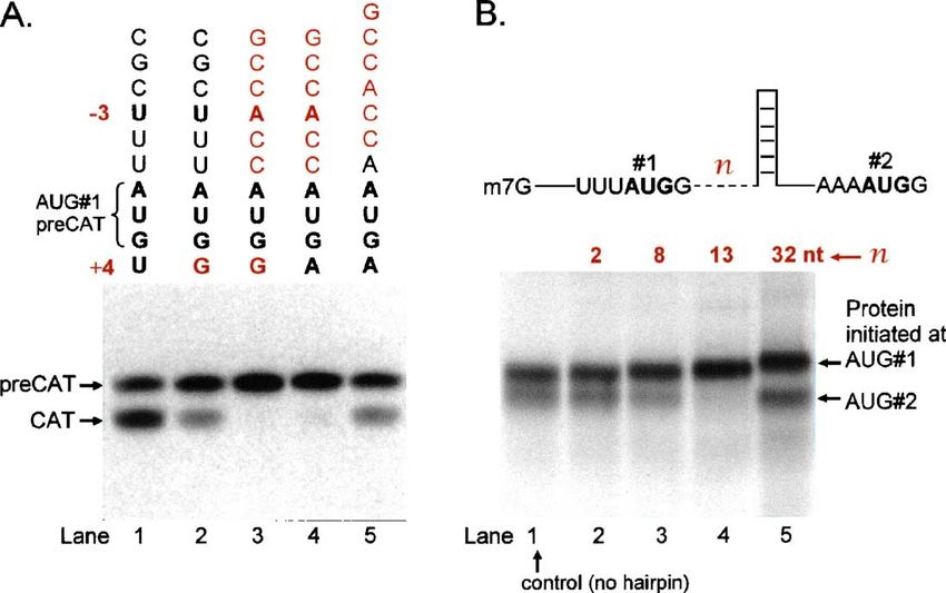
18                                                             M. Kozak / Gene 361 (2005) 13–37

Table 1
Translational repressor proteins in prokaryotes a
Organism                     Repressor protein             Targeted mRNA                  Binding site on mRNA                          References
Coliphage MS2/R17           Coat protein                   Replicase cistron              Hairpin encompasses RBS.                      Bernardi and Spahr, 1972;
                                                                                                                                        Carey et al., 1983
T4 phage                    DNA polymerase                  Gene 43 mRNA                  Extends across SD                             Pavlov and Karam, 2000
                            (gene 43)                                                     (dependent on upstream hairpin).
T4 phage                    DNA binding                     Gene 32 mRNA                  Begins at upstream pseudoknot                 Shamoo et al., 1993
                            protein (gene 32)                                             and extends across RBS.
T4 phage                    RegA                            Numerous                      Unstructured domain includes AUG.             Brown et al., 1997
Bacillus subtilis           TRAP b                          trpE, trpG, trpP              Protein binds far upstream in trpE            Du and Babitzke, 1998;
                                                                                          mRNA which refolds and blocks SD;             Du et al., 1997;
                                                                                          binds directly to SD in trpG and trpP.        Yakhnin et al., 2004
Lactococcus lactis          Intron-encoded                  LtrA                          Stem-loop structure includes RBS.             Singh et al., 2002
                            protein LtrA
E. coli                     CsrA c                          glgC, pgaA                    Binding site includes SD.                     Baker et al., 2002;
                                                                                                                                        Wang et al., 2005a
E. coli                      Thr-tRNA synthetase            thrS                          Binding to hairpin (just 5′ of SD)            Jenner et al., 2005
                                                                                          occludes RBS.
E. coli                     Ribosomal protein L1           L11 cistron                    Protein binds just 5' of SD.                  Said et al., 1988
                                                           (in same operon as L1)
E. coli                     Ribosomal protein S7           S7 cistron                     Protein binds adjacent to SD.                 Robert and
                                                                                                                                        Brakier-Gingras, 2001
E. coli                     Ribosomal protein S8           L5 cistron                     Hairpin d includes AUG codon.                 Merianos et al., 2004
                                                           (in same operon as S8)
E. coli                     Ribosomal protein S4 e         S13 cistron                    Pseudoknot spans RBS; mRNA                    Schlax et al., 2001
                                                           (in same operon as S4)         refolds into inactive conformation.
E. coli                      Ribosomal protein S15 e       S15 cistron                    Pseudoknot spans RBS.                         Philippe et al., 1993
 a
    This is not a complete list; some additional examples are mentioned in the text. The binding site for each protein was determined by mutational analysis,
biochemical tests (e.g. protection against RNase or chemical reagents), or iterative in vitro selection.
  b
    Along with inhibiting translation, the trp RNA-binding attenuation protein (TRAP) causes attenuation of transcription of the trpEDCFBA operon in response to
changes in the intracellular concentration of tryptophan. TRAP is neutralized by interacting with another protein which is also translationally regulated (Chen and
Yanofsky, 2004).
  c
    The function of CsrA is antagonized by small RNAs (CsrB, CsrC) which sequester the protein. CsrA affects mRNA stability as well as translation.
  d
    The base-paired element in L5 mRNA looks strong enough to inhibit translation on its own, but it does not; repression requires binding of ribosomal protein S8.
  e
    Ribosomal proteins S4 and S15 inhibit translation by trapping rather than competing with ribosomes; see text.

   Control of translation by repressor proteins is sometimes                        pairing in or near the initiation region can block translation (de
regulated by, and other times works in conjunction with,                            Smit and van Duin, 1994b; Hall et al., 1982). In the case of
cleavage of the mRNA. In the case of the E. coli spc operon, the                    MS2, however, the hairpin structure at the start of the R cistron
repressor protein binds at the start of the third cistron, shutting                 (Fig. 2C) is not stable enough on its own to block ribosome
off translation of all downstream cistrons, while the two cistrons                  entry (Berkhout and van Duin, 1985); only when the repressor
upstream from the repressor binding site are silenced via                           protein binds is translation inhibited. At the other extreme, the
degradation of the 5′ fragment (Mattheakis et al., 1989).                           base-paired structure that sequesters the M initiation site (Fig.
Cleavage of coliphage λ N mRNA by RNase III, on the other                           2B) is so stable that one might think there could be no way
hand, is part of a mechanism for activating translation (Wilson                     around it. This complicated structure apparently folds slowly,
et al., 2002). In the uncut mRNA, N protein represses                               however; and that provides a narrow window for translation of
translation by binding upstream from, but close to, the SD                          M protein (Poot et al., 1997). Studies with a related phage make
sequence. Cleavage by RNase III separates the N protein                             the additional point that competition between strong and weak
binding site from the RBS, and thus elevates translation.                           initiation sites can be a factor when translation occurs from
   Use of repressor proteins in conjunction with other                              a polycistronic mRNA (Priano et al., 1997).
mechanisms allows fine tuning of gene expression. In the case
of coliphage MS2, a single mRNA encodes four proteins, one of                       3.3. Novel regulatory mechanisms
which–the major coat protein–is required in much larger
amounts than the other three. Fig. 2 outlines how repressor                            Proteins that repress translation are more numerous and
proteins, conformational constraints, and coupled translation                       better studied than proteins that activate translation. The BipA
work together to ensure that each viral protein is produced at the                  protein in E. coli might be an example of the latter. Owens et al.
correct time and in the correct amount.                                             (2004) postulate that BipA, which displays ribosome-dependent
   The MS2 story helps us recognize the limits and                                  GTPase activity, activates the translation of an mRNA which
complications of regulation via base-paired structures. We know                     has a stronger-than-normal SD interaction. Some but not all of
from other examples that a remarkably small amount of base-                         the proffered data support this interesting hypothesis.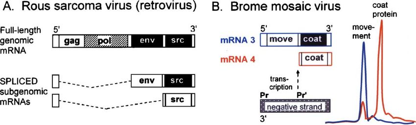
M. Kozak / Gene 361 (2005) 13–37                                                                        19

Fig. 2. Translation of coliphage MS2 mRNA is regulated by conformational constraints and RNA binding proteins. The single-stranded RNA genome encodes four
proteins: coat protein (CP), the major structural component; a minor capsid protein (maturase, M); RNA polymerase (replicase, R); and a small protein (L) which
promotes lysis of the host cell late in infection. Cistrons that are open for translation are shown as white blocks; those dependent on coupled translation are
crosshatched; cistrons shown in black are silent. The coupled translation is the result of base pairing between the initiation site of L or R and the CP coding domain
(Klovins et al., 1997; Licis et al., 2000). (A) (i) Infection begins with translation of mRNA released from parental virions. This brief phase ends when newly
synthesized R protein binds near the start of the CP cistron, shutting off translation of CP and all downstream cistrons. [The evidence for repression by R comes from
a related phage (Meyer et al., 1981); this point has not yet been verified for MS2.] The input mRNA next serves as template for RNA replication (not depicted), which
generates a burst of new plus-strands able to serve as mRNA. Slow folding of the 5′ end of these new transcripts provides a brief interval (ii) during which M can be
translated before base-pairing blocks access to the start codon (Poot et al., 1997). [The folded structure which eventually forms and shuts off translation of M is
depicted in (B).] (iii) Progeny mRNA primarily directs translation of CP. L is also translated, but only via an inefficient coupled mechanism which keeps the yield low
(Klovins et al., 1997). Translation of R is soon shut off by CP which, upon reaching a sufficiently high concentration, binds near the start of the R cistron in a way that
blocks ribosome entry. The binding site for the coat repressor protein is shown in (C). The net effect of these translational controls is that each protein is produced in the
required amount and at the appropriate time. Although viruses can replicate when some of these controls are lost, there is a significant reduction in efficiency (Licis et
al., 2000).

    The 5′ untranslated region (UTR) of certain mRNAs confers                            prokaryotes. Translational regulation is thought to underlie the
preferential translation during cold shock, when bulk protein                            interesting phenomenon wherein a block in production of one
synthesis is drastically diminished (Giuliodori et al., 2004;                            subunit causes disappearance of all the subunits in a given
Yamanaka et al., 1999). The general decrease in translation                              photosynthetic complex. Results described by Wostrikoff et al.
appears to be mediated by a cold-shock induced “protein Y”                               (2004), for example, can be explained by a model wherein
which binds the 30S ribosomal subunit in a way that blocks the                           absence of one subunit (psaB) causes the other unassembled
A and P sites and competes with initiation factors (Vila-Sanjurjo                        subunit (psaA) to block translation of its own mRNA. The
et al., 2004). Whether and how protein Y might account for the                           inhibition is assumed to be at the initiation step, inasmuch as
preferential translation of cold-shock mRNAs, however, awaits                            translation of a chimeric reporter gene that carries the psaA 5′
investigation.                                                                           UTR was also blocked. Cytochrome f, which is part of a different
    It is not out of place here to mention chloroplasts, inasmuch as                     photosynthetic complex, also inhibits translation of its own
the mechanism of translation in that system is very similar to                           mRNA in the absence of its assembly partners (Choquet et al.,
20 M. Kozak / Gene 361 (2005) 13–37
Fig. 3. Some plant and animal virus mRNAs are structurally polycistronic but functionally monocistronic, underscoring the rule that initiation of translation in
eukaryotes is restricted to the 5′ end of the mRNA. Translationally silent cistrons are shown as black blocks. (A) Rous sarcoma virus genomic mRNA encodes four
proteins (Gag, Gag-Pol, Env, and Src), but only the 5′-proximal gag initiation site is accessible to ribosomes. This full-length mRNA thus produces only Gag and (via
frameshifting) Gag-Pol proteins; the downstream cistrons are silent. Translation of Env and Src occurs from spliced, subgenomic mRNAs wherein each cistron is
moved closer to the 5′ end (Pawson et al., 1977; Purchio et al., 1977; Stacey et al., 1977). In similar fashion, many other viruses use splicing to restructure mRNAs and
thus turn on translation of downstream cistrons (reviewed in Kozak, 2002a). (B) Other viruses generate the required subgenomic mRNAs via internal transcriptional
promoters. Brome mosaic virus, shown here as an example, is historically important as the system in which silent downstream cistrons were first discovered. Genomic
RNA3 is structurally dicistronic: the 5′ cistron encodes a protein required for cell to cell movement of the virus, and the 3′ cistron encodes the capsid (coat) protein. In
vitro translation of RNA3 produces only the movement protein, as the gel electrophoresis profile (blue) shows. Coat protein can be translated only from subgenomic
RNA4 (red profile). (These gel profiles are tracings of Fig. 6 in Shih and Kaesberg, 1976.) Initiation of transcription from an internal promoter (Pr′) in the negative
RNA strand generates mRNA4 (Choi et al., 2004).
1998). Some evidence suggests the repression might be understanding of the stop-scanning step, we know nothing
mediated by an unidentified ternary effector rather than by direct about the mechanism that actually propels the 40S subunit/
binding of cytochrome f to the 5′ UTR (Choquet et al., 2003). factor complex. Hints that scanning might be dependent on ATP
hydrolysis (Kozak, 1980) suggest involvement of eIF4A, the
only initiation factor that binds ATP; but this awaits verification.
4. Structural elements in eukaryotic mRNAs that control No real evidence underlies the often repeated claim that eIF4A
initiation of translation unwinds the 5′ end of the mRNA prior to binding of the 40S
subunit; secondary structure might be disrupted only as the 40S
4.1. Preamble subunit/factor complex advances.
Initiation is not always restricted to the AUG codon nearest
Whereas co-transcription of contiguous genes in prokaryotes the 5′ end. The scanning model specifies certain conditions–
produces polycistronic mRNAs, eukaryotic cellular genes are described below under context-dependent leaky scanning
transcribed individually, producing monocistronic mRNAs. (Section 4.3) and reinitiation (Section 5)–which allow limited
This fundamental difference in gene expression follows from escape from the first-AUG rule. In these cases, translation still
a fundamental difference in the mechanism of translation: initiates at the first AUG, but not exclusively.
prokaryotic ribosomes can enter and initiate at multiple sites Some investigators believe that, contrary to the restrictions
within an mRNA, but eukaryotic ribosomes routinely enter only imposed by the scanning mechanism, eukaryotic ribosomes can
at the 5′ end. enter directly at internal positions in certain mRNAs. This idea
The scanning mechanism of initiation (Kozak, 2002a) is not discussed in detail herein because the so-called internal
postulates that the 40S ribosomal subunit enters at the 5′ end of ribosome entry sites (IRES) have not been defined structurally
the mRNA and migrates linearly until it encounters the first (candidate IRES elements share no common sequence) or
AUG codon, which is recognized by base-pairing with the mechanistically. Evidence said to support the internal initiation
anticodon in Met-tRNAi (Cigan et al., 1988a). eIF2, the factor hypothesis is described in other reviews (Hellen and Sarnow,
that escorts Met-tRNAi onto the 40S subunit, is a latent GTPase; 2001; Jackson and Kaminski, 1995), but serious questions have
an associated factor (eIF5) activates GTP hydrolysis by eIF2 been raised about much of this evidence (Kozak, 2001a, 2003a).
only when there is a sufficiently long pause in scanning. In other The absence of natural dicistronic mRNAs (two full-length
words, the eIF5-mediated step is a timing device that helps to nonoverlapping cistrons) in eukaryotic cells is prima facie
distinguish authentic AUG start codons (long pause) from other evidence against the internal initiation hypothesis. Occasional
contenders (e.g. short pause at UUG) (Das and Maitra, 2001; exceptions only underscore the rule: some viral transcripts are
Huang et al., 1997).5 In contrast with our growing structurally dicistronic but only the 5′ proximal cistron gets
5
translated; thus, even these mRNAs are functionally mono-
eIF5 interacts also with eIF1 and eIF3, and genetic evidence suggests these cistronic (Fig. 3). In one case where the 3′ cistron appeared to be
factors can influence the eIF5-mediated GTPase reaction (Valášek et al., 2004).
One possibility is that eIF1 serves as a brake on eIF5. The stop-scanning step translated (Stacey et al., 2000), the interpretation was revised
controlled by these factors is followed by joining of the 60S subunit, which when a second, spliced transcript was found (Zheng et al.,
requires yet another protein factor (Lee et al., 2002; Shin et al., 2002). 2004). Other claims of dicistronic mRNAs were simplyM. Kozak / Gene 361 (2005) 13–37 21
mistaken: the two proteins turned out to be generated by between m7G and eIF4E strongly stimulates translation of most
proteolysis following translation of a single large open reading mRNAs.7
frame (ORF) (Hänzelmann et al., 2002; Ritchie and Wang,
1997, corrected in Feng et al., 1998).6 4.3. Context effects on recognition of AUG (or other) start
In addition to eukaryotic mRNAs being basically mono- codons
cistronic, four other structural features are important vis-à-vis
initiation of translation. These are explained next. As in The optimal context for initiation of translation in mammals
prokaryotes, the eukaryotic ribosome protects ∼15 nt on each is GCCRCCaugG. In experimental tests, the biggest reduction
side of the AUG codon (Kozak, 1977), but it is not appropriate in efficiency was seen when the purine (R) in position − 3 or the
to use the term “ribosome binding site” for eukaryotes; because G in position + 4 was mutated (Kozak, 1986a, 1997). Thus,
of the scanning mechanism of initiation, structural elements that initiation sites are usually designated “strong” or “weak” based
affect initiation can be dispersed throughout the 5′ UTR. (The on those two positions. A start codon flanked by A − 3 and G +4
latter term also is not appropriate, inasmuch as small ORFs can function N10-fold more efficiently than an AUG codon in
located within the 5′ “untranslated region” do get translated, as the weakest context. The GCCRCC motif augments initiation
explained in Section 5; but we are stuck with the term). only when it directly abuts the AUG codon (Fig. 4A, lane 4 vs.
The following discussion does not include structures at the 3′ lane 5; Kozak, 1987a).
end of the mRNA. Despite abundant evidence implicating 3′ Ribosomes will initiate at the first AUG codon to a limited
UTR elements in translational control of developmentally extent even when the context is weak, but the poor context allows
regulated genes (Bashaw and Baker, 1997; Kuersten and some ribosomes to bypass the first AUG and thus reach a start
Goodwin, 2003; Wickens et al., 2000), the mechanisms are not codon farther downstream. This is called leaky scanning. Fig. 4A
yet clear. A recent review explains why some proposed shows a test case wherein initiation was restricted to the first
mechanisms require rethinking (Kozak, 2004). AUG when it resided in the optimal context (lane 3), while
a weaker context allowed initiation from the first and second
AUG codons (lanes 1, 2). The leaky scanning seen when the first
4.2. m7G cap AUG codon resides in a suboptimal context can be suppressed by
downstream secondary structure, as demonstrated in Fig. 4B.
The 5′ end of all cellular and most viral mRNAs is capped Because this depends on precise positioning of the hairpin
with 7-methylguanosine (Furuichi and Shatkin, 2000). Via structure (compare lanes 2, 4 and 5), a reasonable interpretation
interaction with eIF4E (Gingras et al., 1999; von der Haar et al., is that the structured element slows scanning and that recognition
2004), the m7G cap strongly promotes ribosome binding. This of a weak start codon improves when the 40S subunit pauses
was demonstrated directly by varying the 5′ terminal structure with its AUG-recognition center right over the AUG codon.
on mRNAs used for in vitro translation (Both et al., 1975) and When the first and second start codons are in the same
indirectly by the inhibitory effect of soluble cap analogues reading frame, context-dependent leaky scanning generates
(Hickey et al., 1976). In vivo experiments confirmed long and short forms of the protein which can be targeted to
a substantial reduction in translational efficiency (≥ 10-fold) different compartments in the cell (Leissring et al., 2004; Melén
when mRNAs lack the m7G cap (Horikami et al., 1984; Lo et et al., 1996; Shang et al., 2001; Souciet et al., 1999). When the
al., 1998). first and second start codons are in different reading frames,
Initiation was shown to occur exclusively at the first AUG leaky scanning enables one mRNA to produce two completely
codon even in the absence of the cap (Kozak, 1998, Fig. 6) or in different proteins. Many bifunctional mRNAs that use this
the absence of initiation factors required for cap function (Ali et mechanism have been identified in plant and animal cells and
al., 2001). These experiments underscore an important point: it viruses,8 and occasional examples have been found in yeast
is not because of the m7G/eIF4E interaction that ribosomes (Outten and Culotta, 2004). (Recognition of start codons in
enter at the 5′ end. Rather, it is because eukaryotic ribosomes yeast is not sensitive to context in all cases; Cigan et al., 1988b).
enter at the 5′ end that the m7G/eIF4E interaction can augment
initiation. The inability of eukaryotic ribosomes to bind 7
a circularized mRNA (Kozak, 1979) supports the hypothesis Stimulation means simply that more protein is produced because more of
the mRNA is engaged by ribosomes. It merits repeating that selection of the
that entry occurs via the 5′ end. correct start site (the first AUG codon) is not augmented by eIF4E, contrary to
In the aforementioned experiments, the cap might have been what some textbooks say (Alberts et al., 2002). Reports of initiation occurring
dispensable because the 5′ end of the mRNA was relatively free at internal sites upon removal of the cap (Brown et al., 2000) are probably
of secondary structure and thus accessible to ribosomes. Most attributable to partial degradation of the mRNA by 5′ exonucleases.
8
natural mRNAs have considerable secondary structure near the Table 3 (Kozak, 2002a) lists 33 examples with full references. In many of
these examples, operation of the leaky scanning mechanism was verified by
5′ end, however. For this or other reasons, the interaction showing that mutations which improve the context around the first AUG codon
reduce initiation from the second AUG. In rare cases, production of a viral
protein does not respond to changes in start codon context, apparently because
6
Even picornaviruses (e.g. poliovirus), which are deemed the prime example translation is limited at a step other than initiation (Fajardo and Shatkin, 1990).
of internal initiation, do not employ dicistronic mRNAs. The full set of viral Note that leaky scanning can occur, irrespective of context, when the first AUG
proteins is derived by proteolysis from a “polyprotein” which is translated from codon is positioned very close to the cap (Kaneda et al., 2000; Slusher et al.,
a single large cistron. The IRES (if such it is) is at the 5′ end of the mRNA. 1991).22 M. Kozak / Gene 361 (2005) 13–37
Fig. 4. Flanking sequences augment recognition of the AUG codon in eukaryotes. The autoradiograms show [35S]Met-labeled proteins synthesized in vitro from
capped mRNAs that encode chloramphenicol acetyltransferase. (A) Initiation at AUG#1 generates an N-terminally extended protein, labeled preCAT. A suboptimal
context around AUG#1 allows some ribosomes to scan past that site and initiate instead at AUG#2. This leaky scanning produces a shorter protein, labeled CAT. (B)
All mRNAs have a suboptimal context (U in position −3) around the first AUG codon and, except for the control in lane 1, a moderately stable base-paired structure
between the first and second AUGs. The only variable is the distance (n) between AUG#1 and the base of the hairpin structure. When properly positioned, the
downstream base-paired structure apparently suppresses leaky scanning (lane 4). A full description of these constructs and the adjustments required for the rabbit
reticulocyte translation system to work properly are given in Kozak (1990a,b).
Whereas initiation at a codon other than AUG is common in rule involved reiterating a block of nucleotides comprising the
prokaryotes, use of CUG, GUG or UUG as the primary initiation AUG start codon and nearby sequences in rat preproinsulin
site is exceedingly rare in eukaryotes.9 These nonstandard start mRNA. Analysis of proteins produced by this mRNA in vivo
codons are usually weak even when supported by the optimal revealed that translation initiated exclusively at the first of four
context, as shown by an increase in protein production upon tandemly repeated sequences (Kozak, 1983). An important
experimentally changing the codon to AUG. The weakness followup test showed that the first AUG codon was used
explains why non-AUG codons are usually used only as exclusively even when the second AUG was very close (2 or
supplementary initiation sites; i.e. ribosomes initiate at an 5 nt downstream from the first AUG) and in the same favorable
upstream non-AUG codon in addition to initiating at the first context (Kozak, 1995).
AUG (Carroll and Derse, 1993; Chang and Wang, 2004; Fütterer Selection-based-on-position was also verified in yeast via
et al., 1997; Fuxe et al., 2000; Portis et al., 1994). The non-AUG a clever experiment that involved changing the anticodon of
initiated protein serves a useful function in those examples; but Met-tRNAi to 3′-UUC-5′, whereupon initiation shifted to an
in some other cases, the N-terminally extended form of the AAG codon located upstream from the normal AUG start codon
protein has no biological relevance (Miles et al., 2003). (Cigan et al., 1988a).
Meaningless initiation events at upstream CUG codons might In the aforementioned tests, start codons were added or
occur by accident when scanning is slowed by a GC-rich leader removed experimentally. When restructuring of mRNAs
sequence. happens naturally via mutations, the pattern of translation again
reveals the dominant role of position. Several examples are
4.4. Position determines which AUG functions as the start described in Fig. 5. In the first two cases, the mutation
codon introduces an AUG codon upstream from the normal initiation
site, whereupon the new AUG takes over. In the third case,
The strongest evidence for the scanning mechanism is the a point mutation ablates the normal start codon, whereupon the
position rule, which simply means that translation initiates at next downstream AUG codon which had been silent becomes
whichever AUG codon is closest to the 5′ end. (In the following the new start site for translation. A few other naturally occurring
examples, the first AUG was in a favorable context, thus mutations along these lines have been described (Cai et al.,
allowing an uncomplicated test.) The earliest test of the position 1992; Liu et al., 1999; Mével-Ninio et al., 1996).
Elimination of the start codon via a mutation is a rare event,
9
Examples and full documentation are provided in Kozak (2002a). but the everyday production of alternative transcripts illustratesM. Kozak / Gene 361 (2005) 13–37 23
Fig. 5. Proximity to the 5′ end dictates which AUG functions as the start codon. White lettering indicates silent AUG codons. Only the portion of the mRNA relevant to
understanding how an AUG codon is added (A, B) or removed (C, D) is shown. The resulting shifts in initiation have clinical consequences. (A) The G→A mutation
that creates an upstream AUG codon, thereby shutting off translation of hepcidin, was found in patients with juvenile hereditary hemochromatosis (Matthes et al.,
2004). Hepcidin is an important negative regulator of iron absorption. (B) Restructuring of the c-akt gene adds an upstream in-frame AUG codon which takes over as
the initiation site, producing an N-terminally extended form of AKT. This was found in a retrovirus-induced mouse leukemia which undergoes regression due to
recognition of the novel N-terminal peptide by cytotoxic T-lymphocytes (Wada et al., 1995). (C) This point mutation causes loss of only the first four amino acids, but
the shortened polypeptide folds improperly and vasopressin is not produced (Beuret et al., 1999). (D) Activation of only the upstream promoter in colon cancer
prevents modulation of LEF1 activity, which is accomplished normally by balanced production of long and short forms of the protein (Hovanes et al., 2001).
the same principle: when the normal AUG codon is the 5′ end of the mRNA (Berthelot et al., 2004). Thus, it is
eliminated via a change in splicing or a switch in the a mistake to think that a long leader sequence contravenes
transcriptional start site, a downstream AUG which had been operation of the scanning mechanism.
silent becomes the new initiation site. Fig. 5D depicts one
example. There are many, many others.9 This is important 4.5. Base-paired structures in 5′ UTR
biologically because it enables a single gene to produce
different forms of the protein which sometimes have Although leader length per se is not a problem, scanning can
complementary functions, as indicated in Fig. 5D for LEF1. be difficult when a long leader sequence contains secondary
From a theoretical perspective, the need to produce a second structure. The GC-richness of mammalian 5′ UTR sequences
form of mRNA to activate an internal start codon is strong predicts a considerable amount of secondary structure. Yeast
evidence for the scanning mechanism. mRNAs, in contrast, have remarkably AU-rich leader sequences
The position rule asks only “which AUG is first?” The actual (Shabalina et al., 2004).
distance from the 5′ end is irrelevant. The scanning mechanism Base-paired structures are most inhibitory when their
was shown to operate with no measurable reduction in proximity to the 5′ end blocks ribosome entry (Goossen and
efficiency even when the first AUG codon was N 1000 nt from Hentze, 1992; Kozak, 1989; Wang and Wessler, 2001). Once24 M. Kozak / Gene 361 (2005) 13–37
bound to mRNA, the scanning 40S subunit/factor complex has next AUG codon cannot be recognized. Reinitiation can occur
some ability to disrupt base-pairing, although this has limits only if there is enough time (distance) for the scanning 40S
(Kozak, 1986b). The bottom line is that a long 5′ UTR which subunit to pick up Met-tRNAi before arriving at the next AUG.
contains substantial secondary structure can greatly reduce Reacquiring Met-tRNAi requires the help of eIF2 (i.e. a ternary
translational efficiency but does not completely preclude complex must assemble consisting of Met-tRNAi, eIF2, and
scanning (Short and Pfarr, 2002; van der Velden et al., 2002). GTP), and therefore, reinitiation can be regulated by
An often-repeated idea is that increased expression of manipulating eIF2·GTP levels. The significance of this is
initiation factor eIF4E might selectively elevate translation of explained in Section 5.2.
mRNAs that encode critical growth-regulatory proteins (Graff Reinitiation is affected by certain changes in mRNA
and Zimmer, 2003). The selectivity is attributed to these structure, enabling us to formulate working rules even though
mRNAs having leader sequences which are GC-rich, hence the biochemical mechanisms are uncertain.
highly structured. One problem with the hypothesis is that the One rule is that eukaryotic ribosomes can reinitiate following
mRNAs produced by most housekeeping genes also have the translation of a small ORF but not following the translation
extremely GC-rich (N 70%) leader sequences. Thus, there is no of a full-length protein-coding ORF. Naturally occurring
structural basis for the idea that increased expression of eIF4E upORFs usually are only a few codons long. With experimental
specifically elevates translation of growth-regulatory genes. constructs, reinitiation was found to decrease as the upORF was
When increased production of critical regulatory proteins is lengthened, reaching a barely detectable level when the upORF
needed, a proven solution in some cases is to change the was 35 codons long (Kozak, 2001b; Rajkowitsch et al., 2004).
structure of the 5′ UTR via alternative splicing or activation of This might be explained if reinitiation depends on retention of
a downstream transcriptional promoter. In other words, the certain initiation factors which gradually dissociate from 80S
block to translation is relieved by truncating the long, GC-rich ribosomes during the course of elongation. This idea is
5′ UTR (Charron et al., 1998; Han et al., 2003a,b; Sasahara et somewhat supported by experiments wherein manipulations
al., 1998).9 designed to slow elongation through the upORF (e.g. depleting
Occasionally, experimental manipulations of 5′ UTR tRNA levels or introducing a base-paired structure into the
sequences do not produce the expected effects on translation. In mRNA; Kozak, 2001b; Rajkowitsch et al., 2004) reduced the
cases where a long, GC-rich leader inhibits translation in vitro efficiency of reinitiation. The hypothesis cannot really be tested
but not in vivo (Hoover et al., 1997; Nikolcheva et al., 2002; until we understand more about the cycling of initiation
van der Velden et al., 2002), the simplest explanation might be factors.11
that the sequence harbors a cryptic promoter which generates Another rule is that eukaryotic ribosomes cannot backup to
a better-translated short-leader transcript in vivo.10 The lesson is reinitiate at an AUG codon positioned far upstream from the
that, when pondering the function of 5′ UTR structures, it is termination site. It follows that translation of the major coding
a mistake to think only in terms of direct effects on initiation of domain (the longest ORF) should be profoundly inhibited by an
translation. overlapping upORF. Many observations verify this prediction.9
The bottom line is that, when an upORF is relatively small
5. Reinitiation as a device for regulating translation in and terminates before the start of the main coding domain,
eukaryotes reinitiation can occur; but reinitiation is never efficient. The
next section describes some consequences.
5.1. Mechanism of reinitiation
We know little about the molecular mechanisms underlying 11
Cauliflower mosaic virus produces a transactivator protein called TAV
reinitiation. Ribosomes initiate in the normal way at the first which is postulated to interact with eIF3, retaining the initiation factor on
AUG codon, producing the peptide encoded in the small ribosomes and purportedly allowing reinitiation even after translation of a full-
upstream ORF (upORF). At the terminator codon (where the length cistron (Park et al., 2001). Evidence for this idea is not convincing,
60S subunit presumably dissociates), the 40S subunit however. Although TAV and eIF3b appear to co-sediment with small polysomes
in a sucrose gradient, this interpretation was not verified by showing a shift in
apparently remains bound to the mRNA, resumes scanning, and sedimentation of TAV/eIF3b upon disruption of polysomes. As for whether
initiates again at a downstream site. TAV indeed enables a dicistronic mRNA to be translated by reinitiation, the
Whereas reinitiation in prokaryotes is facilitated when the results from DNA transfection experiments are ambiguous. One study showed
stop codon of the first cistron is closely apposed to the next strong stimulation of expression of the downstream cistron by TAV (Park et al.,
2001, Fig. 6). In another study, the efficiency of translation of the 3′ cistron was
start codon, that arrangement works poorly, if at all, in
not revealed; the yield from a particular construct in the presence of TAV was
eukaryotes. Reinitiation in eukaryotes improves as the distance simply called “100%” (Fütterer and Hohn, 1991, Fig. 2). In a third study–the
increases between the stop codon and the re-start site (Kozak, only one in which mRNA structure was checked–the yield of protein from the
1987b; Abastado et al., 1991). This reflects a requirement for downstream cistron in the presence of TAV was only about 5% of the yield
the 40S subunit to reacquire Met-tRNAi, without which the from a monocistronic control mRNA; and the analysis of mRNA structure was
not sensitive enough to rule out production of a monocistronic mRNA at 1/20th
the level of the dicistronic mRNA (Bonneville et al., 1989). Inasmuch as TAV is
present in both the nucleus and cytoplasm of infected cells (Haas et al., 2005),
10
Recent experiments confirm this prediction for the pim-1 gene (Wang et al., the possibility that TAV might augment splicing or transport of viral mRNAs,
2005b). rather than directly promoting translation, needs to be considered.You can also read