Mito-Nuclear Communication in Hepatocellular Carcinoma Metabolic Rewiring - MDPI
←
→
Page content transcription
If your browser does not render page correctly, please read the page content below

cells
Review
Mito-Nuclear Communication in Hepatocellular
Carcinoma Metabolic Rewiring
Tommaso Mello 1, * , Irene Simeone 1,2 and Andrea Galli 1
 1    Clinical Gastroenterology Unit, Department of Biomedical Clinical and Experimental Sciences “Mario Serio”,
      University of Florence, V.le Pieraccini 6, Florence 50129, Italy; irene.simeone@student.unisi.it (I.S.);
      andrea.galli@unifi.it (A.G.)
 2    University of Siena, 53100 Siena, Italy
 *    Correspondence: tommaso.mello@unifi.it; Tel.: +39-055-2758115

 Received: 22 March 2019; Accepted: 1 May 2019; Published: 5 May 2019

 Abstract: As the main metabolic and detoxification organ, the liver constantly adapts its activity
 to fulfill the energy requirements of the whole body. Despite the remarkable adaptive capacity of
 the liver, prolonged exposure to noxious stimuli such as alcohol, viruses and metabolic disorders
 results in the development of chronic liver disease that can progress to hepatocellular carcinoma
 (HCC), which is currently the second leading cause of cancer-related death worldwide. Metabolic
 rewiring is a common feature of cancers, including HCC. Altered mito-nuclear communication is
 emerging as a driving force in the metabolic reprogramming of cancer cells, affecting all aspects of
 cancer biology from neoplastic transformation to acquired drug resistance. Here, we explore relevant
 aspects (and discuss recent findings) of mito-nuclear crosstalk in the metabolic reprogramming of
 hepatocellular carcinoma.

 Keywords: HCC; PPAR; SIRT; PGC-1; NRF; HIF; liver; mitochondria; metabolism

1. Introduction
      A tumor is a very harsh environment to live in. Poor oxygenation, low nutrient levels, high
concentration of waste metabolites, and acidic pH are inevitable consequences of a crowded and
disorganized mass of fast-growing cells. Moreover, the tumor microenvironment can change
dramatically within the growing mass, because of the defective tumor vasculature, necrosis, immune
response and therapeutic treatments. This environment operates an enormous selective pressure
that, combined with the poor genomic stability of cancer cells, leads to cancer cell evolution and the
acquisition of a progressively malignant phenotype. An early-enabled characteristic of the malignant
transformation of cancer cells is the reprogramming of their energy metabolism in order to support the
cell fast growing rate. It has been long noted that cancer cells rely primarily on glycolysis for adenosine
triphosphate (ATP) production, even in the presence of oxygen (Warburg effect) [1]. However, only
more recently the significance of this “metabolic reprogramming”, its plasticity, its implications in
cancer biology and response to treatment have begun to emerge [2]. Otto Warburg proposed that
“aerobic glycolysis” was due to defective mitochondria respiration that forces cancer cells to rely on
an alternative pathway for energy production [3]; it is now clear that mitochondria are not simply
dysfunctional in cancer cells. Rather, they are reprogrammed to serve as “biosynthetic factories”
to supply the building blocks for lipids, DNA and protein synthesis required to support cancer
cell proliferation [4,5]. Mitochondria are unique organelles in many ways. Besides being the main
site of cellular respiration and ATP production through oxidative phosphorylation (OXPHOS), they
are crucial for fatty acid catabolism through the β-oxidative pathway, for anabolic metabolism of
lipids, aminoacids and heme; they also participate in Ca2+ homeostasis, connect signaling pathways

Cells 2019, 8, 417; doi:10.3390/cells8050417                                          www.mdpi.com/journal/cells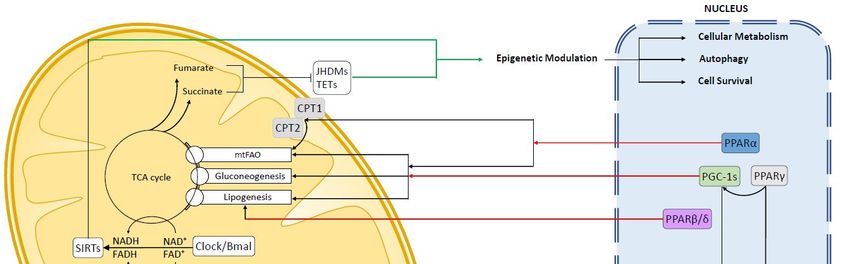
Cells 2019, 8, 417                                                                                 2 of 30

and apoptotic cascades. A tight coordination of nuclear and mitochondrial functions is required
to maintain proper mitochondria functionality and to adjust mitochondrial activity to the energetic
and biosynthetic requirements of the cell. A clear example of this coordination is the assembly of
the respiratory complexes of the electron transport chain (ETC). Mitochondria have a circular DNA
genome of 16.6 Kb that encodes for 13 subunits of complexes I, III, IV and V of the ETC, along with two
ribosomal RNA and 22 mitochondria-specific t-RNA. The ETC complex assembly, therefore, requires a
regulation of both nuclear-encoded and mitochondrial-encoded subunits, which need to be in proper
stoichiometric ratios. Failure to maintain this proportion leads to the mito-nuclear protein imbalance,
which could result in reduced mitochondrial respiration and ATP synthesis [6].
      Mito-nuclear communications are exerted through the “anterograde signaling”, through which
the nucleus regulates mitochondrial activity and number, and the “retrograde signaling”, which
allows mitochondria to inform the nucleus about the onset of oxidative stress, ATP and metabolites
levels, OXPHOS impairments, membrane potential disruption, accumulation of unfolded protein,
thereby activating the proper nuclear transcriptional response [6,7]. It is becoming increasingly clear
that transient and sub-lethal levels of mitochondrial oxidative stress elicit an adaptive response,
termed “mitohormesis” that allows the cell to withstand more harmful stimuli, thus enhancing the
cell resistance to apoptosis and prolonging lifespan [6–8]. Accumulating evidence is highlighting the
importance of the mito-nuclear communication and mitohormesis in the onset and progression of
metabolic, cardiovascular, neurological diseases, ageing and cancer.
      Indeed, mitohormesis is a clear paradigm of the importance of mito-nuclear communications,
since the stress-induced signaling originating from mitochondria elicit a nuclear response aimed at
increasing the antioxidant defenses, to promote the mitochondrial turnover through mitophagy and
biogenesis, and to remodel mitochondrial metabolism.
      Remarkably, a transient increase in mitochondrial oxidative stress during fetal development
triggers a stable hormetic response in the adult liver that heightens the basal level of mitochondrial
antioxidant defense. This mitohormetic adaptive response requires the activation of nuclear
respiratory factor (NRF2), peroxisome proliferator-activated receptors (PPARs) and the peroxisome
proliferator-activated receptor gamma coactivator 1 (PGC1α) pathways [9]. While reactive oxygen
species (ROS) are by far the more studied mitohormetic triggers, other stressors can elicit this adaptive
response, working both together or independently of ROS, such as ions, metabolites, lipids, or nucleic
acids [10–12]. Collectively, these signaling factors can act as “mitokines” promoting mitohormesis in
an autocrine, paracrine and even endocrine manner [13].
      As the main metabolic organ, in order to regulate the body energy metabolism, the liver needs
to adapt its metabolic activity constantly, integrating different input signals such as nutrient and
hormones levels, neuronal signaling, physical activity and circadian rhythms. Moreover, the liver is
the main site of detoxification from xenobiotics and alcohol, which poses additional requirements to
withstand oxidative and metabolic stressors. Despite the remarkable adaptive capacity of the liver,
prolonged alcohol abuse, viral infections, genetic or metabolic disorders (non-alcoholic fatty liver
disease, NAFLD and non-alcoholic steatohepatitis, NASH) can result in the exhaustion of the liver
anti-oxidant defense, leading to chronic liver disease that can eventually progress to hepatocellular
carcinoma (HCC). Hepatocellular carcinoma accounts for 75%–85% of total cases of primary liver
cancer, it is currently the fifth most common cancer in men, the ninth in women and the second leading
cause of cancer-related death worldwide [14]. The very high ratio of mortality to incidence (0.95)
is indicative of the absence of effective therapeutic strategies, even if many treatment options have
been developed during the last years, including hepatic resection, liver transplantation and molecular
targeted therapies.
      Metabolic reprogramming is a key event in hepatocellular carcinoma onset and progression [15].
Aberrant up-regulation of the mammalian target of rapamycin (mTOR) pathway occurs in up to 50%
of HCC [16–19]. The mTOR pathway coordinates cellular metabolism and proliferation according to
nutrient availability, to hormones and to growth factor signaling, by promoting the lipid, nucleotide
Cells 2019, 8, 417 3 of 30
and protein synthesis required for biomass growth. Increased de novo lipogenesis is a key metabolic
reprogramming associated with HCC [20,21]. Activation of PI3K/AKT/mTOR signaling in HCC promotes
lipogenesis, which exploits both mTORC1 and mTORC2 activation [22–24] and shuts-down lipid
catabolism [25]. Importantly, the lipogenic program activated by mTOR through induction of sterol
regulatory element-binding protein 1 (SREBP-1) is crucial for cell proliferation [26,27]. Indeed high
expression of SREBP-1 correlates with increased cell proliferation of reduced survival in HCC patients [28].
Mitochondria defects are well documented in HCC. Many studies have identified frequent
mutations of mtDNA [29], in particular in the D-loop [30–34], as well as reduced mtDNA content [34–36],
resulting in dysfunctional mitochondria that are characterized by decreased OXPHOS [34,37] and
increased ROS production [35,38]. Moreover, depletion of mtDNA in HCC has been correlated with
increased resistance to pharmacological treatments [39–41]. A recent study by Li et al. highlighted the
intra tumoral heterogeneity of mtDNA somatic mutations in Hepatitis B Virus (HBV)-related HCC.
They found that HCC samples showed a higher degree of mtDNA mutations with respect to matched
non-HCC tissues and that mtDNA mutations within HCC had a higher heteroplasmy than those
in paratumoral tissue [42]. Moreover, all identified mtDNA mutations in theparatumoral samples
were private (i.e., unique to that sample), whereas, on average, only ≈24% mtDNA mutations were
private in HCC samples, suggesting a positive selection of mtDNA mutations in HCC. Interestingly,
the burden of mtDNA somatic mutation in non-HCC samples of HBV-infected patients was found
to be twice as much as that of healthy liver samples, suggesting that mtDNA may be a feature of
HBV infection [42], although the pathogenic role of mtDNA mutations in HCC pathogenesis remains
controversial [34,35,39,43,44]. Despite the growing number of studies identifying mitochondrial defects
and mtDNA mutations in HCC, it is still debated if and to what extent these defects are positively
selected and actively promote the progression of HCC or rather that merely reflect the higher oxidative
damage and relaxed negative selection typical of tumors [34,35,39,43,44].
Nevertheless, active research is being conducted to disentangle the intricate connections between
mito-nuclear communications and metabolic rewiring in HCC pathogenesis and progression. This
review summarizes the recent advances in relevant aspects of the mitochondrial-nuclear communication,
in the context of the metabolic reprogramming of hepatocellular carcinoma.
2. Anterograde Signaling
Nuclear control of mitochondria is actuated mainly through the modulation of nuclear-encoded
mitochondrial proteins, regulation of mitochondria translation, mitochondrial biogenesis, autophagy
and dynamics. Nuclear receptors and transcription factors integrate intra- and extra-cellular signals,
such as nutrient levels, hormones, stress signals, redox status, to drive the appropriate nuclear response.
Within the nuclear receptor superfamily, PPARs and PGC-1s are well-known to play a key role in
mitochondria biology (Figure 1).
2.1. Peroxisome Proliferator-Activated Receptors (PPARs)
PPARα, PPARβ/δ and PPARγ are the three members of the Peroxisome Proliferator Activated
Receptors (PPARs) family. PPARs are potent regulators of glucose and lipid metabolism and the
target of several synthetic drugs, such as fibrates (PPARα), TZD (PPARγ) and recently developed dual
agonists (either PPARα-PPARβ/δ or PPARα-PPARγ).
2.1.1. PPARα
In the liver PPARα is by far the most expressed PPAR isoform and is the master regulator of fatty
acid (FA) disposal through the mitochondrial and peroxisomal β-oxidation (fatty acid oxidation, FAO),
regulates ketogenic response and lipoprotein trafficking [45,46]. PPARα modulates mitochondrial
metabolism by directly inducing the transcription of fatty acid transporters located in the outer (carnitine
palmitoyltransferase 1, CPT-1) and inner (carnitine palmitoyltransferase 2, CPT-2) mitochondrial
membrane [46,47], thereby facilitating FA translocation to the mitochondria. Here, FA are degradedCells 2019, 8, 417 4 of 30
through the β-oxidative pathway, a multi-step process that produces acetyl-CoA molecules from
longer acyl-CoAs. The genes coding β-oxidative enzymes are direct target of PPARα transcriptional
activity [46]. During fasting, PPARα promotes acetyl-CoA utilization in liver mitochondria as a
substrate for the synthesis of ketone bodies, which are used in the tricarboxylic acid cycle (TCA) by
peripheral tissues, via the induction of β-Hydroxy β-methylglutaryl-CoA Synthase (HMG-CoA) [48].
PPARα, as the other PPAR isoforms, induce the expression of mitochondrial uncoupling proteins
(UCP1-3), which promote energy expenditure by futile FA oxidation [49]. The beneficial effect of
the increased mitochondrial fatty acid disposal mediated by PPARα activation is well established
in experimental models of NAFLD/NASH [50–52]. In humans, the dual PPARα-PPARβ/δ agonist
Elafibranor was recently shown to ameliorate NASH in a subgroup of patients [53]. Increased disposal
of FA
Cellsdecreases
2019, 8, x hepatocellular damage and inflammation, ameliorating the NASH phenotype.4 of 30
Figure 1. General overview of the mito-nuclear communication in liver cells. Anterograde signaling
Figure 1. General overview of the mito-nuclear communication in liver cells. Anterograde signaling
pathways highlighted in red, retrograde pathways in green.
pathways highlighted in red, retrograde pathways in green.
Whether remodeling of mitochondrial metabolism by PPARα plays a beneficial or detrimental role
2.1. Peroxisome Proliferator-Activated Receptors (PPARs)
in HCC is still debated. A protective role of PPARα in HCC development has been suggested in several
PPARα,
experimental PPARβ/δ
models. PPARα −/− mice
and PPARγ areare
themore
threeprone
members of the Peroxisome Proliferator
to diethylnitrosamine Activated
(DEN)-induced HCC
than WT mice, due to the reduced activation of the pro-apoptotic Bcl2 cascade, mediated byand
Receptors (PPARs) family. PPARs are potent regulators of glucose and lipid metabolism the
nuclear
target of several synthetic drugs, such as fibrates (PPARα), TZD (PPARγ)
factor kappa B, NF-kB [54]. PPARα prevents pyruvate entry in the mitochondria by PDK4-mediatedand recently developed
dual agonists
inhibitory (either PPARα-PPARβ/δ
phosphorylation or PPARα-PPARγ).
of pyruvate dehydrogenases [55], thus blocking glucose utilization in the
TCA for energy production and lipid synthesis [56]. Anaplerosis from glutamine, by which cells
2.1.1. PPARα
replenish TCA intermediates that are consumed by biosynthetic processes, is commonly activated
in cancer In cells,
the liver PPARα HCC
including is by far thewhich
[57], most mayexpressed PPAR isoform
be completely and is the
dependent on master regulator
glutamine of
for their
fatty[58,59].
growth acid (FA) disposal
PPARα throughthe
represses theexpression
mitochondrial and peroxisomal
of glutaminase β-oxidation
and glutamate (fatty acid oxidation,
dehydrogenase, thereby
FAO), regulates ketogenic response and lipoprotein trafficking [45,46]. PPARα
blocking anaplerosis from glutamine [55]. Importantly, actively proliferating hepatocytes, either HCC modulates
mitochondrial
cells metabolism after
of normal hepatocytes by directly
partialinducing the transcription
hepatectomy, were shown of fatty acid transporters
to suppress PPARα located in
expression
the outer (carnitine palmitoyltransferase 1, CPT-1) and inner (carnitine palmitoyltransferase
and FA β-oxidation through a mechanism regulated by CyclinD1. Suppression of CyclinD1 restored 2, CPT-
2) mitochondrial membrane [46,47], thereby facilitating FA translocation to the mitochondria. Here,
both PPARα expression and FAO, thereby directly linking hepatocyte proliferation to inhibition of
FA are degraded through the β-oxidative pathway, a multi-step process that produces acetyl-CoA
PPARα-mediated β-oxidation [60]. These data are in accordance with the observation that, in human
molecules from longer acyl-CoAs. The genes coding β-oxidative enzymes are direct target of PPARα
HCC samples, reduction of mitochondrial FAO due to downregulation of PPARα regulated genes such
transcriptional activity [46]. During fasting, PPARα promotes acetyl-CoA utilization in liver
mitochondria as a substrate for the synthesis of ketone bodies, which are used in the tricarboxylic
acid cycle (TCA) by peripheral tissues, via the induction of β-Hydroxy β-methylglutaryl-CoA
Synthase (HMG-CoA) [48]. PPARα, as the other PPAR isoforms, induce the expression of
mitochondrial uncoupling proteins (UCP1-3), which promote energy expenditure by futile FA
oxidation [49]. The beneficial effect of the increased mitochondrial fatty acid disposal mediated byCells 2019, 8, 417 5 of 30
as hydroxyacyl-CoA dehydrogenase trifunctional multienzyme complex subunit alpha (HADHA) was
shown to correlate with less differentiated cancers [61].
Liver lipid content is decreased by PPARα not only through FA disposal in the β-oxidative pathway,
but also through the repression of lipid biosynthesis. PPARα induces the expression of MLYCD gene,
which code for the malony-CoA degrading enzyme malonyl-CoA decarboxylase. Malonyl CoA is
a precursor of FA biosynthesis and, in turn, prevents FA disposal by inhibiting the mitochondrial
transporter CPT-1 [62]. The importance of this inhibitory regulation is highlighted by the very recent
paper of Lally and colleagues, which elegantly showed how reducing lipogenesis by targeting the
malonyl-CoA-carboxylase (the enzyme that convert acetyl-CoA into malonyl-CoA) effectively prevents
HCC development in mice [63]. Moreover, point mutation in acetyl-CoA carboxylase 1 gene (ACC1)
that prevents its inhibitory phosphorylation by AMPK, results in constitutive lipogenesis that enhance
human HCC cell growth [63]. However, very recent work shows that in β-catenin activated HCC
(Apchep−/− mice), PPARα-induced FAO is the driving force for energy production though OXPHOS
and deletion of PPARα was sufficient to prevent HCC initiation and progression in the Apchep−/−
model [64]. PPARα was shown to be a direct target of β-catenin (CTNNB1) in human HCC, and PPARα
expression was higher in CTNNB1-mutated human HCC than in non-mutated tumors. Differently
from other HCC molecular subtypes, such as AXIN1-mutated, β-catenin-activated HCC did not rely
on lipogenesis for cell growth. On the contrary, Apc-HCC had a reduced acetyl-CoA flux into the
lipogenic pathway, reduced levels of malonyl-CoA and reduced expression of lipogenic enzymes
Acetyl-CoA carboxylase (Acac), fatty acid synthase (Fasn) and lipin1 (Lpin1) [64]. Therefore, different
molecular subtypes of HCC may have divergent (even opposite) metabolic requirements for cell growth
and precise characterization of their metabolism will be of crucial importance to develop effective
therapeutic strategies.
2.1.2. PPARβ/δ
In the liver, PPARβ/δ promotes glucose uptake and utilization (by inducing GLUT2, GK, pyruvate
kinase) either to increase glycogen storage or to promote de novo lipogenesis (by inducing FAS,
ACC1, ACC2, SCD1, SREBP-1c and PGC-1β), while coordinately prevents gluconeogenesis by
inhibition of phosphoenolpyruvate carboxykinase (PEPCK) and hepatocyte nuclear factor alpha
(HNF-4) [65]. PPARβ/δ was shown to be required for mitochondrial biogenesis and differentiation
into hepatic-like tissue of mouse ES cells [66]. Indeed, transient induction of PPARα at the beginning
of the differentiation process triggered PGC-1α induction activating mitochondrial biogenesis, while
acquisition of terminal differentiation was dependent upon stable and sustained expression of PPARβ/δ,
paralleled by the acquisition of high mitochondrial membrane potential and albumin expression [66].
In keeping with its role in hepatocyte differentiation, mice deleted of PPARβ/δ showed impaired
liver regeneration after partial hepatectomy, lacked transient steatosis and induction of Akt and E2F
signaling, which is associated with liver regeneration [67]. In particular, E2F factors are increasingly
being recognized as coordinators of the glycolytic/oxidative metabolism switch, cell proliferation
and apoptosis [68–70]. In HCC samples, PPARβ/δ expression was found reduced compared to
adjacent non-tumoral tissue [71]. Moreover, by screening the expression of all nuclear receptors
during liver regeneration, the authors found that PPARβ/δ expression was consistently associated
with the non-proliferative status of hepatocytes. Pharmacological activation of PPARβ/δ in hepatoma
cells reduced the expression of CyclinD1 and proliferation [71]. However, PPARβ/δ was recently
described to be involved in the acquisition of resistance to sorafenib in HCC cells. The authors found
that sorafenib-resistant cells acquired high glutamine metabolism and elevated PPARβ/δ expression.
Glutamine anaplerosis was used to foster nucleotide synthesis through the Pentose-Phosphate-Pathway
(PPP) and lipid biosynthesis [72], promoting cell proliferation and redox homeostasis. Increased
expression of PPARβ/δ and Glutaminase, (GLS1) were detected in human HCC that acquired resistance
to sorafenib, while pharmacological targeting of PPARβ/δ sensitized HCC cells to sorafenib in vitro
and in xenograft models.Cells 2019, 8, 417 6 of 30
2.1.3. PPARγ
PPARγ is the master regulator of lipogenesis and adipogenesis, but it is also a potent modulator
of the inflammatory response, in particular in macrophages and endothelial cells. PPARγ promotes
glucose uptake by inducing glucose transporters (GLUTs) and by modulating insulin sensitivity (IRS-1
and -2, PI3K) [73]. Glucose is then directed to the de novo lipogenesis pathway and triglyceride
synthesis, by induction of acyl-CoA synthetase, glycerol kinase, PEPCK, among others [73,74]. Fatty
acids uptake and mobilization are also modulated by PPARγ, through the regulation of transporters
and lipases (FAT/CD36, fatty acids binding proteins aP2, lipoprotein-lipase). Importantly, PPARγ
regulates whole-body glucose and lipid homeostasis by coordinate action on the liver, adipose tissue
and muscle, which is mediated by a complex inter-organ communication network of circulating lipids,
adipokines, hepatokines and inflammatory cytokines.
The role of PPARγ in HCC is still debated, as both pro- and anti- tumoral function were
reported. As many studies were conducted in the past using PPARγ agonists (TZDs) which also elicit
PPAR-independent anti-cancer effects [75,76], the interpretation of the experimental results in the
literature is quite complicated. Indeed, we showed that TZD anti-tumoral effect on liver cancerogenesis
in vivo was significantly higher in PPARγ−/− mice that in wild-type ones [76].
Focusing on PPARγ-related function in mitochondria metabolism and HCC, it is worth noting
that PPARγ is a bona fide target of the mTOR pathway [27], which is often deregulated in HCC [17].
Consistently, PPARγ is found to be induced in mTOR-driven HCCs [77]. Interestingly, in PTEN null
mice PPARγ directly induce the expression of glycolytic gene hexokinaseand oncogenic pyruvate
kinase M2 (PKM2), inducing hepatocyte steatosis, hypertrophy and hyperplasia [78]. This finding
is consistent with the observation that the mitogenic MEK/ERK signaling promotes an activating
phosphorylation of PPARγ at Ser84, resulting in the direct induction of glycolitic gene PFKFB4 and
in HCC cell proliferation [79]. Alternative mechanism of activation of PPARγ, leading to increased
glycolysis in HCC, are recently emerging and involve lcnRNAs and miRNAs [80,81]. Recently, novel
PPARγ variants have been discovered which appear to directly modulate mitochondrial metabolism.
A C-terminal truncated PPARγ2 isoform was found to stably associate with the D-loop of mtDNA in
differentiating brown adipocytes. Forcing PPARγ2 localization in mitochondria, resulted in enhanced
ETC gene expression and OXPHOS, showing that in brown adipocytes PPARγ2 can directly induce
mtDNA expression [82]. Somewhat similarly, a recent study by Niu and collaborators shows that
tumor -associated macrophage differentiation is promoted by caspase-1 cleavage of PPARγ. The
cleaved PPARγ translocate to mitochondria, where it directly interacts with medium-chain acyl-CoA
dehydrogenase (MCAD), thereby attenuating FAO and promoting lipid accumulation [83]. Of note,
inhibition of the caspase-1/PPARγ/MCAD axis reduced tumor growth in a transgenic mouse model of
breast cancer. Besides these new mechanisms, whose relevance in HCC still needs to be addressed, most
of the established mitochondrial functions mediated by PPARγ are exerted through the recruitment of
its coactivators PGC-1α and -1β, as discussed in the following section.
2.2. Peroxisome Proliferator Activated Receptor Co-Activators (PGC-1s)
Peroxisome proliferator activated receptor co-activators (PGC-1s), comprising PGC-1α, PGC-1β
and PRC, are the main regulators of mitochondrial biogenesis, integrity and function. Modulation of
virtually every aspect of mitochondrial biology is mediated by PGC-1s [84]. PGC-1s associate with
several transcription factors (such as PPAR, ERR, NRF, GR, ER) and modulate their target specificity,
coordinating the gene expression response in a cell-, tissue- and program- specific manner. Both
PGC-1α and PGC-1β are potent inducers of mitochondria biogenesis; however, their functions in the
liver seem to overlap only in part, since these PGC-1s are recruited by different pools of transcription
factors or, alternatively, the same TF can be preferentially co-activated by PGC-1α or PGC-1β depending
on different contexts. For example, PGC-1α co-activate the estrogen receptor and protects hepatocytes
from the metabolic and oxidative damage of an obesogenic diet (HFD+30% Fructose in drinking
water) [85] and in PGC-1α−/− liver, compensatory increased of PGC-1β exerted a protective role. InCells 2019, 8, 417 7 of 30
a different experimental setting, estrogen effect on mitochondrial biogenesis seems to be selectively
mediated by PGC-1β and not PGC-1α [86]. In the liver, PCG1α is induced by fasting, paralleling
PPARα activation, and promotes gluconeogenesis, a process mediated by PPARβ/δ [87].
PGC-1s modulates the expression of the nuclear encoded ETC subunits and expression of mtDNA
genes, by inducing the expression of NRF1 and NRF2 [88,89]. In turn, NRFs upregulates the expression of
the nuclear-encoded mitochondrial transcription specificity factors (TFB1M and TFB2M) and TFAM, the
latter being essential for transcription, replication and packaging of mtDNA [88,90–92]. Therefore, PGC-1α
and -1β coordinate the expression of both nDNA and mtDNA encoded ETC proteins, directly regulation
OXPHOS and mitochondrial biogenesis [93,94]. Importantly, PGC-1α and PGC-1β in the liver regulate
metabolic functions that are largely divergent. PGC-1α co-activation of HNFα, forkhead box O1 (FOXO1),
CAMP responsive element binding protein (CREB) and glucocorticoid receptors (GRs) is responsible
for the induction of FA β-oxidation and gluconeogenesis [87,95], through transcriptional regulation of
PEPCK and G6PD expression. However, it should be noted that PGC-1α has also been reported to induce
genes involved in the de novo lipogenesis, such as ACC and FASN [96]. Conversely, PGC-1β is a poor
activator of hepatic gluconeogenesis [95]; it is required for SREBP-1c induction of FA and cholesterol
synthesis (through FASN and HMG-CoA Reductase, respectively), it is induced by dietary fatty acids and
promotes lipoprotein secretion from the liver through activation of liver X receptor alpha (LXRα) [97].
Reflecting their divergent metabolic role, PGC-1α and -1β show antiphasic circadian regulation, with
PGC-1α being upregulated at night and by fasting, while PGC-1β is induced by dietary FA intake [97]
and show a diurnal rhythm [98]. Consistently, PGC-1β−/− mice have greatly reduced activity during the
dark cycle (mice are nocturnal animals and preferentially feed at night) [99]. Moreover, PGC-1α was
shown to induce core clock genes, thus integrating metabolism and circadian regulation [100].
Given the key role of PGCs in metabolic reprogramming and mitochondrial homeostasis, several
studies have focused on the role of PGCs in cancer development, including hepatocellular carcinoma.
Whether PGC-1α acts as a tumor promoter or a tumor suppressor is highly debated, not only in
HCC but also in several other cancer types [101,102]. Several lines of evidence support the role of
PGC-1s in hepatocyte proliferation and HCC progression. Induction of PGC-1α is required to promote
mitochondrial biogenesis and compensatory proliferation of hepatocytes surrounding the necrotic areas
in the acetaminophen model of liver toxicity [103]. Mice PGC-1α−/− were protected from DEN-induced
HCC, as well as azoxymethane induced colon carcinogenesis [96]. Interestingly, in this paper the
pro-tumoral effect of PGC-1α was found to be associated with the induction of lipogenic genes ACC
and FASN [96]. PGC-1α was shown to mediate the adaptation of HCC cells to hypoxia by promoting
mitochondrial biogenesis [104] and mitochondrial biogenesis activated by Sirtuin-1(SIRT1)/PGC-1α
was found to foster EMT and HCC metastasis [105]. Activation of PGC-1α is well-known to promote
HBV replication [106–111], thus possibly promoting HCC development.
Other studies have pointed out a tumor suppressor role of PGC-1α. Adenoviral-mediated
expression of PGC-1α induced E-cadherin expression and reduced HepG2 migration [112], while
in another study, overexpression of PGC-1α in the same HepG2 cell line was shown to induce
apoptosis [113]. Silencing PGC-1α in L02 cells promoted a more de-differentiated phenotype, and
PGC-1α was found to be down-regulated in human HCC samples [113]. In a mouse model of
NASH-HCC (CDE diet), PGC-1α expression was reduced within the tumors, as well as in human
HCC samples [114].The authors have shown that PGC-1α and other gluconeogenesis genes were
reduced by miR-23a in experimental and human HCC, however, they did not investigate the effect of
PGC-1α-targeting by miR-23a on mitochondrial biogenesis [114]. Interestingly, miR-23a is significantly
up-regulated in human HCC vs. cirrhosis and its expression correlates with larger tumor size
and progression [115]. Whether mitochondria biogenesis is regulated by miR-23a in hepatocellular
carcinoma still needs to be defined. In other experimental settings, PGC-1α targeting by miR-23a was
shown to impair mitochondrial function and promote mitochondria-mediated apoptosis [116–118].
Consistently, activation of AMPK-PGC-1α axis induces apoptosis of HCC cell lines [119].Cells 2019, 8, 417 8 of 30
Down-regulation of SIRT1 was shown to mediate the reduction in PGC-1α activity and consequent
mitochondrial dysfunction in a model of glycogen storage disease 1a deficient in G6Pase-a, a progressive
liver disease that can result in hepatocellular adenoma and hepatocellular carcinoma [120].
Inhibition of PGC-1α expression and reduction of gluconeogenesis was shown to be required for the
tumor promoting activity of the Yes-associated protein 1 (YAP) in hepatocellular carcinoma. However,
yes-associated protein 1 (YAP-1) repression of PGC-1α did not affect the expression of mitochondrial
genes, suggesting that inhibition of gluconeogenesis, rather than remodeling mitochondrial function,
promotes tumor growth by diverting substrates away from the energy-consuming processes of
gluconeogenesis and toward anabolic pathways [121]. Interestingly, YAP was found to promote HCC
cell migration by preventing JNK activation of Bnip3, a protein involved in excessive mitophagy,
mitochondrial dysfunction and ATP shortage [122]. Mitochondrial dysfunction triggers intracellular
calcium overload, activation of Ca2+ /calmodulin-dependent protein kinases II (CaMKII) and inhibitory
phosphorylation of cofilin, ultimately impairing F-actin polymerization and lamellipodium-based
migration. Indeed, contrary to the glycolytic switch of primary tumor cells, PGC-1α mediated
mitochondrial biogenesis and high OXPHOS seem to be a general requirement for metastatic cells [123].
Therefore, it seems that impairment of mitochondrial biogenesis and function could either promote
or impair hepatocellular carcinoma development. The acquired resistance to the diverse stressors
mediated by the mitohormetic response may contribute to the heterogeneity of response observed in
HCC. Moreover, since telomere dysfunctions trigger a profound inhibition of mitochondrial biogenesis
through p53-mediated suppression of both PGC-1α and PGC-1β [124], loss of function of this tumor
suppressor adds a layer of complexity to the amount of stress that a cancer cell can adapt to before
triggering mitochondria-mediated apoptosis.
The promoting role of PGC-1β in hepatocellular carcinoma is more consistent. In an elegant
paper by Piccinin et al. the contribution of PGC-1β to hepatocarcinogenesis was recently highlighted.
Overexpression of PGC-1β promoted hepatic carcinogenesis induced by DEN or by the genetic
background (Abcb4−/− ). Conversely, hepatocyte conditional PGC-1β−/− mice were protected from DEN
induced HCC [125]. The authors found that PGC-1β promoted the de novo lipogenesis and boosted
the expression of mitochondrial ROS scavengers, thereby limiting oxidative stress-induced apoptosis of
cancer cells [125]. As recalled above, increased de novo lipogenesis is a key metabolic reprogramming
associated with HCC [20,21]. Interfering with de novo lipogenesis by pharmacologically mimicking
the AMPK inhibitory phosphorylation of ACC1 and ACC2 effectively reduces DEN-induced HCC and
the growth of HCC cells, in a cell-autonomous manner, in vitro and in vivo [63]. However, it should
be noted that completely blocking de novo lipogenesis by ACC1/ACC2 deletion actually enhances
DEN-induced HCC, by a mechanism likely dependent on the increased pool of NADPH and reduced
glutathione, which improves survival to the oxidative damage of DEN [126].
Given the important extra-mitochondrial metabolic function of PGCs, it is not entirely clear to
what extent their role on mitochondria is relevant to cancer cell oncogenic transformation, proliferation
and chemoresistance. Indeed, metabolic adaptation (i.e., downregulation of gluconeogenesis and
enhancement of lipid synthesis) could be a major driver in hepatocarcinogenesis, at least in part
unrelated to mitochondria reprogramming. On the other hand, an hormetic response triggered by
mitochondrial defects, such as loss or mutation of mtDNA caused by oxidative stress, would result
in PGCs-mediated compensatory mitochondrial biogenesis. The up-regulation of PGCs required
for the hormetic response could then, in principle, promote also their extra-mitochondrial functions.
Supporting this scenario, PGC-1β was found to mediate the adaptive chemoresistance response
associated with mtDNA mutations [93]. Both PGC-1α and PGC-1β were induced by cisplatin following
mtDNA damage and mediated compensatory mitochondrial biogenesis in resistant cells; however,
only PGC-1β was necessary for the acquired chemoresistance. Strikingly, the chemoresistance function
of PGC-1β were found to be independent on the mitochondrial function of the co-activator [93].
A regulatory mechanism of PGC-1s activity that is relevant to hepatocarcinogenesis is their
regulation by post-translational modifications, including phosphorylation by AMPK [127] andCells 2019, 8, 417 9 of 30
inhibitory acetylation. In particular, PGC-1α deacetylation by Sirtuins seems to play a role in
several HCC-promoting mechanisms.
3. Mitochondrial Retrograde Signaling in Hepatocellular Carcinoma (HCC)
Under specific metabolic conditions, cells need to activate specific programs as an attempt to
compensate for the on-going biological changes. In order to adjust to these conditions, cells can
stimulate the transcription of nuclear target genes by mitochondrial retrograde signals. The retrograde
communication can be triggered by fluctuations in metabolite levels, oxidative stress, energetic stress,
and altered Ca2+ release.
As a result, epigenetic regulation programs, energetic and oxidative stress response in nucleus are
activated to adapt cellular functions to the new metabolic requirements.
3.1. Reactive Oxygen Species (ROS)-Dependent Retrograde Signaling
Aerobic metabolism processes, like oxidative phosphorylation (OXPHOS) and ATP production,
produce in mitochondria reactive oxygen species (ROS).
Although high levels of mitochondrial ROS are known to produce detrimental effects to the cell, it
is now recognized that a controlled production of ROS plays a key role in regulating redox-sensitive
proteins and activating downstream signaling pathways [6,128,129].
As recalled above, mtROS represent the main triggers that activate mito-nuclear communication
in order to promote the mitohormetic response.
3.1.1. Nuclear Factor Erythroid 2-Related Factor 2 (Nrf2)
Nuclear factor erythroid 2-related factor 2 (NRF2) is the best-known transcription factor regulating
ROS-dependent retrograde signaling (Figure 1). Increased mitochondrial oxidative stress activates
NRF2, which moves into the nucleus and binds consensus DNA sequences termed antioxidant response
elements (AREs) on the promoter of target genes, thus increasing the transcription of detoxification
and antioxidant enzymes [130–133]. Physiologically, Nrf2 is sequestered in the cytosol by his inhibitor
Kelchlike ECH-associated protein 1 (Keap1) which regulates the availability of Nrf2 by acting as an
adaptor for the CUL3/RBX1 E3 ubiquitin ligase complex, thereby mediating the rapid ubiquitination and
proteasomal degradation of Nrf2 [134–137]. During redox-stress conditions, such as high mitochondrial
ROS production, Keap1 is oxidized at redox sensitive cysteine residues and undergoes a conformational
change that, ultimately, prevents Nrf2 ubiquitination and proteasomal degradation. The molecular details
of the redox-sensitive Keap1-Nrf2 interaction under stress conditions are complex and the proposed models
involve either the dissociation of the CUL3 complex from Keap1-Nrf2, or a cycling conformation “hinge and
latch”, in which the Keap1 dimer interacts with Nrf2 with one monomer (open conformation) or with both
monomers (closed conformation). In the closed conformation, the lysin residues of Nrf2 are not properly
oriented for ubiquitination by the CUL3 complex, resulting in decreased Nrf2 degradation [138,139]. An
evolution of this model proposes that the closed conformation impairs Keap1 recycling, leaving de novo
synthetized Nrf2 free to accumulate and translocate into the nucleus [140].
Post-translational modifications such as phosphorylation by PKCδ [141] and AMPK [142] or
acetylation by the CREB-binding protein acetylase promote the nuclear localization of NRF2 and its
transcriptional activity, whereas deacetylation by SIRT1 increases NRF2 retention in the cytoplasm [143].
In the nucleus Nrf2 partner with small Maf proteins (sMaf) and the Nrf2-sMaf heterodimer then
bind to ARE-containing promoters, thus activating the transcription of genes involved in antioxidant
response and phase II detoxification enzymes such as NADPH quinone oxidoreductase (NQO-1),
glutathione S-transferases (GSTs), heme oxygenase-1 (HMOX1), and glutamate-cysteine ligase catalytic
subunits [144–147].
Similarly to other adaptive mechanism to stress, Nrf2 shows a dual role in the onset and progression
of cancer [148]: on one hand it suppresses the malignant transformation by protecting cells from
oxidative damage [149], on the other cancer cells can exploit the same mechanism to adapt andCells 2019, 8, 417 10 of 30
proliferate in the harsh tumor microenvironment [150–152]. Oxidative stress is a key determinant
in the development of HCC causing DNA damage, accumulation of protein adducts, membrane
lipo-peroxidation and multi-organelle damage which further increases ROS production [129,153,154].
The expression levels of Nrf2 in end-stage liver disease and HCC has been discrepantly reported.
NRF2 mRNA expression was reduced in HCC tissues compared to matched non-tumoral samples while
KEAP1 expression was generally conserved, leading to a decreased NRF2/KEAP1 ratio [147,155]. Chen
and coworkers reported an increased level of phosphorylated and not-phosphorylated Nrf2 protein in
a larger series of HCC after curative resection. Patients with higher nrf2 and lower keap1 expression
were found to have a significantly reduced overall survival(OS) and disease-free survival(DFS) [156],
Accordingly, Zhang et al. found an increased Nrf2 protein expression in a series of 65 HCC samples,
where Nrf2 expression positively correlated with metastasis at distal sites and lower OS and DFS.
In vitro, they found that Nrf2 expression promoted proliferation and invasion of HCC cell lines [157].
These conflicting reports may reflect the differences in mRNA vs. protein analysis, considering that
Nrf2 is tightly regulated at the post-translational level, as well as the heterogeneity of HCC samples.
Interestingly, the Nrf2 target gene NQO-1 directly interacts with hypoxia inducible factor 1 subunit
alpha (HIF-1α) and inhibits its degradation [158], suggesting a potential mechanism for the increased
angiogenesis and malignity observed in some NRF2 positive tumors [159,160].
The activation of the NRF2/KEAP1/ARE transcriptional pathway plays a crucial role in glycolytic
metabolic switch, increasing the glucose utilization as principal energy source. It has been reported
that NRF2 indirectly induces G6PD expression by down-regulating miR-1 [161]. In several human
HCC there is a significant upregulation of G6PD. Microarray analysis of HCC biopsies confirm an
increased G6PDH expression in association with a reduction of miR-1 expression levels. Moreover,
in vitro studies demonstrate that NRF2-silenced HCC cells down-modulate hexokinase 2 (HK II),
citrate synthase (CS), TNF receptor associated protein 1 (TRAP1) and HIF-1α, further indicating a
central function of NRF2 as in metabolic rewiring [162].
3.1.2. Hypoxia-Inducible Transcription Factor (Hif1-α)
One of the most important features that characterizes the cancer microenvironment is low O2
levels environment (i.e., hypoxia) [163]. In normally oxygenated tissues, O2 levels ranges somewhat
from 4 to 7.5%, while in tumors, due to fast growing rates and poor vasculature supply, O2 levels drop
around and often below 1% [164]. Despite being an highly vascularized organ, hypoxia can occur in
the liver as a result of the tissue remodeling caused by fibrosis and cirrhosis and HCC are reported to
be among the more hypoxic tumors even though are able to potently induce neoangiogenesis [165].
As normal cells rely on oxygen for energy production by OXPHOS, they have evolved conserved
mechanism to adapt to the hypoxic environment by extensively remodeling their energetic metabolism.
Hypoxia-inducible transcription factors (HIFs) are oxygen sensitive transcription factors that play a
key role in this adaptive response [166]. HIF heterodimers consist of HIF-1α and HIF-1β subunits;
despite both are constitutively expressed under physiological O2 levels HIF-1α undergoes a quickly
ubiquitination-dependent proteasomal degradation [167]. The targeted degradation of HIF-1α is
a two-step process that requires an initial hydroxylation by the α-ketoglutarate-dependent prolyl
hydroxylase 2 (PHD2) and subsequent polyubiquitination by the von Hippel-Lindau (VHL) ubiquitin
ligase [168]. Hypoxia inhibits PHD2 activity, resulting in reduced degradation and stabilization of
HIF-1α. Stabilized HIF-1α accumulates and translocates into the nucleus, where it dimerizes with
HIF-1β. HIF-1 dimers binds to Hypoxia Response Elements (HREs) in the promoters of target genes,
activating the hypoxic response (Figure 1) [167].
The transcriptional response to hypoxia regulated by HIFs activate genes involved in angiogenesis
and O2 supply, cell proliferation, stemness, EMT, apoptosis and resistance to apoptosis, invasion and
metastasis [169]. One of the larger cluster of genes regulated by HIF-1α is related to the induction
of glycolytic genes and glucose utilization and repression of oxygen consuming processes such as
OXPHOS [169].Cells 2019, 8, 417 11 of 30
Many cancer types, including HCC, exploit HIF-1α-mediated metabolic reprogramming
independently of hypoxia. Of note, HIF-1α is a transcriptional target of the mTORC1 complex [27]
while, under hypoxic conditions, HIF-1α inhibit mTOR signaling to reduce oxygen utilization [170].
Indeed, cancer cells can activate a pseudo-hypoxic response, mediated by the reduction
of α-ketoglutarate and the accumulation of succinate which occurs, for instance, in succinate
dehydrogenase-mutated cells. Succinate is the end-product formed by PHD during the
α-ketoglutarate-dependent hydroxylation of HIF-1α, and as such, inhibits PHD activity [171]. Therefore,
accumulation of succinate (and to lesser extent fumarate) in mitochondria leads to HIF-1α stabilization
independently of hypoxia [172].
In human HCC samples, high levels of HIF-1α protein are associated with poor prognosis [173–175].
Moreover, HIF-1α activation of glycolysis was found to significantly correlate with a more aggressive
behavior of HCC [176–178]. As for several other cancer types, HIF-1α promote the resistance to
drug-induced apoptosis chemoresistance of HCC cells [179–185].
Mitochondrial ROS have a main role HIF stabilization [186]. Indeed, many studies report
an inability of cells lacking mitochondrial DNA (ρ◦ cells) to stabilize HIF-1α subunit in hypoxic
conditions [187,188].
Since ρ◦ cells are depleted of mtDNA, they are unable to produce key ETC proteins and are
therefore highly defective in OXPHOS, resulting in very low mtROS production [188]. Accordingly,
mitochondria-replete cells fail to stabilize HIF-1α under hypoxia if treated with OXPHOS inhibitors [189].
Restoring the mtDNA content in ρ◦ cells rescue OXPHOS, mtROS production and HIF-1α stabilization
under hypoxia, further substantiating the essential role of mtROS for HIF-1α activation [187]. Indeed,
mitochondria could maximize mtROS production from complex III in order to satisfy cell requests
under hypoxic conditions [190]. mtROS generated specifically at complex III are required for HIF
activation, as suggested in several studies that show a failure of HIF-1α stabilization when cells lose
their ability to generate mtROS from complex III [191–195]. Of importance, HIF-1α stabilization by
complex III-derived ROS does not require OXPHOS [191,193]. Mechanistically, mtROS were shown to
stabilize HIF-1α through the inhibition of PHD2 enzymatic activity [195–197].
3.2. NAD+ -Dependent Retrograde Signaling
Sirtuins (SIRT-1 to -7) are a family of class III NAD+ -dependent histone deacetylases (HDAC)
homolog to the yeast Sir2. SIRTs are able to deacetylate non-histone targets, including several
transcription factors and signaling proteins. Members of the SIRT family are localized in different
organelles: SIRT-3, -4, and -5 are found in the mitochondrial matrix [198], SIRT-6 and -7 are nuclear
proteins, SIRT-1 and SIRT-2 are mainly nuclear and cytoplasmic, respectively, but able to shuttle
between the two compartments [199] and, at least for SIRT1, possibly also to mitochondria [200]. As the
deacetylation activity of sirtuins depends on NAD+ their activity is intrinsically linked to mitochondrial
metabolism and NAD+ /NADH ratio (Figure 1). NADH is produced by glycolysis and TCA cycle, while
NAD+ is regenerated through oxidation of NADH by the ETC complex I (NADH dehydrogenase),
through oxidation by lactate dehydrogenase (LDH) in glycolytic cells, de novo synthetized from
precursor (tryptophan or nicotinic acid) or recycled through salvage pathways [201]. Cytosolic and
mitochondrial NAD Kinases convert NAD+ to NADP+ which is essential (in the reduced form NADPH)
for anabolic reactions, detoxifications and mitochondrial antioxidant defenses [202].
SIRT1 is able to deacetylate a plethora of non-histone targets, many of which are key regulators
of cellular metabolism (PPARγ, SREBP1c, FXR, LXR, FOXO1, AMPK) mitochondrial biogenesis
(PGC-1α), autophagy (Atg5, Atg7, Atg8/LC3), circadian clock (CLOCK, BMAL, PER2) and cell
fate (p53) [201,203,204]. PGC-1α transcriptional activity is tightly controlled by post-translational
modifications, including phosphorylation and acetylation. Under high-nutrient conditions and low
NAD+ levels, General control of amino acid synthesis 5 (GCN5), represses PGC-1α by acetylation on
multiple lysine residues and sequestering it in punctate nuclear speckles [205] (the same occurs forCells 2019, 8, 417 12 of 30
PGC-1β [206]). Upon-fasting, NAD+ levels increase, promoting SIRT1 activity and deacetylation of
PGC-1α, allowing its full transcriptional activity [207].
SIRT1 is expressed at very low levels in normal liver, but it is overexpressed in HCC cell lines and
in a subset of HCC, where its expression correlates with tumor stage [208,209]. However, SIRT1 was
shown to act as a tumor suppressor in a large series HBV-related p53-mutated HCC [210]. Activated
(phosphorylated) SIRT1 was a prognostic factor for longer relapse-free survival in p53-mutated tumors
and significantly correlated with active AMPK. In vitro, the authors showed that SIRT1 is required
for the activation of AMPK and consequent inhibition of mTOR signaling in p53-mutated HCC cells,
resulting in growth arrest. Moreover, metformin (an activator of AMPK), was specifically effective in
reducing the growth of tumors with mutant p53 and inactive SIRT1. These data suggest that the pro-
vs. anti–oncogenic functions of SIRT1 depends on p53 mutation status, although the molecular details
of SIRT1-AMPK-p53 interaction are not clear and require further investigations [210].
In the liver, SIRT-1 also deacetylates mitofusin-2 (but not MFN-1) [211], a major regulator of
mitochondrial shape and fission/fusion dynamics, thus allowing efficient mitophagy and protecting
from I/R injury [211,212]. However, in HCC mitochondrial remodeling by mitofusin-2 seems to produce
a rather different outcome. Indeed, overexpression of mitofusin-2 reduces mitochondrial fission
and triggers Ca2+ release, activating the Bax/Cytochrome-c mediated apoptotic program [213,214].
Mitofusin-2 expression was also found downregulated in HCCs samples respect to adjacent non-tumor
tissue [213,215,216]. Down-regulation of MFN-2 correlated with disease progression and worse
survival. Interestingly, gene expression profiling revealed that focal adhesion and PI3K-AKT pathway
were significantly related to MFN-mediate signaling [215]. Disruption of mitochondrial dynamics
toward fission and mitophagy is operated by HBV to promote cell survival and viral persistence. HBV
induces dynamin-1-like protein (drp1) translocation to mitochondria and Parkin-mediated degradation
of mitofusin-2, thereby promoting mitochondrial fission [217]. Consistently, the major regulator of
mitochondrial fission dpr-1, was found significantly associated with distant metastasis in human HCCs,
while mitofusin-1 showed an opposite trend [218]. Mechanistically, mitochondrial fission promotes
lamellipodia-mediated migration of HCC cells through typical Ca2+ /CaMKII/ERK/FAK pathway [218].
Since Drp1 is transcriptionally regulated by p53 [219], which is inhibited by SIRT1, it is tempting to
speculate that mitochondrial dynamics and mitophagy may be regulated by SIRT1 depending on the
mutation status of p53.
The expression of the mitochondrial SIRT3 is consistently reported to be downregulated in HCC
samples, where its decreased expression correlates with reduced overall survival, tumor progression
and recurrence [220–225]. Mechanistically, several lines of evidence support the tumor-suppressor
activity of SIRT3 in HCC. First, SIRT3 promote the mitochondrial translocation of Bax via activation of
the glycogen synthase kinase 5 beta (GSK-5b) pathway thereby promoting apoptosis [221]. Secondly,
SIRT3 expression correlates with superoxide dismutase 2 (SOD2), a major mitochondrial ROS scavenger,
pointing towards a protective role of this sirtuin from oxidative damage [226]. Interestingly, Ca2+
uptake into mitochondria inhibited the SIRT3/SOD2 pathway and activated JNK/MMP2, promoting
cancer cell invasion and metastasis [226]. Moreover, loss of SIRT3 is involved in HCC chemoresistance
to sorafenib and other chemotherapeutic agents. Mechanistically, SIRT3 downregulates the expression
of Glutathione S-transferase pi 1 (GSTP1), an enzyme involved in cellular detoxification and drug
resistance [227]. Therefore, loss of SIRT3 promotes HCC survival and resistance to treatments.
Intriguingly, SIRT3 knockout (KO) mice were shown to have defective β-oxidation under fasting,
calories restriction or cold exposure, due to hyperacetylation of acetyl-coenzyme A synthetase,
long-chain acyl-coenzyme A (acyl-CoA) dehydrogenase (LCAD), and 3-hydroxy-3-methylglutaryl
CoA synthase 2 [228,229]. In these mice, HFD accelerated the onset of obesity, insulin resistance and
hyperlipidemia due to hyperactivation of the lipogenic enzyme stearoyl-CoA desaturase 1 [230], thus
reinforcing again the link between mitochondrial fatty acid metabolism and HCC development.
Recently, the tumor suppressor role in HCC of another mitochondrial sirtuin was highlighted.
Decreased SIRT4 expression in HCC patients correlates with shorter disease-free survival, and itsCells 2019, 8, 417 13 of 30
deficiency promoted HCC lung metastasis in xenograft and DEN-treated SIRT4 KO mice [231]. SIRT4
does not have a strong deacetylase activity, rather, it acts as ADP/ribosyltransferase under nutrient-rich
conditions, inhibiting glutamine catabolism by repressing glutamate dehydrogenase, thus preventing
glutamine entry into the TCA cycle [59,232]. Wang and collaborators found that loss of SIRT4 promotes
glutamine utilization for mitochondrial energy production by HCC cells, in accordance with the known
function of SIRT4 [232]. Moreover, they found that loss of SIRT4 promoted the activation of mTOR
pathways and inhibition of AMPK activity, while overexpressing SIRT4 elicited the opposite effect,
substantiating a loop of reciprocal regulation between SIRT4 and mTOR pathway [59,231].
SIRT5 is involved in the regulation of multiple post-translational lysine modifications, including
acetylation, succinylation, malonylation, and glutarylation [233–235]. Analysis of the succinylated
proteome in SIRT5 KO mice liver revealed that although the majority of the proteins localized to
mitochondria, a significant proportion were cytoplasmic and also nuclear [234]. Aminoacid catabolism,
TCA and fatty acids metabolism are among the metabolic processes known to be regulated by SIRT5 in
mitochondria. Recently accumulating evidences point towards a role of SIRT5 in HCC, although both
pro- and anti- tumoral effect are being reported, along with several non-mitochondrial targets of this
sirtuin. SIRT5 mRNA was found overexpressed in HCC samples compared to adjacent non-tumoral
tissue and its expression correlated with tumor size, lymph node metastasis and TNM stage [236]. In vitro,
down-regulation of SIRT5 decreased cell proliferation and invasion in HCC cell lines. The authors found
that the pro-proliferative and migratory effects of SIRT5 were mediated, at least in part, by induction of
E2F1. Direct binding of SIRT5 to the E2F1 promoter highlights extra-mitochondrial mechanism of action
of SIRT5 in HCC [236]. Accordingly, SIRT5 was shown to be a direct target of miR-229-3p, which lower
expression in HCC correlated with disease progression and poor survival. Mechanistically, miR-229-3p
reduced HCC cell migration, invasion and proliferation through the downregulation of SIRT5, as shown
by rescue experiments [237]. In another study, SIRT5 expression was found significantly downregulated
in the LIHC cohort of the Cancer Genome Atlas (RNA-seq data) and in a small series of HCC and paired
adjacent non-tumoral tissue (protein expression data) [238]. In this work, SIRT5 was found to suppress
EMT in HCC cell lines and to inhibit cell migration by directly deacetylating vimentin at K120. The
involvement of SIRT5 in another extra-mitochondrial pathway relevant to HCC has been recently shown
by Chen and collaborators. They found that acyl-CoA oxidase1 (ACOX1), the rate-limiting enzyme of
peroxysomal fatty acid β-oxidation, is suppressed by desuccinylation by peroxisomal SIRT5, thereby
reducing H2 O2 production and DNA oxidative stress damage [239]. Reduced expression of SIRT5
(protein data) was found in 78 paired HCC samples, with respect to adjacent normal tissue. In tissue
microarray of 316 HCC samples the authors could establish that reduced SIRT5 expression correlated with
worse overall survival and increased recurrence of HCC. Finally, SIRT5 protein expression negatively
correlated with the DNA damage marker histone H2AX in a separate cohort of 116 HCC samples, further
substantiating its protective role with respect to DNA oxidative damage in HCC [239].
The different pro- vs. anti- cancer functions of mitochondrial sirtuins may be possibly interpreted
on the basis of the recently defined sirtuin interactome by the seminal work of Yang and colleagues. By
using systematic proteomic approach, they were able to build a high-confidence network of protein
interactions between SIRT-3, -4, -5 and proteins with validated mitochondrial localization [240]. The
emerging picture is that each sirtuin interact with distinct clusters of mitochondrial proteins, suggesting
non-redundant roles for these sirtuins. However, the partial-overlapping interactome of SIRT-3 and
SIRT-4 suggest that these proteins may work in concert regulating common partners, although through
separate physical association, while SIRT5 mitochondrial interactome, which is the smallest, appear to
be clearly divergent from SIRT-3 and-4 [240].
3.3. Mitochondrial Metabolism and Epigenetic Regulation in HCC
In recent years several studies have suggested that epigenetic changes and alterations may be
the main driving mechanisms of HCC development and promotion. These modifications cause aYou can also read