Role of Amine Neurotransmitters and Their Receptors in Skin Pigmentation: Therapeutic Implication - MDPI
←
→
Page content transcription
If your browser does not render page correctly, please read the page content below
International Journal of
               Molecular Sciences

Review
Role of Amine Neurotransmitters and Their Receptors in Skin
Pigmentation: Therapeutic Implication
Enkhmend Enkhtaivan and Chang Hoon Lee *

                                          College of Pharmacy, Dongguk University, Seoul 04620, Korea; enhmend.1771@gmail.com
                                          * Correspondence: uatheone@dongguk.edu; Tel.: +82-10-9755-1746

                                          Abstract: Skin pigmentation can occur due to increased melanin, including melanocyte proliferation,
                                          melanin biosynthesis, or melanocyte migration. There are many factors that influence the melanin
                                          production process, but the role of neurotransmitters in this process is still unclear. We found that
                                          histamine and serotonin influence the different stages of melanogenesis and melanogenesis, which
                                          increase melanogenesis. Since then, several related papers have been published, and from these
                                          papers, it has been recognised that the role of neurotransmitters in skin-pigment-related diseases
                                          needs to be summarised. By introducing the role of neurotransmitters in the regulation of various
                                          pigment disorders, including vitiligo and melasma, through this review, many researchers can be
                                          expected to try to apply neurotransmitter-related agonists and antagonists as treatments for skin
                                          pigment disorders.

                                          Keywords: serotonin; histamime; acetylcholine; dopamine; vitiligo; melanogenesis; skin pigment ab-
                                          normality


Citation: Enkhtaivan, E.; Lee, C.H.
                                          1. Introduction
Role of Amine Neurotransmitters and
Their Receptors in Skin Pigmentation:
                                                The skin is the first barrier that separates and protects internal organs from the external
Therapeutic Implication. Int. J. Mol.     environment. Of course, the skin has various functions, including sensory, immune,
Sci. 2021, 22, 8071. https://doi.org/     and nerve systems [1]. Given that skin covers the external surface of the body, it is
10.3390/ijms22158071                      continually subjected to sunlight, physical impact, and environmental threats such as
                                          bacteria [2,3]. Environmental factors are believed to be involved in several skin diseases,
Academic Editor: Lucie Germain            including inflammatory skin diseases, benign hyperplastic disorders, hair growth disorders,
                                          malignant processes, and pigmentation issues.
Received: 30 May 2021                           One of the critical environmental factors that affect the skin is sunlight. Sunlight is
Accepted: 24 July 2021                    essential for vitamin D synthesis. However, sunlight also causes DNA damage within
Published: 28 July 2021                   cells. Therefore, skin cells develop a protective mechanism for DNA through pigmenta-
                                          tion. This involves the synthesis of melanin by epidermal melanocytes and its transfer
Publisher’s Note: MDPI stays neutral      to keratinocytes. This pigmentation can occur in several stages, including melanocyte
with regard to jurisdictional claims in   proliferation, differentiation, melanin synthesis, migration, or dendritic growth [4].
published maps and institutional affil-         Skin pigmentation is an important concern from the standpoint of human beauty
iations.
                                          and skin diseases. Many factors influence skin pigmentation [4]. Recently, the neuroen-
                                          docrine function of melanocytes has been reported by several researchers. For example,
                                          melanocytes produce various stress neurotransmitters, neuropeptides, and hormones that
                                          are triggered by UV rays, biological factors, and other mediators that play an important role
Copyright: © 2021 by the authors.         within the neuroendocrine skin system [5–7]. While the relationship between skin pigmen-
Licensee MDPI, Basel, Switzerland.        tation and neural elements, is significant, there do not seem to be many well-established
This article is an open access article    theories relating these to research on skin pigmentation. Therefore, in this review, we
distributed under the terms and           examine the role of neurotransmitters and their receptors in skin pigmentation in order to
conditions of the Creative Commons        boost the possibility of potentially applying receptor-modulating agonists or antagonists to
Attribution (CC BY) license (https://
                                          treat skin pigment disorders.
creativecommons.org/licenses/by/
4.0/).

Int. J. Mol. Sci. 2021, 22, 8071. https://doi.org/10.3390/ijms22158071                                      https://www.mdpi.com/journal/ijms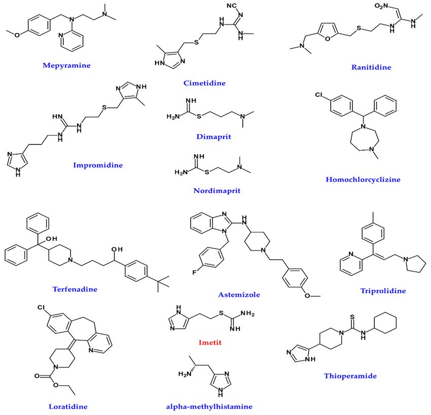
Int. J. Mol. Sci. 2021, 22, 8071                                                                                           2 of 28

                                   2. Skin Pigmentation
                                        Pigmentation of the skin is related to melanin. It occurs via the transfer of melanin
                                   produced from melanocytes to keratinocytes [8–10]. This process can be affected by a
                                   variety of factors. For example, pregnancy, Addison’s disease, and sun exposure can all
                                   make the skin darker [11–14]. Too little melanin in the body brightens skin. Vitiligo is a
                                   disease that causes soft light spots on the skin [15]. Albinism is a genetic condition that
                                   affects a person’s skin [16]. People with albinism may have lighter-than-normal skin tones.
                                   Infections, blisters, and burns can also make skin lighter [17,18].

                                   2.1. Types of Melanin
                                        In humans, melanin is a significant determinant of skin colour. Hair, pigmented tissue
                                   under the eye’s iris, and the vascular striatum in the inner ear are sources of melanin. In
                                   the brain, melanin-containing tissues include the medulla and pigment-bearing neurons
                                   within brainstem regions (such as trajectories). It also occurs in the reticular tissue of the
                                   adrenal gland [19,20].
                                        The melanocytes in the basal layer of the epidermis in the skin produce melanin.
                                   Humans have similar melanocytes in their skin, but some individuals and ethnic groups
                                   have melanocytes that produce varying amounts of melanin. Some humans have little or
                                   no melanin synthesis in their bodies; this is known as albinism [16,21].
                                        Different types of melanin differ in the proportions and binding patterns of certain
                                   component molecules, i.e., pheomelanin and eumelanin. The latter is the most abundant
                                   form of melanin in humans and is the most likely form of deficiency in albinism [22,23].

                                   2.1.1. Eumelanin
                                        Eumelanin polymers have long been thought to consist of polymers consisting of
                                   several cross-linked 5,6-dihydroxyindoles (DHI) and 5,6-dihydroxyindole-2-carboxylic
                                   acids (DHICA) [24,25]. Eumelanin is divided into brown eumelanin and black eumelanin.
                                   These two types of eumelanin have chemically different polymer bonding patterns. Small
                                   amounts of black eumelanin without other pigments cause grey hair. Small amounts
                                   of brown eumelanin without other pigments cause yellow (blond) hair. As humans get
                                   older, the human body continues to produce black eumelanin but stops producing brown
                                   eumelanin, resulting in grey hair, which is common in older people [26,27]. Of course,
                                   the grey of human hair may occur for other reasons. If you are interested, please see
                                   O’Sullivan’s recent review [28].

                                   2.1.2. Pheomelanin
                                        Pheomelanin comes in various colours, from yellow to red [29]. Pheomelanin is
                                   particularly concentrated in certain body parts such as the lips, nipples, glans penis, and
                                   vagina [30]. When small amounts of brown eumelanin, which can cause blond hair, are
                                   mixed with red pheomelanin, the result is orange hair, commonly referred to as “red” or
                                   “ginger” hair. As pheomelanin is also present in the skin, redheads often have a more
                                   pinkish skin tone. Please note that skin indicating pinkish tone can be attributed to skin
                                   vascularisation and blood, especially as a result of inoxification by carboxyhemoglobin
                                   following carbon monoxide poisoning. The chemical structure of pheomelanin differs from
                                   that of eumelanin in that its oligomeric form contains benzothiazine and benzothiazole
                                   units [31,32].

                                   2.1.3. Neuromelanin
                                        Neuromelanin (NM) is a dark polymer pigment found in a population of specific
                                   catecholaminergic neurons in the brain. Humans have the highest NM, which is present in
                                   small amounts in other primates, but not in many other species [33,34]. Although human
                                   NM has been shown to bind efficiently to transition metals such as iron and other potentially
                                   toxic molecules, its biological function is still unknown. It may play an essential role in
                                   apoptosis and related Parkinson’s disease [35,36]. In addition, trichochrome (formerly
Int. J. Mol. Sci. 2021, 22, x FOR PEER REVIEW 3 of 29
Int. J. Mol. Sci. 2021, 22, 8071 3 of 28
potentially toxic molecules, its biological function is still unknown. It may play an essen-
tial role in apoptosis and related Parkinson’s disease [35,36]. In addition, trichochrome
(formerly
called called trichosiderin)
trichosiderin) is a produced
is a pigment pigment produced in the
in the same same metabolic
metabolic pathwayspathways as
as eumelanin
eumelanin and pheomelanin, but unlike those molecules, it has a lower molecular weight.
and pheomelanin, but unlike those molecules, it has a lower molecular weight. It occurs in
It occurs
red in red hair [37,38].
hair [37,38].
2.2. Melanogenesis
2.2. Melanogenesis
2.2.1. Melanocytes
2.2.1. Melanocytes
Melanogenesis is
Melanogenesis is defined
definedas asthe
theproduction
productionof ofmelanin
melaninpigments.
pigments.These
Theseare aremost
mostof-often
ten produced
produced by cells
by cells calledcalled melanocytes
melanocytes [2,4,8].
[2,4,8]. Melanocytes
Melanocytes areare dendritic
dendritic cells
cells derivedfrom
derived
from
the the neuroectoderm
neuroectoderm [2,5,19,39,40].
[2,5,19,39,40]. Melanoblasts,
Melanoblasts, thethe premelanocytes,
premelanocytes, arepigmentless
are pigmentlesscells
cells derived
derived fromfrom embryonic
embryonic neural
neural crestcrest
cellscells [22,41,42].
[22,41,42].
After closing
After closingthe theneural
neuraltube
tube[23],
[23],melanocytes
melanocytes move
moveto various
to various parts of the
parts body
of the and and
body
develop into melanocytes and peripheral nervous systems, bone and
develop into melanocytes and peripheral nervous systems, bone and cartilage in the head,cartilage in the head,
and membrane
and membrane cells cellsininthe
theeye
eye[2,4,24].
[2,4,24].Melanocytes,
Melanocytes, differentiated
differentiated from melanoblasts,
from melanoblasts, are are
found mainly in the skin epidermis and hair follicles [26,29]. Tyrosinase (TYR),
found mainly in the skin epidermis and hair follicles [26,29]. Tyrosinase (TYR), tyrosinase- tyrosinase-
related protein
related protein1 1(TRP1),
(TRP1), DOPAchrome
DOPAchrome tautomerase
tautomerase or tyrosinase-related
or tyrosinase-related protein-2
protein-2 (TRP2),
(TRP2), premelanosome protein 17 (Pmel 17/gp1000), T cell 1 (MART-1),
premelanosome protein 17 (Pmel 17/gp1000), T cell 1 (MART-1), and microphthalmia- and microphthal-
mia-related
related transcription
transcription factor
factor (MITF)
(MITF) areare
thethe melanocyte-specific
melanocyte-specific markers.
markers.
The melanocytes of the skin are surrounded by keratinocytes (one melanocyte is sur-
The melanocytes of the skin are surrounded by keratinocytes (one melanocyte is
rounded by about 30–40 keratinocytes) [31,37,43] and transfer melanin pigments to
surrounded by about 30–40 keratinocytes) [31,37,43] and transfer melanin pigments to
keratinocytes via direct from melanocytes or keratinocytes [33,37,44]. The molecular struc-
keratinocytes via direct from melanocytes or keratinocytes [33,37,44]. The molecular
ture of melanin is very suitable for the absorption of ultraviolet (UV) and visible light,
structure of melanin is very suitable for the absorption of ultraviolet (UV) and visible light,
which prevents ultraviolet radiation (UVR) from sunlight [45,46]. Melanocytes are also
which prevents ultraviolet radiation (UVR) from sunlight [45,46]. Melanocytes are also
found in other body tissues, such as the central nervous and cardiovascular systems, the
found in other body tissues, such as the central nervous and cardiovascular systems, the
uvea of the eye, the cochlea and adipose tissue issue [47,48].
uvea of the eye, the cochlea and adipose tissue issue [47,48].
Melanocytes contain melanosomes, which are organelles similar to lysosomes. Mel-
anin Melanocytes
pigments arecontain melanosomes,
synthesised and stored which
beforearedistribution
organelles similar to lysosomes.
in surrounding Melanin
keratino-
pigments are(Figure
cytes [20,49] synthesised and stored before
1). Melanosomes require distribution
many specificin surrounding
enzymes and keratinocytes
structural pro- [20,49]
(Figure 1). Melanosomes require many specific enzymes and structural
teins to mature, which gives them the ability to produce melanin. TYR and TYRP2 are proteins to mature,
which gives
structural them the
proteins whereability to produce
Pmel17 and MART1melanin. TYR andfor
are essential TYRP2 are structural
the quantity proteins
and quality
where Pmel17 and MART1 are essential for the quantity and quality
of melanin [50,51]. AP-3, BLOC-1, and OCA2 play an important role in classifying and of melanin [50,51]. AP-3,
BLOC-1, and OCA2 play
trapping melanosomes [52–54]. an important role in classifying and trapping melanosomes [52–54].
Figure 1. Biosynthesis of melanin.
Figure 1. Biosynthesis of melanin.
2.2.2. Biosynthesis of Melanin
The most well-known function of melanin is to absorb and scatter UV rays to protect
DNA in cells from UV damage. UV irradiation (100–400 nm) is classified into three
categories: UVA (320–400 nm), UVB (280–320 nm), and UVC (100–280 nm). The UVC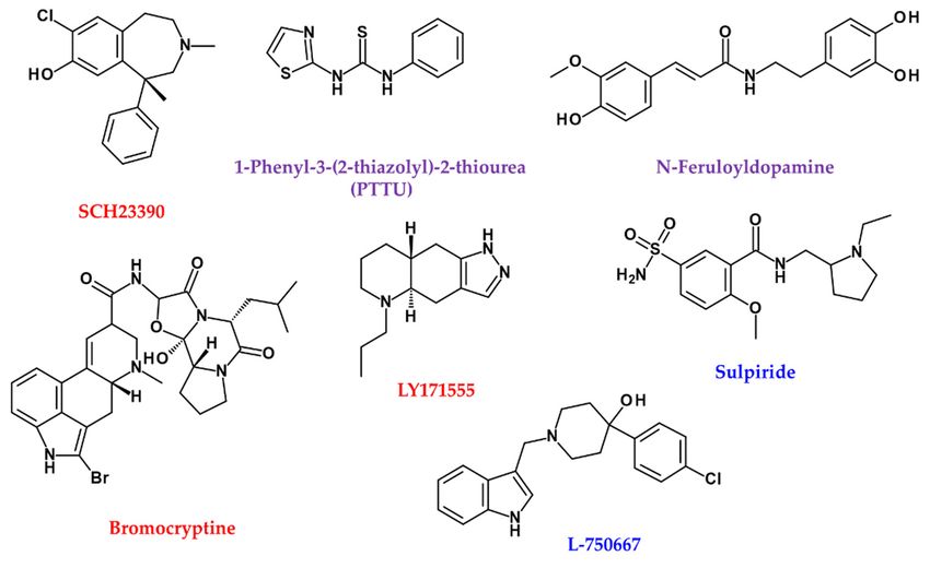
Int. J. Mol. Sci. 2021, 22, 8071                                                                                         4 of 28

                                   portion of the light spectrum is blocked by the atmosphere’s ozone layer and cannot reach
                                   the earth’s surface. Naturally, melanin forms a “cap” or parasol on the nucleus [22,55,56].
                                         Eumelanin is dark and much more polymerised, whereas pheomelanin contains
                                   sulphur and is lighter and less polymerised. Eumelanin and pheomelanin come from
                                   the same precursor, dopaquinone (DQ). DQ is formed by the oxidation of L-tyrosine
                                   through TYR (TYR; Figure 1). The first step in eumelanin production in DQ production
                                   involves the spontaneous cyclisation of quinones that produce cyclodopa. This cyclodopa
                                   rapidly generates one molecule each of DOPAchrome (DOPAC) and DOPA through a redox
                                   exchange with other DQs [26].
                                         DOPAC decomposes through decarboxylation at neutral pH to form DHI and DHICA
                                   in a ratio of 70:1 [24]. However, in the presence of TRP2, DOPAC is only converted
                                   to DHICA via tautomerisation [29]. Finally, DHI and DHICA are further oxidised and
                                   polymerised to form eumelanin (Figure 1).
                                         Pheomelanogenesis consists of several distinct stages at the monomer level. The
                                   first step involves the reductive addition of cysteine to DQ to generate 5-S-cysteine Dopa
                                   (5SCD) and 2-S-cysteine dopa (2SCD). The second step is the redox exchange reaction
                                   between cysteine monodopa (CD) and DQ to produce CD-quinone and DOPA. The third
                                   step is CD-quinone cyclisation via dehydration to form ortho-quinonimine (QI). QI is then
                                   converted to a 1,4-benzothiazine intermediate with or without decarboxylation. These
                                   intermediates are finally polymerised to pheomelanin [30] (Figure 1).

                                   2.3. Players in Melanogenesis
                                        The main enzyme involved in synthesising all types of melanin in tyrosine is TYR
                                   (EC 1.14.18.1), a single-chain type I membrane glycoprotein [31]. TYR require copper for
                                   catalytic function [37]. This oxidase is responsible for the hydroxylation of monophenols to
                                   quinone (monophenolase or cresolase activity, EC 1.14.18.1) and DOPA to dopaquinone
                                   (diphenolase or catechol oxidase activity, EC 1.10.3.1) in these ortho-diphenols
                                   (Figure 1) [15,57,58].
                                        TRP1 and TRP2 belong to the family of Cu++ /Zn++ metalloenzymes that feature a
                                   large amount of sequence homology, are expressed in melanocytes, and are mainly localised
                                   in unique organelles melanosomes, which play an essential role in melanogenesis. The two
                                   proteins share the same signal sequence comprising two cysteine-rich domains and one
                                   transmembrane domain [35].
                                        Human TRP1 is encoded by the TRP1 gene (human homologation of the mouse brown
                                   gene) found in chromosome 9 (9p23) [45]. A mature form of TRP1, also called gp75, is a
                                   protein per 75 kDa membrane transverse [46]. The TRP1 amino acid sequence has a 58 kDa
                                   MW. It undergoes glycosylation and acquisition of copper ionic bonds, producing a 17 kDa
                                   protein and converting it into a mature active form [22].
                                        The activity of the TRP1/gp75 enzyme in melanogenesis is still quite unclear despite
                                   43% homology with TYR [47]. A series of in vitro biochemical experiments suggested that
                                   TRP1/gp75 serves as catalase and weak dihydroxyindolcarboxylic acid oxidase [48,59].
                                   In addition to the unclear enzyme activity, TRP1/gp75 can help stabilise TYR and form
                                   xenobiotics in vivo. This complex formation can also weaken cytotoxicity caused by TYR
                                   reactions, preventing premature melanocyte death [49].
                                        TRP2, also called dopachrome tautomerase (DCT, EC 5.3.3.12), promotes keto-enol
                                   tautomerisation of dopachrome to the relatively more stable intermediate DHICA [29].
                                   In another pathway, spontaneous decarboxylation of dopachrome produces DHI. This
                                   metabolic pathway, involving TYR specific to melanocytes, leads to the synthesis of black
                                   eumelanin [31,60] (Figure 1).
                                        All three melanin enzymes (TYR, TRP1, and TRP2) that play an important role in
                                   melanogenesis are transcription targets of the microphthalmia-associated transcription
                                   factor (MITF). The promoters of TYR, TRP1, and TRP2 are activated by MITF. MITF plays
                                   a key role in mammalian pigmentation, which is regulated by environmental factors,
                                   including UV, and factors secreted by keratinocytes, fibroblasts, and other cells. MITF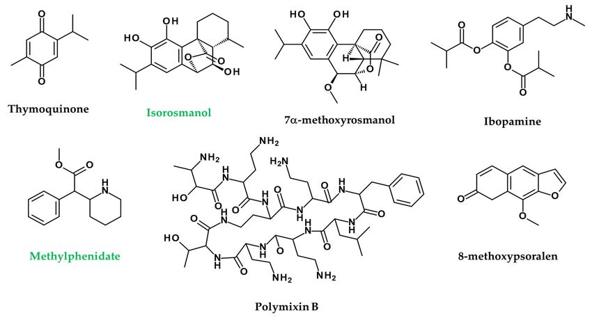
Int. J. Mol. Sci. 2021, 22, 8071                                                                                            5 of 28

                                   controls melanogenesis and differentiation, density, proliferation, and cell death through
                                   various pathways and mechanisms [4,61].
                                        The activation of PKC-β in the signalling pathway of melanogenesis is involved in
                                   regulating TYR activity by phosphorylating serine residues in the cytoplasmic domain of
                                   TYR [22].
                                        Several factors released from surrounding cells, such as keratinocytes, fibroblasts,
                                   and nerve cells, drive melanin production through receptors. Several receptors have
                                   been reported to be involved in melanogenesis. Progress in this area has been extensively
                                   reviewed by eminent researchers [4,24,37]. Recent advances are well explained by Park [22].

                                   3. Pigmentation Abnormality in Skin Disease
                                   3.1. Vitiligo and Albinism
                                         Vitiligo is the loss of melanocytes, resulting in white skin spots. It affects up to 2% of
                                   people [15]. The cause of vitiligo is unknown. However, it can be accompanied by an attack
                                   of the immune system on the cells that produce skin pigment melanin (melanocytes) [62].
                                         Vitiligo tends to develop either within a family or spontaneously in an individual.
                                   Vitiligo can occur with certain other diseases. Vitiligo is associated with autoimmune
                                   diseases (when the body attacks its tissues), among which thyroid disease is the most
                                   common. It is most closely related to hyperthyroidism (especially when due to Graves’
                                   disease) and hypothyroidism (especially when due to Hashimoto’s thyroiditis) [63]. People
                                   with diabetes, Addison’s disease, and pernicious anaemia are also somewhat more likely
                                   to develop vitiligo [64]. However, the relationship between these disorders and vitiligo is
                                   not clear.
                                         Melanocytes exist in the leptomeninges of the human brain, especially over the ven-
                                   trolateral surfaces of the medulla oblongata [65]. These leptomeningeal melanocytes also
                                   appear to be involved in vitiligo. Aseptic meningitis, observed in Harada syndrome, is
                                   likely due to the destruction of leptomeningeal melanocytes [66]. This observation appears
                                   to be an interesting example of the presence of a neuro-immune axis in melanocytes.
                                         Oculocutaneous albinism is a rare hereditary disorder in which little or no skin
                                   pigment melanin is formed [16]. Only the skin, hair, and eyes, or sometimes only the eyes,
                                   are affected [16]. There are four main types of OCA-OCA1 (TYR), OCA2 (OCA2), OCA3
                                   (TRP1), and OCA4 (SLC45A2) [67]. In addition, albinism is observed as a symptom of
                                   various genetic diseases. In the case of the WS2 variant of Waardenburg syndrome (WS1-4),
                                   the MITF gene exists as a variant, causing albinism [68]. In addition to causing pigmentation
                                   of the skin (abnormally small amounts of melanin) or depigmentation (complete loss of
                                   pigmentation), it causes decreased vision, misalignment of the eyes (strabismus), and
                                   involuntary eye movements (nystagmus) [69]. A type of albinism, called albino, affects the
                                   eyes but usually does not affect the skin and hair. A different kind of albinism occurs with
                                   bleeding disorders [70].

                                   3.2. Hyperpigmentation
                                        Hyperpigmentation is a darkening of the skin, mainly caused by an abnormally large
                                   skin pigment melanin [71]. When exposed to sunlight, special skin cells increase melanin,
                                   causing the skin to darken or tan. In some people with light skin, certain melanocytes
                                   respond to sunlight to produce more melanin than others-this uneven production of
                                   melanin results in pigment spots known as freckles. Freckle tendencies occur in families.
                                   Factors other than sunlight can increase melanin in spots (topical) or large areas of the
                                   skin [72]. In rare cases, the skin darkens because of substances other than melanin.
                                        Local hyperpigmentation can be caused by skin damage, skin inflammation, reaction
                                   to sunlight, and abnormal skin growth [73]. Hyperpigmentation can also occur after
                                   injuries such as cuts and burns, or inflammation caused by disorders such as acne and
                                   lupus [74–76].
                                        Hyperpigmentation is often accompanied by other diseases or symptoms. For ex-
                                   ample, hyperpigmentation can occur in conjunction with blemishes, freckles, black spots,
Int. J. Mol. Sci. 2021, 22, 8071                                                                                             6 of 28

                                   and caffeore spots (flat spots or brown spots), as well as abnormal skin growths such
                                   as melanomas [77,78]. People with a disorder called acanthosis nigricans develop dark,
                                   thickened skin in the armpits, nape of the neck, and wrinkles of the skin [79]. Melanocytosis
                                   can be a symptom of diabetes [80].
                                        Lentigines (commonly referred to as age spots or liver spots (not related to liver
                                   problems)) are flat, tan to brown oval spots on the skin [81]. There are two types of
                                   lentigines, namely, solar and non-solar lentigines. The solar-type is caused by sun exposure
                                   and is the most common type [81]. It occurs most often on the face and the back of the
                                   hand exposed to the sun. It usually first appears in middle age and increases in number
                                   with age. Lentigines are noncancerous (benign), but people with them may have a higher
                                   risk of developing melanomas.
                                        Non-solar lentigines are not due to sun exposure. The non-solar type sometimes
                                   occurs in people with certain rare genetic disorders, such as Peutz–Jeghers syndrome
                                   (characterised by many black spots on polyps in the lips and intestines), pigmentation, and
                                   multiple black spot syndrome (LEOPARD syndrome) [82,83].
                                        Extensive hyperpigmentation can be caused by hormonal changes, internal diseases,
                                   drugs, and exposure to heavy metals [19,84]. Hormonal changes can increase melasma in
                                   pregnancy or the use of hormonal contraceptives and hyperpigmentation in Addison’s
                                   disease [11,85,86]. A liver disease called primary biliary cholangitis (previously called
                                   primary biliary cirrhosis) can also increase melanin production [87].
                                        Hyperpigmentation can be caused by drugs, including amiodarone, hydroquinone,
                                   antimalarial drugs, tetracycline antibiotics, phenothiazine, cancer chemotherapy drugs,
                                   and tricyclic antidepressants. Heavy metals (such as silver, gold and mercury, which can be
                                   toxic) can also cause hyperpigmentation [88]. The pigmentation can be purple, bluish-black,
                                   tan, or shades of blue, silver and grey, depending on the drug or metal, and is concentrated
                                   on the skin [89]. In addition to the skin, the teeth, nails, the whites of the eyes (sclera) and
                                   the inside of the mouth (mucous membranes) may be discoloured [90].

                                   4. Amines Neurotransmitters and Their Receptors in Skin Pigmentation Diseases
                                   4.1. Role of Neurotransmitters and Their Receptors in Melanogenesis
                                   4.1.1. Acetylcholine
                                             Acetylcholine (ACh) is an organic compound that acts as a neurotransmitter in the
                                       brain and body and is usually a chemical message released by nerve cells to signal other
                                       cells, such as neurons, muscle cells, and glands (Figure 2) [91]. Acetylcholine receptors
Int. J. Mol. Sci. 2021, 22, x FOR PEER REVIEW                                                                              7 of 29
                                       (AChR), which recognise and bind the neurotransmitter acetylcholine, are integrated
                                       membrane proteins [92].

                                                 Figure 2.
                                                 Figure    Amine neurotransmitters.
                                                        2. Amine neurotransmitters.

                                        There are
                                        There   aretwo types
                                                     two      of acetylcholine
                                                           types               receptors:
                                                                   of acetylcholine       nicotinenicotine
                                                                                      receptors:   (N) and muscarine
                                                                                                            (N) and (M)AChRs.
                                                                                                                      muscarine
                                   MAChR    is located at the neuromuscular     junction that causes skeletal
                                   (M)AChRs. MAChR is located at the neuromuscular junction that causes       muscle contraction
                                                                                                                skeletal muscle
                                   contraction through the end-plate potential (EPP). NAChR causes depolarisation in the
                                   autonomous ganglia, causing post-neural impulses (Figure 3) [91].
Figure 2. Amine neurotransmitters.
Int. J. Mol. Sci. 2021, 22, 8071 7 of 28
There are two types of acetylcholine receptors: nicotine (N) and muscarine
(M)AChRs. MAChR is located at the neuromuscular junction that causes skeletal muscle
contraction
through thethrough
end-platethe end-plate
potential potential
(EPP). NAChR(EPP). NAChR
causes causes depolarisation
depolarisation in the
in the autonomous
autonomous ganglia,
ganglia, causing causingimpulses
post-neural post-neural impulses
(Figure (Figure 3) [91].
3) [91].
Figure
Figure 3.
3. Receptors
Receptors of
of amine
amine neurotransmitters.
neurotransmitters.
NAChR
NAChR causescauses thethe release
release of catecholamines
catecholamines from the adrenal medulla and causes
site-specific
site-specific excitation
excitation or or inhibition
inhibition in the brain. M and NAChRs are joined joined by Na++ and
by Na
Ca 2+
Ca2+ channels,
channels,but butNAChR
NAChRisisalso connected
also connected bybyadditional
additionalK+ K +
channels.
channels. NAChRs
NAChRsare lig-
are
ligand–gate + +
and–gate ionion channels,
channels, andand the diffusion
the diffusion of and
of Na + Na Kand+ K across
across the receptors
the receptors causescauses
depo-
depolarisation, i.e., endplate potentials, opening voltage-gated + channels to allow firing
larisation, i.e., endplate potentials, opening voltage-gated Na+Nachannels to allow firing of
of action
action potentials
potentials and,
and, potentially,
potentially, muscle
muscle contraction
contraction [91].
[91].
AChR, in
AChR, in contrast,
contrast, belongs
belongs toto aa superfamily
superfamily of of G-protein-binding
G-protein-binding receptors
receptors that
that acti-
acti-
vate other ionion channels
channels through
through aa second
second messenger
messenger cascade
cascade rather
rather than
than ion
ion channels
channels [93].
[93].
MAChRs activate G-proteins
MAChRs activate G-proteins when when they bind to an extracellular ligand, ACh. G-protein
alpha activates
alpha activates thethe guanylate
guanylate cyclase
cyclase (suppressing
(suppressing the effect of intracellular cAMP), while
the beta-gamma subunit activates
the beta-gamma subunit activates the K the K++ channel
channelto tohyperpolarise
hyperpolarise the the cell.
cell.
In skin cells, including keratinocytes and MCs, ACh synthesis is catalysed by choline
acetyltransferase (ChAT), and degradation is catalysed by acetylcholine esterase (AChE) [94].
The vesicular ACh transporter (VAChT) is responsible for the transport of ACh and choline
using a proton electrochemical gradient generated by a vacuole-type H+ ATPase and
exchanges two luminal protons for one cytoplasmic ACh or choline [95].
Several reports suggest the role of ACh, AChRs, and enzymes related to ACh metabolism
in melanogenesis. That is, sunlight, for example, promotes the release of ACh from
keratinocytes in the skin, and this increased ACh and AChE inhibitor also suppressed
the increased melanogenesis by light [96]. These results suggest the possibility that ACh
inhibits melanogenesis and the possibility that AChE may play an important role in the
promotion of melanogenesis.
MITFs upregulate AChE expression during melanin production in murine melanoma
cells [97]. This report states that AChE is expressed in melanocytes and melanoma cells,
and the tetrameric (G4) form is the main AChE isoform in these cells. During melanin
production in B16F10 murine melanoma cells, AChE levels decrease significantly. In
contrast, ACh stimulates the release of alpha melanocyte-stimulating hormones (α-MSH)
from frog pituitary melanotropes through the activation of M and NAChRs [98]. MAChRs
mediate nerve-induced pigmentation in free catfish melanophores [99]. The quick changesInt. J. Mol. Sci. 2021, 22, 8071 8 of 28
in skin darkness in lower vertebrates are due to melanosomes concentration or dispersion in
the cytoplasm of melanophores, similar to the transfer of melanosomes from melanocytes to
the keratinocytes. Despite their neurogenic character, they cannot be considered a response
of the melanogenic apparatus to neurotransmitter stimuli, as the net content of melanin
does not change.
Normal human skin melanocytes express the MlR, M2R, M3R, M4R, and M5R sub-
types of MAChRs in the cell membrane, which controls concentrations of intracellular
free Ca2+ . This can play an essential physiological role in melanocyte behaviour and skin
pigmentation [100]. However, there was no result on the role of receptors in melanogenesis
in this report. In the following paper, M4R KO mice did not exhibit hair follicle melanogen-
esis and failed to produce pigmented hair shafts [101]. This suggest that M4R is involved
in hair follicle pigmentation.
ACh seems to inhibit melanogenesis, and thus AChE, which induces the degradation
of ACh, appears to be involved in the promotion of melanogenesis. Although various
receptors mediating the action of ACh exists, there is not much information related to
the involvement of these receptors in human melanogenesis. However, M4R seems to be
involved in melanogenesis, and M4R is also involved in the pigmentation phenomenon
in catfish melanophores, as mentioned above. Therefore, it seems necessary to study the
role of AChR in pigmentation. Compared to the results of studies on the role of AChE
in pigmentation, the presence of ChAT in melanotrope cells has been reported. Still,
its presence in melanocytes does not seem to have been reported [102]. Therefore, the
possibility of the existence of ChAT in melanocytes and a study on its role in melanogenesis
are also needed.
4.1.2. Dopamine
Dopamine (DA, contraction of 3,4-dihydroxyphenethylamine) is a neurotransmitter
that plays several essential roles in the brain and body [103]. Dopamine comprises ap-
proximately 80% of the catecholamine content in the brain [104]. Dopamine is an amine
synthesised by removing carboxyl groups from molecules of L-DOPA, a precursor chemical
synthesised in the brain and kidneys [105].
The actions of dopamine are mediated by five specific cell surface receptors belong-
ing to G-protein-coupled receptor families, D1-like receptors, and D2-like receptors [106].
Dopamine receptors D1 (DRD1) and D5 (DRD5) are members of the D1-like subfamily,
while DRD2, DRD3, and DRD4 are members of the D2-like receptor subfamily. G-protein-
binding dopamine receptors mediate all the physiological functions of the catecholamine
neurotransmitter dopamine, from voluntary exercise and compensation to hormonal con-
trol and hypertension [107].
The DRD1 and DRD2 are expressed in human skin [108]. α-MSH competes for the
action of DRD1 agonists, SCH 23390 (Figure 4) [109]. DA resulted in reduced melanocyte
survival at concentrations ranging from 0.01 to 100 M (0.1 and 1 µM, p < 0.05; 10 µM, p < 0.05;
100 µM, p < 0.01). Furthermore, DA-induced melanocyte apoptosis has been demonstrated
to increase the ratio of sub-G1 cells from 7.71 ± 0.28% (control) to 12.22 ± 1.05% (0.1 µM
DA) (p < 0.005) and N-acetylcysteine (NAC), which reverses DA-induced apoptosis [110].
DA treatment induces ROS production, which can be prevented by pre-treatment with NAC.
Although DA induces apoptosis of melanocytes, DA can produce melanin in vitro, which
is enhanced by pro-oxidant hydrogen peroxide (EC50 = 500 µM) and Fe3+ but reduced by
antioxidants such as ascorbates (IC50 = 10 µM) and glutathione (GSH; IC50 = 5 µM) [111].strated to increase the ratio of sub-G1 cells from 7.71 ± 0.28% (control) to 12.22 ± 1.05% (0.1
μM DA) (p < 0.005) and N-acetylcysteine (NAC), which reverses DA-induced apoptosis
[110]. DA treatment induces ROS production, which can be prevented by pre-treatment
with NAC. Although DA induces apoptosis of melanocytes, DA can produce melanin in
Int. J. Mol. Sci. 2021, 22, 8071
vitro, which is enhanced by pro-oxidant hydrogen peroxide (EC50 = 500 μM) and Fe3+9 of but
28
reduced by antioxidants such as ascorbates (IC50 = 10 μM) and glutathione (GSH; IC50 = 5
μM) [111].
Figure 4.
Figure Chemical structures
4. Chemical structures of
of agonist
agonist (red),
(red), antagonist
antagonist (blue),
(blue), and
and enzyme
enzyme inhibitors
inhibitors or
or others
others
(purple) related to DA neurotransmitters.
(purple) related to DA neurotransmitters.
Therefore, derivatives containing agonists and antagonists of DA and its metabolism
Therefore, derivatives containing agonists and antagonists of DA and its metabolism
regulators are also expected to affect melanocytes. While N-nicotinoyl dopamine does
regulators are also expected to affect melanocytes. While N-nicotinoyl dopamine does not
not inhibit TYR and melanin synthesis in B16F10 mouse melanoma cells, it inhibits
inhibit TYR and melanin synthesis in B16F10 mouse melanoma cells, it inhibits melano-
melanosome transfer in normal human melanocyte–keratinocyte co-culture systems [112].
some transfer in normal human melanocyte–keratinocyte co-culture systems [112]. 1-Phe-
1-Phenyl-3-(2-thiazolyl)-2-thiourea (PTTU) is a well-characterised dopamine β-hydroxylase
nyl-3-(2-thiazolyl)-2-thiourea (PTTU) is a well-characterised dopamine β-hydroxylase in-
inhibitor that suppresses degenerative neurological diseases caused by 6-hydroxydopamine
hibitor that suppresses degenerative neurological diseases caused by 6-hydroxydopamine
(Figure 4) [113]. Interestingly, PTTU also reduces the enzyme activity and stability of TYR
(Figure 4) [113]. Interestingly, PTTU also reduces the enzyme activity and stability of TYR
in normal human epidermal melanocytes [113]. N-Feruloyldopamine inhibits human TYR
in normal human epidermal melanocytes [113]. N-Feruloyldopamine inhibits human TYR
with higher efficacy than arbutin (Figure 4) [114].
with higher efficacy than arbutin (Figure 4) [114].
Bromocriptine, a dopamine agonist that blocks α-MSH secretion, inhibits melanin
Bromocriptine, a dopamine agonist that blocks α-MSH secretion, inhibits melanin
production in hair follicular melanocytes of adolescent C3H-HeAvy mice (Figure 4) [115].
production
Specific DRD2 in hair follicular
agonist melanocytes
LY171555 of adolescent
also inhibits C3H-HeAvy
TYR activity mice
on the skin’s (Figurein4)a[115].
explants dose-
Specific DRD2 agonist LY171555 also inhibits TYR activity on the skin’s explants
related manner, and its effect is blocked by sulpiride, the DRD2 antagonist (Figure 4) [115]. in a dose-
related
Onmanner,
the otherand its effect
hand, DRD4 is blocked
antagonistby L-750,667,
sulpiride, the DRD2
inhibits antagonist
melanin (Figure through
production 4) [115].
On the other hand, DRD4 antagonist L-750,667, inhibits melanin production
transcriptional downregulation of MITF via ERK signals (Figure 4) [116]. When the melanin through
transcriptional
precursor molecule downregulation
DHI (2C) is of MITF via ERK
methylated signals (Figure 4) [116]. When(COMT),
by catechol-O-methyltransferase the mela- it
nin precursor molecule DHI (2C) is
cannot be incorporated into melanin [117]. methylated by catechol-O-methyltransferase (COMT),
it cannot be incorporated
Therefore, the resultsinto melanin
so far indicate[117].
that DA acts in the direction of inhibiting melano-
Therefore,
genesis. the results
In particular, so far indicate
although the action that
of DA
DA acts in the has
receptors direction of inhibiting
not been verified atmel-
the
anogenesis. In particular, although the action of DA receptors has not been
genetic and molecular level, it seems certain that DRD2 activation inhibits melanogenesisverified at the
and DRD4 activation promotes melanogenesis. Since many compounds with DA moieties
affect melanogenesis, it is clear that DA plays an important role in regulating melanogenesis.
However, recent research results show that DA can promote melanin production in vitro,
but it is not yet confirmed whether this can happen even under intracellular conditions.
4.1.3. Epinephrine and Norepinephrine
Norepinephrine (NE) or epinephrine (EP) is an organic chemical of the family cate-
cholamine that functions as a hormone and neurotransmitter in the brain and body [118].
NE release is the lowest in sleep, increases when awake, and reaches even higher levels
during the so-called fight-or-flight reactions in stressful or dangerous situations [119].
EP is produced in NE by N-methylation and is catalysed by phenylethanolamine N-
methyltransferase (PNMT) using methyl donor S-adenosyl methionine [120]. EP is usually
made by a small number of neurons in the adrenal glands and medulla oblongata. EP plays
a vital role in the fight-or-flight response by enhancing blood flow to muscles, increasingInt. J. Mol. Sci. 2021, 22, 8071 10 of 28
heart output by acting on sinoatrial nodes, and increasing pupil dilatation and blood
glucose levels [121].
Adrenergic receptors (ARs), or adrenaline receptors, are a type of G-protein-binding
receptor targeting many catecholamines, such as NE and EP produced by the body [122].
There are nine ARs: α1A, α1B, α1D, α2A, α2B, α2C, β1, β2, and β3 [123]. Human
epidermal melanocytes express α1-ARs and β2-ARs [124,125], autocrine catecholamine
biosynthesis and β-adrenergic receptor signals promote pigmentation in human epidermal
melanocytes [126]. This paper reports that human melanocytes express functional β2-AR
(4230 receptors per cell) of Bmax at 129.3 and 3.19 nM of Kd but lack β1-AR expression. On
the other hand, activation of β1-ARs is not affected [124]. β1- and β2-ARs are expressed in
tissues of benign melanomas, atypical naevi, and malignant melanomas, and their expres-
sion was significantly higher in malignant tumours [127]. The impact of epinephrine on
melanin pigment has been observed not only in mammals but in other animals too. An
epinephrine-treated frog showed a significant colour change from brown to yellow within
5 min [128]. β-ARs are involved in producing proopiomelanocortin-derived peptides and
prolactin induced by histamine in rats [129].
Abundant NE as an exogenous factor causes direct damage to differentiated melanocytes
in neural crest cells and suppresses crosstalk between c-kit receptors within melanocytes
and the stem cell factor of keratinocytes [130]. NE is secreted into the epidermal microenvi-
ronment by nerve endings or keratinocytes, directly inhibiting TYR activity of melanocytes,
causing mitochondrial calcium ion malabsorption and free radical production [131].
The secretion of NE due to stress differentiates most of the hair pigment melanocyte
stem cells (MeSCs) into melanocytes, leading to the rapid depletion of the pigment-
producing cells in the hair follicle to mature melanocytes undergoing apoptosis, and
the MeSCs are eventually eliminated. Blocking NE sympathetic nerve system (SNS) signals
preserves hair pigmentation in stressed animals. Increased SNS activation has a severe
irreversible effect on homeostasis in greying of hair and Alzheimer’s disease [132].
NE seems to act to inhibit melanogenesis and, in the case of EP, to promote melanogenesis,
suggesting that PNMT, an enzyme that catalyses the conversion of NE to EP, may influence
melanogenesis. However, since this enzyme is not detected well in melanocytes, studies on
melanogenesis mainly by PNMT likely require co-culture with keratinocytes [126,133].
4.1.4. Gamma-Aminobutyric Acid
Gamma-aminobutyric acid (GABA) is an inhibitory neurotransmitter that acts on
the central nervous system (CNS) of mammals. GABA is responsible for controlling
nerve excitation in the nervous system. In humans, GABA directly regulates muscle
condition [134]. Although it is difficult to regard it as a neurotransmitter of amines such as
dopamine and serotonin (5-HT), GABA must be mentioned in this review because it has an
amino group.
GABA receptors are a family of receptors that respond to the neurotransmitter GABA,
a major inhibitory neurotransmitter in the mature vertebrate CNS. There are two types of
GABA receptors: GABAA and GABAB . GABAA receptors are ligand–gate ion channels
(also known as ionotropic receptors). GABAB receptors are G-protein-coupled receptors,
also called metabotropic receptors. GABAA receptor exists in the epidermal keratinocytes
and accelerates cutaneous barrier recovery [135,136]. Although reports of direct expression
of GABA receptors in human skin melanocytes have been challenging to find, the action
of GABA receptors in melanocytes has been reported in model animals such as larval
zebrafish [137]. For example, inhibition of GABAA function, especially GABAA ρ subtype,
induces excessive melanocyte population in larval zebrafish [137].
By enhancing GABAA -mediated anion transport, benzodiazepines depolarise melanoma
cells and impair their viability. Benzodiazepine alone reduces tumour growth in vivo and
enhances the effect of radiation therapy and α-PD-L1 anti-tumour activity (Figure 5). The
combination of benzodiazepine, radiotherapy, and α-PD-L1 results in almost completedirect expression of GABA receptors in human skin melanocytes have been challenging
to find, the action of GABA receptors in melanocytes has been reported in model animals
such as larval zebrafish [137]. For example, inhibition of GABAA function, especially
GABAA ρ subtype, induces excessive melanocyte population in larval zebrafish [137].
By enhancing GABAA-mediated anion transport, benzodiazepines depolarise mela-
Int. J. Mol. Sci. 2021, 22, 8071 11 of 28
noma cells and impair their viability. Benzodiazepine alone reduces tumour growth in
vivo and enhances the effect of radiation therapy and α-PD-L1 anti-tumour activity (Fig-
ure 5). The combination of benzodiazepine, radiotherapy, and α-PD-L1 results in almost
complete regression of treated
regression tumours
of treated andand
tumours a powerful
a powerfulabscopal effect
abscopal mediated
effect by by
mediated in-increased infil-
creased infiltration of of
tration multifunctional CD8
multifunctional + +
CD8T cells [138].
T cells [138].
Figure 5. Chemical structures
Figure 5. Chemicalof structures
agonist (red), antagonist
of agonist (blue),
(red), and enzyme
antagonist (blue),inhibitors or others
and enzyme (purple)
inhibitors or related
others to GABA,
glutamate neurotransmitters.
(purple) related to GABA, glutamate neurotransmitters.
Midazolam
Midazolam prevents cancerprevents
metastasiscancer metastasis by by
by hyperglycaemia hyperglycaemia by inhibiting intracel-
inhibiting intracellular
events and subsequent vascular leakage from the lungs of diabetic mice through GABAA mice through
lular events and subsequent vascular leakage from the lungs of diabetic
GABA
(Figure 5) [139]. A (Figure
Diazepam 5) [139].
improves Diazepam improves
melanogenesis melanogenesis
and melanocyte dendriticand
andmelanocyte
mela- dendritic
and melanosome transport through the peripheral benzodiazepine
nosome transport through the peripheral benzodiazepine receptor/cAMP/PKA pathway receptor/cAMP/PKA
pathway
(Figure 5) [140]. (Figure 5)have
Benzodiazepines [140]. Benzodiazepines
high-affinity bindinghave
sites high-affinity bindingpro-
and induce melanin sites and induce
melanin production
duction in B16/C3 melanoma cells [141].in B16/C3 melanoma cells [141].
It seems
It seems that there are that there are
not many not many
studies on thestudies
effect ofonGABA
the effect of GABA on melanogenesis.
on melanogenesis.
However, these results suggest that GABA promotes melanogenesis. GABA receptor-
However, these results suggest that GABA promotes melanogenesis. GABA receptor-re-
related compounds seem to suppress melanoma progression. In the future, studies on
lated compounds seem to suppress melanoma progression. In the future, studies on which
which receptors are involved in melanogenesis and how they are involved in melanogenesis
receptors are involved in melanogenesis and how they are involved in melanogenesis
seem to have to be verified at the molecular level, beyond the studies using compounds.
seem to have to be verified at the molecular level, beyond the studies using compounds.
Additionally, research on how GABA is altered in skin pigmentation disorders and whether
Additionally, research on how GABA is altered in skin pigmentation disorders and
related compounds can have therapeutic effects in skin pigmentation disorders may also
whether related compounds can have therapeutic effects in skin pigmentation disorders
be needed.
may also be needed.
4.1.5. Glutamate
Glutamic acid is one of the typical neurotransmitters in the CNS that primarily acts as
an excitatory synaptic, with several types of receptors reported in the nervous system [142].
Glutamate, like GABA, has an amino group, so it was included in this review.
GRM1 is part of the glutamate receptor family, which is divided into two main groups:
ionic glutamate receptors (iGluR) and metabolic glutamate receptors (mGluR). mGluR is
a seven-membrane spanning-domain G-protein coupled-receptor. The combined recep-
tors are further subdivided into three groups based on sequence-wise and downstream
signals. GRM1 and GRM5 belong to groups I mGluR. GRM1 expression was not detected
in melanocytes, while GRM5 expression was detected in melanocytes. iGluR contains
glutamate–gate ion channels, such as N-methyl-D-aspartate (NMDA) type (NMDAR) or
α-amino-3-hydroxy-5-methyl-4- isoxazolepropionic acid type receptors (AMPAR).
GRM1 mediates melanocyte transformation to melanoma through transactivation of
insulin-like growth factor 1 receptors [143]. Glutamate receptors such as GRM2 (AMPAR)
and NMDAR2A in human melanocytes regulate the expression of MITF [144]. After 24 h
treatment with the AMPAR inhibitor CFM-2 at 50 µM, the expression of the key melanocyte
differentiation and proliferation factor, MITF, was drastically reduced.
Expression of GRM1 occurs in 60% of human melanomas and cytoplasm but not in be-
nign or normal human melanocytes, suggesting that GRM1 may be involved in melanoma
formation [126,127]. Expression of GRM5 promotes melanomas in transgenic mice [145].
GRM6 signalling increases TRPM1 calcium channel function and enhances melanin produc-
tion in human melanocytes [146]. Stimulation of NMDAR enables filopodia transmission
and promotes direct morphological effects on melanocytes to induce melanosome transfer.
Whereas 100 µM NMDAR antagonist, MK-801, causes intracellular β-tubulin redistribu-Int. J. Mol. Sci. 2021, 22, 8071 12 of 28
tion and affects the delivery between filopodia between melanocytes and keratinocytes
(Figure 5) [147].
Heterotetrameric NMDARs are cationic channels that are primarily permeable to Ca2+ .
The NR1 and NR3 subunits bind to glycine, while the NR2 subunit binds to glutamate
for full activation [148]. The NMDAR complex, consisting of NR1-NR3B, is present in the
nucleus of melanoma cells. This phenomenon was not observed in melanocytes.
In addition, the NMDAR appears to be involved in the activation of TYR and the
promotion of melanin synthesis in the ink gland of Sepia officinalis, a cuttlefish [149]. The role
of the glutamate transporter rather than the receptor has also been reported. For example,
solute carrier family 7 member 11 (SLC7A11) is a cysteine/glutamate exchanger also known
as xCT and plays an essential role in synthesising pheomelanin. MITF, MC1R, SLC24A5,
Agouti, and CREB1 expression were significantly downregulated after the suppression
of SLC7A11 [149]. From these results, it seems clear that glutamate neurotransmitter and
related signalling mechanisms are involved in melanogenesis.
4.1.6. Histamine
Histamine (HA) is well known as a substance involved in the local immune response.
Still, it also regulates the physiological functions of the intestine and acts as a neurotrans-
mitter for the brain, spinal cord and uterus [150,151].
HA exhibits its biological effects by binding to and activating four different G-protein-
coupled receptors (GPCRs) (H1 R, H2 R, H3 R, and H4 R) [152]. H1 R and H2 R exist in ker-
atinocytes and H1 R in fibroblasts [153]. H1 R and H2 R exist in human melanocytes and
melanoma cells [154].
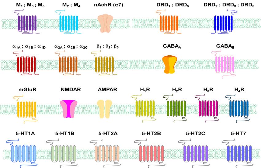
The possible involvement of HA receptors in melanogenesis has been suggested to take
place through the agonists and antagonists of each HA receptor. That is, it was reported that
H1 R antagonist (mepyramine) and H2 R antagonists (cimetidine, ranitidine, impromidine)
increased TYR activity in cultured human melanoma cells, whereas H2 R agonists (dimaprit,
nordimaprit) decreased the activity (Figure 6) [155]. It could be expected that these receptors
would inhibit the activation of melanogenesis, but subsequent studies showed opposite
results. The H1 R antagonist homochlorcyclizine (HC) dose-dependently inhibited melanin
production in B16 cells stimulated by the α-MSH or 3-isobutyl -1-methylxanthine (IBMX)
(Figure 6) [156]. H1 R antagonists such as terfenadine, astemizole, and triprolidine induce
apoptosis in all four melanoma cell lines (Figure 6) [157]. Significantly, H1 R antagonist
therapy did not adversely affect the viability of normal human melanocytes and mouse
fibroblasts at the same dose and exposure time [157]. H1 R antagonist loratadine inhibits
melanin production via downregulating MITF in human melanocytes (Figure 6) [158].
Similarly, H2 R agonists have been reported to decrease the activity of TYR, but HA
induces melanogenesis and morphological changes by activating G-protein kinase A
through H2 R in normal human melanocytes [155,159]. HA had a more significant ef-
fect on melanocytes proliferation than melanin production [160]. This occurred through
H2 R with complex signals for ERK, CREB, and Akt activation, stimulating melanocyte
migration [160]. H2 R-mediated growth differentiation factor-15 (GDF-15) expression is in-
volved in histamine-induced melanogenesis. Gene silencing of GDF-15 inhibited histamine-
induced proliferation, melanogenesis, TYR activity, and chemotropic migration in SK-MEL-
2 cells. Histamine-induced expression of TYR, TRP1, and TRP2 was also inhibited by
growth differentiation factor-15 gene silencing [41].
TYR was inhibited by the H3 R agonist imetit but not by alpha-methylhistamine or the
H3 R antagonist thioperamide (Figure 6) [161].
HA partially inhibits proliferation through the stimulation of H4 R and induces cellular
senescence and melanin production in 1205 Lu metastatic melanoma cells expressing
H4 R [162].pendently inhibited melanin production in B16 cells stimulated by the α-MSH or 3-isobu-
tyl -1-methylxanthine (IBMX) (Figure 6) [156]. H1R antagonists such as terfenadine, aste-
mizole, and triprolidine induce apoptosis in all four melanoma cell lines (Figure 6) [157].
Significantly, H1R antagonist therapy did not adversely affect the viability of normal hu-
Int. J. Mol. Sci. 2021, 22, 8071 man melanocytes and mouse fibroblasts at the same dose and exposure time [157]. H113 R of 28
antagonist loratadine inhibits melanin production via downregulating MITF in human
melanocytes (Figure 6) [158].
Figure 6.
Figure Chemicalstructures
6.Chemical structuresofof agonist
agonist (red),
(red), andand antagonist
antagonist (blue)
(blue) of histamine.
of histamine.
AccordingHto
Similarly, 2R reports
agonistssohave
far, HA
beenreceptor-related agonists
reported to decrease the seem
activityto of
inhibit
TYR, TYR
but HAactivity
in vitro, but histamine promotes melanogenesis,
induces melanogenesis and morphological changes by activating and H 1 R, H 2 G-protein4 kinase Ato be
R, and H R seem
involved in this process. The possible involvement of
through H2R in normal human melanocytes [155,159]. HA3 had a moreH R is still unclear.
significant effect
on melanocytes proliferation than melanin production [160]. This occurred through H2R
4.1.7.complex
with Serotoninsignals for ERK, CREB, and Akt activation, stimulating melanocyte migra-
tion [160]. H2R-mediated
Serotonin growth differentiation
(5-hydroxytryptamine, 5-HT) isfactor-15 (GDF-15)
a ubiquitous expression
monoamine is involved
that acts as a neu-
in histamine-induced
rotransmitter melanogenesis.
in the synapses Gene[163].
of neurons silencing of GDF-15
The 5-HT pathway inhibited
existshistamine-in-
in the skin of hu-
duced
mans andproliferation, melanogenesis,
mice [164,165]. 5-HT itself TYR
wasactivity,
detectedandusing
chemotropic migration in SK-MEL-
immunocytochemistry in immor-
2talised
cells. HaCaT
Histamine-induced
keratinocytesexpression of TYR,
and confirmed TRP1, and TRP2
by reverse-phase was also inhibited
high-performance bychro-
liquid
growth differentiation factor-15 gene silencing [41].
matography using the electrochemical detector, which also detects 5-hydroxyindoleacetic
acid (5HIAA) [166]. Immunocytochemistry studies of the human scalp have shown 5-HT
immunoreactivity in cells of the dermal compartment and epidermal and appendage struc-
tures. Significant expression of the 5-HT immune response was detected in cutaneous mast
cells. This observation is consistent with the immune detection of 5-HT in perivascular
human mast cells in the adrenal cortex [167].
The action of 5-HT is mediated by its interaction with membrane-bound receptors,
which can be classified into seven families (5HTR1-7), including at least 21 subtypes [168,169].
Skin cells have 5-HT receptor genes that encode 5-HTR1A, 5-HTR1B, 5-HTR2A, 5-HTR2B,
5-HTR2C and 5-HTR7, and 5-HT has various effects on the proliferation of skin cells
(Figure 3) [170]. Several 5-HT receptor (5-HTR) subtypes interact three-dimensionally
with the calmodulin via the C-terminal and/or intracellular loops. These interactions can
regulate phosphorylation and the consequent desensitisation of the receptor [171].
Studies have shown that these receptors are involved in the physiological function
of the skin. For example, the culture of human skin and skin cells expresses mRNAInt. J. Mol. Sci. 2021, 22, 8071 14 of 28
species coding for receptors for 5-HTR1A, 5-HTR1B, 5-HTR2A, 5-HTR2B, 5-HTR2C, and
5-HTR7 [172]. Expression of 5-HTR1A and 5-HTR2A was observed in basal epidermal
melanocytes and the epidermis of normal and eczema-like human skin [173]. In addition,
5-HTR3 is expressed in the proliferative base layer of the epidermis [174].
The enzyme tryptophan hydroxylase 1 (TPH1), which catalyses the step of determining
the rate of 5-HT synthesis, is expressed throughout human skin. TPH1 transcriptomes of
the expected sequence were found in skin samples containing normal skin and basal cell
carcinoma, cultured melanocytes, melanoma cell lines, normal keratinocytes, squamous
cell carcinoma cells, and fibroblasts (skin and dermal follicles) [175]. Ultraviolet rays (UVR)
also inhibit TPH1 expression in squamous cell carcinoma C1-4 cells and human melanoma
cells [166].
It was initially reported that 5-HT inhibits melanin production [19]. However, it
is not clear which subtype of the 5-HT receptor is involved in this process. The 5-HT
concentration used in previous reports was relatively high. Therefore, the effect of 5-HT
on melanogenesis has been extensively investigated in three melanocyte-related cell lines,
including B16F10, SK-MEL-2, and Melan-a cells [21]. 5-HT increased melanin synthesis
levels in B16F10, SK-MEL-2, and Melan-a cells with or without 12-O-tetradecanoylphorbol-
13-acetate, which was used as a melanin-producing stimulator in Melan-a cells. 5-HT
(100 µM) treated cells showed an increased dendritic network and densely pigmented
granules in the cytoplasm. 5-HT dose-dependently increased the migration of B16F10 cells,
SK-MEL-2 cells, and Melan-a cells up to four folds compared to untreated cells.
Int. J. Mol. Sci. 2021, 22, x FOR PEER REVIEW 15 of 29
The effects of 5-HT agonists, including (+)-8-hydroxy-DPAT (5-HTR1A), 2,5-dimethoxy-
4-iodoamphetaminehydrochloride (DOI; 5-HTR2A) and 1- (3-chlorophenyl) biguanide
hydrochloride (5-HTR3A), on the melanin content of SK-MEL-2 melanoma cells were inves-
were investigated
tigated (Figure 7). (Figure 7). DOI, aagonist,
DOI, a 5-HTR2A 5-HTR2A agonist,melanin
increased increased melanin
content content in cells.
in SK-MEL-2 SK-
MEL-2 cells. DOI
DOI increased increased melanogenesis
melanogenesis and TYR
and TYR activity activity in cells
in SK-MEL-2 SK-MEL-2 cells in a dose-
in a dose-dependent
dependent
manner. Atmanner. AtDOI
72 h after 72 htreatment,
after DOI treatment,
multipolarmultipolar branchednetworks
branched dendritic dendritic and
networks
dense
and densegranules
pigment pigment appeared
granules appeared in the cytoplasm
in the cytoplasm of the cells
of the treated treated
in acells in a dose-de-
dose-dependent
manner. manner.
pendent Chemotactic migrationmigration
Chemotactic induced by DOI showed
induced by DOIa showed
maximum 2.7-fold increase
a maximum 2.7-foldin
migrating
increase in SK-MEL-2A cells compared
migrating SK-MEL-2A cells to the control.
compared to the control.
Figure 7.
Figure Chemicalstructures
7. Chemical structuresof
of agonist
agonist (red),
(red), antagonist
antagonist (blue),
(blue), and
and enzyme
enzyme inhibitors
inhibitors or
or others
others
(purple) related to serotonin neurotransmitters.
(purple) related to serotonin neurotransmitters.
The 5-HTR2A
The 5-HTR2A receptor
receptorantagonist
antagonistketanserin dose-dependently
ketanserin inhibited
dose-dependently 5-HT-induced
inhibited 5-HT-in-
duced melanin pigmentation in SK-MEL-2 cells (Figure 7). DOI induced TYR andinTRP1
melanin pigmentation in SK-MEL-2 cells (Figure 7). DOI induced TYR and TRP1 a dose-
in
a dose-dependent manner. The induction of TYR, TRP1 and TRP2 by DOI was blocked by
the 5-HTR2A receptor antagonist E-HT16a (Figure 7). Interestingly, 5-HTR2B agonist,
BW723C86, decreased melanin synthesis in melan-A cells and human melanocytes (Figure
7) [176]. However, the molecular mechanism of how 5-HTR2B is directly involved in in-You can also read