Function of prokaryotic and eukaryotic ABC proteins in lipid transport
←
→
Page content transcription
If your browser does not render page correctly, please read the page content below
Biochimica et Biophysica Acta 1733 (2005) 29 – 52
                                                                                                                     http://www.elsevier.com/locate/bba

                                                                        Review

  Function of prokaryotic and eukaryotic ABC proteins in lipid transport
                                 Antje Pohla,b, Philippe F. Devauxb, Andreas Herrmanna,*
                                 a
                                    Humboldt-University Berlin, Institute of Biology, Invalidenstr. 42, D-10115 Berlin, Germany
                      b
                          Institut de Biologie Physico-Chimique, UMR CNRS 7099, 13 rue Pierre et Marie Curie, 75005 Paris, France

                              Received 2 August 2004; received in revised form 8 November 2004; accepted 16 December 2004
                                                            Available online 31 December 2004

Abstract

    ATP binding cassette (ABC) proteins of both eukaryotic and prokaryotic origins are implicated in the transport of lipids. In humans,
members of the ABC protein families A, B, C, D and G are mutated in a number of lipid transport and metabolism disorders, such as Tangier
disease, Stargardt syndrome, progressive familial intrahepatic cholestasis, pseudoxanthoma elasticum, adrenoleukodystrophy or
sitosterolemia. Studies employing transfection, overexpression, reconstitution, deletion and inhibition indicate the transbilayer transport of
endogenous lipids and their analogs by some of these proteins, modulating lipid transbilayer asymmetry. Other proteins appear to be involved
in the exposure of specific lipids on the exoplasmic leaflet, allowing their uptake by acceptors and further transport to specific sites.
    Additionally, lipid transport by ABC proteins is currently being studied in non-human eukaryotes, e.g. in sea urchin, trypanosomatides,
arabidopsis and yeast, as well as in prokaryotes such as Escherichia coli and Lactococcus lactis. Here, we review current information about
the (putative) role of both pro- and eukaryotic ABC proteins in the various phenomena associated with lipid transport. Besides providing a
better understanding of phenomena like lipid metabolism, circulation, multidrug resistance, hormonal processes, fertilization, vision and
signalling, studies on pro- and eukaryotic ABC proteins might eventually enable us to put a name on some of the proteins mediating
transbilayer lipid transport in various membranes of cells and organelles.
    It must be emphasized, however, that there are still many uncertainties concerning the functions and mechanisms of ABC proteins
interacting with lipids. In particular, further purification and reconstitution experiments with an unambiguous role of ATP hydrolysis are
needed to demonstrate a clear involvement of ABC proteins in lipid transbilayer asymmetry.
D 2004 Elsevier B.V. All rights reserved.

Keywords: ABC protein superfamily; Flippase; Cholesterol; Lipid asymmetry; Lipid exposure; Molecular mechanism

1. Introduction                                                                 pointed out that ABCB1 (MDR1 Pgp) behaved like a
                                                                                bflippaseQ which would be able to transport amphiphilic
   The ATP binding cassette (ABC) protein superfamily                           molecules (potentially also lipids) from the inner to the outer
comprises transporters for a whole variety of organic and                       leaflet of the plasma membrane. Since then, there have been
inorganic compounds. In 1992, Higgins and Gottesmann [1]                        many indications for lipid transport mediated by ABC
                                                                                proteins in cellular membranes, and a few reports on lipid
    Abbreviations: ABC, ATP binding cassette; APLT, aminophospholipid           transport by purified ABC proteins in reconstituted systems.
translocase; Cer, ceramide; FA, fatty acid; GlcCer, glucosylceramide; HDL,      Lipid transport by human ABC proteins has been the subject
high density lipoprotein; LTC, leukotriene C; nbd, [N-(7-nitrobenz-2-oxa-
1,3-diazol-4-yl)amino]; NBD, nucleotide binding domain; PAF, platelet           of several review articles in the past [2–5]. In the following,
activating factor; PC, phosphatidylcholine; PE, phosphatidylethanolamine;       we will first give a general introduction on lipid transbilayer
PS, phosphatidylserine; PXE, pseudoxanthoma elasticum; SL, spin labeled;        movement and assays used for its determination, before
SM, sphingomyelin; TM, TMD, transmembrane (domain); VLCFA, very                 reviewing data accumulated on the involvement of eukary-
long-chain fatty acids; X-ALD, X-linked adrenoleukodystrophy                    otic and prokaryotic ABC proteins in lipid transport. We
    * Corresponding author. Tel.: +49 30 2093 8860; fax: +49 30 2093
8585.
                                                                                will then discuss their putative role in lipid transbilayer
    E-mail addresses: antje.pohl@web.de (A. Pohl)8                              transport and exposure, and show models proposed for
andreas.herrmann@rz.hu-berlin.de (A. Herrmann).                                 mechanisms of lipid transbilayer transport by ABC proteins,
1388-1981/$ - see front matter D 2004 Elsevier B.V. All rights reserved.
doi:10.1016/j.bbalip.2004.12.007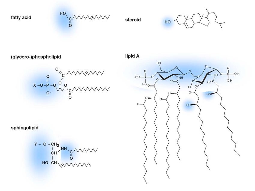
30                                           A. Pohl et al. / Biochimica et Biophysica Acta 1733 (2005) 29–52

before relating some concluding remarks. Considering the                           lipid classes are (glycero-) phospholipids, sphingolipids,
broad span of disciplines contributing to this topic, it                           steroids, lipopolysaccharides and triacylglycerols (for some
appears to be important in this article to draw a clear line                       examples see Fig. 1). The phospholipids phosphatidylcho-
between the transport of endogenous lipids and lipid analogs                       line (PC), phosphatidylethanolamine (PE), phosphatidylser-
carrying a reporter group or other modifications. Similarly,                       ine (PS) and phosphatidylinositol (PI), the sphingolipid
phenomena which appear to be related to the action of a                            sphingomyelin (SM) and the steroid cholesterol are the
particular protein need to be distinguished from those for                         major lipids found in mammalian membranes [6,7].
which a specific transport has been proven unequivocally.                              The lipid transbilayer distribution in the eukaryotic
On the one hand, most attempts in reconstituted systems                            plasma membrane is asymmetrical, generally with the
have revealed only very small effects of ABC proteins on                           majority of PC and sphingolipids in the exoplasmic leaflet,
lipid transport so far, with an influence of ATP hydrolysis                        and the aminophospholipids PE and PS mainly in the
hardly significant.                                                                cytoplasmic leaflet (reviewed in [8]). This asymmetry raises
    In experiments on whole cells, on the other hand, the                          several questions about the biological mechanisms by which
involvement of various transport steps must be taken into                          it is established (Fig. 2A), and about the putative biological
account, complicating transport rate quantification, and                           functions coupled to it.
hence the evaluation of their physiological significance.
                                                                                   2.1. Spontaneous transbilayer movement

2. Lipid transbilayer movement                                                        The rate of spontaneous transbilayer movement (flip-
                                                                                   flop) in a pure lipid membrane differs for each lipid,
   Lipids form a vast group of chemically different                                depending on its structure (headgroup, backbone) and its
amphiphilic or hydrophobic substances containing a sub-                            environment (reviewed in [9]). Small, uncharged lipids
stantial portion of aliphatic or cyclic hydrocarbon. Abundant                      (including cholesterol) and negatively charged lipids in their

Fig. 1. Lipid substrates of ABC proteins (examples). Hydrophilic parts are indicated by blue clouds. Examples for the phospholipid moiety X are ethanolamine
(in PE), choline (in PC) or serine (in PS). Examples for the sphingolipid moiety Y are hydrogen (in ceramide), phosphorylcholine (in SM), glucose (in GlcCer),
galactose (in GalCer) or lactose (in LacCer). The fatty acid shown is oleic acid, the steroid shown is cholesterol. Lipid A structure as predominant in E. coli
[218].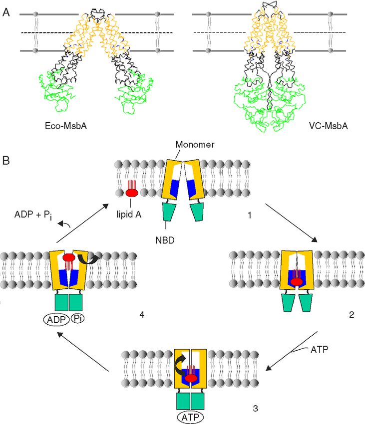
A. Pohl et al. / Biochimica et Biophysica Acta 1733 (2005) 29–52 31
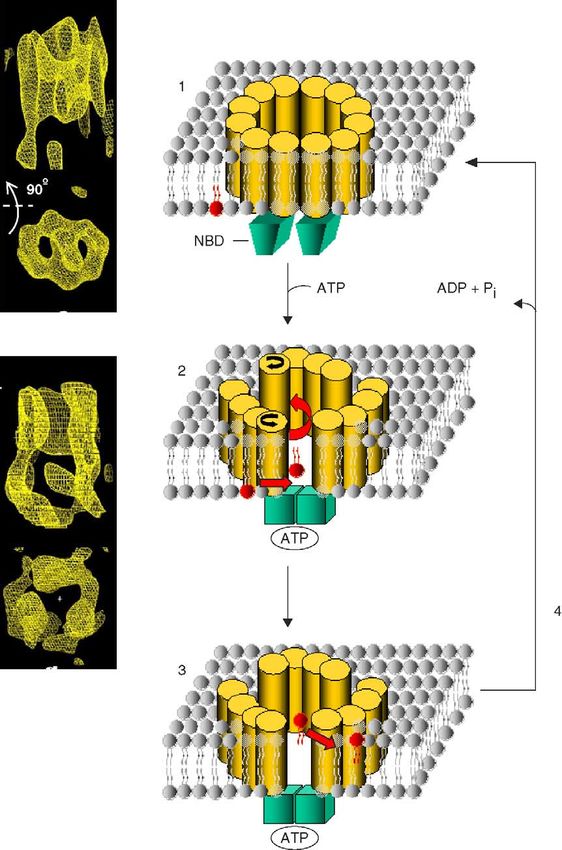
Fig. 2. Transbilayer lipid movement in biological membranes and models for function of lipid transporting ABC proteins (A). Lipids can move across a lipid
bilayer by passive flip-flop or mediated by proteins. Protein-mediated transbilayer movement could be energy-independent, unspecific and bidirectional (e.g.
scramblase), or energy-dependent and monodirectional (e.g. inward transport by P-type-ATPases, and outward transport by ABC proteins). The lipid specificity
varies among transporters (for details, see text). The number of lipids indicates the efficiency of lipid movement at a qualitative level. (B) Models for ABC
protein function in lipid transport and/or exposure to an acceptor A (for details, see text).
protonated form can flip across pure lipid bilayers within Already in 1980, it was shown that the mere presence of
seconds or minutes. In contrast, lipids with highly polar membrane proteins facilitates lipid flip-flop [22]. Thus, it is
headgroups move only slowly from one leaflet of a lipid not clear whether a dedicated flippase or a family of
bilayer to the other (half-times of the order of hours to days selective flippases is involved in bidirectional lipid move-
[10,11]). Cholesterol has been shown to decrease the ment across ER and Golgi membranes [23,24].
transbilayer movement of phospholipids [12,13]. Energy-independent, bi-directional transbilayer move-
ment of all major phospholipids has equally been shown in
2.2. Energy-independent and energy-dependent flippases the eukaryotic plasma membrane. This flip-flop (half time of
the order of 1 min) [25,26], activated by cell stimulation and
In eukaryotic cells, most lipids are synthesized asym- the subsequent increase in intracellular calcium, has been
metrically in the membranes of the endoplasmic reticulum ascribed to the lipid scramblase protein. Effort has been made
(ER) and the Golgi system from which they reach, e.g. via to isolate and clone the potential scramblase PLSCR1 [27].
vesicle traffic, the plasma membrane or other organelles Experiments with fluorescent (nbd1) phospholipids in the
(reviewed in [9]). Because lipid movement across cellular rabbit intestine brush border have indicated ATP- and Ca2+-
membranes is essential for cell growth and survival, lipid independent transbilayer movement of monoacyl phospho-
transporting proteins (flippases) are required for efficient lipids and lipids with short chains, supposedly constituting a
transbilayer lipid movement. Although some proteins have mechanism for the intestinal uptake of phospholipid
been identified as candidate lipid transporters over the last digestion products as lysolipids [28].
years (reviewed in [9,14,15]), the protein vehicles respon- While energy-independent flippases allow lipids to
sible for many lipid transport phenomena have not been equilibrate rapidly between the two bilayer leaflets, energy-
identified yet (Table 1). dependent flippases are responsible for a net transfer of
In the mammalian ER, energy-independent flippases were specific lipids to one leaflet of a membrane. In the plasma
shown to mediate rapid bidirectional, rather unspecific membrane of eukaryotic cells, spontaneous flip-flop of
phospholipid flip-flop (half times of the order of minutes or phospholipids is limited, possibly due to the high cholesterol
less) to ensure balanced growth of this membrane [16–18]. content, allowing the generation of a stable asymmetric lipid
Similarly, rapid protein-mediated flip-flop has been demon-
strated for phospholipids and glucosylceramide (GlcCer, a 1
(Typically, the abbreviation for [N-(7-nitrobenz-2-oxa-1,3-diazol-4-
precursor for complex glycosphingolipids) in the Golgi [19]. yl)amino] is written in capital letters (NBD). However, in order to avoid
Lipid flippase activity was also found in the bacterial plasma confusion with the abbreviation for nucleotide binding domain, this style of
membrane [20,21]. abbreviation was chosen).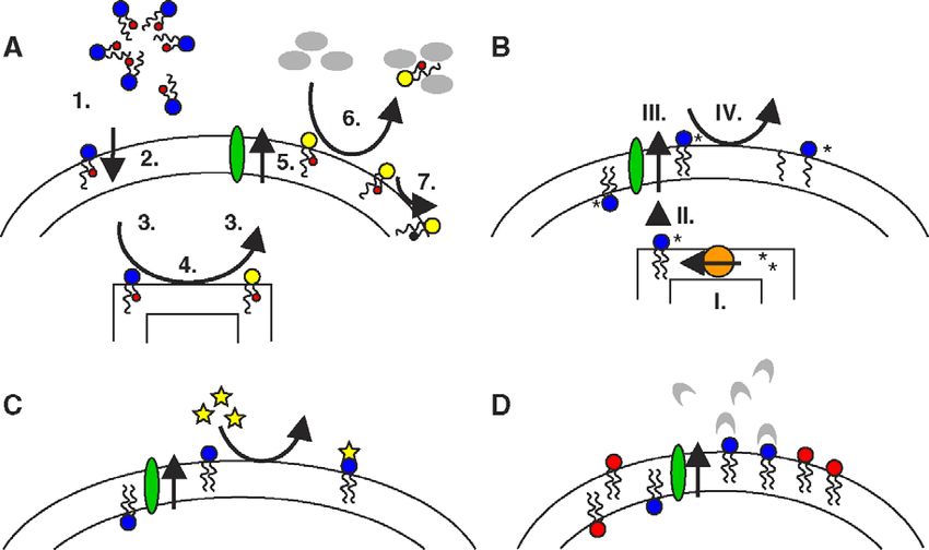
32 A. Pohl et al. / Biochimica et Biophysica Acta 1733 (2005) 29–52
Table 1
ABC proteins involved in lipid transport
Organism Family Name Trivial name Involvement Lipid analogs Endog. lipids
Human ABCA ABCA1 ABC1 macrophage lipid homeostasis SL-PS cholesterol
HDL deficiencies phospholipids
Tangier disease
phagocytosis
ABCA2 ABC2 macrophage lipid homeostasis? steroids?
neural development?
ABCA3 ABC3 lung surfactant synthesis? PC?
ABCA4 ABCR dark adaptation N-retinylidene-PE
Rim Stargardt disease
ABCA6 macrophage lipid homeostasis?
ABCA7 ABCX hematopoiesis? cholesterol
macrophage lipid homeostasis? phospholipids
keratinocyte differentiation? ceramide
ABCA9 hematopoiesis?
macrophage lipid homeostasis?
ABCA10 macrophage lipid homeostasis?
ABCB ABCB1 MDR1 Pgp detoxification C6-nbd-PC steroids
multidrug resistance C6-nbd-PE cholesterol
adrenal secretion C6-nbd-PS GlcCer, PS,
dendritic cell migration C6-nbd-SM SM, PAF
C6-nbd-GlcCer
ABCB4 MDR2/3 Pgp bile formation C6-nbd-PC PC
progessive familiar
intrahepatic cholestasis
ABCC ABCC1 MRP1 detoxification C6-nbd-SM
multidrug resistance? C6-nbd-GlcCer
LTC inflammatory response C6-nbd-PC
C6-nbd-PS
ABCC6 MRP6 pseudoxanthoma elasticum
lipid transport and metabolism
ABCD ABCD1 ALDP beta oxidation VLCFAs?
X-Adrenoleukodystrophy
peroxisome biogenesis
ABCD2 ALDRP beta oxidation VLCFAs?
ALDL1
ABCD3 PMP70 beta oxidation VLCFAs?
PXMP1 peroxisome biogenesis
ABCD4 PXMP1L beta oxidation VLCFAs?
P70R
PMP69
ABCG ABCG1 WHITE macrophage lipid homeostasis cholesterol, PC
ABCG2 BCRP1 detoxification multidrug resistance bodipy-Cer steroids? PS
MXR1 C6-nbd-PS
ABCP C6-nbd-PC
ABCG5 WHITE3 bile steroid secretion cholesterol
sitosterolemia steroids
ABCG8 WHITE4 bile steroid secretion cholesterol
sitosterolemia steroids
Sea urchin ABCA SuABCA sperm acrosome reaction? phospholipid?
cholesterol?
Leishmania ABCA LtrABC1.1 parasite–host interaction vesicular transport C6-nbd-PC
C6-nbd-PE
C6-nbd-PS
ABCB LPgp-lp multidrug resistance bodipy-PC
alkyl-lyso-
phospholipids
Arabidopsis PMP Ped3p COMATOSE beta oxidation FA CoAs?
(ABCD) CTS PXA1
Yeast PDR/CDR Pdr5p drug resistance C6-nbd-PE PE, steroids
Cdr1p drug resistance C6-nbd-PC PE
C6-nbd-PE
C6-nbd-PSA. Pohl et al. / Biochimica et Biophysica Acta 1733 (2005) 29–52 33
Table 1 (continued)
Organism Family Name Trivial name Involvement Lipid analogs Endog. lipids
Cdr2p drug resistance C6-nbd-PC phospholipid?
C6-nbd-PE
C6-nbd-PS
Cdr3p C6-nbd-PC phospholipid?
C6-nbd-PE
C6-nbd-PS
MDR Ste6p pheromone transport lyso-PC?
(ABCB) drug resistance?
MRP/CFTR Yor1p drug resistance C6-nbd-PE
(ABCC) efflux organic anions?
ALDp Pat1p Pxa2p peroxisomal lipid transport VLCFAs?
(ABCD)
Pat2p Pxa1p peroxisomal lipid transport VLCFAs?
E. coli ABCB MsbA lipid A transport lipid A
phospholipids
L. lactis ABCB LmrA drug resistance C6-nbd-PE lipid A
lipid A transport?
F. novicida ? ValA lipid A transport? lipid A-linked
polysaccharide
Substrates confirmed in reconstitution experiments are in bold print. Question marks indicate functions or substrates for which an involvement has not been
proven experimentally.
distribution between the two leaflets. The depletion of fusion competence, protein association and activity, as well
cholesterol leads to an enhanced spontaneous flip-flop of as various biochemical pathways (reviewed in [9]). Because
phospholipid analogs in the red blood cell membrane [12]. of the low compressibility of lipid monolayers, the inward
PE and PS are subject to an efficient and rather rapid and outward transport of lipids requires a subtle balance. In
energy-dependent inward transport from the outer to the the absence of a compensatory flux, unidirectional lipid
inner leaflet mediated by a protein, the aminophospholipid transport by energy-coupled transporters might create an
translocase [29,12]. Some cell types display similar trans- area imbalance between the two membrane leaflets, building
port of PC across the plasma membrane and may contain up surface tension eventually relaxing by membrane
either a PC-specific translocase in addition to the amino- budding or invagination [36,37] (see also Section 6). This
phospholipid translocase, or an inward translocase of phenomenon was suggested to be a molecular motor for the
different specificity which transports both aminophospholi- first stage of endocytosis [38,39]. The balance between
pids and PC (see [15]). The aminophospholipid translocase, inner and outer membrane leaflet has been generally
not yet identified on the molecular level, very likely belongs overlooked when the activity of potential lipid transporters
to the novel DrS2p P-type-ATPase subfamily with over a was assessed in large unilamellar vesicles (LUVs), where
dozen members in eukaryotes from yeast to plant cells surface tension generated by lipid transport could be
[30,31,15]. Two members of this family, Dnf1p and Dnf2p, sufficient to eventually block the transporter itself (dis-
have been shown to be essential for ATP-dependent cussed in [37,39,40]).
transport of fluorescent analogs of PS, PE, and PC from
the outer to the inner leaflet of the yeast plasma membrane 2.3. Determination of lipid transbilayer distribution
[32].
Recently, it was reported that the fluorescent analog nbd The techniques used to determine the transbilayer
PS is a preferred substrate of Drs2p localizing to the trans- distribution of lipids have been described and critically
Golgi-network [33]. evaluated in the literature [7,41–46]. In Fig. 3, some assays
However, it is still unclear what permits the accumulation for lipid analogs as well as for endogenous lipids are shown
of the majority of PC and sphingolipids in the mammalian schematically. Since methods for the rapid quantification of
outer plasma membrane leaflet. ABC proteins were sug- endogenous lipids are still very limited, spin-labeled (SL) or
gested to be involved in the outward transport of phospho- fluorescent lipid analogs are frequently employed to
lipids [34]. Alternatively, it was proposed on theoretical determine lipid transbilayer distribution. The relevance of
grounds that the inward transport of aminophospholipids, such probes has been discussed by Devaux et al. [42]:
together with passive fluxes, would be sufficient to Briefly, bulky reporter moieties and a short fatty acid chain
accumulate choline containing lipids in the outer leaflet replacing one of the natural long chains to facilitate
[35]. membrane incorporation may affect lipid polarity and cause
Changes in the distribution of a lipid species between the perturbations, somewhat modifying the absolute values of
membrane leaflets can influence membrane curvature and the transbilayer distribution of a lipid. However, compar-34 A. Pohl et al. / Biochimica et Biophysica Acta 1733 (2005) 29–52
Fig. 3. Assays for the detection of lipid transbilayer distribution. (A) BSA-extraction, dithionite and ascorbate assays for fluorescent and spin-labeled lipid
analogs. The short-chain lipid analog precursor integrates into the outer membrane leaflet (1), crosses the plasma membrane (e.g. by passive flip-flop) (2) and
distributes to different intracellular membranes (e.g. by monomeric transport) (3). ER or Golgi enzymes convert part of the lipid analog precursor to the lipid
analog of interest (4), which can distribute back to the plasma membrane, where it becomes available to outward transport by transporter proteins (5). Lipid
analog is extracted from the outer plasma membrane leaflet by BSA (6), followed by the separation of cells and media. In a variant of this assay, the cells can be
directly labeled with the lipid analog of interest, if it is able to reach the inner plasma membrane leaflet by passive flip-flop or active transport, analog remaining
on the outer leaflet being removed by BSA extraction prior to outward transport incubation. Alternatively to BSA extraction, fluorescene of lipid analogs can be
quenched using dithionite, and the spin-label signal be reduced using ascorbate (7). (B) Phospholipase/sphingomyelinase assay for endogenous lipids.
Endogenous lipid is synthesized in the presence of radioactive precursors in the cell (I) and localizes to the various cellular membranes due to vesicular or
monomeric transport (II), to modification, and to the presence of transporter proteins (III). Phospholipase A2 treatment converts lipids in the outer plasma
membrane leaflet to lysolipid and fatty acid (IV). Lipid products are then analyzed by chromatography and can be compared to samples untreated with enzyme.
An analogous technique is used for SM, employing sphingomyelinase. (C) Chemical modification assays for endogenous lipids. Endogenous lipids present on
the outer plasma membrane leaflet are typically modified on the level of the headgroup. Reagents frequently used for modification are trinitrobenzene sulfonic
acid (TNBS, specific for PE) and fluorescamine. (D) Antibody, peptide or protein binding assay for endogenous lipids. Specific antibodies, respectively
peptides (e.g. Ro09-198, binding to PE [45,46]) or proteins (e.g. Annexin V, binding to PS) with affinity for a particular lipid headgroup, bind to endogenous
lipids present on the outer plasma membrane leaflet; the amount of bound antibody or binding protein is quantified.
isons between lipids with different headgroups, but with the for a particular set of substrates (the multispecific ABCB1
same fatty acid chains, can be very revealing about the (MDR1 Pgp) probably represents an exception in this
specificity of the lipid transport. Recently, various choles- respect). Substrates can be amino acids, sugars, inorganic
terol analogs have been studied showing large variations in ions, peptides, proteins, lipids and various organic and
the potential to mimic endogenous cholesterol [44]. Never- inorganic compounds. ABC proteins consist of nucleotide
theless, new assays will have to be developed to rapidly binding domains (NBDs), and of transmembrane (TM)
determine the transbilayer distribution of endogenous lipids. domains (TMDs) of usually six alpha-helices. NBDs and
TMDs can occur as separate proteins (frequently found in
prokaryotes) or fused together. The transmembrane domains
3. General features of the ABC protein superfamily vary considerably between different ABC proteins, whereas
the nucleotide binding domains are highly conserved (e.g.
The ATP-binding cassette (ABC) protein superfamily Walker motifs). The substrate specificity is believed to be
comprises a large number of transporters, channels and determined by the transmembrane domains, including the
regulators in pro- and eukaryotes [reviewed in [47–49]]. loops connecting the individual helices [47].
Their functions range from the acquisition of nutrients and
the excretion of waste products to the regulation of various
cellular processes. Generally, ABC proteins are low 4. Eukaryotic ABC proteins
capacity, but high affinity transporters, able to transport
substrates against a concentration gradient of up to 10 000 Structurally, eukaryotic ABC proteins typically possess
fold. Hydrolysis of ATP is required for substrate transport. two nucleotide binding domains and two TM domains,
ABC proteins are mainly either import or export pumps, probably representing the minimal functional unit (full-size
bidirectional ABC proteins appear to be rare exceptions protein). In some ABC proteins, deviating organizations of
[50]. Import pumps seem to be limited almost exclusively to the domains can prevail [49] (Fig. 4), e.g. half-size proteins
prokaryotes. Typically, ABC proteins are relatively specific with one NBD and one TM domain each, thought to requireA. Pohl et al. / Biochimica et Biophysica Acta 1733 (2005) 29–52 35
Fig. 4. Domain organization of human ABC proteins involved in lipid transport. Transmembrane domains (TMDs) are shown as membrane-spanning helices,
nucleotide binding domains are marked NBD. Intracellular domains (ICDs) and posttranslational modifications (e.g. glycosylations) are not shown.
dimerization in order to be functional. ABC proteins with ABCA1 (ABC1), studied intensely with regards to lipid
nucleotide binding domains only (families E, F) are likely transport, is expressed in numerous tissues such as the
not directly involved in membrane transport, and are trachea, lung, adrenal gland, spleen and uterus [52], in some
thought to have regulatory functions. of which its expression is steroid-dependent [53]. ABCA1-
GFP chimeras localize to the plasma membrane and to
4.1. Human ABC proteins intracellular vesicles in transfected HeLa cells [54]. ABCA1
has been implicated with the transport of cholesterol and
The 49 human ABC proteins currently known can be phospholipids (see Section 6).
classified into 7 families (A–G) according to sequence In mammals, the majority of cholesterol is synthesized de
similarity [49,51]. An overview can be found on Michael novo in the liver, and delivered to peripheral cells by
Mqller’s website (http://nutrigene.4t.com/humanabc.htm). lipoproteins. Since peripheral cells are unable to degrade
Several human ABC proteins found to be mutated in lipid- cholesterol, any surplus of cholesterol must either be stored
linked diseases (families A, B, C, D, and G) were suggested to in the cytosol in the form of esters, or released from the cell.
be involved in lipid transport. Although diseases due to ABCA1 mutations can be responsible for some cases of
complete loss of function of single ABC proteins do not familial high density lipoprotein (HDL) deficiency, e.g.
appear to have a very high incidence in the population, Tangier disease [55–57], characterized by impaired efflux
polymorphisms in ABC genes are likely to play a role in of cholesterol and phospholipids from peripheral cells onto
medically relevant phenomena. At present, direct transport of apolipoproteins such as apoA-1. Cholesterol accumulation in
lipid substrates has only been shown for a small number of macrophages and apolipoprotein degradation lead to tissue
human ABC proteins. The ABC proteins identified in deposition of cholesterol esters and increase the risk of
mammals so far (e.g. in mouse, rat, pig) are highly similar arteriosclerosis in the patients. While Tangier cells typically
to members of the human ABC protein families, and will fail to bind nascent apoA-I [58,59], the expression of ABCA1
therefore not be discussed separately in this work. in cultured cells has been found to enhance the binding of
apoA-I to the plasma membrane [60], and to increase the
4.1.1. Human ABCA (ABC1) family efflux of cellular phospholipid and cholesterol to this
Out of the 12 ABCA family members, all of which are apolipoprotein [61]. Both sequential and parallel mechanisms
full-size proteins, 8 are assumed to transport lipophilic have been proposed for the transport of phospholipids and
substrates. For a number of these, an involvement in lipid cholesterol by ABCA1 (see also Section 6).
transport was deduced from lipid dependent expression, and ABCA1 has been suggested to directly transport the
homology to ABCA1, but has not yet been confirmed aminophospholipid PS (typically restricted to the cytoplas-
experimentally. mic leaflet of mammalian plasma membranes) to the36 A. Pohl et al. / Biochimica et Biophysica Acta 1733 (2005) 29–52 exoplasmic leaflet [61], where the presence of PS facilitates involved in dark-adaptation through the transport of the apoA-1 binding, leading thus indirectly to cholesterol efflux lipid product all-trans-N-retinylidene-PE across the disc [61–63]. Indeed, the exposure of endogenous PS (detected membrane following the photobleaching of rhodopsin. This via prothrombinase activity and binding of Annexin V) transport allows all-trans-retinal to be reduced to all-trans- upon Ca2+ induced stimulation was found to be low in red retinol on the surface of the disc membrane. Thus, ABCA4 blood cells, resp. thymocytes, derived from mice lacking mediated transport is an important step in the recycling of ABCA1, and could be partially restored upon ABCA1 all-trans-retinal to 11-cis-retinal for the regeneration of transfection [61]. A recent study on apoptotic murine cells rhodopsin. Studies on the ATPase activity of reconstituted confirmed elevated levels of exposed PS to cause a strong ABCA4 strongly suggested PE to be required to couple the increase in apoA-I binding, although not sufficient to trigger binding of retinoids to ABCA4 ATPase activity [76]. phospholipid and cholesterol efflux to apoA-I [64]. Defective ABCA4 can cause the degeneration of the macula In contrast, Wang et al. [65] recently proposed direct lutea and consequent deterioration of vision (Stargardt transbilayer transport of both cholesterol and phospholipids disease), presumably through the accumulation of the via ABCA1, following experiments in which phospholipid/ lipofuscin fluorophore N-retinylidene-N-retinyl ethanol- apoA-I particles made by ABCA1 were unable to stimulate amine (A2E) [77,78]. passive cholesterol efflux when added to a second set of cells. ABCA6 shows expression in the lung, heart, brain, liver In addition to a role in lipid loading of apolipoproteins, and ovaries. As ABCA6 expression is suppressed by ABCA1 has been implicated with PS exposure on the outer cholesterol loading [79], the protein was attributed a plasma membrane leaflet of apoptotic cells and phagocytiz- potential role in macrophage lipid homeostasis. ing macrophages [66]. In the absence of apoliposomes, Ca2+ ABCA7 is expressed mainly in myelo-lymphatic tissues. induced externalization of spin-labeled (SL) analogs of PS, The protein is localized either in the plasma membrane or but not of PC, was reduced in mice lacking ABCA1 [61]. the endoplasmic reticulum [80], and was implicated with the The large ABC protein ABCA2, found in high levels in specification of hematopoietic cell lineages during develop- human brain (possibly in oligodendrocytes), colocalizes ment [81]. As the loading of macrophages with steroids with a lysosomal/endosomal marker [67]. Vulevic et al. increases, and unloading decreases the expression of the [67] have associated ABCA2, which contains a signature ABCA7 gene, Kaminski et al. proposed ABCA7 to be motif for lipocalins (protein family binding small hydro- involved in macrophage transbilayer lipid transport [82]. phobic molecules), with the transport of steroids and lipids ABCA7 overexpression in HeLa cells increased the due to colocalization with an analog of the steroid exposure of endogenous ceramide (Cer) on the outer plasma estramustine, and increased estramustine resistance upon membrane leaflet, and raised levels of endogenous PS [83]. ABCA2 gene overexpression. As ABCA7 is upregulated during keratinocyte differentia- Kaminski et al. suggested a role for ABCA2 in macro- tion, this led to speculations about a regulator role for phage lipid metabolism and neural development, after ABCA7 in lipid transport during terminal keratinocyte having found an induction of ABCA2 mRNA during steroid differentiation. Very recently, apolipoprotein-mediated loading [68]. Based on these observations and the unique release of cellular cholesterol and phospholipids was expression profile, Schmitz and Kaminski [69] inferred a reported from HEK293 cells transiently or stably expressing role of ABCA2 in transbilayer lipid transport of neural cells. ABCA7 [80,84]. Furthermore, the transfection of GFP- ABCA3 is expressed exclusively in type II epithelial lung tagged ABCA7 of L929 cells triggered apolipoprotein- cells expressing the gene surfactant protein A. It is mediated assembly of cholesterol-containing HDL. In hypothesized to play a role in the formation of pulmonary contrast, Wang et al. [65] reported ABCA7-mediated efflux surfactant [70], a mixture containing PC and various of phospholipid and SM, but in contrast to ABCA1 not of surfactant proteins, which reduce surface tension on the cholesterol, from HEK293 cells. surface of the alveoli. Mutations in ABCA3 were found in ABCA9, highly homologous to ABCA6, shows ubiqui- several cases of infants with fatal surfactant deficiencies tous expression, the highest levels being found in the heart, [71]. ABCA3 is localized in the plasma membrane and in brain and fetal tissues. ABCA9 is induced during macro- the limiting membrane of lamellar bodies, in which phage differentiation. Different from ABCA7, the expres- surfactant is stored prior to exocytotic delivery into the sion of ABCA9 in macrophages decreases upon steroid alveolar space. Mulugeta et al. have therefore speculated loading of the cells. Piehler et al. suggested that ABCA9 that ABCA3 might transport PC into or other lipids out of might act on monocyte differentiation and macrophage lipid lamellar bodies, which are highly enriched in PC [72]. homeostasis [85]. ABCA4 (ABCR or Rim protein) [73] is localized in the ABCA10, as ABCA9 highly homologous to ABCA6, is photoreceptor outer segment disc membranes of the retina ubiquitously expressed, with high gene expression levels in [74,75]. Reconstitution studies, in which its ATPase activity the heart, brain, and the gastrointestinal tract. As its gene was stimulated by retinal, lead to the hypothesis that expression in macrophages is suppressed by cholesterol ABCA4 may function as an active retinoid transporter loading, Wenzel et al. hypothesized on the involvement of [76]. ABCA4 appears to be highly substrate-specific, being ABCA7 in macrophage lipid homeostasis [86].
A. Pohl et al. / Biochimica et Biophysica Acta 1733 (2005) 29–52                               37

4.1.2. Human ABCB (MDR/TAP) family                                      inner plasma membrane leaflet, such as PS and PE.
    The 4 full-size and 7 half-size proteins of the ABCB                Reconstitution experiments with ABCB1 have thus far
family show highly varied specificities (e.g. amphiphilic               yielded ambiguous results: While Romsicki and Sharom
compounds, peptides, iron, phospholipids, bile salts) [51].             [104] found a low ATP dependent increase in reoriented
The proteins associated with antigen processing ABCB2, 3                short-chain nbd analogs of PC, PE, PS and SM, Rothnie et
(TAP1, 2), and the bile salt export pump ABCB11 (BSEP)                  al. [40] observed low reorientation of short-chain nbd
are ABCB protein family members.                                        analogs of PC, PE, Cer and of short-chain SL analogs of
    ABCB1 (Multidrug Resistance 1 P-glycoprotein, MDR1                  PC, PE, GlcCer, and SM which was ATP independent.
Pgp) [87] is a full-size protein with two transmembrane                 Interestingly, the activity of ABCB1 was dependent on
domains and two nucleotide binding domains, which                       cholesterol. Due to the small size of the vesicles containing
appears to be functional as a monomer [88]. It occurs in                the reconstituted protein, lateral pressure (surface tension)
the apical membrane domain [89] of epithelia with secretory             might have prevented substantial transport of lipids via
functions (e.g. adrenal gland, kidney), at the pharmacolog-             ABCB1 [105] (see Section 2). The structure and potential
ical borders of the body (intestine, blood–brain barrier, feto-         mechanisms of ABCB1 will be discussed in Section 7.
maternal barrier) [90], and in many tumor tissues [91].                    ABCB4 (MDR2/3 Pgp), a full-size protein, is a close
    ABCB1 has a surprisingly broad spectrum of (mainly                  relative of ABCB1 (MDR1 Pgp), sharing 75% of its amino
cationic or electrically neutral) amphiphilic substrates [92].          acid sequence [49]. Unlike ABCB1, ABCB4 is highly
One of its important physiological roles appears to be the              substrate specific, exclusively transporting (short-chain nbd
protection of the organism against toxins by exporting these            analogs of) PC, as observed in ABCB4 transfected porcine
into the bile, urine, or gut. In tumors, the overexpression of          cells [97]. The secretion of PC into the bile appears to be the
ABCB1 is one of the principal factors responsible for                   physiological function of ABCB4 [103] (see also Section 6),
multidrug resistance (MDR) against a variety of structurally            the protein being present in high amounts in the canalicular
unrelated drugs. ABCB1 is involved in other phenomena as                membranes of hepatocytes. In mice lacking ABCB4, PC
well, in which, interestingly, lipid transport often seems to           secretion into the bile is abolished [103], and the trans-
be implicated: It was found to mediate the secretion of the             bilayer movement of radioactively labeled endogenous PC
steroid aldosterone by the adrenals [93], and its inhibition            appears to be slightly enhanced in fibroblasts from mice
blocked the migration of dendritic immune cells [94],                   overexpressing the ABCB4 gene [106]. In some cases of
possibly related to the outward transport of the lipid platelet         progressive familiar intrahepatic cholestasis (type III) in
activating factor (PAF, see below). Ueda et al. reported                humans, ABCB4 has indeed been found to be defective
ABCB1 mediated transport of the steroids cortisol and                   [107].
dexamethasone, but not of progesterone in ABCB1 trans-
fected cells [95]. Inhibition studies have also led to                  4.1.3. Human ABCC (CFTR/MRP) family
speculations about the transport of cholesterol by ABCB1                   All 13 ABCC family members are full-size proteins.
[96] (see also Section 6). Short-chain and long-chain (nbd              However, they differ in the number of transmembrane
and radiolabeled) analogs of PC, PE, PS, SM, and GlcCer                 domains (two for ABCC4, ABCC5, ABCC11 and
were found to be expelled from ABCB1 overexpressing                     ABCC12, while all others possess a third TM domain,
cells [97–99]. Among the endogenous lipids, the short-chain             TMD0 (Fig. 4)). The major functions of the ABCC proteins
PC PAF [100] is an ABCB1 substrate (ABCB1 antisense                     are, among others, the protection against toxic compounds
oligonucleotides blocking PAF secretion in human mesan-                 and the secretion of organic anions [108]. Examples for
gial cells). Other potential substrates are GlcCer (rescued             ABCC family members are the cystic fibrosis transmem-
from cytosolic hydrolysis in the presence ABCB1, and                    brane conductance regulator ABCC7 (CFTR), defective in
strongly reduced in the absence of ABCB1) [101] and PS                  mucoviscidosis, and the sulfonylurea receptors ABCC8,9
(as found by Annexin V binding experiments in ABCB1                     (SUR 1, 2), regulating associated potassium channels.
overexpressing human gastric carcinoma cells) [99]. Inter-              ABCC8 is defective in patients with familial persistent
estingly, the exposure of endogenous SM to the outer                    hyperinsulinemic hypoglycemia of infancy. The ABCC
plasma membrane leaflet of human myeloblastic cells was                 family members differ in substrate specificity, tissue and
reduced upon ABCB1 inhibition, a possible hint for ABCB1                organelle distribution [109].
mediated transport of this lipid involved in signalling [102].             ABCC1 (MRP1) [110] is located in the basolateral domain
While the inability of the ABCB1 mouse homologs Mdr1a/                  of polarized cells [111], and displays wide tissue distribution
1b to restore the transport of PC into the bile of mice lacking         [109]. ABCC1 transports a large variety of toxins across the
Mdr2 (homologous to human ABCB4) [103] could suggest                    plasma membrane, either unconjugated or conjugated with
that natural long-chain PC is not an ABCB1 substrate, it is             glutathione, sulfate or glucuronate [112]. While it is unclear
also conceivable that Mdr1a/1b activity was too low in this             whether ABCC1 contributes significantly to multidrug
system. Multispecific transport of diverse endogenous lipids            resistance in tumor cells [109], it protects particularly
via ABCB1 could affect the transbilayer distribution of                 sensitive organs by expelling toxins into the blood (the
lipids, in particular of species normally predominant on the            internal environment [113], in contrast to the apically located
38 A. Pohl et al. / Biochimica et Biophysica Acta 1733 (2005) 29–52
ABCB1 (MDR1 Pgp) which exports toxins into the external system white matter and the adrenal cortex [130], leading to
environment). Additionally, ABCC1 mediates the leuko- neuron demyelinization, renal insufficiency and testicular
triene C (LTC) dependent inflammatory response by the dysfunction. Increased VLCFA levels have been attributed
transport of the arachidonic acid derivative LTC4 [114]. to impaired peroxisomal beta-oxidation, and transfection
ABCC1 transfected pig kidney epithelial cells showed with ABCD1 cDNA was shown to restore beta-oxidation in
increased outward transport of short-chain nbd analogs of X-ALD fibroblasts [131] (see also yeast homolog Pat1p;
SM, and GlcCer [115], and the transport of short-chain (PS, Section 4.5). Recently, mitochondrial beta-oxidation was
PC) and long-chain PC nbd analogs in erythrocytes was reported to affect peroxisomal beta-oxidation, and a role of
equally attributed to ABCC1 in an inhibition study [116]. ABCD1 in the interaction of peroxisomes with mitochon-
Upon reconstitution, ABCC1 transported an nbd PC analog dria was suggested [132]. ABCD1 has been implicated with
[117]. However, thus far, no endogenous lipids have been peroxisome biogenesis, restored upon ABCD1 overexpres-
found to be ABCC1 substrates. sion in Zellweger cells (having a defect in the peroxisomal
Despite similarities in the substrate spectrum of ABCC1 protein Pex2p) [133].
and several other ABCC proteins, Raggers et al. did not ABCD2 (ALDRP) is closely related to ABCD1 (63%
observe translocation of short-chain nbd lipid analogs by amino acid identity). Its mRNA is highly expressed in human
other ABCC proteins tested besides ABCC1 [101]. brain and heart [128]. Similar to ABCD1, transfection with
ABCC6 (MRP6) is expressed exclusively in the liver and ABCD2 cDNA restored beta-oxidation in X-ALD (ABCD1
kidney, where the gene product is localized on the defect) fibroblasts [134], and peroxisome proliferation could
basolateral domain of the plasma membrane [118]. ABCC6 be induced upon pharmacologically increased ABCD2
defects result in pseudoxanthoma elasticum (PXE) expression in X-ALD cells [135]. In X-ALD fibroblasts,
[119,120], a disorder characterized by a calcification of ABCD2 induction by steroid depletion was found to reduce
the elastic fibers of the eye, skin, and vasculature, leading to the accumulation of VLCFA [136]. While defects in ABCD2
decreased visual acuity, characteristic skin lesions, and as a cause for X-ALD were considered to be unlikely,
peripheral vascular disease. Additionally, frequently found ABCD2 could be a heterodimeric partner for ABCD1 in some
high plasma triglyceride levels and low plasma HDL tissues, acting as a modifier gene accounting for the high
cholesterol in PXE patients suggest an involvement of phenotypic variability of X-ALD [127].
ABCC6 in lipid transport and metabolism [121]. ABCD3 (PMP70, PXMP1) shows 36% amino acid
The molecular basis of this disease is not solved, and identity with ABCD1. The mRNA of its mouse homolog
PXE might be a primary metabolic disorder with secondary is highly expressed in the liver, kidney, heart, lung and
involvement of elastic fibers [122]. intestine [128]. The overexpression of ABCD3 was found to
The transport of glutathione conjugates upon ABCC6 restore beta-oxidation in X-ALD fibroblasts and to normal-
gene expression in Sf9 insect cells point to a role for ABCC6 ize peroxisome biogenesis in Zellweger cells [133] (see also
in the transport of organic anions [123], thought to confer yeast homolog Pat2p, Section 4.5).
low levels of resistance to certain anticancer agents [124]. ABCD4 (PXMP1L, P70R, PMP69) shares 25% of
ABCD1 amino acid sequence. ABCD4 mRNA is highly
4.1.4. Human ABCD (ALD) family expressed in human kidney, spleen and testis [128]. As
All four known members of the ABCD family are half-size ABCD2 and 3, it has been suggested to act as a modifier
proteins found in peroxisomes, single-membrane organelles gene contributing to X-ALD phenotypic variability, possibly
involved in beta-oxidation of long and very long chain fatty heterodimerizing with the other members of this protein
acids, synthesis of bile acids, cholesterol plasmalogens and family [137].
metabolism of amino acids and purines. ABCD proteins have
been implicated with the transport of fatty acids (FA), 4.1.5. Human ABCG (WHITE) family
coenzyme A (CoA), or FA-CoA, although ATP independent The five characterized ABCG proteins are half-size
transport of very long chain fatty acids (VLCFA) into proteins. In contrast to other proteins, the ABC-domain is
peroxisomes [125] might argue against direct VLCFA trans- N-terminal, followed by the transmembrane domain (Fig. 4)
port via ABCD proteins. ABCD half-size proteins can form [138]. A number of ABCG members are thought to be
not only homodimers (ABCD1–ABCD1), but also hetero- involved in the transport of steroids (ABCG1, 5, 8),
dimers (ABCD1–ABCD2, ABCD1–ABCD3, ABCD2– additionally, some appear to transport phospholipids
ABCD3) [126]. Although the heterodimers might be func- (ABCG1) and toxins (ABCG2).
tional, the differing tissue gene expression of ABCD ABCG1 (WHITE) [139] is thought to be active either as a
members argues against obligatory heterodimerization [127]. homo- or a heterodimer (possibly with ABCG2) [140]. It
ABCD1 (ALDP) mRNA is highly expressed in human shows an ubiquitous gene expression pattern [138] and
liver, heart, skeletal muscle, lung and intestine [128]. localizes primarily to ER and Golgi [140]. ABCG1 derives
Defects in ABCD1 result in the inherited neurometabolic its trivial name from its homology to the Drosophila white
disorder X-linked adrenoleukodystrophy (X-ALD) [129], protein, which transports guanine and tryptophane as
characterized by elevated levels of VLCFA in nervous precursors for eye pigments [141]. ABCG1 itself appearsA. Pohl et al. / Biochimica et Biophysica Acta 1733 (2005) 29–52 39
to serve a different function, presumably in the transport of implicated in the sperm acrosome reaction during fertiliza-
phospholipids and steroids out of macrophages, as its gene tion. Due to its close homology to human ABCA3,
expression in macrophages was found to be induced during Mengerink and Vacquier suggested SuABCA to be involved
cholesterol influx, and suppressed by lipid efflux via HDL. in phospholipid or cholesterol transport [160].
The inhibition of ABCG1 expression resulted in reduced
HDL-dependent efflux of cholesterol and PC from these 4.3. Eukaryotic parasite ABC proteins
cells [140].
ABCG2 (BCRP, MXR, ABCP) [142] is found in the ABC proteins found in parasites are of particular clinical
placenta, intestinal epithelium, liver canaliculi, breast ducts relevance due to the occurrence of multidrug resistant
and lobules, as well as in veinous and capillary endothelium strains. Specific attention will be given here to ABC
[143]. Recent studies suggest the protein to be active as a proteins found in unicellular eukaryotes of the Trypanoso-
homotetramer [144] in the plasma membrane [145]. It is matidae family, causing Leishmaniasis and Trypanosomia-
assumed to prevent the tissue uptake of xenobiotics [143], sis, major and globally widespread parasitic diseases.
transporting various drugs across the plasma membrane in In Leishmania spp., three different families of ABC
an ATP dependent process [146]. Transfection studies proteins are known (reviewed in [161,162]), homologous to
proved the overexpression of ABCG2 to induce multidrug the mammalian ABC families ABCA, ABCB, and ABCC,
resistance in a previously drug sensitive cell line [147]. In respectively.
addition to drug transport, the reduced accumulation of a
short-chain Bodipy analog of ceramide in ABCG2 over- 4.3.1. Leishmania ABCA family
expressing cells has given hints for a transport of lipid LtrABC1.1 [161], a full-size protein containing two
analogs [146]. Recently, we have found increased outward transmembrane domains and two nucleotide binding
movement of short-chain nbd analogs of PS and PC, but not domains, is found mainly in the plasma membrane and
of PE, and increased exposure of endogenous PS in a human flagellar pocket of Leishmania [163]. LtrABC1.1 expres-
gastric carcinoma cell line overexpressing ABCG2 [148]. In sion appears to be uncorrelated with multidrug resistance. In
Lactococcus lactis transfected with human ABCG2, a Leishmania tropica cell line overexpressing LtrABC1.1,
ABCG2-associated ATPase activity was significantly stimu- the accumulation of short-chain nbd analogs of PC, PE, and
lated by cholesterol and estradiol, pointing to a possible role PS was found to be reduced [163], regardless of the nature
of ABCG2 in the transport of steroids [149]. of the phospholipid head group. Furthermore, LtrABC1.1
ABCG5 (WHITE3) and ABCG8 (WHITE4) are encoded overexpression reduced vesicular transport. Parodi-Talice et
by neighbouring genes [150]. Their mouse homologues are al. have suggested a role for LtrABC1.1 in lipid movement
expressed in the liver and intestine [151]. Very recently, across the plasma membrane and in vesicle trafficking.
ABCG5 and ABCG8 have been demonstrated to function as Finally, Legare et al. noted the potential importance of
an obligate heterodimer to promote steroid secretion into the members of the Leishmania ABCA family in the interaction
bile [152–154]. Mutations in either ABCG5 or ABCG8 of the parasite with its host, an engulfing macrophage cell
result in an identical clinical phenotype; and the expression [162].
of both genes is required for either protein to be transported
to the plasma membrane. Defects in ABCG5 and 8 can 4.3.2. Leishmania ABCB family
result in sitosterolemia, a disorder characterized by Leishmania Pgp-like proteins (LPgp-lp), about 37%
increased uptake of steroids (among them the plant steroid identical to mammalian ABCB1s, are full-size ABC
beta-sitosterol) in the intestine, combined with decreased proteins [161]. They have been shown to transfer multidrug
biliary excretion, resulting in cholesterol deposits in skin resistance upon transfection [164]. In Leishmania enriettii,
and tendons, and in premature coronary artery disease Leishmania Pgp-like protein is located mainly in different
[150,155,156]. intracellular vesicles [165]. Upon the overexpression of the
The overexpression of human ABCG5 and 8 in mice LPgp-lp gene in L. tropica, the parasite cells accumulated
promoted bilary cholesterol secretion, reduced cholesterol lower amounts of a short-chain Bodipy analog of PC and the
absorption, and increased hepatic cholesterol synthesis [157]. antiproliferative alkyl-lysophospholipids miltefosine and
Furthermore, no significant sitosterol transport by edelfosine than did the controls [166].
ABCB1 (MDR1 Pgp), ABCC1 (MRP1), and ABCG2
(BCRP) was found [158].Taken together, these data 4.4. Plant ABC proteins
demonstrate a central role of ABCG5 and ABCG8 in in
vivo cholesterol excretion [159] (see also Section 6). Ped3p (peroxisome defective protein 3, also designated
COMATOSE, CTS, PXA1), predicted to be a full-size protein
4.2. Sea urchin ABC proteins [167,168], is found in arabidopsis glyoxysomes, specialized
peroxisomes occurring in cells of storage organs (endo-
SuABCA is a full-size ABC protein found abundantly in sperms, cotyledons) and senescent organs [168]. Both halves
the sea urchin sperm plasma membrane, and is possibly of Ped3p show significant sequence homology to the human40 A. Pohl et al. / Biochimica et Biophysica Acta 1733 (2005) 29–52
half-size protein ABCD1 (ALDP) [169]. In Ped3p defective 4.5.3. MRP/CFTR family (ABCC homolog)
plants, fatty acid beta-oxidation is impaired [168,169], Yor1p is a plasma membrane located [182] full-size
causing a defect in gluconeogenesis that severely inhibits protein in S. cerevisiae. It is related to the human ABCC
seedling germination in the absence of sucrose [170]. As family [183]. Besides conferring drug resistance, Yor1p has
Ped3p mutants accumulate fatty acyl CoAs, Footitt et al. been suggested to be involved in the efflux of organic
proposed the protein to be a transporter of fatty acyl CoAs anions. The deletion of Yor1p in S. cerevisiae resulted in
with little specificity concerning chain length [169]. increased accumulation of a short-chain nbd analog of PE
[174].
4.5. Yeast ABC Proteins
4.5.4. ALDp family (ABCD homolog)
In yeast, the 31 ABC proteins identified so far have been Pat1p (also designated Pxa2p) and Pat2p (also desig-
classified into 6 clusters, including 10 subclusters, according nated Pxa1p) [184,185] are S. cerevisiae half-size proteins
to predicted topology, binary amino acid sequence compar- related to human ABCD3 (PMP70) and ABCD1 (ALDP),
ison and phylogenetic classification. Both half-size and full- respectively. Pat1 and Pat2 are thought to heterodimerize
size proteins exist in yeast. Besides I.1 and VI, all [185] in the peroxisomal membrane, and are required for the
subclusters seem to have human homologues [171]. import of long-chain fatty acids into peroxisomes, Pat1 and
Pat2 deletion causing a partial deficiency in long-chain fatty
4.5.1. PDR/CDR family acid beta-oxidation [184]. These data also support the
Pdr5p is a full-size protein located in the plasma hypothesis that ABCD1 and ABCD3 are involved in
membrane [172] of Saccharomyces cerevisiae, mediating VLCFA transport (see Section 4.1).
multidrug resistance. It confers resistance to progesterone
and deoxycorticosterone, which are also inhibitors of Pdr5p
drug transport, suggesting these steroids to be direct 5. Prokaryotic ABC proteins
transport substrates of Pdr5p [173]. Pdr5p deletion in S.
cerevisiae leads to an increased accumulation of short-chain ABC proteins exist in both Gram-positive and Gram-
nbd PE [174]. Notably, the depletion of Yor1p and Pdr5p negative bacteria (reviewed in [48]). Most of them are
causes reduced surface exposure of endogenous PE [32]. concerned with the import of small solutes, depending on
Cdr1p and Cdr2p, candida drug resistance proteins of specific binding proteins, but exporters exist as well. In
the human pathogenic yeast C. albicans, are full-size prokaryotes, ABC proteins can possess either separate NBD
proteins composed of two homologous halves, each and TM domains, or fused domains.
comprising a TM domain and a nucleotide binding domain
[175]. Cdr3p, highly homologous to Cdr1p and Cdr2p, 5.1. Prokaryotic ABCB family
shows the same domain organization, but appears not to be
involved in drug resistance [176]. In C. albicans strains with MsbA is a half-size ABC protein [186,187] active as a
a disrupted CDR1 gene, the (already low) exposure of homodimer [188] in the Escherichia coli inner membrane. It
endogenous PE on the outer plasma membrane leaflet, is a close bacterial homolog of ABCB1 (MDR1 Pgp) as was
revealed via TNBS labeling, was reported to be further deduced from protein sequence homology [189,190]. It is an
reduced [177]. However, taking into account the limitations essential ABC protein in prokaryotes, conserved in all
of the TNBS labeling approach and the very low percentage bacteria, with more than 30 orthologs identified today [191].
of labeled PE, the results have to be taken with caution. MsbA plays an important role in the transport of lipid A
Furthermore, gene disruption may also affect other pro- from the inner to the outer membrane of Gram-negative
cesses involved in PE exposure, for example, intracellular bacteria. Lipid A, a hexa-acylated disaccharide of glucos-
lipid trafficking. In transfected S. cerevisiae cells, Cdr1p amine unique to Gram-negative bacteria, is a major
and Cdr2p elicited in-to-out transport of short-chain nbd component of the outer membrane, representing the hydro-
analogs of PE, PC and PS. In contrast and rather excep- phobic anchor of lipopolysaccharides on the outside of the
tional, Cdr3p mediated the out-to-in transport of these outer membrane. MsbA defects cause the accumulation of
analogs [178]. lipid A and phospholipids in the inner membrane, lethal to
E. coli [187,192]. The stimulation of the ATPase activity of
4.5.2. MDR family (ABCB homolog) MsbA reconstituted into liposomes by lipid A [193], but not
Ste6p, a S. cerevisiae full-size ABC protein transporting by short-chain nbd phosphatidylglycerol [23], provides
the farnesylated dodecapeptide mating pheromone a-factor further indications for specific transport of lipid A by MsbA
[179], was reported to confer resistance against the lyso-PC [23]. Very recently, newly synthesized lipid A, and possibly
analog ET-18-OCH3 (Edelfosine) in transfected yeast cells, PE, has been shown to accumulate on the cytoplasmic half
presumably by outward transport of the drug [180]. of the E. coli inner membrane upon the inactivation of
However, failure to reproduce these results led to retraction MsbA in a temperature-sensitive mutant [194] arguing for
of the original article [181]. acceleration of transbilayer movement by MsbA.A. Pohl et al. / Biochimica et Biophysica Acta 1733 (2005) 29–52 41
The structure and potential mechanisms of MsbA will be require therefore transport to other membranes, onto
discussed in Section 7. lipoproteins, or into the extracellular lumen.
LmrA, a half-size protein in L. lactis forming homo-
dimers, extrudes various drugs [195]. Like MsbA, LmrA is So far, there is no indication for an involvement of ABC
homologous to both halves of human ABCB1 (MDR1 Pgp) proteins in the generation of an asymmetric transbilayer
[196,189], which it can complement functionally in human distribution of abundant lipids such as phospholipids or
lung fibroblasts, causing multidrug resistance [189]. cholesterol. All available quantitative data indicate that the
Recently, Reuter et al. could show functional substitution activity of ABC transporters in cellular membranes cannot
of temperature-sensitive mutant MsbA in E. coli, suggesting compete with the inward-directed activity of the flippases
transport of lipid A by LmrA [190]. described in Section 2 to affect transbilayer distribution on a
Reconstituted LmrA has been found to mediate ATP- qualitative level. However, the activity of ABC proteins
dependent transport of fluorescent short-chain nbd PE [197]. may not be negligible. Several studies indicate that outward-
However, short-chain nbd PC was not recognized, suggest- directed lipid transport mediated by ABC proteins can
ing headgroup-specificity, different from the low substrate modulate the transbilayer distribution even of abundant
specificity of ABCB1 despite the high sequence conserva- phospholipids inward transported by energy-dependent
tion. It is not known whether LmrA also transports flippases in the mammalian plasma membrane. An example
endogenous phospholipids of L. lactis. In the light of the is the enhanced exposure of aminophospholipids on the
rapid, ATP-independent lipid flip-flop mediated by proteins outer plasma membrane leaflet of ABCB1 (MDR1 Pgp),
in the inner membrane of bacteria [20,21,198–200] with Yor1p, Pdr5p and ABCA1 expressing cells. ABC proteins
half-times of the order of one minute, it remains open may thus affect physiological functions associated with
whether lipid transport by LmrA is of physiological asymmetric transbilayer lipid distribution. Their outward-
relevance for L. lactis. directed lipid transporting activity may also counteract
vesicle budding driven by energy-coupled flippases (see
5.2. Val A Section 2.2): While the overexpression of ABC genes in
yeast can lead to endocytosis defects [202,174], the loss of
ValA is an ABC protein in Francisella novicida with ABCA1 function in Tangier fibroblasts is associated with
high homology to E. coli MsbA, able to rescue MsbA enhanced endocytosis [203], supporting a functional link
defective E. coli [201]. Due to this finding and decreased between lipid transport and vesicle biogenesis.
cell surface exposure of a lipopolysaccharide epitope in the Furthermore, ABC proteins may play a role in the
absence of functional ValA, McDonald et al. suggested transbilayer distribution of marginal, but physiologically
ValA to be required for the transport of lipid A molecules relevant lipids exhibiting slow passive transbilayer move-
linked to core polysaccharide across the inner membrane. ment. In Section 7, models for lipid transbilayer transport by
ABC proteins will be discussed.
Most examples of ABC proteins implicated in lipid
6. Putative functions in lipid transbilayer transport and transport indicate a role not primarily in lipid asymmetry,
exposure but rather in the exposure of specific lipids on the
exoplasmic leaflet, allowing their uptake by acceptors and
The studies summarized above document the physiolog- further transport to specific sites: The lipopolysaccharide
ical importance of ABC proteins in lipid transport, their precursor lipid A has to be delivered from the outer leaflet
malfunction being able to cause severe diseases. This raises of the inner membrane to the outer membrane of bacterial
the question of the specific function of ABC proteins in lipid cells, and PC transported across the canalicular membrane
transport (Fig. 2B). by ABCB 4 (MDR3 Pgp) is taken up by bile salts in the bile
Protein-mediated lipid transport can serve essentially two canalicular lumen. The necessity of appropriate exposure of
functions, which may not exclude each other: membrane-bound lipid to a cognate acceptor can maybe best
be illustrated for cholesterol: Deeply buried in the mem-
(i) Lipid transbilayer transport, establishing, preserving or brane with the polar OH group facing the aqueous phase and
perturbing a distinct asymmetric transbilayer lipid the alkyl chain oriented toward the bilayer center, choles-
distribution (asymmetric distribution of lipid species terol has recently been shown to experience rapid flip-flop
between the two leaflets, or asymmetry in the number in PC vesicles, and membranes of erythrocytes and
of lipid molecules per leaflet, resulting in an area presumably most other cells (reviewed in [204]). The
difference between the two leaflets) primary function of ABCG5/8 may therefore very likely
(ii) Exposure of lipids to acceptors: Besides being mem- not be cholesterol transbilayer transport across the canal-
brane constituents, some phospholipids are constituents icular membrane, but rather facilitation of lumenal choles-
of bile and of pulmonary surfactant, and various terol uptake (e.g. by mixed bile salt and PC micelles),
steroids act as hormones. In addition to movement possibly by pushing it partly into the aqueous phase, as
from one membrane leaflet to the other, some lipids suggested recently [205].You can also read