THE LIGHT-HARVESTING ANTENNA OF HIGHER PLANT PHOTOSYSTEM I - ULRIKA GANETEG - DIVA PORTAL
←
→
Page content transcription
If your browser does not render page correctly, please read the page content below
The light-harvesting antenna of higher
         plant photosystem I

              Ulrika Ganeteg

             Umeå Plant Science Centre
           Department of Plant Physiology
                 Umeå University
                      Sweden

              Dissertation Umeå 2004
© Ulrika Ganeteg, 2003 Umeå Plant Science Centre Department of Plant Physiology Umeå University SE-901 87 Umeå Sweden ISBN 91-7305-625-1 Printed by VMC, KBC, Umeå University, Umeå, 2002 Front cover by Lottie Eriksson
Till minne av min mamma
Table of contents

List of papers ………..………………………….………… 7
Abbreviations ...............…..........................…................ 8
Preface ........................................................….............. 9
Introduction .................................................….............. 10
   The beginning ...................................................................…..................... 10
   Photosynthesis .................................................................…..................... 11
       Light-harvesting .............................................................…..................... 12
           Pigments ..................................................................…..................... 12
           Energy transfer through the antenna ........................….................... 14
       The light reactions ................................................................................. 14
           Non-cyclic electron transport                  ........................................................... 14
          Cyclic electron transport ................................................................... 16
       Architecture of the photosynthetic membrane ............…........................ 16
   The light-harvesting complex .........................................................…...... 18
       The Lhc super gene family .........................................…......................... 18
       Structure ................................................................................................ 19
       Additional members of the Lhc family .................................................... 20
       Evolution ................................................................................................ 20
   The photosystem I holocomplex ............................................................. 21
       Supermolecular organisation ....................….......................................... 21
       The red chlorophylls             .............................................................................. 23
   Regulatory mechanisms .......................................................................... 24
       Photo-oxidative damage                 .................................................……............... 25
       Avoiding absorption of excess light ....................................................... 25
           Lhc gene expression ........................................................................ 25
           State transitions           ............................................................................... 26
       Eliminating excess light energy ............................................................. 27
       Repair ..........................................…....................................................... 28
Aims .........................…..................................................30

                                                            5
Experimental procedures .…......................................... 31
Plant material ............................................................................................. 31
Methods ..................................................................................................... 32
Reverse genetics ................................................................................... 32
Antisense inhibition .......................................................................... 32
T-DNA tagged mutants ..................................................................... 33
Fluorescence ........................................................................................ 33
Low temperature steady state fluorescence .................................... 34
Modulated Fluorescence Measurements ......................................... 34
Fitness assay ........................................................................................ 36
Results and discussion ..................................................38
A collection of LHCI deficient plants ...................................................... 38
Efficient antisense repression ............................................................... 38
Methodological precautions .................................................................. 39
Lhca proteins are dependent on each other for stability ….................. 40
Red pigments are present on all Lhca proteins .................................... 42
Model of PSI .............................................................................................. 43
Location of Lhca polypeptides …………………..………………………… 43
Location of chlorophyll species ………………….………………………… 44
Does this model reflect the in vivo situation? ……….…………………… 45
LHCI deficiency affects whole plant performance …………..……..…… 46
Every LHC protein is necessary for plant fitness .................................. 47
Lhca5, another member of the PSI antenna .......................................... 48
Reverse genetics – A tool for photosynthesis research ...................... 49
Conclusions …………………...................................…...50
Future perspectives ……………………………………….52
Ljusantennen hos fotosystem I …………………………..53
Acknowledgments .............................….........................56
Literature cited ..............…….........…............................57
6List of papers
This thesis is based on the following papers, which will be referred to by their Roman
numerals.
I
Ganeteg U, Strand Å, Gustafsson P and Jansson S
(2001) The properties of the chlorophyll a/b-binding
proteins Lhca2 and Lhca3 studied in vivo using
antisense inhibition. Plant Physiol 127:150-158
II
Ganeteg U, Külheim C, Andersson J and Jansson S
(2004) Is each light-harvesting complex protein
important for plant fitness? Plant Physiol 134: 502-509
III
Ganeteg U, Klimmek F and Jansson S (2004) Lhca5 -
an LHC-type protein associated with photosystem I.
(submitted)
IV
Ganeteg U, Klimmek F, Ihalainen J, Ruban A, Benson
S, van Roon H, Scheller HV, Horton P, Dekker J and
Jansson S (2004) Structure and function of the light-
harvesting complex of higher plant photosystem I.
(manuscript)
Papers I and II are copyrighted by the American Society of Plant Biologists and are reprinted
by kind permission of the publishers.
7Abbreviations
Arabidopsis Arabidopsis thaliana
ATP; ADP adenosine triphosphate; adenosine diphosphate
cyt b6/f cytochrome b6/f complex
Fd ferrredoxin
Fm, Fo, Fv maximal, minimal, variable fluorescence
LHC/Lhc light-harvesting complex/corresponding gene
LHCI light-harvesting complex I (Lhca1-Lhca4)
LHCII light-harvesting complex II (heterotrimers of Lhcb1-Lhcb3)
Lhca/Lhca light-harvesting proteins of PSI/corresponding genes
Lhcb/Lhcb light-harvesting proteins of PSII/corresponding genes
MSR membrane spanning region
NADPH nicotine adenine dinucleotide phospate
NPQ non-photochemical quenching of chlorophyll fluorescence
P680 reaction centre of PSII
P700 reaction centre of PSI
PC plastocyanin
PCR polymerase chain reaction
PQ plastoquinone
PSI; PSII photosystem I; photosystem II
T-DNA the transferable part of the Ti plasmid of Agrobacterium tumefasciens
qE feed-back de-excitation = DpH dependent NPQ
VDE violaxanthin de-epoxidase
8Preface
Photosynthesis is one of the most important chemical processes on Earth. In addition to
providing us with air to breathe, and fossil fuels as an energy source, it is the major
nutritional basis of life on Earth and has generated an ozone layer that protects the planet’s
surface and life upon it from lethal UV radiation. Understanding photosynthesis is of vital
importance for mankind, both for nutritional reasons and because it is essential for
understanding and preserving our environment. These considerations (together with the
possibility photosynthesis offers of providing the ultimate, clean, inexhaustible energy
source) are reason enough to conduct research on this intricate and wonderful subject. Since
the 18th century, when it was found that leaves were the primary sites for oxygen evolution
and that light was required for this process, the mechanisms of photosynthesis have been
extensively researched. The discovery of photosynthetic phosphorylation, the development
of Z-scheme theory and the discovery of the Calvin-Benson cycle are some major landmarks
in photosynthesis research.
The light-harvesting antenna is an important part of the photosynthetic machinery. LHCII,
the major antenna complex of photosystem II, was first isolated in the 1960s. This marked
the beginning of research on the LHC proteins of higher plants. Since then, at least ten
members of the multi-gene family coding for these proteins, as well as a number of close
relatives, have been found in all plant species examined so far. The subject of this thesis is
the light-harvesting antenna of higher plant photosystem I. Hopefully, it will enhance our
understanding of the photosynthetic antenna, and shed a few more quanta of light on the
complex process of photosynthesis.
Ulrika Ganeteg
April 2004
9Introduction
The beginning
At the outer rim of one of our galaxy’s spiral arms a beautiful blue-green planet is orbiting a
small yellow sun. The lush planet is a wonderful place with diverse natural habitats, which
have proved suitable for the evolution of a variety of different life forms. But it has not
always been so hospitable. The habitat and nature of early life are excellently reviewed in
Nisbet and Sleep (2001). Our solar system was formed after supernova explosions about 4.6
Gyrs ago (1 Gyr = 1 x 10⁹ years). Materials in the accretion ring orbiting the pale young sun
collided and coalesced to form planetesimals and eventually planetoids. During the first 0.5
Gyrs of Earth’s history, in the Hadean age (Figure 1), Earth was a harsh, barren, possibly
glacial world, subjected to continuous meteorite bombardments, some of which were capable
of evaporating the entire ocean and causing fiery volcanic infernos. At the end of the Hadean
age the bombardments decreased, and Earth’s crust solidified, marking the beginning of the
Archean age. It should be noted that our understanding of these ancient events, and many of
the evolutionary developments discussed
later, is based of necessity on theoretical
analyses rather than direct observations.
Nevertheless, the evidence for the
assertions is strong, as outlined in the
cited references.
The origin of life is subject to much
debate. It could have originated in a
number of habitats, such as warm pools
near geothermal vents as well as in cool
places adjacent to glaciers. It is also
possible that life did not originate on
Earth at all. Mars appears to have been a
much less violent world at this time and
would have provided a suitable
environment for the earliest evolution of
life, life that could have been transferred
later to the other inner planets after
ejection into space by meteor impacts.
Whether or not life arose de novo on
Figure 1. History of life on Earth. Schematic
representation of the geological eras and rise in Earth, possible evidence of biological
atmospheric oxygen levels. Arrows on the left x-axis carbon fixation has been found in rocks
indicate major evolutionary events. 3.8 Gyrs old (Schidlowski, 1988).
10There are several theories on the origin of photosynthesis (for a review on the evolution of
photosynthesis, see Xiong and Bauer (2002). The consensus idea is that chemoautotrophic
organisms accidentally evolved pigments (i.e. not in response to a specific selection
pressure), which were perhaps initially used for infrared thermotaxis, and eventually to
exploit light as an additional source of energy. Studies have found that pigments most
probably arose first in purple bacteria and were transferred through lateral gene transfer to
other bacteria, creating the photosynthesising bacterial branches that we know today.
Similarly, the different types of reaction centres seem to have evolved from the cytochrome
b subunit of the cytochrome b/c1 complex and also to have been transferred to other bacteria
through lateral gene transfer. Later during evolution oxygenic photosynthesis emerged when
the manganese water oxidation complex was developed. In the middle of the Archaean age,
some 2.7 Gyrs ago, the cyanobacteria dominated Earth (Des Marais, 2000) and one of the
world’s greatest environmental catastrophes took place. Photosynthetic bacteria released
oxygen (O2) into the atmosphere, creating toxic levels of O2 (25% of current levels) for
anaerobic species and giving the cyanobacteria a massive advantage in evolutionary
competition (Nitschke et al., 1998).
Fossil records suggest that eukaryotes appeared 1.8 Gyrs ago (Nitschke et al., 1998), or even
earlier (Brocks et al., 1999). In a single phagocytosis event, a heterotrophic eukaryote
engulfed a cyanobacterium, which over the next 0.6-0.8 Gyrs became incorporated as the
chloroplast, and generated the atmospheric O2 levels of today. Land plants, the descendants
of algae, appeared on Earth 0.5 Gyrs ago, and created conditions allowing the development
of the world as we know it today.
Photosynthesis
Using solar energy, photosynthetic organisms assimilate atmospheric carbon dioxide (CO2)
into organic carbon compounds such as sugars, fatty acids and amino acids, which are used
to sustain growth and development. Photosynthesis can be divided into two major parts. In
the first, the light reactions, photonic energy is captured and bound into the energy-rich
chemical ATP and the reducing agent NADPH. These compounds are subsequently used in
the second part of photosynthesis, where CO2 is incorporated into organic macromolecules.
In higher plants, photosynthesis takes place in cell organelles, chloroplasts (Figure 2). The
chloroplasts have two-envelope membranes, which encompass the aqueous stroma where the
most abundant soluble protein on earth, Rubisco, is located and the carbon fixation process
of the Calvin-Benson cycle takes place. The stroma is also the matrix for an intricate
continuous membrane system, the thylakoids, which enclose a single aqueous phase, the
lumen. Multi-protein complexes embedded in the thylakoid membrane are constituents of the
light-harvesting antenna and the reaction centres which are involved in the light reactions.
11Light-harvesting
The molecules of the light-harvesting
antenna of photosynthesis are extremely
important, since they allow optimisation
of photosynthesis. Even though
prokaryotic and eukaryotic
photosynthesising organisms have
evolved different types of antenna
systems, there are some common criteria
that must be met for the antenna to
function within acceptable parameters.
Clearly, the antenna must absorb visible
or near infrared light strongly. Also, the
excited states generated by the light-
absorption must be long lived, so that
the energy can be transferred from the
antenna before being dissipated. The
antenna molecules must be stable and
Figure 2. The chloroplast. A. Thin section electron have a structure that allows tight packing
micrograph of a tobacco chloroplast. The two into an array, enabling effective energy
envelope membranes (EM) encompass the
transfer. However, efficient light
chloroplast stroma (S) in which grana (GT) and
stroma (ST) thylakoids and lipid vesicles, so called harvesting is not enough. During energy
plastoglobuli (PG) can be seen. Bar: 1µm B. Three- transfer, destructive side products such
dimensional model of two grana stacks surrounded as triplet states and singlet oxygen are
by unstacked stroma thylakoids. (Staehelin and van
formed, which must be deactivated to
der Staay, 1996) Printed with kind permission of
Kluwer Academic Publishers. prevent damage to the photosynthetic
apparatus.
Pigments
The initial light-harvesting components of the photosynthetic antennae are pigment
molecules. In nature, only three classes of pigments are found, (bacterio)chlorophylls,
phycobilins and carotenoids, all of which fulfil the criteria for the antenna mentioned above.
In higher plants the antenna pigments consist of chlorophyll a, b and carotenoids. However,
the most important function of carotenoids is to act as quenchers, converting excess absorbed
energy to heat (Cogdell and Frank, 1987; Demmig-Adams, 1990; Havaux and Niyogi, 1999).
Some photosynthetic proteins cannot assemble correctly in the absence of chlorophylls and
carotenoids, suggesting that pigments are also important for maintaining the structure of
these proteins (Plumley and Schmidt, 1987; Kühlbrandt et al., 1994). Chlorophylls a and b,
which have absorption maxima in solution of 430/660 nm and 460/650 nm, respectively, are
the primary light-harvesting pigments, (Figure 3A). Upon excitation by blue or red light, an
12electron in the chlorophyll molecule is elevated to a higher orbital (Figure 3B). This
excitation state is called singlet, since only one electron is present in this orbital. Since blue
light has higher energy than red light, excitation with it will result in a more highly excited
state (second singlet, S2, Soret transition) compared to red light (first singlet, S1, Qy). The
Soret transition is very unstable and relaxes to Qy, losing the energy as heat. The energy
differences between these two main excitation states and ground state are the sources of the
blue and red absorbance pattern of chlorophyll, which covers much of the visible sunlight
spectrum. Chlorophylls do not absorb much green light, which is instead reflected, giving the
plants their green colour. However, the gap in green light absorbance is filled to some degree
by the carotenoids, which function as accessory antenna pigments.
Figure 3. The photosynthetic
pigments of higher plants and the
energy levels in the chlorophyll
molecule. A. Absorption spectra of
chlorophyll a (chl a ), chlorophyll b
(chl b ) and carotenoids dissolved in
non-polar solvents. Upon association
with proteins the spectroscopic
properties of the pigments are
altered, as shown in the figure by the
absorption spectrum of a thylakoid
preparation. The visible spectrum of
the light is shown at the top of the
figure. B. Simplified scheme of the
energy levels in a chlorophyll
molecule showing the ground state
and the two main absorption
maxima, the first and second singlet
states (S1, S2). Thin horizontal lines
represent vibronic energy levels.
Return to the ground state can occur
via a number of mechanisms shown
in the figure (for details, see the text).
The molecule can also attain an
energy level lower than S1, the first
triplet state, from which it returns to
the ground state through
phosphorescence (not shown).
Absorption of blue and red light is
responsible for the characteristic
absorption spectra of chlorophyll,
shown at the right of the figure.
Because of internal conversions, the
energy of the fluorescent light is
lower than that of the excitation light.
13Energy transfer through the antenna
There are several ways whereby a chlorophyll molecule in the first singlet state can return to
its ground state. The molecule will eventually reach the ground state in all cases, but the
energy released may be converted into a number of forms. One mechanism involves
dissipation of the energy as heat through internal conversions or intersystem crossing
(relaxation). Upon releasing energy as heat, the chlorophyll molecule can reach a lower
energy, the first triplet state, from which it returns to the ground state by emitting
phosphorescent light. In the triplet state, the chlorophyll molecule can excite oxygen to a
singlet state yielding harmful reactive oxygen species, which can irreversibly damage the
cell. The energy may also be released through the emission of a photon in a mechanism
called fluorescence. Because energy is lost through relaxation preceding fluorescence, the
emitted light will have slightly lower energy than the absorbed light (Figure 3B).
Alternatively, the absorbed energy can be transferred to another chlorophyll molecule
(energy transfer), providing the basis for light harvesting. Upon excitation, the energy is
transferred to neighbouring chlorophyll molecules through resonance transfer. The excitation
energy of the chlorophylls is determined by their chemical structure (chlorophyll b has
higher excitation energy than chlorophyll a) and by their interaction with the chemical
environment. The closer the pigments are to the reaction centre, the lower the energy
threshold for excitation of the chlorophylls, creating a shallow funnel, which directs the
energy towards the reaction centre. However, it has been shown that the energy gradient is so
shallow that there is a considerable probability that the energy will move away from the
reaction centre (Schatz et al., 1988). Finally, the excited molecule can donate the electron to
a nearby electron acceptor (charge separation). Here the energy is converted to chemical
work used in photosynthesis.
The light reactions
Non-cyclic electron transport
The light energy absorbed by the light-harvesting antenna must be transformed into a more
stable form before it can be used for biomass production. This is achieved through a series of
redox reactions. Two multi-protein complexes, photosystem (PS)I and PSII, operate in
tandem to transfer electrons from water to NADP+ via the cytochrome b6/f complex (cyt
b6/f), resulting in the release of O2 and the production of NADPH (Hill and Bendall, 1960).
Concomitantly, protons are imported from the stroma into the lumen, creating a proton
motive force that is used in non-cyclic photosynthetic phosphorylation by ATPase to yield
ATP (Figure 4).
The two photosystems use light of different quality. PSII and PSI require energy
corresponding to light with wavelengths of 680 and 700 nm, respectively, to induce charge
14separation. Hence, their reaction centres are called P680 and P700. The reaction centre of
each photosystem contains a special pair of chlorophyll a molecules. After excitation of
PSII, charge separation occurs and an electron is transferred from chlorophyll a to a quinone
via pheophytin. After accepting two electrons, the quinone acquires two protons from the
stroma to form plastoquinol (PQH2), which is released from PSII. Electrons derived from a
tyrosine residue of the D1 protein of PSII compensate for the electron deficit in the reaction
centre of PSII. In turn, this tyrosine is re-reduced by the manganese cations of the oxygen-
evolving complex, which splits water into O2 and protons that are released into the lumen.
The PQH2 released from PSII migrates through the membrane to cyt b6/f, where it is oxidised
through the Q-cycle and the protons are released into the lumen, after which the resulting
plastoquinone (PQ) is recycled to PSII. Plastocyanin (PC), which is located in the lumen, is
reduced by cyt b 6/f and diffuses to PSI. Light energy absorbed by the PSI antenna is
transferred to P700 causing charge separation. The electron is relocated via phylloquinone
and a number of FeS centres to Ferredoxin (Fd). Here NADP+ is reduced to NADPH with
electrons from Fd and protons from the stroma. Electrons from PC are used to re-reduce
P700+. For each electron transported from water to NADP+, three protons are released into
the lumen. To produce one molecule of O2, a total of four electrons are required. The total
amount of 12 protons will be delivered into the lumen thereby generating the proton gradient
used in ATP synthesis.
Figure 4. The photosynthetic electron transport chain. Two multi-protein complexes, photosystem
(PS) I and PSII, operate in tandem to transfer electrons from water to NADP+ via cyt b6/f , resulting in
the release of O2 and the production of NADPH. Protons imported from the stroma into the lumen and
released from the cleavage of water by PSII create a proton motive force used in non-cyclic
photosynthetic phosphorylation by ATPase to yield ATP. For details and for information regarding the
ATP:NADPH ratio, see the text.
15Cyclic electron transport
Besides non-cyclic photophosphorylation, plants can also circulate electrons around PSI. In
this process, electrons from Fd are transferred back to PC via the cyt b6/f complex. PSII is
not involved in this reaction and does not produce any O2 or NADPH. Instead, only ATP is
produced. Since non-cyclic photophosphorylation is the dominating route, and since the
cyclic pathway has been proposed to account for only about 3% of the linear pathway under
normal conditions (Bendall and Manasse, 1995), the importance of cyclic electron transport
has been subject to debate. The Calvin-Benson cycle requires an ATP:NADPH ratio of 3:2
for CO2 fixation, and ATPase of beef-heart mitochondrial ATPase has a 12-fold CFo
component (Abrahams et al., 1994). With 12 protons being pumped into the lumen and used
by a 12-fold CFo to produce three ATP per two NADPH, the magic ratio of 3:2 can
apparently be achieved through linear transport alone, and the cyclic pathway seems to be
redundant, or perhaps even an experimental artefact (Allen, 2003). However, it has been
shown that spinach CFo actually is 14-fold (Seelert et al., 2000). This stoichiometry should
give a 9:7 ratio, implying that there is indeed a need for additional ATP in the chloroplast
(Allen, 2003). If this is supplied by cyclic electron transport it would mean that PSI would
have to recycle every fifth electron and, hence, that plants would need 20% more PSI than
PSII, which is in accordance with recent estimates (Albertsson, 2001). Another possible
source of ATP is through pseudocyclic electron transport, in which molecular oxygen in the
stroma replaces NADP+ as the electron acceptor. In this reaction, also called the Mehler
reaction or water-water cycle (Asada, 1999), superoxide is formed and eliminated by
superoxide dismutase and ascorbate peroxidase to yield water. If this is the source of extra
ATP, 14% of the electrons must be accepted by O2 instead of NADP+ (Allen, 2003).
Besides being an additional source for ATP, cyclic and pseudocyclic photophosphorylation
are thought have protective functions. Feedback de-excitation (see below) by PsbS (Li et al.,
2000) involves the xanthophyll cycle and a decreased pH in the lumen. Both cyclic and
pseudocyclic electron transport will yield a transthylakoid DpH, which may increase the
dissipation of excess energy in PSII (Asada, 1999; Munekage et al., 2002). These processes
may also have further functions in winter needles, in which PSI has high rates of cyclic
electron transport, when extra ATP is required to preserve the chloroplast’s functional
integrity (Ivanov et al., 2001). It is possible that changes in ATP demand are compensated
for by flexibility in the ATP:NADPH ratio provided by a combination of cyclic and non-
cyclic photophosphorylation (Allen, 2003).
Architecture of the photosynthetic membrane
The thylakoid membrane of higher plants is laterally differentiated into distinct domains,
consisting of the non-appressed stroma-exposed lamellae and the appressed grana regions.
Many models of the thylakoid membrane’s ultra-structure have been proposed, but the
16general view is that cylindrical grana stacks are surrounded by multiple right-handed helices
of stroma lamellae (Figure 2B; Staehelin and van der Staay, 1996; Staehelin, 2003; Mustárdy
and Garab, 2003). The components of the light-reaction machinery are spatially separated in
the membrane (Andersson and Anderson, 1980). Photosystem II is mainly present in the
grana stacks, while PSI is present in grana end membranes and stroma-exposed thylakoids.
There is also heterogeneity among the photosystems. The antenna sizes of PSI and PSII vary
in the different thylakoid sub-domains. Photosystems in the grana (PSIa/PSIIa) have larger
antennae than in the stroma (PSIb/PSIIb; Andreasson et al., 1988; Svensson et al., 1991;
Lavergne and Briantais, 1996; Danielsson et al., 2004). The cyt b 6/f complex is dispersed
throughout the thylakoid membrane, whereas ATPase has a similar distribution to PSI. The
lateral segregation of the multi-protein complexes and the topology of the photosynthetic
membrane are due to differences in the structure and surface properties of the thylakoid
macromolecules. Protein complexes such as PSI and ATPase, with protruding stromal
structures, will be omitted from the grana stacks by steric hindrance (Allen and Forsberg,
2001). The major antenna protein complex LHCII has been suggested to be responsible for
the stacking of grana (Allen and Forsberg, 2001). However, chlorina-f2 mutants of barley
(Król et al., 1995) and Lhcb2 antisense Arabidopsis plants (Andersson et al., 2003a; Ruban
et al., 2003), which lack LHCII, can still form grana stacks.
The photosynthetic membrane is an extremely flexible structure. Grana membrane
proportions are not constant, but vary with differences in irradiance. Plants grown in low
light have more appressed membranes than do plants adapted to high light (Anderson, 1999).
The change in grana content is not only a long-term adaptation. Changes in thylakoid
appression due to fusion or separation of grana can occur within minutes in response to
fluctuations in incident light (Rozak et al., 2002).
There are many theories on the functional significance of grana stacks. Since PSI reaction
centres absorb light of lower energis than PSII, quanta absorbed by the antenna would be
drained from PSII and migrate to PSI if the photosystems were not separated physically.
There is similar adaptive pressure to separate PSII from PSI to avoid quenching of PSII by
PSI, since trapping in PSI is three times faster than in PSII (Trissl and Wilhelm, 1993). Also,
more appressed membranes are needed to pack economically the increased amounts of
chlorophyll and its associated proteins in low light (Anderson, 1999). It has also been
hypothesised that grana are necessary for regulation of light-harvesting (Horton, 1999) and
for providing increased protection of PSII from photoinhibition (Anderson and Aro, 1994).
The different types of PSI have been suggested to have special functions. It is believed that
PSIa cooperates with PSIIa in the grana during linear electron transport, and PSIb in the
stroma performs cyclic and pseudocyclic electron transport. The separation of the different
photophosporylation pathways is important if they are to be regulated separately (Allen and
17Forsberg, 2001). Moreover, there is a PSII activity gradient in the thylakoid membrane with
different PSII functions in the different thylakoid domains probably due to PSII synthesis
and repair after photoinhibition (Mamedov et al., 2000).
Table I. The Lhc super-gene family of Arabidopsis
Gene name Protein name Number of Size of mature TAIR Ref
ESTs found protein accession-
(Jansson 1999) (amino acids) number
Lhca1 Lhca1 15 197 At3g54890 1
Lhca2 Lhca2 15 213 At3g61470 2
Lhca3 Lhca3 30 232 At1g61520 3
Lhca4 Lhca4 15 199 At3g47470 4
Lhca5 Lhca5 1 211 At1g45474 2
Lhca6 Lhca6 1 220 At1g19150 4
Lhcb1.1 Lhcb1 5 232 At1g29920 5
Lhcb1.2 Lhcb1 5 232 At1g29910 5
Lhcb1.3 Lhcb1 80 232 At1g29930 5
Lhcb1.4 Lhcb1 25 231 At2g34430 6
Lhcb1.5 Lhcb1 40 232 At2g34420 6
Lhcb2.1 Lhcb2 7 228 At2g05100 2,7
Lhcb2.2 Lhcb2 8 228 At2g05070 2
Lhcb2.3 Lhcb2 1 228 At3g27690 2
Lhcb3 Lhcb3 10 223 At5g54270 2
Lhcb4.1 CP29 20 258 At5g01530 8
Lhcb4.2 CP29 15 256 At3g08940 2
Lhcb4.3 CP29 1 244 At2g40100 2
Lhcb5 CP26 30 243 At4g10340 2
Lhcb6 CP24 20 211 At1g15820 2
Psbs Psbs 15 205 At1g44575 2
Lil1.1 ELIP 4 149 At3g22840 2
Lil1.2 ELIP 1 151 At4g14690 2
Lil2 OHP1 1 59 At5g02120 2
Ohp2 OHP2 ? 130 At1g34000 9
Lil3.1 ? 2 ? At4g17600 2
Lil3.2 ? 1 ? At5g47110 2
Lil4 SEP1 1 103 At4g34190 10
Lil5 SEP2 1 181 At2g21970 10
FC Ferrochelatase 1 ? At2g30390 11
1. Jensen et al., 1992; 2. Jansson, 1999; 3. Wang et al., 1994; 4 Zhang et al., 1991; 5.
Leutwiler et al., 1986; 6. McGrath et al., 1992; 7. Andersson et al., 2003a; 8. Green and
Pichersky, 1993; 9. Andersson et al., 2003b; 10. Heddad and Adamska, 2000; 11. Chow et
al., 1998
The light-harvesting complex
The Lhc super gene family
In my empirical work, I studied the light-harvesting antenna of higher plant PSI. The
photosynthetic pigments are bound to specific proteins that co-ordinate the molecules into
defined antenna structures. The different pigment-environments created by differences in
18pigment-protein interactions also modify the properties of the pigments, giving a further
means of optimising light harvesting (Figure 3A). The antenna proteins of plants and green
algae belong to a superfamily of chlorophyll-carotenoid binding proteins, which are
constituents of membrane-intrinsic light-harvesting complexes. In higher plants, a multi-gene
family of nuclear genes encode at least ten light-harvesting complex (LHC) proteins (Table
I; Dunsmuir, 1985; Jansson, 1994; Jansson, 1999), which are translated on free ribosomes in
the cytoplasm and subsequently imported into the chloroplast to associate with PSI and/or
PSII. The light-harvesting antenna of PSI consists of Lhca1-4 (LHCI) proteins, whereas
Lhcb4-6 proteins associate with PSII. Trimers of Lhcb1-3 form the major light-harvesting
complex of PSII (LHCII). Also, trimers of Lhcb1-2 can associate with PSI.
Figure 5. Structure of LHCII. A. The three-dimensional structure of LHCII. The three a-helices of the
membrane-spanning regions (A, B and C), the amphiphatic helix (D) and the location of the pigments are
shown. B. Structural map of the Lhcb1 protein showing the location of the chlorophylls and the
connections to their ligands. (Kühlbrandt et al., 1994). Copyright, Nature Publishing Group,
(http://www.nature.com/), printed with permission.
Structure
The atomic structure of LHCII has been determined at 3.4 Å resolution in electron
crystallography studies (Figure 5A; Kühlbrandt et al., 1994). The LHCII polypeptides fold
into three a-helical membrane-spanning regions (MSRs), the first and third of which are held
together by reciprocal ion pairs. The derived structure includes 12 chlorophylls, two
carotenoids and eight chlorophyll-binding residues (Figure 5B).
Since the first and third MSRs, as well as all the pigment-binding residues, are highly
conserved in all members of the protein family studied to date (Pichersky and Jansson, 1996)
they are predicted to have similar membrane topologies. Aminoacid identity between
homologous LHC proteins from different plant species (orthologous proteins from different
19plants) is very high, around 80-90% (Jansson and Gustafsson, 1990, 1991). Between the
different LHC proteins within the same species there is up to 65% divergence, but the MSRs
are still highly conserved. Even so, minute changes in pigment-protein interactions create
differences in the biochemical and spectroscopic properties of each LHC, and it is
hypothesised that these differences form the basis for the specific function of each
polypeptide (Morosinotto et al., 2002).
Additional members of the Lhc family
Based on the similarity of the MSR helices, additional members of the Lhc super-gene
family have been found (Table I; Jansson, 1999). However, the function of some of these
proteins remains to be established. Two of the genes, Lhca5 and Lhca6, encode LHC
proteins that are assumed to associate with PSI, even though the corresponding proteins have
not yet been found. The other new members are distant relatives of the LHC proteins, most
of which are involved in photoprotection. PsbS was recently shown to be essential for
protective feedback de-excitation (qE; Li et al., 2000) and the ELIPs, which are transiently
induced under various stress conditions, have been considered to participate in protective
mechanisms (Montané and Kloppstech, 2000; Adamska, 2001). The one membrane-spanning
helix protein, OHP, also called HLIP, which is found in cyanobacteria too (Dolganov et al.,
1995), is also up-regulated under high-light conditions (Jansson et al., 2000). Very recently,
another Ohp gene was found. The corresponding protein (OHP2), which associates with PSI,
was found to be light-stress induced (Andersson et al., 2003b). No proteins corresponding to
Lil3.1 and Lil 3.2 have been reported yet, but SEP1 and SEP 2, encoded by Lil4 and Lil5,
respectively, have been found to be expressed in response to stress (Heddad and Adamska,
2000). The gene encoding the chloroplastic ferrochelatase in Arabidopsis has also been
included in the family because of LHC sequence similarities (Jansson, 1999).
Evolution
All light-harvesting proteins are thought to be derived from a common ancestral gene
(Dolganov et al., 1995). Two HLIP type gene duplications during plastid evolution gave rise
to a four-helix intermediate, the ancestor of PsbS. Pre-LHC forms with three MSRs appeared
through the subsequent loss of one helix (see, for example, Durnford et al., 1999). The
divergence of red and green algae occurred very early during evolution. Since red algae, in
which phycobilisomes are the main light-harvesting antennae, have LHCs that are only
associated with PSI, the first membrane-intrinsic antenna protein was probably PSI-
associated (Wolfe et al., 1994). It is hypothesised that pre-LHCs resulting from duplications
of an Lhca gene may have served as antennae for PSII (or provided protection for it) and
that, after the loss of phycobilisomes, this pre-LHC evolved into LHCII (Durnford et al.,
1999). In an analysis of available sequence information, at least 28 Lhc genes were found in
Chlamydomonas reinhardtii by Elrad et al. (2002). While Lhcb4 and Lhcb5 orthologues
20were found in Chlamydomonas, none of the other Lhca or Lhcb genes were found to
correspond to the genes in Arabidopsis. This shows that the Lhca genes as well as the genes
encoding Lhcb1, Lhcb2, Lhcb3 and Lhcb6 diverged into specific genes following the
separation of the green algal and vascular plant lineages. It also suggests that the last
common ancestor of Chlamydomonas and higher plants had genes encoding Lhcb4, Lhcb5
and a major LHCII polypeptide, as well as at least one gene coding for LHCI polypeptides.
Other subtypes, lost during evolution, might also have been present in the genome. Since the
same set of LHC proteins are present in all higher plants, they must all have evolved before
the separation of angiosperms and gymnosperms 300-350 million years ago (Jansson, 1994).
The Photosystem I holocomplex
Supermolecular organisation
PSI is a large multi-subunit protein complex that mediates electron transfer from PC through
the thylakoid membrane to Fd, which reduces NADP+ (Figure 4). The PSI holocomplex
consists of a chlorophyll a binding core complex and a chlorophyll a/b-binding peripheral
antenna (LHCI). The PSI core complex is composed of at least 14 polypeptides (12 in
cyanobacteria), two of which (PsaA and PsaB) coordinate most of the core antenna pigments
and the reaction centre, P700. In addition PSI binds a number of small subunits named PsaC-
PsaL, PsaN and PsaO (in cyanobacteria PsaC-PsaF, PsaI-PsaM and PsaX) that perform
different functions in PSI (Scheller et al., 2001). Plant PSI is in many respects similar to
cyanobacterial PSI, and the crystal structure of Synechococcus elongatus PSI (Jordan et al.,
2001) has been used as a model for PSI of higher plants, even though there are also
considerable differences. It has been shown that PSI in cyanobacteria can occur as both
monomers and trimers (Boekema et al., 1987; Hladik and Sofrova, 1991) and that PsaL is
essential for trimerisation. However, plant PSI exists as monomers both in vivo and in vitro
(Scheller et al., 2001; Ben-Shem et al., 2003b).
In contrast to the antenna of plants and green algae (see above) the antenna of cyanobacteria
consists of membrane-extrinsic antenna proteins, phycobilisomes, which increase light
harvesting under low-light conditions. In addition, cyanobacteria respond to iron deficiency
by accumulating the membrane protein IsiA in an 18-mer ring around PSI (Bibby et al.,
2001; Boekema et al., 2001a). Recently, the crystal structure of plant PSI was determined at
4.4 Å resolution (Figure 6A; Ben-Shem et al., 2003a) confirming that it has a similar
structure to cyanobacterial PSI. The positions of almost all cyanobacterial chlorophylls are
very highly conserved. In higher plants, 93 chls were found as opposed to 96 in
cyanobacteria. In addition, 18 plant-specific chlorophylls can be seen, eight in the core and
ten in the gap between PSI and LHCI. From an evolutionary perspective, it is intriguing that
after a billion years of separate evolution the chlorophyll organisation in cyanobacteria and
higher plants is still similar. In order to adapt to energy transfer from the LHCI antenna, only
21ten additional chlorophylls were required, at the contact regions between PSI and LHCI
(Ben-Shem et al., 2003a).
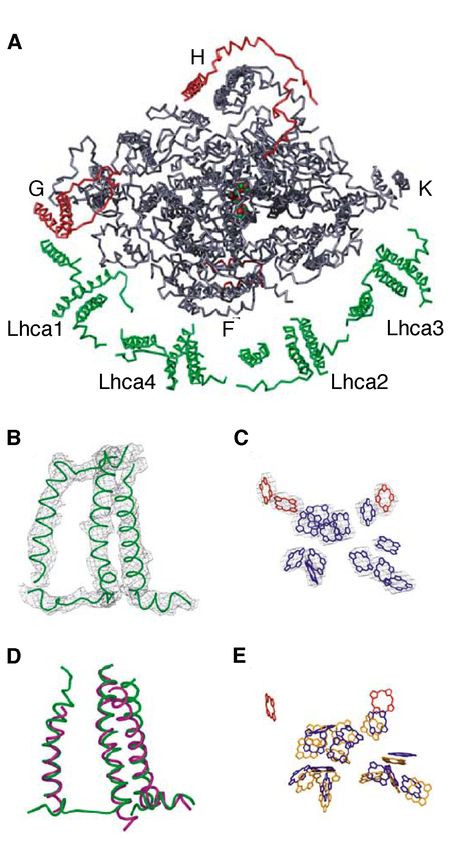
Figure 6. Higher plant PSI. A. Structural
model at 4.4 Å of higher plant PSI seen from
the stroma. The positions of subunits F, G, H
and K of the reaction centre are indicated.
The light-harvesting antenna proteins Lhca1-
Lhca4 are coloured green. Structural
elements not present in cyanobacterial PSI
are in red. The three Fe4-S4 clusters are
shown as red (Fe) and green (S) balls. B-E.
Structural comparison of Lhca and LHCII
monomers. B. Ca backbone of Lhca4. C .
Relative locations of the chlorophylls of
Lhca4. Chlorophylls with parallells in LHCII
are shown in blue and additional linker
chlorophylls in red. D. Superposition of
Lhca2 (green) and LHCII (magenta) Ca
backbones. E. Comparison of the chlorophyll
positions in Lhca2 (blue and red) and LHCII
(yellow) (Ben-Shem et al 2003). Printed with
permission.
The peripheral antenna of plant PSI is
composed of four polypeptides, Lhca1-4,
which bind to one side of the core
complex (Boekema et al., 2001b). The crystal structure confirmed the attachment of LHCI as
a half-moon-shaped belt, consisting of two moieties, binding to the PsaF side of the complex
(Ben-Shem et al., 2003a). The antenna of PSI can be separated in sucrose gradients into two
sub-fractions with differing protein composition and content, denoted LHCI-730 and LHCI-
680, based on their spectroscopic properties (Lam et al., 1984; Knoetzel et al., 1992). LHCI-
730 consists of heterodimers of Lhca1 and Lhca4 (Jansson et al., 1996; Schmid et al., 1997)
and LHCI-680 of Lhca2 and Lhca3. The oligomeric state of LHCI-680 has not been
established, but it seems to occur as dimers in vivo (Jansson et al., 1996; Croce et al., 2002;
Ben-Shem et al., 2003a). It has been suggested earlier that six to eight LHC proteins bind
each PSI (Bassi and Simpson, 1987; Jansson et al., 1996; Croce et al., 2002), while
crystallographic studies indicate that only one heterodimer each of Lhca1/Lhca4 and
Lhca2/Lhca3 is associated with PSI (Figure 6A; Ben-Shem et al., 2003a). However, this
model may be a simplification of the native nature of LHCI, since it has been previously
shown that the Lhca composition may vary (Paper I; Paper II; Bossman et al., 1997;
Andersson et al., 2003a) and it also changes according to the light regime (Bailey et al.,
2001). The pre-solubilization treatments and/or size separation prior to PSI crystallisation
22might have caused any additional LHC proteins to be detached from the complex in the cited
investigations. The pigment composition of the different Lhca proteins has been extensively
studied in preparations of native LHCI complexes and reconstitution studies (see, for
example, (Croce et al., 2002; Schmid et al., 2002), but no consensus has been reached. The
crystallisation studies (Ben-Shem et al., 2003a) have shown that the hypothesized structural
similarity among LHC proteins is correct, at least, for Lhca proteins (Figure 6B), and that
LHCI collectively binds 56 chlorophylls, nine of which do not have counterparts in LHCII.
These linker chlorophylls were identified between the individual Lhca monomers as well as
between monomers and the core. They can probably be lost during LHCI particle preparation
procedures, and not incorporated during reconstitution experiments, causing some of the
discrepancies in results concerning pigment stoichiometry.
The red chlorophylls
In addition to the bulk antenna chlorophylls, which have absorption maxima at about 680
(chlorophyll a) and 650 (chlorophyll b) nm, PSI also contains a small number of “red”
chlorophylls with shifted maxima caused by pigment-protein interactions and/or tighter
pigment-pigment interactions due to dense chlorophyll packing in LHCI. The red
chlorophylls in higher plants are present both in the core and in LHCI, red forms in the core
fluorescing at about 720 nm and in LHCI at about 735 nm (for a review, see Gobets and van
Grondelle, 2001). About 80% of the red chlorophylls are thought to be associated with LHCI
(Croce et al., 1998). These pigments, even though in a minority, contribute significantly to
the spectroscopic features of PSI.
It has been shown that the presence of the red chlorophylls significantly slows the rate of
charge separation (Gobets et al., 2001). Therefore, the red forms may at first seem to be
flaws in the system. However, since the rate of charge separation is higher than the rate at
which excitons are lost, the quantum efficiency of charge separation is not significantly
affected by the red chlorophylls. The biological function of the red pigments has been
debated, and a number of functions have been proposed (see for example Gobets and van
Grondelle, 2001). A possible role is in photoprotection. Since slowing down the overall
trapping in PSII enhances the efficiency of non-photochemical quenching mechanisms in
PSII, (Jennings et al., 1996) the same might be true for PSI. If so, the red pigments may have
a photoprotective role. However, there is no evidence at present suggesting that a process
similar to feedback de-excitation in PSII occurs in PSI. The red chlorophylls absorb light at
energies lower than that of the reaction centre, P700, raising questions about whether these
pigments participate in light harvesting. Even so, at room temperature enough thermal
energy is available to allow “uphill” transfer at a considerable rate to the bulk antennae and
P700. In normal daylight, when red pigments have low absorption intensity, this will not
have a significant effect on light harvesting. However, in dense vegetation systems, with
23light enriched in wavelengths higher than 690 nm, the presence of red chlorophylls increases
the absorption cross-section, which should significantly enhance the energy capture capacity
of the PSI complex. In fact, it has been shown that under shade conditions, red pigments are
responsible for about 40% of the total absorption flux in photosynthetic systems (Rivadossi
et al., 1999).
Regulatory mechanisms
Because of their sessile nature, the ability of plants to respond and acclimate to the
environment is imperative, and the development of plants is to a great extent under the
control of environmental cues. Light is an important variable for plant development,
influencing morphological parameters such as leaf size and thickness, leaf tissue structure,
stem elongation, flowering and senescence. The quality of light and the diurnal rhythm are
sensed by photoreceptors, phytochrome (Neff et al., 2000) and cryptocrome (Cashmore et
al., 1999), and the quantity of light is
believed to be sensed through the redox state
of the PQ pool (Pfannschmidt, 2003). Even
though light is a prerequisite for
photosynthesis, too much light is harmful
for photosynthesising organisms. The
intermediates and by-products of the
photosynthetic process, for example reactive
oxygen species, can damage the components
of the photosynthetic apparatus (photo-
oxidative damage). This process occurs at
all light intensities. However, the risk of
photo-oxidative damage is greatly
intensified under conditions of excess light
when the absorbed energy exceeds the
plant’s photochemical capacity. This is
likely to occur in situations with increased
or highly fluctuating irradiation, chilling
temperatures or limiting CO2 levels. In
nature, where the light intensity can vary
over several orders of magnitude in a matter
Figure 7. Schematic representation of photo-
of seconds due to shading by trees and
protective mechanisms in plants. clouds, optimisation of the photosynthetic
machinery requires precise regulation.
Because of the obvious adaptive benefits of being able to acclimate to such changes, a
number of photoprotective mechanisms have evolved (Figure 7; Niyogi, 1999).
24Photo-oxidative damage
During photosynthesis a number of oxidising molecules are formed, such as singlet oxygen
(1O2), superoxide radicals (O2-l), hydrogen peroxide (H2O2) and hydroxyl radicals (l OH).
These are chemically aggressive molecules, capable of causing irreversible damage to the
cell. Reactive oxygen species can arise in several ways. In the antenna singlet chlorophylls
(1Chl) are formed upon excitation, and the excess energy is subsequently transferred through
the light-harvesting complex. From 1Chl, triplet chlorophyll (3Chl) can be formed in the
antenna through intersystem crossing. In comparison with 1Chl, 3Chl is relatively long-lived
and in interactions with O2 it can produce 1O2. Formation of oxidising molecules also occurs
in PSII. Inevitably, given its ability to split water into protons and O2, P680+ has an
extremely high oxidizing potential, which can cause damage to the reaction centre (Anderson
et al., 1998). 1O2 can also be formed by interaction between O2 and 3P680, which can occur
when the electron transport chain is obstructed. In addition, at very low light levels, if the
time between consecutive higher-light flashes is too long, back-flow of electrons from QB- to
P680 may generate 3P680 (Keren et al., 1997). Through the Mehler reaction, the acceptor
side of PSI can reduce O2 to O2-, which can be metabolised to H2O2. Diffusion of O2- and
H2O2 through the chloroplast will destroy sensitive molecules such as metal proteins.
Subsequently released Cu2+ or Fe3+ ions can interact with H2O2, catalysing the formation of
the extremely toxic radical lOH (Asada, 1999).
Avoiding absorption of excess light
Lhc gene expression
The most effective step to avoid damage to the photosynthetic apparatus is to decrease the
amount of light absorbed by the antenna. Long-term light avoidance mechanisms include
minimizing the irradiated leaf surface through leaf movements, and enhancing leaf
reflectance, for example by epicuticular wax layers. Light absorption can also be decreased
by internal mechanisms, including relocation of chloroplasts within the cells and the
accumulation of screening compounds such as anthocyanins (Steyn, 2002). Growth
irradiance influences the reaction centre content (Walters et al., 1999; Bailey et al., 2001) as
well as the functional antenna size. Changes in the chlorophyll a/b ratio in response to
different levels of growth irradiance occur mainly through changes in LHCII content
(Anderson, 1986; Larsson et al., 1987a; Mäenpää and Andersson, 1989; Bailey et al., 2001).
It has been found that the levels of the other LHC proteins also vary according to the growth
light intensity (Bailey et al., 2001). These changes in protein content can be regulated by
modulating translation (Flachmann and Kühlbrandt, 1995), protein degradation (Lindahl et
al., 1995) or Lhc gene expression.
25Lhc genes respond to a number of stimuli (see for example, Grossman et al., 1995) and
references therein; Anderson and Kay, 1995; Hamazato et al., 1997; Piechulla, 1999;
Mazzella et al., 2001) During the development of a plant, there are remarkable changes in
Lhc gene expression. This process is partly controlled by a family of photoreceptors,
phytochromes, whose biological activity is modulated by red and far-red light. In addition,
Lhc gene transcription is controlled by blue light. The developmental stage of the plant
influences Lhc gene activity by increasing Lhc mRNA levels during hypocotyl emergence
and decreasing levels during senescence. Plant growth regulators, such as abscisic acid and
methyl jasmonate, also modulate Lhc gene expression, as well as organ- and tissue- specific
sequence elements. Lhc genes are also regulated in a circadian fashion: expression increasing
at or shortly prior to the beginning of the light period. The mRNA levels rise to a maximum
at midday and decrease to a minimum after midnight. Oscillating Lhc transcript levels allow
the coordination of LHC synthesis with light availability (Piechulla, 1999).
The expression of the Lhc genes is also regulated by signals originating in the plastid.
Multiple processes in the plastid influence these signals, which regulate the expression of
nuclear genes, but the nature of the signals and the signalling pathways involved are not well
understood (Surpin et al., 2002). There is considerable evidence suggesting that at least two
independent pathways are involved, mediated by tetrapyrroles and the redox state of the PQ
pool. It has been shown that expression of a number of photosynthetic genes, including Lhc
genes, is repressed by accumulation of the chlorophyll intermediate Mg-protoporphyrinIX
(Strand et al., 2003). The redox state of the PQ pool can be manipulated by adding the
inhibitors DCMU and DBMIB, which oxidise (at low excitation pressure) and reduce (at
high excitation pressure) PQ, respectively. Using this approach, Lhc gene expression was
found to be high at low excitation pressure and low at high excitation pressure in Dunaliella
tertiolecta in a study by Escoubas et al. (1995). It has also been shown that the
phosphoenolpyruvate/phosphate translocator, which is required for PQ synthesis, is
necessary for plastid-dependent nuclear gene expression (Streatfield et al., 1999). The
thylakoid protein TSP9, which is phosphorylated and released from the membrane upon
illumination, has been suggested to participate in cell signalling in response to changes in
light conditions (Carlberg et al., 2003). In addition, reactive oxygen species have been
proposed to be involved in the regulation of photosynthetic nuclear genes (Rodermel, 2001;
Mullinaux and Karpinski, 2002).
State transitions
The relative effective antenna size of PSII and PSI can also be altered over a short period of
time through state transitions (reviewed in Haldrup et al., 2001; Kruse, 2001; Wollman,
2001). The outer antenna of PSII has been shown to have two pools of LHCII, an inner and a
peripheral pool, the latter of which participates in state transitions (Larsson et al., 1987b).
26Since PSI and PSII absorb light of different wavelengths, their excitation must be balanced
to ensure efficient photosynthetic performance. Light regimes favouring PSII will generate
redox conditions in the thylakoid that activate a protein kinase which phosphorylates the
peripheral pool of LHCII. Upon phosphorylation, phospho-LHCII detaches from PSII and
associates with PSI, thereby increasing the functional PSI antenna size. Whether or not PSI
participates in state transitions has been debated. However, Arabidopsis plants that are
deficient in the PsaH subunit of PSI cannot perform state transitions, demonstrating the
importance of PSI in state transitions (Lunde et al., 2000). In addition, LHCII is still
phosphorylated in these plants and continues to transfer energy to PSII, showing that LHCII
phosphorylation per se does not cause LHCII migration. Nevertheless, there is evidence that
phosphorylation is a prerequisite for state-transitions. It has been shown that the capacity for
state transitions is reduced in plants with reduced activity of the kinase responsible for
LHCII phosphorylation (Snyders and Kohorn, 2001) and a Chlamydomonas mutant defective
in state transitions has been shown to be devoid of phosphorylated LHCII (Fleischmann et
al., 1999; Kruse et al., 1999).
Even though state transitions have been suggested to play a role in photoprotection, there is
no evidence that they are photoprotective in excess light. It has been shown that the LHCII
kinase is active when a plastoquinol molecule is bound to the Qo site of cyt b6/f (Vener et al.,
1997). However, there is evidence that the LHC kinase is inactivated in high light (see for
example Rintamäki et al., 1997; Pursiheimo et al., 1998) and also that the regulation of the
kinase is modulated by thiol reagents, which inhibit LHCII phosphorylation (Rintamäki et
al., 2000). Therefore, state transition probably regulates excitation energy distribution
between PSI and PSII rather than protecting the plant against excess light.
Eliminating excess light energy
Excess absorbed light can be thermally dissipated in the antenna of PSII through feedback
de-excitation, qE. Absorption of excess light will cause a build-up of the trans-thylakoid
DpH, which may also be maintained by cyclic and pseudocyclic electron transport (Asada,
1999; Munekage et al., 2002). The increased acidification of the thylakoid lumen results in
protonation of several antenna proteins, and the activation of violaxanthin de-epoxidase
(VDE; Rockholm and Yamamoto, 1996). VDE participates in the conversion of violaxanthin
associated with LHCs to zeaxanthin via antheraxanthin in the so-called xanthophyll cycle
(Demmig-Adams, 1990). Both of these mechanisms are thought to cause steric changes in
several antenna proteins, which induce qE. The PsbS protein has been shown to be
indispensable for qE, since Arabidopsis plants lacking PsbS cannot perform feed back de-
excitation (Li et al., 2000). In addition, a study of the Arabidopsis npq1 mutant, which is
deficient in VDE, has shown that a functional xanthophyll cycle is required for inducing qE
(Niyogi et al., 1998). The requirement of zeaxanthin for qE has been proposed to be due to
27You can also read