Obesity and COVID-19: Molecular Mechanisms Linking Both Pandemics - MDPI
←
→
Page content transcription
If your browser does not render page correctly, please read the page content below
International Journal of
            Molecular Sciences

Review
Obesity and COVID-19: Molecular Mechanisms
Linking Both Pandemics
Andreas Ritter * , Nina-Naomi Kreis , Frank Louwen and Juping Yuan *
 Division of Obstetrics and Prenatal Medicine, Department of Gynecology and Obstetrics, University Hospital
 Frankfurt, J.W. Goethe-University, Theodor-Stern-Kai 7, D-60590 Frankfurt, Germany;
 Nina-Naomi.Kreis@kgu.de (N.-N.K.); Frank.Louwen@kgu.de (F.L.)
 * Correspondence: Andreas.Ritter@kgu.de (A.R.); yuan@em.uni-frankfurt.de (J.Y.); Tel.: +49-69-6301-83297

 Received: 24 July 2020; Accepted: 10 August 2020; Published: 12 August 2020

 Abstract: The coronavirus disease 2019 COVID-19 pandemic is rapidly spreading worldwide and is
 becoming a major public health crisis. Increasing evidence demonstrates a strong correlation between
 obesity and the COVID-19 disease. We have summarized recent studies and addressed the impact of
 obesity on COVID-19 in terms of hospitalization, severity, mortality, and patient outcome. We discuss
 the potential molecular mechanisms whereby obesity contributes to the pathogenesis of COVID-19.
 In addition to obesity-related deregulated immune response, chronic inflammation, endothelium
 imbalance, metabolic dysfunction, and its associated comorbidities, dysfunctional mesenchymal
 stem cells/adipose-derived mesenchymal stem cells may also play crucial roles in fueling systemic
 inflammation contributing to the cytokine storm and promoting pulmonary fibrosis causing lung
 functional failure, characteristic of severe COVID-19. Moreover, obesity may also compromise motile
 cilia on airway epithelial cells and impair functioning of the mucociliary escalators, reducing the
 clearance of severe acute respiratory syndrome coronavirus (SARS-CoV-2). Obese diseased adipose
 tissues overexpress the receptors and proteases for the SARS-CoV-2 entry, implicating its possible roles
 as virus reservoir and accelerator reinforcing violent systemic inflammation and immune response.
 Finally, anti-inflammatory cytokines like anti-interleukin 6 and administration of mesenchymal
 stromal/stem cells may serve as potential immune modulatory therapies for supportively combating
 COVID-19. Obesity is conversely related to the development of COVID-19 through numerous
 molecular mechanisms and individuals with obesity belong to the COVID-19-susceptible population
 requiring more protective measures.

 Keywords: COVID-19; obesity; adipose-derived mesenchymal stem/stromal cells; cytokine storm;
 immune response; inflammation

1. Introduction
     The severe acute respiratory syndrome coronavirus (SARS-CoV) in Asia in 2002/2003 and the
Middle East respiratory syndrome coronavirus (MERS-CoV) in the Middle East in 2012 caused
severe respiratory diseases [1,2]. Currently, a novel SARS-like coronavirus 2 (SARS-CoV-2) is rapidly
spreading over the world, resulting in the global coronavirus disease 2019 (COVID-19). COVID-19
causes high morbidity and mortality worldwide and the World Health Organization (WHO) officially
declared this outbreak as pandemic [3]. Advancing progression of the infected lung leading to SARS is
recognized as the most common complication of this disease. As of August 8, 2020, there have been
19,187,943 confirmed cases of COVID-19, including 716,075 deaths worldwide (WHO dashboard) [4].
The survival of severe COVID-19 might be followed by multiple long-term complications including lung
insufficiency, vascular dysfunction, and heart failure. The COVID-19 pandemic poses a devastating
challenge to the global health system and the economy.

Int. J. Mol. Sci. 2020, 21, 5793; doi:10.3390/ijms21165793                         www.mdpi.com/journal/ijms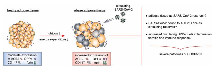
Int. J. Mol. Sci. 2020, 21, 5793                                                                       2 of 28

     The dramatic increase in the prevalence of obesity represents another urgent global challenge.
Over the last decades, obesity has become a major epidemic in most industrialized countries,
with extensive consequences for their health care systems [5]. This is exemplified by the fact that
the percentage of children and adults with overweight has doubled, and even tripled in adolescents
over the last three decades [6]. Obesity, commonly defined by the body mass index (BMI), is induced
by an imbalance between food intake and energy expenditure [7]. Importantly, obesity is highly
associated with a variety of disorders like cardiovascular diseases, insulin resistance, type 2 diabetes
mellitus (T2DM), and various cancers [8,9]. These associations are likely connected to the hallmarks of
obesity, which are characterized by a dysfunctional energy homeostasis promoting the development
of the metabolic syndrome [10], diseased adipose tissues associated with local inflammation [11],
hypoxia and cellular stress [11], altered release of cytokines [12], chronic systemic inflammation [13],
insulin resistance [14], impaired immune response [15], atherosclerosis, and altered cardiac structure
and function connected to a highly increased risk of cardiovascular diseases (CVD) [16]. Most of
these hallmarks have direct or indirect negative impact on the well-balanced immune surveillance
system leading to an impaired immune response, flawed chemotaxis, and deregulated immune cell
differentiation [17]. Consequently, it is well documented that obesity is associated with an enhanced risk
as well as worse course of infectious diseases with increased complications, prolonged hospitalization,
and elevated critical illnesses [18–20].
     The present coincidence of the COVID-19 and obesity pandemics exponentiates the aforementioned
problems. Increasing evidence demonstrates that obesity is reversely associated with the development
of COVID-19. In this review, we summarize the data of recent studies regarding the relationship
between obesity and COVID-19 in terms of various clinical parameters and discuss the potential
molecular linkages between these two pandemics, with a focus on a special cell population, namely,
mesenchymal stem/stromal cells (MSCs).

2. Clinical Correlation of COVID-19 with Obesity
     Studies concerning the H1N1 influenza A virus (H1N1) infection in 2009 identified obesity as a
classified risk factor for infected patients for the first time, showing an increased severity of the disease
and elevated mortality of patients [18]. This negative correlation between H1N1 infection and obesity
was mainly associated with a delayed and reduced antiviral response towards the viral infection [18,21],
poor effectiveness of antiviral drugs and vaccines [18,22,23], and boosted viral shedding/transmission
as well as impaired clearance [18]. The correlation between obesity and COVID-19 was not clear at
the beginning of this pandemic. Early COVID-19 studies, for example, from the “Chinese Center of
Disease Control and Prevention” with 72,314 cases, did not provide sufficient patient data like body
weight and height for calculating the BMI of patients [24]. This lack of data has changed recently,
when multiple studies with large patient collectives were published from the UK [25], Mexico [26,27],
and USA [28]. We have collected clinical reports (Table 1), which are supplemented with body weight,
height, or the BMI, based on the publications in several online databases like PubMed, bioRxiv, Web of
Science, etc. as of 24 July 2020.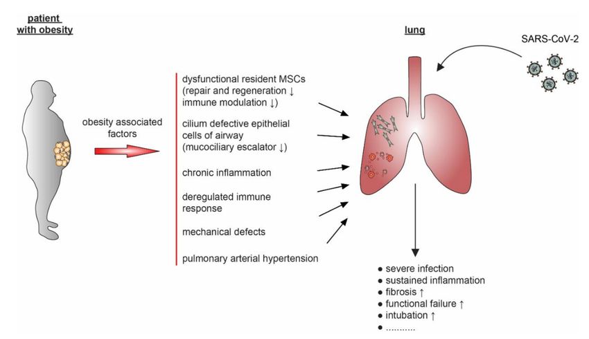
Int. J. Mol. Sci. 2020, 21, 5793                                                                                                                                                                    3 of 28

                                                                     Table 1. Clinical reports of COVID-19 patients with obesity.

   Case              BMI (kg/m2 )      Hazard Ratio (HR)/                 Country/City      Clinical Relevance                                                               Author/Year
   Number                              p-Value
      20,133         lean
     14,396          obese             1.33 (1.19–1.49, p < 0.001)        UK/multicenter    Obesity increased significantly the risk for hospitalization and mortality.      Docherty et al. 2020
       1685
       3003          40                                                                    any cardiovascular or pulmonary disease.
      51,633         lean              Lethality:                                           Obesity raises the risk of an infection with COVID-19. It increases mortality
      40,925         obese             1.25 (1.17–1.34, p < 0.001)        Mexico/Mexican    and diseases severity.                                                           Bello-Chavolla et al. 2020
     10,708
                                                                                            High frequency of obesity among patients admitted in intensive care.
                     25–30             1.69 (0.52–5.48) 0.22                                Disease severity increased with BMI.
        124          30–35             3.45 (0.83–14.31) 0.48             France/Lille      Obesity was a risk factor for COVID-19 severity.                                 Simonnet et al. 2020
                     ≥35               7.36 (1.63–33.14) 0.021                              Patients with obesity required more mechanical ventilation (BMI ≥ 35 at
                                                                                            85.7%).
                     age ≥ 60 years
        141          30–34             0.9 (0.6–1.2) 0.39                                   Obesity was a risk factor for hospital admission and patients with obesity
         99          ≥35               0.9 (0.6–1.3) 0.59                 USA/New York      needed more critical care.                                                       Lighter et al. 2020
        173          age < 60 years    2.0 (1.6–2.6) < 0.0001                               Obesity in people < 60 years is a newly identified epidemiologic risk factor,
        134          30–34             2.2 (1.7–2.9) < 0.0001                               which may contribute to increased morbidity rates documented in USA.
                     ≥35
                     24.4 (all)
                     26.0                                                                   Deceased patients had a slightly yet not significant increased BMI
        102          (non-survivors)   0.088                              China/Wuhan       compared to survived patients.                                                   Cao et al. 2020
                     24.3
                     (survivors)
        383          18.5–23.9
        203          24.0–27.9         1.86 (1.00–3.46) 0.05              China/Shenzhen    Obesity, especially in men, significantly increased the risk for developing      Cai et al. 2020
        123          ≥28               3.42 (1.42–8.27) 0.006                               severe pneumonia in COVID-19 patients.
         41
                                                                                            Obesity and diabetes mellitus were associated with greater OR of needing
        422                            2.04 (1.14–3.65) 0.016             USA/              hospitalization and increased risk of pneumonia.
         71          ≥30               association with age:              Los Angeles       Obesity, diabetes, or an elevated overall comorbidity index were                 Ebinger et al. 2020
Int. J. Mol. Sci. 2020, 21, 5793 4 of 28
Table 1. Cont.
Case BMI (kg/m2 ) Hazard Ratio (HR)/ Country/City Clinical Relevance Author/Year
Number p-Value
1158 < 25
266 30–34.9 BMI > 40: Kuwait/Kuwait Overweight, obesity and diabetes were associated with intensive care and
98 35–39.9 3.95 (1.00–15.20, p = 0.046) city poor outcomes of patients with COVID-19. Al-Sabah et al. 2020
40 >40
19
mortality:
200Int. J. Mol. Sci. 2020, 21, 5793 5 of 28
Table 1. Cont.
Case BMI (kg/m2 ) Hazard Ratio (HR)/ Country/City Clinical Relevance Author/Year
Number p-Value
387 30 p < 0.001 Italy/Milan significant higher BMI above 29.9. Acute respiratory distress and male sex Chiumello et al. 2020
41 are associated with obesity class I to III.
mortality
176 30 higher age of surviving Greece/Athen mortality in critically ill COVID-19 patients. Halvatsiotis et al. 2020
59 patients
p = 0.007
BMI, the body mass index; ICU, intensive care unit, OR, odds ratio; MV, mean value.Int. J. Mol. Sci. 2020, 21, 5793 6 of 28
Docherty and colleagues showed that obesity was significantly associated with a higher mortality
(HR (Hazard Ratio): 1.33 (1.19–1.49, p < 0.001)) in a patient cohort of overall 20,133 cases [25].
This observation is supported by the New York study with 10,544 COVID-19 patients demonstrating
that patients with a BMI of 30–40 kg/m2 had a significantly increased risk for hospitalization (HR: 4.26
(3.5–5.2, p < 0.001)) and clinical progression (HR: 1.38 (1.03–1.85, p = 0.029) compared to patients with a
BMI below 30 kg/m2 [28]. Further support comes from a Mexican study with 4103 COVID-19 cases
showing that patients with a BMI of ≥30 kg/m2 had a significantly reduced survival (HR: 1.74 (1.35–2.26,
p < 0.001)) and increased hospitalization rate (HR: 1.64 (1.37–1.95, p < 0.001)) [26]. Another Mexican
study with 51,633 patients showed similar results, with a significant increased lethality rate in patients
with obesity (HR: 1.25 (1.17–1.34, p < 0.001) [27]. These reports with large patient cohorts are further
strengthened by multiple clinical studies with smaller collectives worldwide [29–44]. Interestingly,
Lighter et al. and Ebinger et al. showed that middle aged patients with an age below 52 and up to
60 years were more affected by obesity (BMI 30–34 and ≥35) with an HR: 2.0 (1.6–2.6, p < 0.0001)) and
HR: 2.2 (1.7–2.9, p < 0.0001), respectively, resulting in increased morbidity rates compared to >60 years
old patients [31,43]. Surprisingly, in the study by Ebinger and colleagues, obesity displayed the highest
association with overall COVID-19 illness severity (p < 0.0001) correlated with patient age (Int. J. Mol. Sci. 2020, 21, 5793 7 of 28
inflammation as well as energy homeostasis [51]. Adipocytes in healthy AT are insulin-sensitive,
a trait essential for adipocyte glucose uptake and for the modulation of hepatic gluconeogenesis,
which enables the maintenance of normal blood glucose levels [52]. AT consists of lipid-filled mature
adipocytes and several types of stromal cells including fibroblasts, adipose-derived mesenchymal
stromal/stem cells (ASCs), endothelial cells, and various immune cells [53]. Interestingly, nearly all
immune cells, such as resident macrophages, mast cells, monocytes, dendritic cells, natural killer cells,
B cells, T cells, neutrophils, and eosinophils, have been found in AT [54].
Adipose Tissue in Obesity
Obesity changes the composition, structure, and function of AT. Responding to excessive calorie
intake, AT undergoes expansion via two processes: hyperplasia (increase in adipocyte number)
and hypertrophy (increase in adipocytes size), and it is also influenced by endoplasmic reticulum
stress, metabolic endotoxemia, or adipocyte death [55]. Expansion of adipocytes and insufficient
vascularization lead to hypoxia; adipocyte apoptosis/necrosis; irregular fatty acid flux; and enhanced
secretion of inflammatory adipokines, cytokines, and chemokines. This causes a massive immune
cell infiltration that further promotes inflammation, stimulates lipolysis, and fuels insulin resistance,
resulting in adipocyte dysfunction [56]. As a consequence, AT develops a local low-grade inflammatory
microenvironment, which recruits inflammatory M1 macrophages, T cells, B cells, neutrophils, and
mast cells. By contrast, the populations of T helper type 2 (Th2), M2 macrophages, and regulatory
T cells (Treg) remain or even decrease in later stages of obese AT [11,57,58]. This shifts the balance
from a regulatory anti-inflammatory immune state with the secretion of immunoregulatory cytokines
including interleukin-4 (IL-4), IL-5, IL-10, IL-13, and IL-33 to a highly inflammatory state causing the
secretion of monocyte chemoattractant protein-1 (MCP-1), tumor necrosis factor α (TNF-α), IL-1β,
interferon γ (IFN-γ), and IL-6, leading to the development of a chronic and systemic inflammation [17].
Among proinflammatory cytokines, IL-1 family cytokines, as IL-1 and IL-18, are highly associated
with obesity induced metabolic complications [59]. The production of these proinflammatory cytokines
is mediated by NLRP3 (NOD-, LRR-, and pyrin domain-containing protein 3) inflammasome, which is
crucial in immune responses, glucose homeostasis, lipid metabolism, and adipocyte functions.
Multiple studies have reported that NLRP3 is activated in obese AT and associated with metabolic
disorders [60]. Elevated inflammatory cytokines IL-6 and TNF-α are majorly produced by increased M1
macrophages in obese AT [61] and their altered circulating levels have been reported in patients with
overweight and obesity [62], contributing to a local as well as systemic chronic inflammation. Further,
the “trans-signaling” of the soluble form of the IL-6 receptor was shown to recruit macrophages into
AT [63] and Mauer et al. highlighted the important role of IL-6 in the polarization of macrophages into
the M2 subtype [64,65], both mechanisms are likely involved in the inflammatory maladaptation of
obese AT.
This process is likely connected to a gradual loss of functional ASCs, a type of MSCs, in AT by
reducing their differentiation, motility and immunomodulation capacity as well as impairing their
ciliogenesis and pro-angiogenic ability [66]. Inflammation and its associated inflammatory cytokines,
including IL-6, TNF-α, IL-1β, and inflammatory factors like C-reactive protein (CRP), are all known
to induce endothelial dysfunction in AT [67]. Moreover, the deregulated expression of adipokines
like leptin and resistin in AT of obese patients causes increased expression of vascular cell adhesion
molecule 1 (VCAM-1) and intercellular adhesion molecule 1 (ICAM-1), both leading to vascular
dysfunction and oxidative stress [68]. These factors contribute to dysfunctional/damaged endothelial
cells and reduced angiogenesis worsening the hypoxic state of AT [69]. The hypoxic state induces
the upregulation of hypoxia-inducible factor 1α that promotes fibrosis in an AT-specific manner
by crosslinking collagen [57,70]. As a result, the extracellular microenvironment loses its flexibility,
increases mechanical stress, restrains adipocyte expansion, and triggers adipocyte cell death and a
sustained immune response in AT.Int. J. Mol. Sci. 2020, 21, 5793 8 of 28
4. Obesity and COVID-19: Pathological Molecular Linkages
A great body of studies demonstrates a strong association between obesity, obesity-related
comorbidities, and the severe outcomes of COVID-19. In the following, we discuss the issues that link
obesity to the severe outcomes of COVID-19.
4.1. ASCs, MSCs, Obesity, and COVID-19
Among various cell types, ASCs are a specialized fundamental cell population in AT. These cells
reside in human AT with multipotent differentiation features [71]. ASCs conduct their complex biological
functions by direct cell–cell interaction and by indirect interaction through secretion of a variety of
bioactive factors and microvesicles, exerting diverse functions including homeostasis, cell renewal,
spontaneous repair, angiogenesis, immune modulation, and inflammation regulation [72–74]. Moreover,
ASCs/MSCs have been shown to trigger changes in the energy metabolism, cell homeostasis,
epithelial-to-mesenchymal transition, and fibrosis by stimulating various cellular pathways [75,76].
Interestingly, over the last two decades multiple investigations have revealed that morbid obesity
compromises almost all ASC functions and properties, and changes their regulatory protective activity
into a hypoxia and inflammation promoting phenotype [11,66]. In obesity, these cells lose their main
features accompanied by a decrease in their multipotent state on account of reduced expression of
pluripotency-associated genes like octamer-binding protein 4 (OCT4), SRY-Box transcription factor
2 (SOX2), homeobox transcription factor Nanog (NANOG), RNA exonuclease 1 homolog (REX1),
and homeobox protein hox-C10 (HOXC10) [77,78]. Furthermore, ASCs/MSCs from obese patients
are characterized by increased secretion of inflammatory cytokines including TNF-α, IL-8, IL-6,
and MCP-1 [77,79]; reduced endothelial cell differentiation evinced by decreased secretion of vascular
endothelial growth factor A (VEGF), hepatocyte growth factor (HGF), fibroblast growth factor 2 (FGF2),
and platelet-derived growth factor (PDGF) [80,81]; and altered immune modulation capacity such as
the inhibition of lymphocyte proliferation, recruitment of inflammatory monocytes and polarization
of M1 macrophages [61,82], changed intracellular metabolism manifested by increased reactive
oxygen species (ROS) production, impaired mitochondria function, and downregulation of sirtuin
1-6 (SIRT1-6) [11,83,84]. Moreover, as we reported, obesity impairs the ASC primary cilium, a unique
signaling organelle on the cell surface, further contributing to their dysfunction in obese patients [66].
Interestingly, obesity-associated cytokines like IL-6 and TNF-α are able to influence many signaling
pathways and induce defects in the ciliogenesis of ASCs [66,77,85]. Ciliary defective ASCs are not
able to fulfill their physiological functions. Instead, these cells fuel inflammation in AT by secreting
multiple inflammatory cytokines as IL-6 and IL-8 [66,85].
AT with its ASCs is present in various locations and in diverse organs. Of importance, ASCs are
resident within the perivascular adipose tissue (PVAT) showing differentiation capacity towards
adipocytes, endothelial cells, smooth muscle cells (SMCs), as well as osteoblasts for repair and
replacement [86–88]. These cells also alter their functionality during the development of obesity,
negatively affecting homeostasis and integrity of the endothelium, one of the major targets
of SARS-CoV-2.
Moreover, patients with obesity may lose their functional ASCs in AT as well as MSCs in
various organs including brain and lung [11,66]. Importantly, current clinical evidence suggests that
pulmonary fibrosis or fibrous stripes is a severe complication observed in COVID-19 patients [89].
Pan and colleagues observed that 17% of hospitalized COVID-19 patients presented fibrous stripes [90].
Multiple studies demonstrate that loss or dysfunctional resident stem/MSC-like cells are responsible
for the development of pulmonary fibrosis [91–93]. It has been revealed that resident lung perivascular
ATP-binding cassette superfamily G member 2+ (ABCG2+ ) MSCs were decreased in human pulmonary
fibrosis [93]. Moreover, perivascular glioma-associated oncogene+ (Gli1+ ) MSC-like cells, residing in
various organs including lung, kidney, liver, and heart, generated myofibroblasts playing a central role
in organ fibrosis after injury [92]. These data are underscored by Xie and colleagues reporting that
transcription factor T-box gene 4 (TBX4)-lineage mesenchymal progenitors are the predominant sourceInt. J. Mol. Sci. 2020, 21, 5793 9 of 28
of myofibroblasts in injured adult lung [91]. In support of this notion, bleomycin-induced fibrosis is
also associated with loss of resident lung MSCs [94]. In addition, Wnt/β-catenin signaling has been
suggested to be an essential mechanism underlying the regulation of myofibroblast differentiation
of lung-resident MSCs participated in the development of pulmonary fibrosis [95]. Thus, COVID-19
patients with obesity may suffer a more severe lung injury with pulmonary fibrosis owing to loss or
dysfunctional lung resident MSCs induced by obesity. These MSCs in individuals with obesity may
not be able to defend SARS-CoV-2 by modulating the immune response, tissue repair and homeostasis,
and anti-inflammation. Conversely, obese ASCs/MSCs may further promote systemic inflammation
and negatively impact the immune response [11].
Interestingly, ASCs/MSCs have been reported as cellular therapy option for pulmonary fibrosis
and bronchopulmonary dysplasia by regulating immune cells inside the lung tissue, modulating
trans-differentiation of different cell types including fibroblasts, and secreting various growth
factors [96,97]. Furthermore, these cells could be used in infectious diseases because of their
immunosuppressive or immune enhancement effects depending on their cytokine production and
on the context of the immune response [98]. Many scientific and clinical trials are still ongoing to
decipher how MSCs/ASCs regulate the immune system and several studies have shown their potential
in regulating T cell proliferation or interacting with pathogens [99,100]. These cells even possess
antimicrobial potential [101], highlighting the crucial roles of ASCs/MSCs in infectious diseases as well
as their possible implications as novel cellular therapy for COVID-19 patients, especially for COVID-19
patients with obesity.
4.2. SARS-CoV-2 Receptors, Proteases, Adipose Tissue, and Obesity
SARS-CoV-2 uses its viral spike (S) protein for the entry into target cells. The spike protein consists
of two functionally distinct subunits. The surface subunit S1 recognizes and binds to the cellular
receptor, whereas the transmembrane subunit S2 facilitates fusion of the viral membrane with the
cell membrane [102,103]. Like SARS-CoV, SARS-CoV-2 spike protein binds to its receptor human
angiotensin-converting enzyme 2 (ACE2) through its receptor-binding domain (RBD) mediating its
entry into host cells [104–106]. ACE2 is a zinc metalloprotease, which shares homology with ACE
in its catalytic domain [107]. It has 805 amino acids including an N-terminal signal sequence and a
C-terminal membrane binding domain [108]. ACE2 contains a single HEXXH zinc-binding motif and
inactivates the potent vasoconstrictive peptide angiotensin II (Ang II) by removing its C-terminal
phenylalanine residue to yield heptapeptide Ang-(1–7) [109]. The most remarkable expression of ACE2
protein was found on lung alveolar epithelial cells, and enterocytes of the small intestine, while ACE2 is
present in arterial and venous endothelial cells, and arterial smooth muscle cells in all organs including
oral and nasal mucosa, nasopharynx, stomach, colon, liver, kidney, and brain [110]. Moreover, based on
bioinformatic and protein docking models, it has been suggested that, like MERS-CoV, the spike RBD
of SARS-CoV-2 binds to human dipeptidyl peptidase 4 (DPP4) with a high affinity in addition to
ACE2 [111,112]. Furthermore, cluster of differentiation (CD147)/basigin is recently proposed to be an
alternative receptor for SARS-CoV-2 binding on the cell surface [113], although it is structurally not
yet validated.
To fulfill its function, SARS-CoV-2 spike protein is proteolytically activated at its S1/S2 cleavage
site by human transmembrane protease serine 2 (TMPRSS2) [104]. Apart from TMPRSS2, SARS-CoV-2
spike protein can be proteolytically activated by a variety of other proteases including furin, elastase,
factor X, and trypsin, indicating the interesting fact that coronaviruses favor as receptors various
protease proteins. These proteases are capable to perform a “priming” proteolysis that initiates the
process of cellular entry [114–116]. Particularly, the spike protein of SARS-CoV-2 harbors a S1/S2
cleavage site containing multiple arginine residues, which is cleaved by the protease furin and is
essential for spike protein mediated cell–cell fusion and entry into target cells [116]. SARS-CoV-2
depends on furin-mediated pre-cleavage of its spike protein at the S1/S2 site for subsequent spike
protein activation by TMPRSS2 in lung cells [117].Int. J. Mol. Sci. 2020, 21, 5793 10 of 28
AT expresses various receptors and enzymes required for SARS-CoV-2 infection. ACE2,
the functional receptor for SARS-CoV and SARS-CoV-2, is highly expressed in AT [118,119]. Its mRNA
was detected in human AT, with higher ACE2 expression in visceral compared to subcutaneous
AT [120,121]. Importantly, its expression is upregulated in adipocytes of patients with obesity and
diabetes [122]. In line with these observations, mouse AT and 3T3-L1 adipocytes display expression
of ACE2 mRNA, protein, and enzymatic activity, and are regulated by high fat (HF) feeding [122].
Fatty acids confer multiple effects on gene expression, potentially related to activation of the peroxisome
proliferator-activated receptor gamma (PPARγ) [123,124]. Interestingly, HF feeding induced an
abundant expression of PPARγ in AT, which is associated with robustly increased ACE2 mRNA [122].
Obesity results in ACE2 upregulation in AT of mice causing mild epicardial AT inflammation [125].
Recently, a study with 5457 COVID-19 patients showed that individuals with obesity demonstrate
significantly higher levels of ACE2 in their blood sera [126]. In sum, ACE2 is highly expressed in
adipocytes and AT, and its expression is increased in obesity, which could turn AT into a potential
target and viral reservoir, as suggested [127].
Other suggested receptors for SARS-CoV-2 are also present in AT. DPP4, the potential SARS-CoV-2
receptor, is multifunctional including its roles in glucose homeostasis, inflammation, and the immune
system [128]. Identified as a novel adipokine in AT [129], DPP4 is strongly expressed on the apical
surfaces of the polarized epithelium of various organs such as lung and liver, and increased DPP4 results
in failures to resolve inflammation and chronic subclinical activation of the immune system [128].
Interestingly, DPP4 is upregulated in obesity, especially in the insulin resistance state [129,130].
Inhibition of DPP4 prevented fibrosis in obese white AT [131]. AT, and specifically adipocytes,
have been proposed to be a significant circulating source of DPP4 [132]. DPP4 secretion from AT was
also demonstrated in vivo with greater release in obese compared to lean individuals [133]. Thus,
AT from obese patients highly expresses DPP4 and possibly is its major circulating source, which may
facilitate the entry of SARS-CoV-2 into cells and also strong inflammation and violent immune response,
important steps leading to the cytokine storm of COVID-19. It will be of high interest to decipher the
roles of DPP4 in mediating SARS-CoV-2 entry as well as in inflammation and immune response in
COVID-19 patients with or without obesity. Interestingly, DPP4 inhibitors were introduced as 2nd to
4th-line treatment option of type 2 diabetes [134], and they might be a suitable combinatory treatment
option for COVID-19 patients. In addition, the expression of CD147, the suggested alternative receptor
for SARS-CoV-2 [113], is positively correlated with the BMI possibly contributing to the COVID-19
morbidity and severity patterns [135].
Figure 1. The illustration indicates that adipose tissue expresses the receptors ACE2, DPP4, and CD147,
and the protease furin for the SARS-CoV-2 entry. These proteins are upregulated in obese adipose
tissues accompanied by an enhanced secretion of ACE2 and DPP4 in the circulation of obese patients.
Diseased adipose tissues could be targeted by SARS-CoV-2 and serve as its reservoir, as well as an
accelerator reinforcing systemic inflammation and immune response, resulting in severe outcome of
COVID-19 in obese patients.Int. J. Mol. Sci. 2020, 21, 5793 11 of 28
Moreover, the protease TMPRSS2 is expressed in AT, though at a low level [136], while furin is
enhanced expressed in obese AT as well as during adipogenesis [137]. Furin supports not only the
entry of SARS-CoV-2 into cells, but also the exit of virus particles from cells by priming the spike
protein [138]. New viral particles can attack the neighboring cells or be released into the circulation.
As AT contains almost all components for SARS-CoV-2 entry into and exit from cells, and some
of these components are highly involved in inflammation and immune response, it is tempting to
suggest that AT, especially AT of obese patients, could serve as a SARS-CoV-2 target organ as well as
its viral reservoir [127]. This reservoir could act as an accelerator reinforcing vigorous inflammation,
fueling a storming immune response, damaging tissues, and causing multi-organ failure, representing
severe complications of COVID-19 (Figure 1). Experimental investigations are required to examine
these hypotheses.
4.3. Obesity-Related Inflammation and Immune Responses
AT is predominantly characterized by inflammatory and dysfunctional immune response in
patients with morbid obesity. The extensive intake of nutrients and saturated free fatty acids has
been shown to stimulate toll-like receptors (TLRs) expression in dendritic cells (DCs) [139], especially
TLR2 and TLR4, known for their role in activating M1 macrophages [140]. Second, increased levels
of lipopolysaccharide and adenosine triphosphate activate the NLRP3 inflammasome, mediating
reactive oxygen species (ROS) activation and the release of IL-1β and IL-18 [141]. Third, the activation
of M1 macrophages outside of AT is further enhanced by increased secretion of AT inflammatory
cytokines TNF-α, IL-6, visfatin, resistin, angiotensin II, and plasminogen activator 1 into the blood [142].
Fourth, the secretion of important adipocytokines in particular leptin impacts the immune response
by augmenting the production of TNF-α, IL-6, and IL-12, leading to a predominantly pathogenic
proinflammatory T helper type 1 cell (TH1) population [143]. Crucial downstream targets of these
cytokines are inhibitor of nuclear factor Kappa-B kinase subunit beta (IKKβ), nuclear factor Kappa B
subunit 1 (NF-κB), and mitogen-activated protein kinase 8 (JNK), triggering endoplasmic reticulum
(ER) stress associated with the activation of the unfolded protein response pathway [144]. Fifth,
the regulated expression of miRNAs is important in immunomodulation and more than 23 circulating
miRNAs were deregulated in obese patients [145]. These miRNAs have implications in immune
cell development, T lymphocyte generation, lipid metabolism, and macrophage proinflammatory
responses [144]. In conclusion, multiple obesity-associated factors have tremendous impact on the
immune response by activating the proinflammatory cell populations of Th1 cells, M1 macrophages,
CD8+ T cells, and DCs.
In accordance with an impaired immune response, several clinical and animal studies showed that
obesity increased the severity and mortality rate of individuals with obesity in infectious diseases like
influenza [18,146]. Obese mice induced either by high fat diet or leptin receptor deficiency had a highly
impaired immune response to influenza virus. Obese mice suffered from decreased numbers of bone
marrow-resident B cells, increased cytotoxic CD8+ T cells, and reduced Treg cells, resulting in increased
inflammation and damage in the lung, a reduced response to adjuvant vaccination, and reduced
virus clearance [147–149]. Additionally, obese mice had altered metabolic profiles after infection with
H1N1 in several organs including lung, liver, AT and blood [150], suggesting a relationship between
impaired tissue metabolism, elevated infection, and mortality rates in obese individuals. In support of
these observations, obese patients had a significant reduced influenza specific antibody titer one year
after postvaccination [151,152]. Furthermore, the peripheral blood mononuclear cells from influenza
vaccinated adults with obesity had less active CD4+ and CD8+ T cells with reduced marker expression
of CD28, CD40 ligand, CD69, IL-12R, and IFN-γ. In line with these findings, another study showed
that vaccinated adults with obesity had twice the risk of influenza or influenza-like illnesses compared
to healthy lean adults [23]. This indicates that obesity diminishes the immune response observed
in healthy adults even after vaccination [23], which should be taken into consideration for potential
SARS-CoV-2 vaccines in the future.Int. J. Mol. Sci. 2020, 21, 5793 12 of 28
4.4. Obesity and Endothelial Dysfunction
The endothelium regulates vascular homeostasis by maintaining a delicate balance between
the secretion of vasodilators and vasoconstrictors. It produces a series of bioactive mediators that
moderate vascular tone, control permeability, modulate proliferation, regulate migration of smooth
muscle cells, decrease leukocyte migration, and regulate platelet adhesion and aggregation [67].
Obesity-associated inflammation causes an imbalance between proinflammatory/pro-coagulant
and anti-inflammatory/anti-coagulant states of the endothelium, thus, contributes to its disturbed
hemostasis. Various studies reveal that endothelial dysfunctions in obesity develop from a chronic
and progressive inflammatory process [153]. Diverse cell types like adipocytes, ASCs, and immune
cells in obesity secrete and release various proinflammatory factors including IL-6, IL-1, TNF-α,
leptin, and MCP-1. These cytokines activate endothelial cells by enhancing leukocyte and monocyte
adhesion to the endothelium and inducing infiltration of proinflammatory macrophages, which in
turn increase levels of inflammatory factors, worsening inflammation in the endothelium [154].
In particular, PVAT plays a crucial role in obesity-induced vascular dysfunction. Hypoxia, inflammation,
and oxidative stress in PVAT lead to an impairment in the release of vasoactive factors from PVAT,
and the normal anti-contractile function of PVAT is lost in obesity [155].
Increased inflammation and dysregulated metabolic processes have a profound influence on
endothelial dysfunction associated with the formation of atherosclerotic plaque [67]. Interestingly,
all cells in atherosclerotic plagues, like endothelial cells, monocytes/macrophages, and smooth muscle
cells, express furin [156], one of the priming proteases for SARS-CoV-2. Furin expression levels correlate
with atherogenesis [157]. Recently, it has been revealed that expressed furin in vascular endothelial
cells promotes the NF-κB activity; the expression of VCAM-1, MCP-1, and monocyte–endothelial
adhesion; and transmigration [158], which results in further inflammation and damage of vascular
endothelium. In addition to furin, vascular endothelium also highly expresses the ACE2 receptor [110]
and the priming protease TMPRSS2 [159], which make the endothelium of patients with obesity highly
susceptible to SARS-CoV-2 infection. The viral infection results in increased cell death of endothelial
cells, triggering an enhanced release of proinflammatory mediators and increased recruitment of
inflammatory/immune cells. In fact, a recent study based on post-mortem analyses of COVID-19
patients demonstrated viral inclusion bodies in endothelial cells, accumulation of inflammatory cells
on the endothelium, congestion of the small lung vessels, and endotheliitis of the submucosal vessels
of the small intestines in patients who died of COVID-19 [160]. The authors further proposed that
induction of apoptosis and pyroptosis might have an important role in endothelial cell injury in
COVID-19 patients [160]. Obesity diseased endothelium may facilitate SARS-CoV-2 infection and
cause widespread endotheliitis, coagulopathy, arterial and venous thromboses.
Moreover, obesity-related inflammation and metabolic dysregulation change the whole landscape
of the endothelium accompanied by a variety of structural, functional, and molecular alterations
including alteration in nitric oxide (NO) and ROS. NO produced by endothelial NOS (eNOS)
relaxes vascular smooth muscle cells; prevents their excessive proliferation; increases blood flow,
suppresses platelet aggregation; inhibits the activation of endothelial cells; and suppresses the release
of mediators that recruit leukocytes, monocytes, and macrophages to the endothelium [161,162].
Unfortunately, endothelial NO concentration and production are suppressed in obesity [163,164].
Instead, inducible NOS (iNOS) produces much higher and toxic levels of NO and is found in
adipocytes and proinflammatory macrophages. NO production by iNOS is elevated in obesity [165].
Moreover, ROS are often greatly elevated in obesity and causes serious pathological alterations. Chronic
hypernutrition induces the production of superoxide (O2 - ), mitochondrial oxidative phosphorylation,
and endothelial dysfunction/eNOS uncoupling [166–168]. Chronic inflammation in obese AT can
further promote the infiltration of inflammatory ROS-producing macrophages [169,170].
Collectively, obesity disrupts the delicate balance and promotes the development of vascular
endothelial dysfunction. Importantly, vascular endothelium in patients with obesity may highly express
ACE2, TMPRSS2, and furin, which make it more vulnerable to SARS-CoV-2 infection, impairing thisInt. J. Mol. Sci. 2020, 21, 5793 13 of 28
balance. Alterations in this balance predispose the vascular endothelium toward pro-thrombotic
and pro-atherogenic states, resulting in platelet activation, impaired coagulation, and thrombosis,
leading subsequently to damage and failure of vital organs [67,171]. Endothelial dysfunction presented
in obesity significantly contributes to obesity-related comorbidities, such as hypertension, diabetes,
and dyslipidemia.
5. Obesity and Its Comorbidities
The development of obesity has a multifactorial etiology including individual genetics and
epigenetics, social and family environment, the nervous system, and psychological and metabolic
factors [172]. The resulting disequilibrium between energy intake and expenditure causes a
disproportional accumulation of AT [173]. This excessive expansion of AT leads to the formation of
class III obesity (morbid obesity, BMI > 40 kg/m2 ), which is associated with at least 18 comorbidities
highlighted in a comprehensive meta-analysis report [174]. These studies indicate that obesity is
significantly correlated with T2DM and bears an increased risk for several cancer entities, hypertension,
coronary heart disease, congestive heart failure, pulmonary embolism, stroke, and asthma [172,174],
resulting in higher mortality rates in individuals with morbid obesity [175]. As this diseased AT of
obese patients has various implications in nearly every system of the human body [176], it is not
surprising that the most impactful comorbidities of COVID-19 are in direct relation to obesity.
5.1. Obesity, Diabetes, and Their Implications in COVID-19
AT is an important contributor to the insulin sensitivity, glucose homeostasis, and dyslipidemia
by regulating lipid metabolism, glucose uptake, and endocrine regulation [177]. Obesity combined
with insulin resistance often leads to the development of T2DM, one of the serious comorbidities for
COVID-19 patients [28,178]. The molecular mechanisms behind the insulin resistance are dependent on
various obesity related factors. The first measurable change after extensive energy intake, for example,
after HF feeding in animal models, is an elevated level of circulating insulin that develops into a
hyperinsulinemia at later stages [179,180]. This is accompanied by a hyperglycemia at least partly
induced by the upregulation of forkhead box O1 (FOXO1) in hepatocytes, increasing the expression
of gluconeogenesis-related proteins and the impaired translocation of GLUT4 in muscle cells [181].
Additionally, the response to insulin is also defective in AT from obese individuals, induced by
high levels of TNF-α that interfere with the insulin signaling transduction by activating IKK and
MAPK, both of which perform inhibitory phosphorylation on insulin receptor substrate 1 and 2 [182].
AT from obese patients produces high levels of retinol-binding protein-4 (RBP4), an antagonist
of the phosphoinositid-3-kinasen (PI3K), resulting in a deregulated expression of gluconeogenic
enzyme phosphoenolpyruvate carboxykinase associated with increased triglyceride levels in the
serum and fat deposition in the liver [183]. Moreover, the secretion of proinflammatory cytokines
like TNF-α, IL-6, and MCP-1 promotes JNK, IKK-β/NF-κB, and inducible iNOS pathways, further
enhancing the inflammatory process induced by obesity, facilitating the development of insulin
resistance [14,184]. In line with this, omental adipose tissue of insulin-resistant patients has a significant
increased infiltration of macrophages compared to insulin-sensitive patients, further enhancing the
inflammation signaling inside the AT [185]. Furthermore, the release of high levels of non-esterified
fatty acids (NEFA) could be a key component by linking extensive adipose tissue mass to insulin
resistance, β-cells dysfunction, and apoptosis [186]. This is ascribed to highly increased plasma level
of NEFA, also connected to lipotoxicity, decreased insulin release from β-cells, increased production
of metabolites such as diacylglycerol (DAG), fatty acyl-coenzyme A (ACLS) interfering with the
downstream activation of insulin receptor signaling [186,187]. The development of insulin resistance is
characterized by hyperglycemia, hyperlipidemia, and hyperinsulinemia [188], indicating that diabetes
is tightly associated with the hallmarks and progression of obesity.
The coherence between obesity, diabetes, and COVID-19 is suggested at least by five
different factors: viral load, immune response, alveolar dysfunction, endothelial dysfunction,Int. J. Mol. Sci. 2020, 21, 5793 14 of 28
and coagulopathy [189]. As ACE2 mediates the entry of SARS-CoV-2 into the host cells, and an increased
expression of ACE2 was detected in lung, kidney, and heart tissue of diabetic mice [190], diabetes could
foster the viral load in COVID-19 patients. Next, the immune response is altered in patients with obesity
and diabetes, both factors are associated with a chronic low-level inflammation [11,191], reduced NK
cell activity, and deregulated numbers of CD4+ and CD8+ T cells [189], which could cause a delayed
immune response and a prolonged hyperinflammation in COVID-19 patients. Moreover, molecular
mechanisms of impaired endothelium in patients with diabetes are complex and connected to oxidative
stress, inflammation, and a change in the hemodynamic balance [82]. Increased expression of eNOS,
elevated production of ROS, activation of NF-kB, and chronic inflammation are some of the key factors
for endothelial dysfunction with a shift to a vasoconstrictor, pro-thrombotic and chronic inflammatory
state [171,192], likely fueling the severe impairment of COVID-19 on the cardiovascular system. Finally,
severely ill COVID-19 patients are often associated with coagulopathy/thrombosis [193] and obesity
as well as diabetes are characterized by a hypercoagulable state [194]. In obesity, this symptom
is associated with the overexpression of multiple proteins associated to coagulation like thrombin,
fibrinogen, coagulation factors (FXa, FVIIa, and FV), and activated protein C in diverse cell types
including adipocytes, endothelial cells, and vascular smooth muscle cells [194]. This is even enhanced
in patients with obesity and diabetes by acute hyperglycemia and hyperinsulinemia that were shown
to trigger coagulation and fibrinolytic activity during inflammatory response [189]. Consequentially,
patients with obesity and diabetes are likely more affected by coagulopathy, but reliable data correlating
these factors are still missing.
5.2. Obesity and Its Connection to Hypertension and Respiratory Diseases
Hypertension, one of the most prominent comorbidities of patients with obesity, is induced by
multiple mechanisms. As aforementioned, damaged endothelium as well as systemic inflammation
in obesity favor the formation of hypertension [195]. Moreover, patients with morbid obesity have
higher plasma concentrations of renin, angiotensinogen, ACE, Ang II, and aldosterone, activating
the renin–angiotensin–aldosterone system (RAAS). Increased natrium chloride reabsorption and
blood pressure as well as reduced arteriolar resistance are all known factors in the development of
hypertension and cardiovascular diseases in obesity [196,197].
Negative impacts of obesity on lung function are versatile and weaken the lung defense against
SARS-CoV-2 (Figure 2). The implication of obesity in respiratory diseases can initially be ascribed
to two pathomechanisms: the mechanical/physiological and the inflammatory/metabolic part [198].
Many studies over the last decades show that disproportional weight gain accompanied with fat
deposition on the chest wall, abdomen, and upper airway are associated with decreased lung volume,
forced expiratory volume in 1 second (FEV1), forced vital capacity (FVC), functional residual capacity
(FRC), and expiratory reserve volume (ERV) [199]. The second factor is again associated with systemic
inflammation triggered by obesity. The increased levels of adipokines including leptin, resistin,
and visfatin; proinflammatory cytokines/chemokines like MCP-1, IL-6, IL-8, and TNF-α; and other
factors such as free fatty acids and triacylglycerol increase the vulnerability of the lung to pulmonary
inflammation [200]. This has implications in enhanced leukocyte recruitment, cytokine production,
microvascular permeability, airway obstruction, and damaged endothelial cells [200] causing a highly
increased risk for several respiratory systemic diseases [201]. Third, in combination with systemic
inflammation, insulin resistance, and oxidative stress, expansion of AT surrounding the pulmonary
artery contributes to the pathobiology of pulmonary arterial hypertension [202], impairing the structure
and function of the lung. Fourth, as discussed, patients with obesity may lose their functional lung
MSCs, vulnerable to the development of pulmonary fibrosis, a severe complication observed in
COVID-19 patients [89]. Fifth, obesity may compromise motile cilia on airway epithelial cells and
impair functioning of the mucociliary escalators, which could render difficulty in clearing the invading
SARS-CoV-2. Motile cilia are traditionally considered to work as mechanical machines responsible for
clearing solutes, debris, and/or pathogens, whereas primary cilia are regarded to modulate sensing andInt. J. Mol. Sci. 2020, 21, 5793 15 of 28
signaling environment [203]. However, studies demonstrate that, like primary cilia, motile cilia have
various receptors, and are capable of sensing and regulating signal transduction in ciliated cells [204].
As primary cilia on a diversity of cells are negatively affected by obesity [66], it is tempting to speculate
that obesity deregulates motile cilia on airway epithelial cells as well. It will be of importance to
compare the morphology and function of motile cilia on airway epithelial cells between lean and obese
individuals, and to examine potential alterations after the exposure to coronaviruses like SARS-CoV-2.
Figure 2. A model showing negative effects of obesity on the pulmonary pathogenesis of COVID-19.
Obesity-associated aspects, including defective immune response, chronic inflammation, dysfunctional
MSCs, compromised ciliated airway epithelial cells, mechanical defects, and pulmonary arterial
hypertension, impair the lung defense system against SARS-CoV-2 infection and cause worse outcome
of obese COVID-19 patients.
6. Obesity and Cytokine Storm in COVID-19
The severe manifestation of COVID-19 is characterized by an uncontrolled extensive production
of soluble inflammatory cytokines, an aberrant systemic inflammatory response, and associated very
often with the acute respiratory distress syndrome (ARDS) that has been described in up to 20% of
COVID-19 patients [205]. This so-called cytokine storm is a commonly induced complication by viral
infections and its incidence rate averages between 3.7% and 4.3% in all sepsis cases [206]. It was
found to be the major cause of morbidity in patients infected with SARS-CoV and MERS-CoV with
elevated IL-6 and other cytokines [2,207]. Cytokine storm is characterized by highly increased levels
of IL-6, TNF-α, G-CSF (granulocyte colony-stimulating factor), IP10 (interferon-γ inducible protein
10, also known as CXCL10), MCP1, MIP1-α (macrophage inflammatory protein 1-α), IL-2, and IL-7
in patient blood [206,208,209]. SARS-CoV-2 may infect monocytes, macrophages, and dendritic cells
resulting in their activation and secretion of IL-6 and other inflammatory cytokines [210]. On the
other hand, patients with obesity already suffer from chronic inflammation accompanied by elevated
inflammatory cytokines, suggesting that these individuals with increased inflammatory cytokines and
dysfunctional immune response are more susceptible to this severe complication, though conclusive
clinical data are still being awaited. Clinical studies reported an increased severity of patients with
obesity, which are infected with the H1N1 virus [211]. Moreover, an animal study reported that,
compared to lean mice, obese mice developed a more intense reaction to proinflammatory cytokines
associated with a cytokine storm and increased morbidity [212]. These phenomena could be presumablyInt. J. Mol. Sci. 2020, 21, 5793 16 of 28
true for COVID-19 patients with obesity, which would make an intense monitoring and therapeutic
intervention mandatory for these patients.
7. Anti-Obesity-Related Therapies: Potential Strategy for Combating COVID-19
No specific drugs or vaccines are currently available to cure patients with COVID-19. Along with
the development of antivirals and vaccines that specifically prevent or ameliorate the SARS-CoV-2
infection, several anti-obesity-related aspects could be considered as a supportive therapy, for example,
anti-inflammatory cytokines like anti-IL-6 and administration of MSCs.
The crucial involvement of IL-6 in COVID-19 is strengthened by a recent report that impaired
immune cell cytotoxicity in severe COVID-19 is IL-6-dependent [213], underlining the importance of
anti-IL-6 in combating severe COVID-19. Interestingly, IL-6 receptor antagonists have been shown to
reduce the mortality of patients with a cytokine release syndrome induced by chimeric antigen receptor
T cell therapy leading to the FDA approval of such therapies [214]. In fact, multiple studies using
tocilizumab, a recombinant humanized anti-human IL-6 receptor monoclonal antibody that binds both
membrane bound IL-6 receptor (mIL6R) and soluble IL-6 receptor (sIL6R), have been carried out to
treat severely ill COVID-19 patients, as recently summarized [215]. Encouraging results were initially
reported on small case series of severe COVID-19 patients in China [216,217]. However, the results
from a recent study with a large cohort of severe COVID-19 patients treated in an off-label access
with tocilizumab were not satisfying [218]. Randomized controlled trials will clarify the efficacy of
tocilizumab and the best timing of its administration in different patient categories. Alternatively,
anti-IL-6 agents majorly targeting sIL6R (IL-6 trans-signaling, endothelial cells) but less targeting
mIL6R (IL-6 cis-signaling, lymphocytes) should be administrated like Olamkicept/glycoprotein 130,
which is already used in phase II clinical trials [219], so that immune cells are less targeted reducing
severe immunosuppressive side effects as observed with tocilizumab. Moreover, as IL-6 works
through the Janus kinase–signal transducer and activator of transcription (JAK/STAT) pathway [219],
selectively targeting this pathway will be useful to regulate the IL-6 activity. To modulate, but not
completely block, the activity of IL-6 may open a new path to treat severely ill COVID-19 patients by
reduced complications.
Second, the administration of MSCs could be considered to modulate dysfunctional immune
response and to regenerate damaged tissues induced by SARS-CoV-2. Obesity-associated factors
compromise functionalities of MSCs in various organs as well as in AT, and MSC restoration is useful
for tissue regeneration, anti-inflammation, and immune modulation [11,66]. There are several studies
with small case numbers of COVID-19 patients treated with MSCs derived from the umbilical cord
(UC-MSCs) [220,221]. Encouragingly, Leng and colleagues reported that in the UC-MSCs treated group,
the clinical parameters, including the oxygen saturation, inflammation and tissue injury biomarkers,
and aspartic aminotransferase, were improved or normalized [220]. Moreover, a positive clinical
outcome was also demonstrated in a severe COVID-19 patient treated with intravenous infusion of
human umbilical cord Wharton’s Jelly-derived MSCs [222]. These limited clinical data imply that
MSCs exert their anti-inflammatory and immunomodulatory actions in COVID-19 patients. Several
clinical trials with lager collectives are ongoing, which hopefully provide more reliable data. MSCs,
especially UC-MSCs, might be useful for preventing and minimizing cytokine storm in the acute phase
of COVID-19 as well as for repairing and regenerating damaged tissues in the recovery phase of severe
COVID-19 survivors, especially in patients with obesity.
8. Conclusions
Multiple studies reveal a strong association between COVID-19 and obesity. COVID-19 patients
with obesity have an enhanced hospitalization rate, more severe progression, and worse clinical
outcomes. In particular, cytokine storm, a severe complication of COVID-19, could be associated with
obesity, though conclusive clinical data are still being awaited. Apart from obesity-related comorbidities,
systemic chronic inflammation, deregulated metabolism, dysfunctional immune system, inflamedInt. J. Mol. Sci. 2020, 21, 5793 17 of 28
endothelium, impaired MSCs, and altered AT play crucial roles in bridging obesity to critically severe
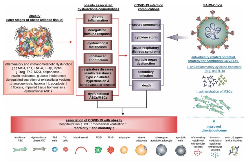
outcomes of COVID-19 (Figure 3).
Figure 3. Schematic illustration presenting that obesity negatively impacts the development of
COVID-19. Obesity is characterized by various pathological features, including systemic chronic
inflammation, deregulated immune response, dysfunctional endothelium, increased comorbidities,
and dysfunctional ASCs/MSCs, which fundamentally influence the progression and outcome of
COVID-19. Anti-inflammatory cytokine therapies for example anti-IL-6 and administration of MSCs
may be useful for supportively treating COVID-19.
In particular, loss or dysfunctional lung resident MSCs may cause more severe lung injury
with pulmonary fibrosis in COVID-19 patients with obesity. Obesity may also compromise motile
cilia on airway epithelial cells and impair the function of the mucociliary escalators responsible for
clearing SARS-CoV-2. Obese diseased adipose tissue that contain all components for SARS-CoV-2
infection could be targeted and even serve as a virus reservoir, and an accelerator reinforcing violent
systemic inflammation and immune response, facilitating the development of a cytokine storm, a severe
complication of COVID-19. This highlights the key role of inflammation and the cytokine network
in COVID-19 patients with obesity. Individuals with obesity are highly susceptible to SARS-CoV-2
infection and more protective measures should be taken for this population. Anti-inflammatory
cytokine therapy like anti-IL-6 and administration of MSCs may have potential for supportively
treating COVID-19.
Author Contributions: Conceptualization, J.Y. and A.R.; writing—original draft preparation, J.Y. and A.R.;
writing—review and editing, J.Y., F.L., and N.-N.K.; visualization, A.R.; funding acquisition, A.R. All authors have
read and agreed to the published version of the manuscript.
Funding: This review was partially funded by the German Research Foundation (Deutsche Forschungsgemeinschaft
(DFG)), project number 413992926.
Acknowledgments: We thank S.C. Hoock for the critical reading of the manuscript.
Conflicts of Interest: The authors declare no conflict of interest. The sponsors had no role in the design, execution,
interpretation, or writing of the review.You can also read