Crosstalk among Calcium ATPases: PMCA, SERCA and SPCA in Mental Diseases - MDPI
←
→
Page content transcription
If your browser does not render page correctly, please read the page content below
International Journal of
Molecular Sciences
Review
Crosstalk among Calcium ATPases: PMCA, SERCA and SPCA
in Mental Diseases
Tomasz Boczek 1 , Marta Sobolczyk 1 , Joanna Mackiewicz 1 , Malwina Lisek 1 , Bozena Ferenc 1 , Feng Guo 2 and
Ludmila Zylinska 1, *
1 Department of Molecular Neurochemistry, Medical University of Lodz, 92215 Lodz, Poland;
tomasz.boczek@umed.lodz.pl (T.B.); marta.sobolczyk@stud.umed.lodz.pl (M.S.);
joanna.mackiewicz1@stud.umed.lodz.pl (J.M.); malwina.lisek@umed.lodz.pl (M.L.);
bozena.ferenc@umed.lodz.pl (B.F.)
2 Department of Pharmaceutical Toxicology, China Medical University, Shenyang 110122, China;
blueforest611@hotmail.com
* Correspondence: ludmila.zylinska@umed.lodz.pl
Abstract: Calcium in mammalian neurons is essential for developmental processes, neurotransmitter
release, apoptosis, and signal transduction. Incorrectly processed Ca2+ signal is well-known to trigger
a cascade of events leading to altered response to variety of stimuli and persistent accumulation of
pathological changes at the molecular level. To counterbalance potentially detrimental consequences
of Ca2+ , neurons are equipped with sophisticated mechanisms that function to keep its concentration
in a tightly regulated range. Calcium pumps belonging to the P-type family of ATPases: plasma
membrane Ca2+ -ATPase (PMCA), sarco/endoplasmic Ca2+ -ATPase (SERCA) and secretory pathway
Ca2+ -ATPase (SPCA) are considered efficient line of defense against abnormal Ca2+ rises. However,
their role is not limited only to Ca2+ transport, as they present tissue-specific functionality and
unique sensitive to the regulation by the main calcium signal decoding protein—calmodulin (CaM).
Citation: Boczek, T.; Sobolczyk, M.;
Based on the available literature, in this review we analyze the contribution of these three types of
Mackiewicz, J.; Lisek, M.; Ferenc, B.;
Ca2+ -ATPases to neuropathology, with a special emphasis on mental diseases.
Guo, F.; Zylinska, L. Crosstalk among
Calcium ATPases: PMCA, SERCA
and SPCA in Mental Diseases. Int. J.
Keywords: calmodulin; calcium; plasma membrane Ca2+ -ATPase; sarco/endoplasmic Ca2+ -ATPase;
Mol. Sci. 2021, 22, 2785. https:// secretory pathway Ca2+ -ATPase; mental diseases
doi.org/10.3390/ijms22062785
Academic Editor: Antonio Villalobo
1. Introduction
Received: 24 February 2021 Ca2+ -ATPases are key components of Ca2+ extrusion machinery and thus are pivotal
Accepted: 8 March 2021
for preservation of neuronal function. Among three main calcium pumps, the plasma mem-
Published: 10 March 2021
brane Ca2+ -ATPase (PMCA) and sarco/endoplasmic Ca2+ -ATPase (SERCA) are known
for decades while the secretory pathway Ca2+ -ATPase has been discovered in 2000s by
Publisher’s Note: MDPI stays neutral
two independent laboratories that described novel mutations leading to Hailey-Hailey
with regard to jurisdictional claims in
disease [1–3]. All pumps have high affinity for Ca2+ and function to restore cytosolic
published maps and institutional affil-
Ca2+ concentration [Ca2+ ]c to the resting, nanomolar level following neuronal stimulations.
iations.
They belong to the superfamily of mammalian P-type ATPases and are characterized by
formation of a phosphorylated enzyme intermediate during catalytic cycle [1]. However,
they have a low (~15%) degree of sequency identity [1], and differ in several other key
features including tissue distribution, regulatory mechanisms, and contribution to neuronal
Copyright: © 2021 by the authors. Ca2+ homeostasis. Each pump is encoded by multiple genes giving rise to a number of
Licensee MDPI, Basel, Switzerland.
isoforms and further splice variants, which often possess distinguishable kinetic param-
This article is an open access article
eters and are dedicated to unique and highly regulated neural processes [4]. Naturally,
distributed under the terms and
the pumps share essential basic properties such as membrane topology, catalytic mech-
conditions of the Creative Commons
anism and probably the general features of 3D structure [5,6], although the structure of
Attribution (CC BY) license (https://
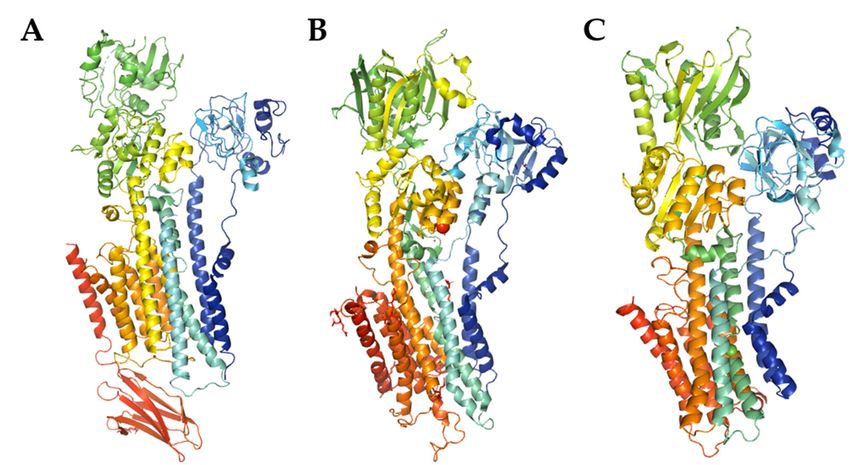
SPCA pump has not been solved yet (Figure 1). The rapid expansion of the knowledge on
creativecommons.org/licenses/by/
4.0/).
pumps peculiar role, which run parallel to the advances in neuronal Ca2+ signaling, led
Int. J. Mol. Sci. 2021, 22, 2785. https://doi.org/10.3390/ijms22062785 https://www.mdpi.com/journal/ijmsInt.J. J.Mol.
Int. Mol.Sci.
Sci.2020, 22,x 2785
2021,21, FOR PEER REVIEW 2 of2323
2 of
signaling, led to the identification of several diseases associated either directly or indi-
rectly
to thewith Ca2+ pumps
identification of malfunction. Most
several diseases of these defects
associated have genetic
either directly background
or indirectly with and
Ca2+
the number of studies have been aimed to characterize their severity,
pumps malfunction. Most of these defects have genetic background and the number ofeffect on neuronal
Ca 2+ homeostasis and signaling as well as neuronal survival. Besides known neuropathol-
studies have been aimed to characterize their severity, effect on neuronal Ca2+ homeostasis
ogies,
and signalinginasCa
defects wellpumps
2+ and alterations
as neuronal survival. in the mechanisms
Besides regulating theirdefects
known neuropathologies, activityin
may also
2+ produce subtle, tissue-specific disturbances that are not clinically
Ca pumps and alterations in the mechanisms regulating their activity may also produce manifested,
yet they tissue-specific
subtle, may affect neuronal machinery
disturbances controlling
that are and processing
not clinically manifested,Ca
2+ signal. In this
yet they may affect
review, we focus on the contribution of PMCA, SERCA
2+ and SPCA
neuronal machinery controlling and processing Ca signal. In this review, we to mental diseases
focus onand
the
give a special emphasis
contribution of PMCA,toSERCA
alteredand
regulation
SPCA to bymental
calmodulin (CaM)
diseases andthat
giveoften accompanies
a special emphasis
pump defects.
to altered regulation by calmodulin (CaM) that often accompanies pump defects.
Figure
Figure1.1.The
Themodel
modelofofPMCA
PMCA((A),
((A),PDB
PDBentry
entrycode
code6A69)
6A69)and
andSERCA
SERCA((B),
((B),PDB
PDBentry
entrycode
code 3W5C)
3W5C) structures, and SPCA structure prediction using SWISS-MODEL (C). The cartoon
structures, and SPCA structure prediction using SWISS-MODEL (C). The cartoon models models
were
were generated with PyMOL.
generated with PyMOL.
2.2.Calmodulin—Ubiquitous
Calmodulin—UbiquitousCa Ca2+2+ Sensor
Sensor inin Neurons
Neurons
Mostcommonly,
Most commonly,detectiondetectionand andtransduction
transductionofofCa 2+2+
Ca signalsininneurons
signals neuronsare areorches-
orches-
tratedby
trated byubiquitous
ubiquitousmessenger
messengercalled
calledcalmodulin.
calmodulin.CaM CaMisisknown
knownasasaarelatively
relativelysmall
small
(149aa;16.7
(149aa; 16.7kDa)kDa)and andhighly
highlyconserved
conservedcalcium-binding
calcium-bindingsensorsensorsynthesized
synthesized in in all
all eukar-
eukary-
otic cells.
yotic cells. ItIt isis particularity
particularity involved
involvedin insynaptic
synapticsignaling
signalingprocesses,
processes,neurotransmitter
neurotransmitter
release and neuroplasticity by modulation (called “calmodulation”) of a alarge
release and neuroplasticity by modulation (called “calmodulation”) of largearray
arrayofof
binding partners such as enzymes (e.g., adenylate cyclase, calcineurin,
binding partners such as enzymes (e.g., adenylate cyclase, calcineurin, cyclic nucleotide cyclic nucleotide
phosphodiesterase,nitric
phosphodiesterase, nitricoxide
oxidesynthase,
synthase,and andcertain
certainkinases),
kinases),transcription
transcriptionfactors
factors(e.g.,
(e.g.,
CREB,NeuroD2,
CREB, NeuroD2,NFAT NFAT and and MEF2)
MEF2) as as well
well as asvarious
variousionionchannels
channelsand andtransporters
transporters [7–10].
[7–
10]. In human, CaM is encoded by three independent genes CALM1, CALM2, CALM3 lo-
cated on chromosomes 14q32.11; 2p21; and 19q13.32, respectively,
In human, CaM is encoded by three independent genes CALM1, CALM2, CALM3 which are collectively
transcribed
located into at least 14q32.11;
on chromosomes eight mRNAs 2p21;using differentrespectively,
and 19q13.32, alternative polyadenylation signals
which are collectively
(reviewed in [7,11]). Next, the resulting protein is susceptible to
transcribed into at least eight mRNAs using different alternative polyadenylation signalsundergo various post-
translational modifications, mainly phosphorylation on tyrosine
(reviewed in [7,11]). Next, the resulting protein is susceptible to undergo various post-(Thr26, Thr29, Thr44,
Thr79, Tyr99,modifications,
translational Thr117, and Tyr138)
mainlyand serine (Ser81, and
phosphorylation Ser101) sites
on tyrosine [12];Thr29,
(Thr26, acetylation
Thr44,of
the N-terminal alanine [13]; trimethylation of the Lys115 [14]; and proteolytic cleavage at
Thr79, Tyr99, Thr117, and Tyr138) and serine (Ser81, and Ser101) sites [12]; acetylation of
the C-terminal domain [15], all collectively regulating CaM biological activity. The crystal
the N-terminal alanine [13]; trimethylation of the Lys115 [14]; and proteolytic cleavage at
structure of mature CaM contains two independently folded lobes (N-lobe and C-lobe)
the C-terminal domain [15], all collectively regulating CaM biological activity. The crystal
connected by a flexible central α-helical linker, that differ by calcium affinity and kinetics of
structure of mature CaM contains two independently folded lobes (N-lobe and2+C-lobe)
calcium dissociation. Each of these globular clusters can bind up to two free Ca ions via
connected by a flexible central α-helical linker, that differ by calcium affinity and kinetics
a pair of helix-loop-helix motives (EF-hands) in a cooperative manner (Kd = 5 · 10−2+7 M to
of calcium dissociation. Each of these globular clusters can bind up to two free Ca ions
5 · 10−6 M) [16–18]. Because of subtle structural differences between these lobes resulting
via a pair of helix-loop-helix motives (EF-hands) in a cooperative manner (Kd = 5 · 10−7 M
from evolutionary processes [19], EF hands in the C-lobe exhibit a three- to five times
to 5 · 10−6 M) [16–18]. Because of subtle structural differences between these lobes resulting
higher affinity for Ca2+ . However, they possess slower rate of ion binding than the regionsInt. J. Mol. Sci. 2021, 22, 2785 3 of 23
of EF hands located in the N-lobe, establishing the broad range of CaM sensitivity to
the changes in calcium concentrations in the intracellular space [20]. CaM is susceptible
to dramatic structural rearrangements via partially exposed hydrophobic patch on the
C-terminal domain which may interact with CaM-binding proteins (CaMBPs) in a Ca2+
-free (apo-CaM) state or in partially calcium-saturated forms (two Ca2+ ions bound to the
C-terminus) [16]. Up to date, over three hundred different calmodulin targets with specific
binding sites and unique affinities for CaM, many of which located in the central ner-
vous system (CNS) neurons [21], have been validated and extensively characterized [22].
The analysis of over 80 CaM complexes compiled in the Protein Data Bank (PDB) has
revealed that CaM binding sites not always contain defined consensus sequence but rather
share some common biochemical and biophysical properties such as high helix-forming
propensities, positively charged binding region and the presence of hydrophobic anchor
residues [8,22]. Thus, the classification of several CaM-binding motifs is determined by
the spacing between these anchor residues as was extensively discussed by Mruk and
colleagues [23]. As observed from sequence analysis of several CaMBPs, their IQ motif
([FILV]Qxxx[RK]Gxxx[RK]xx[FILVWY]) with highly conserved amino acid residues at
positions 1, 2, 5, 6, 11, and 14 or IQ-like ([FILV]Qxxx[RK]Gxxxxxxxx) motif may also bind
CaM in the presence or absence of Ca2+ [23,24].
Considering the diversity of CaM interactions and its abundance in the brain (up to
100 µM range) [25], it seems rational to suspect that disruption of these multifunctional
interactions regulating Ca2+ -dependent intracellular signal transduction cascades may
be implicated in the development of numerous neuropsychiatric disorders. Moreover,
there is increasing evidence suggesting that pathophysiology of these states is intimately
related to the disturbed neuronal calcium homeostasis also mediated by ATP-driven pumps
located in the plasma membrane, in the membranes of the endoplasmic reticulum (ER), or
Golgi compartments.
3. Plasma Membrane Ca2+ -ATPase (PMCA)—The Only Calcium Pump Directly
Regulated by Calmodulin
PMCA is one of the most important and sensitive players in maintaining of low
resting Ca2+ concentration, and ensuring a fast recovery of [Ca2+ ]c to the basal level
following neuronal excitation [26]. The enzyme was first described by Schatzmann in
1960s as ATP-powered mechanism that removes calcium from red blood cells [27], whereas
further studies revealed the presence of PMCA in other cells, including neurons [28–30]
Structurally, PMCA comprises of ten transmembrane segments with N- and C- terminal
tails both located on the cytosolic site [31]. Most of the regulatory regions including acidic
phospholipids, protein kinase C (PKC), protein kinase A (PKA) and the crucial natural
activator—CaM, are located at the C- terminus. The important regulatory role of CaM in
stimulating of PMCA is associated with increasing the affinity of the pump for calcium
and the maximum rate of calcium extrusion. In the activation process, CaM removes
the auto-inhibitory C-terminal domain from the active site and releases the enzyme from
auto-inhibition [32]. It is also worth mentioning that PMCA is so far the only known
calcium pump directly activated by CaM [26].
In mammals, four isoforms of PMCA (PMCA1-PMCA4), structurally similar to each
other, have been found [4] but their expression depends on cell type (Table 1). The
PMCA1 and PMCA4 are widely expressed in virtually all animal tissues and both play a
house-keeping role.Int. J. Mol. Sci. 2021, 22, 2785 4 of 23
Table 1. Properties of PMCA isoforms. Modified based on [1].
PMCA1 PMCA2 PMCA3 PMCA4
Restricted Restricted
Tissue Distribution Ubiquitous Ubiquitous
(brain) (brain)
Developmental Isoform switch Isoform switch Isoform switch Isoform switch
Expression/Switch fetal/adult fetal/adult fetal/adult fetal/adult
Affinity CaM (Kd nM) 40–50 2–4 8 3–40
Expression of PMCA2 and PMCA3 is highly restricted to excitable cells and their
high concentration has been detected in the CNS [4]. PMCA2 is especially abundant
in cerebellar Purkinje cells and granule cells, but it also localizes to the cerebral cortex
and hippocampus [33]. PMCA3, in turn, is present predominantly in cerebellar granule
cells and in the choroid plexus [34] what suggests its role in generation and release of
cerebrospinal fluid. Additionally, PMCA isoforms are characterized by distinct calmodulin
sensitivity (Table 1) and specific kinetic properties. PMCA2 and PMCA3 are referred to
as “fast” isoforms due to their high basal activity and high affinity for CaM, whereas
PMCA1 and PMCA4 are much slower despite their strong stimulation by CaM [1]. It has
been suggested that the cell response to a physiological stimulus depends on significant
differences in the kinetic parameters of the individual isoforms. In the brain, distribution
of PMCA isoforms clearly alters during development, what may indicate their specific role
in embryogenesis and further in postnatal period [35].
In addition to control critical neuronal functions such as synaptic transition and neu-
rotransmitter release, neuronal Ca2+ also participates in the regulation of survival and
differentiation, processes common to other cell types [36]. Early in vitro study on differen-
tiated pheochromocytoma-derived cells, a model frequently used to mimic the physiology
of sympathetic neurons, has shown that PMCA1 knockdown impaired neuritogenesis and
axonal elongation [37]. Similar effect was seen when PMCA2 or PMCA3 expression was de-
creased with isoform-specific antisense oligonucleotides [38,39] suggesting a role of PMCA
in neuronal differentiation. Moreover, cells deficient in neuron-specific PMCA isoforms
were unable to maintain Ca2+ and pH homeostasis which translated into altered activity
of main signaling- and energy-generating pathways [40–42]. There is also a compelling
evidence that PMCA4 is of paramount importance for neuronal survival in the conditions
of Ca2+ overload as pheochromocytoma viability was preserved or impaired when this iso-
form was overexpressed or downregulated, respectively [43,44]. The sections below further
explore the association and the specific role of the PMCA in neurodegenerative disorders.
3.1. PMCA in Neuropathology
3.1.1. PMCA in Aging
Contribution of PMCA to age-related neuropathologies was first suggested by Michaelis
and coworkers [45–47]. These authors showed for the first time that PMCA activity and
abundance in the synaptosomal membranes is reduced with age, similar to the PMCA
activation by aged CaM. Zaidi and coworkers further demonstrated that the decline in
PMCA activity was progressive with increasing biological age and was associated with
lowered maximal velocity (Vmax ) with no apparent changes in the affinity for Ca2+ [48].
The age-dependent alterations in PMCA are likely to be a consequence of oxidative stress
as PMCA was identified to be a target for reactive oxygen/nitrogen species as does for
CaM [49]. For instance, exposure of purified pump protein to H2 O2 inhibited both basal
and CaM-stimulated activity. However, neither CaM binding to the oxidized protein
nor the concentration-dependent CaM effect on PMCA were affected, suggesting that
C-terminal CaM binding domain is not primarily targeted by the oxidant. Pretreatment
of PMCA with CaM almost completely preserved PMCA activity in the presence of H2 O2
indicating that conformational state upon CaM binding may be more resistant to oxida-
tion [50]. Several oxidative agents have been demonstrated to abolish PMCA sensitivity to
CaM in a concentration-dependent manner [51], induce proteolytic degradation in synap-Int. J. Mol. Sci. 2021, 22, 2785 5 of 23
tic membranes [52] or promote internalization and subsequent lack of detectable PMCA
expression in hippocampal neurons [53].
PMCA activity is also affected by lipid-surrounding environment. It was demonstrated
that PMCA and CaM are partitioned to the cholesterol-rich lipid rafts and PMCA activity
in these membrane microdomains is higher than in non-raft regions [54]. Moreover, raft-
localized PMCA is more sensitive to age-dependent loss of the activity [55]. Depletion of
cholesterol drastically inhibited the activity of raft-associated PMCA but did not produce
any effect on non-raft PMCA [54]. Therefore, increasing lipid order may be beneficial for
protection of PMCA activity in the aged membranes but cannot overcome age-dependent
loss of PMCA.
3.1.2. PMCA in Alzheimer’s Disease
Besides brain aging, altered PMCA expression and activity was detected postmortem
in the brain cortex of patients affected by Alzheimer’s disease (AD) [56,57]. One of the
histological hallmarks of this disease are the presence of a senile plaques of the amyloid
β-peptide (Aβ) and accumulation of an abnormal tau protein [58]. Biochemical studies
have revealed that Aβ decreased the activity of purified PMCA and the strongest inhibitory
effect was seen for PMCA4 [57]. Moreover, cholesterol was shown to abolish the inhibitory
effect of Aβ and the level of inhibition was lower in the lipid rafts of synaptosomal
membranes than in non-rafts [57]. The Aβ inhibitory effect can be blocked by CaM, and
the activity of PMCA lacking C-terminal CaM-binding domain was unaffected by Aβ.
This antagonistic action of CaM is due to physical association with Aβ [59] or competing
for PMCA binding [57]. Hence, CaM can protect PMCA activity by masking Aβ-PMCA
interacting sites making them unavailable for Aβ. The accumulated data present a clear
link between Ca2+ , CaM and amyloid plaque formation indicating how the dysregulation
in neuronal Ca2+ homeostasis and Aβ formation affect each other, and how CaM function
in the center of this crosstalk. Interestingly, CaM content in the frontal, temporal, parietal
cortex and subjacent white matter in AD was reduced by nearly 66% compared to the
normal control brains [60]. The mechanism of progressive decline in PMCA and CaM in AD
is unknown. However, the recent study on differentiated pheochromocytoma suggests that
PMCA downregulation may be a trigger initiating calcineurin/NFAT-dependent repression
of CalmII and CalmIII genes [40]. The altered expression of CaM is thus expected to deepen
the physiological decline of PMCA function with increasing age and offer insufficient
protection not only against Aβ, but also proteolytic and oxidative pump deactivation.
Growing body of evidence suggests that soluble forms of Aβ and tau cooperate with
each other to drive healthy neurons into diseased state and the toxicity of Aβ requires
tau [58]. The study of Berrocal and colleagues demonstrated that tau, which hyperphospho-
rylated form is predominant in neurofibrillary tangles, can directly interact with PMCA and
inhibit its activity [56]. In this study, PMCA function was solely affected by this interaction
as neither SERCA not SPCA were targeted. In differentiated pheochromocytoma, tau was
concentrated in growth cones and interacted with PMCA through its N-terminal projection
domain [61]. Overexpression of this amino-terminal fragment, but not full-length protein,
suppressed nerve growth factor (NGF)-induced axonal outgrowth [61] establishing tau as a
mediator of microtubule-plasma membrane interactions during neuritic development. Tau
is also required for Fyn kinase-mediated NMDR receptor activation in the postsynaptic
densities [62], which strengthen the interaction between NMDA receptor and postsynaptic
density protein 95 (PSD-95) [63]. Considering the recruitment of PMCA via PSD-95 to a
close proximity to NMDA receptor-mediated Ca2+ entry, a direct physical interaction be-
tween PMCA and tau in vivo would complement the contribution of PMCA-tau interaction
to Ca2+ homeostasis dysregulation in the pathogenesis of AD.
Several early studies demonstrated that association between tau and CaM in vitro
is Ca2+ -dependent and it prevents tau interaction with microtubules [64,65]. Binding to
CaM also prevents tau phosphorylation by PKC [66]. Although several CaM-dependent
kinases and phosphatases are involved in tau posttranslational modification, for instanceInt. J. Mol. Sci. 2021, 22, 2785 6 of 23
Ca2+ /CaM-dependent protein kinase II (CaMKII), cyclin-dependent kinase 5 or protein
phosphatase 2B (PP2B or calcineurin) [67], no recent studies have further expanded the
functional significant of direct interaction with CaM and tau in the AD.
3.1.3. PMCA in Parkinson’s Disease
Altered PMCA function may significantly contribute to neuronal Ca2+ dyshomeostasis
and increase the duration and frequency of intracellular Ca2+ spikes which may in turn
influence the formation of pathological proteins such as the alpha synuclein in Parkinson’s
disease (PD) [68]. This hypothesis is supported by the studies of Brendel and coworkers
who demonstrated increased Ca2+ level and reduction in PMCA2 expression in primary
midbrain neurons and neuroblastoma SH-SY5Y treated with Parkinsonian mimetic 1-
methyl-4-phenylpyridinium (MPP) [69]. Interestingly, other Ca2+ efflux systems such as
SERCA pump or Na+ /Ca2+ exchanger remained largely unaffected. The same authors
showed that PMCA2 knockdown with siRNA decreased the survival of mesencephalic
neurons, but overexpression significantly increased the resistance of midbrain neurons to
MPP toxicity [69]. These data indicate that PMCA2, which possesses the highest affinity
for CaM, is particularly vulnerable to the inhibition by MPP. The mechanistic explanation
of this phenomenon may lie in the oxidative stress and partial oxidative inactivation of
PMCA, as membrane protein-selective antioxidants fully prevented MPP toxicity [70].
PMCA inactivation or even PMCA2 knockdown are known to irreversibly deprive
neurons of substantial part of their Ca2+ clearing potency leading to dysregulation of Ca2+
homeostasis [39,68,71]. It is known that Ca2+ is a key controller of synuclein formation
and Ca2+ -dependent binding of CaM to α-synuclein accelerates formation of protein fibrils
in vitro [72]. Recent study demonstrated that calcineurin also binds α-synuclein and this
interaction is mediated by Ca2+ /CaM signaling [73]. These and other authors demonstrated
that increased calcineurin activity was associated with α-synuclein toxicity [73,74]. The
activity of calcineurin is known to be regulated by the interaction with PMCA2 [75] and
disruption of this interaction elevates intracellular activity of this phosphatase [76,77].
Although no direct interaction between α-synuclein and PMCA has been established, as
does for SERCA pump [78], PMCA by regulating the activity of Ca2+ -dependent signaling
may potentially contribute to PD pathogenesis.
3.1.4. PMCA in Schizophrenia and Bipolar Disorder
Ca2+ has been placed in the center of dopaminergic hypothesis of schizophrenia,
mainly because of its essential role in dopamine receptors D1 and D2-mediated synaptic
plasticity and signal transduction [79]. Therefore, it is not surprising that many of cal-
cium signaling proteins, including PMCA and CaM, have been found to be differentially
regulated in schizophrenia. In early study, Kluge and Kuhne demonstrated that kinetic
properties of CaM-stimulated PMCA were altered in erythrocytes of patients with affective
psychoses and hyper- or para-kinetic schizophrenics [80]. Unexpectedly, schizophrenia
proteomic studies revealed PMCA4 to be up-regulated in anterior temporal lobe in affected
patients [81] what may be seen as compensatory change to counterbalance elevated [Ca2+ ]c .
On the other hand, proteins such as CaM (CALM1 and CALM2), CaMKII (CAMK2B,
CAMK2D, CAMK2G) and CaM-like proteins were found to be downregulated in the
brain or secretion fluids [81,82]. These findings limit, but not exclude, the special role of
PMCA4 in neuronal signaling in schizophrenia, however the disadvantage of the proteomic
studies is a fact they were performed not on isolated cells but on whole tissue. Therefore, it
is likely that non-neuronal cells could contribute to revealed changes but because of high
sample heterogeneity, these data should be interpreted with caution.
In the subgroup of patients involving mostly those with unipolar maniac and bipolar
psychoses, the activation of human erythrocyte PMCA by CaM measured in the presence
of sub-optimal concentration of lithium was stronger in all individuals with maniac-
depressive episodes [80,83,84]. Interestingly, the activity measured in the conditions of
optimal concentration of monovalent ions (Na+ and K+ ) was higher in lithium-treatedInt. J. Mol. Sci. 2021, 22, 2785 7 of 23
groups than in control and untreated patients suggesting that the CaM-activated PMCA
may be differentially regulated in maniac-depressive patients.
The potential involvement of PMCA in schizophrenia is also supported by data de-
rived from pharmacological in vivo models. Recent study on animals challenged with
30 mg/kg ketamine, a drug that is known to mimic a wide spectrum of psychotomimetic
and cognitive aberrations observed in schizophrenia in humans [85], demonstrated differen-
tial regulation of PMCA expression in functionally distinct brain regions [86]. Moreover, the
basal and CaM-stimulated PMCA activities were reduced in the synaptosomal membranes
mainly due to a direct interaction of the drug within large catalytic loop and competing
with CaM for binding in the C-terminal domain of the pump [87,88]. Similarly, emerging
studies also suggest the interaction of ketamine with CaM-dependent enzymes, in particu-
lar CaMKII [89,90], but no specific functional studies largely limit the conclusions on CaM
and PMCA-mediated Ca2+ regulation in schizophrenia.
3.1.5. PMCA in Cerebellar Disorders
Immunohistochemical studies showed that neuron-specific PMCA2 and PMCA3 are
more abundant in the cerebellum than PMCA1 and PMCA4. Moreover, they are concen-
trated in synaptic terminals of Purkinje cells while PMCA1 and PMCA4 localize mostly to
granular layer [91]. Purkinje cells integrate the excitatory input to the cerebellar cortex be-
ing an essential component for the regulation and coordination of motor movements [92]. It
is therefore not surprising that one of the most visible cerebellar dysfunctions during ataxia
are uncoordinated movements and inability to maintain body balance. The link between
ataxia and PMCA3 became evident when Zanni and coworkers identified a point mutations
in ATP2B3 gene (located on the human chromosome X) in a family affected by X-linked
congenital cerebellar ataxia [93]. The mutation located in exon 20 of ATP2B3/PMCA3 gene
replaces a conserved Gly by Asp in C-terminal CaM-binding domain. The mutated pump
demonstrated decreased ability to extrude Ca2+ what can be a direct consequence of altered
interaction of its C-terminus with CaM. This is supported by mathematical modeling show-
ing reduced ability of CaM to interact with the mutated binding domain what tends to
depress the basal PMCA activity and decrease autoinhibitory interaction of CaM-binding
domain with the main body of the pump. The possible defective interplay between mu-
tated pump and PMCA-interacting signaling molecules (see Table 2) organized within
Ca2+ nanodomains could contribute to the phenotype of cerebellar disease. It is even
more plausible as more than a dozen X-linked gene defects have been demonstrated to
contribute to cerebellar phenotype [94]. Among them, mutation in Ca2+ /CaM-dependent
serine protein kinase (CASK) may be significant [95,96] as CASK interacts with PMCA
through PDZ domain located in the C-terminal end of the pump.
Cali and colleagues [97] identified another mutation in ATP2B3 gene in a patient with
cerebellar ataxia and global developmental delay. The mutation (R482H) significantly
reduced the Ca2+ clearing potency of the pump and resulted in the inability to handle
intracellular Ca2+ transients evoked by cell stimulation. Interestingly, the patient also
carried two additional mutations in the LAMA1 gene encoding laminin 1α. It has been
well-characterized that mutations or deletions in this gene are associated with cerebellar
dysplasia phenotype [98]. Therefore, on the basis of the family pedigree of the patient,
it is reasonable to suspect that mutations in PMCA3 along with those in LAMA1 could
synergistically contribute to the ataxic symptoms.
Recently, a novel mutation in PMCA3 (G733R) has been identified in a patient carrying
a defect in phosphomannomutase 2 (PMM2), indicating a possible link between these muta-
tions in generating ataxia phenotype [99]. PMM2 is an enzyme catalyzing the isomerization
of mannose-6-phosphate to mannose1-phosphate and two missense mutations (R123Q
and G214S) in PMM2 gene are known to be associated with type I congenital disorder of
glycosylation [100,101]. The G733R substitution in the pump disturbed the ability to handle
Ca2+ rises upon Ca2+ release from the ER or influx through the plasma membrane without
affecting pump expression or subcellular targeting. The basal activity of autoinhibitedInt. J. Mol. Sci. 2021, 22, 2785 8 of 23
pump and constitutive active variant of PMCA3 lacking C-terminal domain was also com-
promised. The coexistence of both PMCA3 and PMM2 mutations in the patient affected by
non-progressive ataxia and muscular hypotonia is of special significance as PMM2 was
found to be Ca2+ -regulated enzyme [102]. Therefore, mutated PMCA3 may give rise to
increased Ca2+ concentration in microdomains where PMM2 is located, inhibiting it. This,
along with the mutations in PMM2, would deepen the decline in PMM2 enzymatic activity.
There is also convincing evidence coming from PMCA2 knockout mice that this
isoform, apart from its function in auditory system discussed elsewhere [103–105], plays an
essential role in cerebellar function. PMCA2−/− mice exhibited severe ataxia that became
apparent by 12 days of age and had great difficulty in maintaining body balance [106].
Histological examination showed increased density of Purkinje neurons and reduced
density of granular layer in cerebellum. Similarly, the deafwaddler (dfw) and wriggle mouse
sagami (wri) strains displayed similar phenotypes such as tremor and vestibular/motor
imbalance. Both dfw and wri genes were reported to be associated with a mutation in
PMCA2 gene [107–109]. Interestingly, despite prolonged accumulation of Ca2+ in the
cytosol of Purkinje neurons, heterozygous PMCA2+/− mice exhibited outwardly normal
behavior but presented clear deficits in hindlimb coordination when challenged with
exercise [110].
The first mutation in PMCA2 (V1143F) associated with congenital cerebellar ataxia
has been recently identified by Vicario and coworkers [111]. In contrast to the hearing loss
phenotype [105], the ataxia phenotype was generated without corresponding mutations in
cadherin 23 and the hearing ability was fully retained. As V1143F substitution was located
within CaM binding domain, it affected the interplay between mutated pump and CaM
and the effect was particularly visible for full-length PMCA2 variant. Like other mutations,
one of the consequences was prolonged duration of Ca2+ transients and compromised
ability to maintain Ca2+ homeostasis in neurons. The list of PMCA mutations associated
with cerebellar defects are summarized in Table 2.
Table 2. List of PMCA mutations and associated phenotype. Modified based on [112].
Species Mutation Phenotype Reference
PMCA2 Mouse G283S (Dfw) Vestibular/motor imbalance [107]
Mouse I655N (Elfin) Ataxia [113]
Mouse S877F (Obv) Ataxia [114]
Mouse E629K (Tmy) Ataxia [115]
Mouse E412K (Wri) Abnormal movements [116]
Human V1143F Ataxia [111]
PMCA3 Human G1107D Ataxia [93]
Human R482H Ataxia [97]
Rat R35C Ataxia [117]
Human G773R Ataxia [99]
There is substantial body of evidence to suggest that CaM-regulated PMCA isoforms
play an important role in neuronal survival and synaptic transmission, thus contributing to
several pathological states of the CNS. However, at the present stage of development, the
exact molecular mechanisms by which the defective PMCA function leads to generation of
disease phenotype is still under investigation. Plausibly, the answer lies in the regulation of
Ca2+ within discrete plasma membrane microdomains hosting PMCA-associated signaling
molecules with CaM, due to its universality as a Ca2+ decoding molecule, being central to
the regulation of neuronal signaling.
3.2. PMCA-Interacting Proteins in Mental Diseases
Besides the well-known regulation of PMCA by CaM, the pump is also affected in less
well-studied ways by multiple interacting partners that have been associated with many
of the neurodegenerative diseases (Table 3) [118]. These interactions are thought to target
PMCA to highly-specialized membrane microdomains, regulate pump activity or recruit itInt. J. Mol. Sci. 2021, 22, 2785 9 of 23
to multiprotein complexes—“signalosomes” responsible for orchestration of local Ca2+ sig-
naling. The dynamic and fidelity of these interactions determined by structural differences
further augments functional specialization of particular PMCA isoforms. For instance, it
has been demonstrated that PMCA2b and PMCA4b interact with postsynaptic density
protein-95 (PSD-95, not indicated in the table but widely discussed elsewhere [119–122]),
which tether the pump to microdomains enriched in NMDA receptor [123]. Alterations in
NMDA receptor are, in turn, frequently reported in schizophrenia, mood disorders, Hunt-
ington’s disease (HD), AD and substance-induced psychosis [124]. The functional coupling
between NMDA receptor and PMCA would allow rapid response to local Ca2+ rises due to
bring the pump to a local Ca2+ entry sites. The formation of tertiary PMCA/PSD95/NMDA
receptor complexes could modulate the amplitude of Ca2+ increases and affect the neuro-
transmission. Such modulation has been recently showed for glutamate in a rat model of
ketamine-induced psychosis [87]. As PMCA plays a pivotal role in the regulation of local
Ca2+ fluxes, it cannot be ruled out that interaction with the binding partners may affect
their clustering into large signaling complexes and influence the neurosecretory process.
Table 3. Protein interacting with PMCA. Modified based on [125]. AD—Alzheimer’s disease; BP—bipolar disorder; HD—
Huntington’s disease; PD—Parkinson’s disease; SZ—schizophrenia, MDD—major depressive disorder, ALS—amytrophic
lateral sclerosis, ASD—autism spectrum disorder.
A Protein PMCA Domain A Protein Domain
Functional Importance of
Interacting with Associated Disease Involved in the Involved in the
the Interaction
PMCA Interaction Interaction
AD, BP, MDD, ALS, Decline in NOS activity,
PDZ-domain binding
NOS anxiety, stroke, HD PDZ domain down-regulation of NO
sequence
[126,127] production
Microcephaly with
pontine and cerebellar Down-regulation the T-
PDZ-domain binding
CASK hypoplasia, X-linked PDZ domain dependent transcriptional
sequence
intellectual disability, activity
ASD [128]
Williams-Beuren PDZ-domain binding Pump translocation during
CLP36 PDZ domain
syndrome [129] sequence platelet activation
Biding enables the
AD, PD, stroke,
localization of PMCAs in
X-linked mental PDZ-domain binding
MAGUK PDZ domain specific membrane
retardation, BD, MDD, sequence
domains and local control
SZ [130–132]
of Ca2+ concentration
SZ, ASD, MDD, suicide
Stabilization of PMCA in
attempt, cocaine PDZ-domain binding
Ania3/Homer PDZ domain domains near the sites of
dependence, opiate sequence
calcium influx into the cell
abuse [133]
Inhibition of the
phosphatase activity of
AD, HD, SZ, PD, ALS,
Calcineurin catalytic domain Amino acids 58–143 calcineurin, decrease in the
BD epilepsy [134–136]
activity of the transcription
factor NFAT
The formation of a triple
SZ, mild intellectual complex with PMCA and
Syntrophin α1 catalytic domain Amino acids 399–447
disability [137,138] NOS-1 inhibits the
production of NO
Inhibition of PMCA
ε 14-3-3 AD, BP, PD, SZ [139] the N-terminal region Amino acids 2–92
activityInt. J. Mol. Sci. 2021, 22, 2785 10 of 23
4. The Sarco/Endoplasmic Reticulum Ca2+ -ATPase (SERCA)
The SERCA pump is the product of a multigene family. It is a 110-kDa single polypep-
tide located in the sarco/endoplasmic reticulum (SR/ER) which primary role is to transport
Ca2+ back to the internal stores. Mammals express three isoforms of the pump, called
SERCA1 through SERCA3, and post-translational modifications increase the total number
of identified subtypes to 10 [140]. The expression profile of individual variants is not only
tissue-dependent, but also undergoes changes during development. The distribution of
SERCA1 is limited to fast- and slow-twitch skeletal muscles, and the role of this pump is
to accumulate calcium in SR of skeletal muscle. The alternative splicing of SERCA1 gene
generates two variants which expression pattern is developmentally regulated. SERCA1b
is predominantly expressed in neonatal stages, then is entirely replaced by SERCA1a in
adults [141]. SERCA2 exists in two variants—SERCA2a and SERCA 2b [141]. SERCA2a
is mostly found in cardiac and skeletal muscle with slow contractile characteristics, but
also exhibits minor expression in the brain, where it is almost exclusively restricted to
the Purkinje neurons of the cerebellum [142]. SERCA2a is considered to be involved in
contraction and relaxation of cardiac muscle. SERCA2b is the most abundant variant ex-
pressed widely in all tissue types, including neurons. In the brain, SERCA2b expression has
been identified in both, cerebrum, and cerebellum [143]. Moreover, SERCA2b is the only
SERCA variant present in astrocytes [144]. Further studies confirmed that both variants
of SERCA2 pump are present in substantia nigra—the structure involved in dopamine
release [145]. In humans, there are six possible variants of SERCA3 [141]. However, due
to the complex alternative splicing, their functional and structure aspects remain poorly
understood. SERCA3a-3f are located in most tissues, especially in secretory cells. Because
of predominant presence of SERCA3 in pancreatic β-cells, this isoform is recognized as
being involved in metabolic homeostasis [146].
Despite high structural homology, all isoforms of SERCA possess different affinity for
calcium and unique kinetic properties. For instance, SERCA2a has a two-fold lower affinity
for [Ca2+ ] and shows two-fold higher velocity of Ca2+ transport compared to ubiquitous
SERCA2b [147]. This translates into primary role of SERCA2a in cardiac function, whereas
SERCA2b is considered more as a house-keeping form. In turn, among all SERCA isoforms
the lowest affinity for calcium and the slowest turnover rate for Ca2+ uptake can be ascribed
to SERCA3 [148]. This feature is a consequence of the distribution of this isoform in non-
excitable cells. Accumulating evidence suggests that SERCA2 is ubiquitously present
in different brain areas and therefore may be the isoform of paramount importance for
neuronal function [143,149–151].
4.1. SERCA Pumps in Neuropathology
Disturbances in ER Ca2+ homeostasis can lead, among others, to ER stress and accumu-
lation of unfolded or misfolded proteins, which are detected for most neurodegenerative
diseases, including AD, PD, HD, and ALS [152–154]. It has been reported that truncated
isoform of human SERCA1 (S1T) triggered and amplified ER stress response, leading to
apoptosis [155]. Furthermore, the increase of human S1T protein expression has been
demonstrated in sporadic AD-derived post-mortem brains and in a cellular AD model,
confirming that S1T can induce neuroinflammatory response in vitro and in vivo [156].
An interesting observation was identification of several ATP2A2 mutations in autosomal
dominant skin disorder—Darier’s disease (DD), a disorder frequently associated with sev-
eral mental diseases (bipolar disorder, schizophrenia, affective psychosis, epilepsy) [157–161].
Numerous SERCA2 mutations in DD have been detected, including missense and non-
sense types, which produced the insoluble truncated, misfolded and/or aggregated pro-
teins, finally decreasing the amount of fully active SERCA. All these results indicate that
ATP2A2 mutations may have pleiotropic effects on the brain as well as skin.Int. J. Mol. Sci. 2021, 22, 2785 11 of 23
4.2. Calmodulin-Controlled Regulation of SERCA Pumps
Although the direct regulation by CaM has been shown only for PMCA, a grow-
ing body of evidence indicates that CaM can significantly, but indirectly, participate in
modulation of SERCA activity. The well-recognized mechanism exists in cardiac and
skeletal muscles and is based on regulation of SERCA activity by endogenous molecule—
phospholamban (PLN) [162,163]. This small, 52-amino-acid transmembrane protein,
which is expressed almost exclusively in muscle cells, in non-phosphorylated form binds
SERCA2a, the muscles-specific variant, and lowers its affinity for Ca2+ , thus attenuating
transport rate by ~50% [164]. It was shown that PLN can inhibit SERCA2a and SERCA2b
isoforms to the same extent [165]. Phosphorylation at Ser16 by PKA or by Ca2+ -CaMKII
at Thr17 causes PLN dissociation from SERCA, thereby removes its inhibitory effect, and
increases Ca2+ uptake to the SR [166]. The decrease in the phosphorylation level of PLN by
protein phosphatases PP1 and/or PP2B restores interaction between SERCA2a and PLN,
inhibiting pump activity [167]. Although up to now the presence of PLN protein has not
been confirmed in neurons, its expression was demonstrated in astrocytes what may have
profound implications for the function of CNS.
Astrocytes, the important elements of glia, are an integral part of the CNS that cou-
ple the activity of neurons and blood-brain barrier (BBB). The proper function of BBB
requires cooperation between endothelium, astrocytes, neurons, and extracellular matrix.
An important role in a progression of neurological diseases is a disruption of the BBB
integrity [168,169]. Degeneration of BBB has been confirmed in different disease—AD, PD,
ALS, multiple sclerosis (MS), vascular dementia, stroke, hypoxia, or ischemia [170]. It leads
to altered permeability with subsequent infiltration of serum components what can trigger
abnormal intrinsic signaling pathways, including those involving calcium and/or CaM.
Under physiological conditions astrocytes secrete neurotrophic factors, growth factors and
cytokines that regulate neurogenesis, synaptogenesis, neuromodulation, and neuronal
survival, but abnormal function of astrocytes has been observed in many neurodegen-
erative diseases [171–173]. For example, accumulation of α-synuclein detected in PD
astrocytes clearly indicates their critical role in the course of disease [174]. It has been
recently demonstrated that in α-synuclein-treated brain astrocytes, a leucine-rich repeat
kinase 2 mutant G2019S (LRRK2-GS) can act through SERCA inactivation triggering ER
stress [175]. LRRK2 is a multifunctional protein kinase, localized in the cytoplasm and
associated with cellular membrane structures, that contains many domains capable of
protein–protein interactions [176]. The GS mutation in the kinase domain of LRRK2 is
one of the most common genetic basis of PD [177–179]. SERCA directly interacts with
LRRK2-GS and, after translocation to the ER, forms persistently inactive SERCA–PLN
complex. Disruption of SERCA function causes Ca2+ depletion from ER, finally leading to
the cell death.
5. Secretory Pathway Ca2+ -ATPase (SPCA)—The Golgi-Resident Ca2+ /Mn2+ Pump
SPCA pump, as a member of the P2A subfamily, shares some common structural and
mechanistic properties of SERCA [180], yet in addition to high Ca2+ affinity, SPCA also
represents strong preference for Mn2+ ions. This Ca2+ /Mn2+ transporter possesses unique
structural elements in the N-terminus and transmembrane (TM) region determining orien-
tation and selectivity of the ion transport during phosphoryl-transfer reactions. Particularly,
SPCA pump is crucial for maintaining the sufficient supply of Mn2+ for glycosyltrans-
ferases and sulfotransferases in the Golgi lumen [181]. On the other hand, in the cytosol,
SPCA pump prevents excessive accumulation of Ca2+ and Mn2+ , while the overload of
these ions may trigger neurotoxicity resulting in several neurological disorders [182].
In human, two SPCA isoforms, SPCA1 and SPCA2 are encoded by ATP2C1 and
ATP2C2 genes, respectively. These isoforms share nearly 60% of sequence identity and they
exhibit distinct expression pattern and tissue distributions in the human body [183]. Al-
though SPCA1 is ubiquitously presented in the Golgi membranes of almost all mammalian
tissues [184], it displays predominant localization in the brain where it helps to maintainInt. J. Mol. Sci. 2021, 22, 2785 12 of 23
appropriate physiological features of neurons including reception, conduction, and trans-
mission of signals [185,186]. For SPCA2, its expression is more restricted to respiratory,
gastrointestinal, genitourinary systems, as well as salivary and mammary glands [187,188].
The abundance and the specific role of SPCA2 in the brain is still a contentious issue,
however its subcellular localization was confined mainly to the Golgi apparatus (GA)
and the secretory vesicles [181,187]. Through alternative splicing at the 30 -end of human
ATP2C1 pre-mRNA, four splice variants are generated, leading to additional SPCA1a-d
isoforms with different length and sequence at the COOH-terminal domain. However,
functional differences among these mature variants still remain largely unexplored [189].
SPCA pump is presumed to be composed of ten membrane-spanning helices (M1-M10)
and cytosolic headpiece containing conservative motifs, which are critical for transport
functions in a SERCA-like manner, including the Thr–Gly–Glu loop in A site, the phosphate-
accepting Asp residue, the ATP- and FITC-binding region, and the Asp–Pro–Pro–Arg loop
between the N and P sites. Unlike SERCA, SPCA pump lacks long cytoplasmic tail and
elongated luminal loops linking some of the transmembrane domains, and importantly,
only one calcium-binding region in SPCA protein overlaps with site II in the transmembrane
domains of SERCA [183]. The research on the PMR1 yeast enzyme has shown that interface
between Gln-783 and Val-335 in M6 and M4 domains can be critical for Mn2+ transport by
SPCA pump [180]. Human SPCA1a/2 isoforms exhibit comparable Mn2+ affinity while
SPCA1a displays significantly lower apparent Ca2+ affinity than SPCA2. Remarkably,
SPCA1a displays a two-fold higher maximal ATPase activity in the presence of Ca2+ as
compared to Mn2+ conditions, whereas SPCA2 shows similar maximal turnover rates
for both ions. These differences in biochemical properties of SPCA isoforms seem to be
determined, at least in part, by EF hand-like motif that is present in SPCA1a N-terminus,
but absent in SPCA2. It is believed that such motif influences lower affinity and higher
Ca2+ capacity of this pump promoting stronger Ca2+ -dependent autophosphorylation.
This feature may have physiological implications in cells with a high calcium load and/or
fulfilling secretory functions [190].
5.1. SPCA Pumps in Neuropathology
The GA is associated with post-translational processing of proteins destined for
secretion as well as their incorporation into the ER, lysosomes or plasma membranes. In
the GA, calcium can reach ~300 µM, due to the action of SERCA and SPCA localized in
this organelle [191,192]. Stored Ca2+ can be released mainly by IP3 receptors following
extracellular stimuli. It is now widely accepted that GA, together with ER and mitochondria,
plays an important role in the regulation of cytosolic Ca2+ . It has been shown that the
maintenance of the Ca2+ content provided by SPCA1 appears essential for the correct
structure of the entire GA and for important functions of the secretory pathway.
Early study on SPCA1 indicated its vital role during brain development and closure
of neural tube [185,193]. Additionally, downregulation of SPCA1 disrupted the proper pro-
cesses of neuronal growth and differentiation, leading to altered GA morphology (like its
fragmentation), as well as slowed down protein transport in the Golgi compartments [194].
In neural tissue SPCA1 exhibited an important role in the control of cytoskeletal dynamics
in mice neuroepithelial cells and perturbation of calcium homeostasis impaired apical
constriction during neural tube closure [195]. Since the GA is an important platform for
a number of signaling cascades, inactivation of SPCA1 can also induce the disturbances
in mitochondrial structure and metabolism, increasing their sensitivity to stress condi-
tions [196]. Thereby, the abnormal function of the GA in several neuropathologies can
be initiated by one or more aforementioned mechanisms. Moreover, GA in neurons is
suggested to participate in modification of calcium signaling in some diseases, including
AD, HD, or ALS [197].
A unique SPCA1 expression pattern has been described during ischemic/reperfusion
brain injury (IRI), which was affected by pre-ischemic challenge. Whereas IRI induced
the depression of SPCA-mediated calcium transport in hippocampus, ischemic precondi-Int. J. Mol. Sci. 2021, 22, 2785 13 of 23
tioning (IPC) had a partial protective effect on SPCA activity [198]. Similar relationship
between the expression of SPCA1 and calcium concentration in neuronal cytoplasm and
GA was observed during cerebral ischemia and reperfusion [199]. In naive ischemia,
SPCA1 declined at early reperfusion, but increased in late reperfusion [200]. In rat brain
cortex and hippocampus down-regulation of SPCA2 due to IRI has also been shown [201].
Based on these findings, it can be concluded that both SPCA isoforms may play a vital role
in the GA stress during brain IRI, showing dual type of action. In preconditioned rat brain,
SPCA may reduce calcium overload and the level of oxidative stress, but on the other hand,
SPCA level was downregulated after prolonged ischemia.
Besides Ca2+ transport, SPCA also controls manganese homeostasis, which is impor-
tant for brain development. Moreover, it represents the only known system for cellular
Mn2+ detoxification. In rat brain, SPCA1 was upregulated following Mn2+ exposure,
but impaired regulation of Mn2+ transport may trigger neuronal pathology. Although
SPCA2 shows more restricted distribution, its relatively high level in brain could be more
important taking into account the correlations between manganese neurotoxicity and
PD [202,203]. High extracellular Mn2+ concentration exerted the toxic, inhibitory effect on
SPCA activity in cultured mice neurons and glia [204]. In addition, since Mn2+ and Ca2+
can occupy the same ion-transport site, Mn2+ toxicity may also affect Ca2+ homeostasis. At
elevated levels, Mn2+ can also compete with magnesium-binding sites in many functional
proteins, enhance oxidative stress or lead to accumulation of intracellular toxic metabo-
lites [205]. Finally, disturbances in ions homeostasis can induce ER and mitochondrial
dysfunction, leading to neuronal and/or glial apoptosis. Although glial cells seem to be
more resistant to Mn2+ poisoning than neurons, prolonged dyshomeostasis may result in
fragmentation of the GA. The role of Mn2+ in the etiology of neurodegenerative diseases
has been widely documented. Excess Mn2+ was shown to induce a neurological disorder
with symptoms similar to PD [206]. Moreover, Mn2+ contributed to AD associated with
impaired cognitive function and cognitive decline [207].
5.2. Calmodulin-Controlled Regulation of Calcium in Golgi Apparatus
Changes in the GA morphology observed in a number of neuronal diseases such as
AD, ALS, and stroke include GA fragmentation and intracellular Ca2+ overload, which can
lead to additional injury. These processes are also associated with specific brain areas, as
well as with BBB dysfunction. Mechanisms underlying the neuropathological changes are
observed in different cell types and frequently result in improper communication between
brain structures, including cooperation between neurons and glial cells [208]. Astrocytes,
which represent the prevalent glial cell types in mammalian brain, are responsible for
maintenance of neurotransmitter and ion balance [209]. The active, bidirectional regulation
of synapses by astrocytes has been shown to influence neuronal and synaptic functions
accompanied by the altered Ca2+ circulation. Furthermore, astrocytes are an integral part
of the BBB [210]. Interestingly, many transport systems responsible for balancing cytosolic
Ca2+ concentration are under feedback control of Ca2+ /CaM complex [211–213]. Up to
now, the direct regulation of SPCA by CaM has not be detected, but there are several
indirect CaM-induced processes that can affect the GA function. One of them is linked
with production of nitric oxide (NO). The initial release of calcium forms a Ca2+ /CaM
complex, which binds to nitric oxide synthase (NOS), and actives the enzyme producing
NO from L-arginine. The most important target for NO is guanylate cyclase (GC). High
level of NO can contribute to excitotoxicity following a stroke and neurodegenerative
diseases. In addition, NOS generates superoxide, which is involved in both cell injury and
signaling [214].
It has been demonstrated that Ca2+ released after the GA fragmentation caused
by pathological conditions triggers overactivation of eNOS in a Ca2+ /CaM-dependent
way. Moreover, increased Mn2+ levels in astrocytes elevated the expression of iNOS and
activated soluble guanylate cyclase (sGC), which is suggested to be a causative factor for
PD [215]. sGC expression and activity appear to be higher in the striatum than in anyInt. J. Mol. Sci. 2021, 22, 2785 14 of 23
other brain regions [216]. The second interesting regulation by Ca2+ /CaM comprises the
activation of CaMKII, the enzyme controlling multiple signaling pathways in the brain.
It should be noted that the effects of CaMKII may be reversed by antagonistic action of
protein phosphatases, particularly PP2B, which is also a Ca2+ /CaM-dependent enzyme.
Phosphorylation of nNOS at Ser847 by CaMKII was shown to decrease NO generation, but
increase superoxide generation [217]. Subsequently, reactive oxygen species and reactive
nitrogen species may damage intracellular membranes of ER, GA and mitochondria what
results in the leakage of Ca 2+ into the cytoplasm. Nitrosative/oxidative stress may also
induce BBB rupture and impair metabolic network of astrocytes [218].
Ca2+ in the ER and the GA is mainly released through the inositol-1,4,5-trisphosphate
receptor (IP3 R) and ryanodine receptor (RYR) channels, which are also regulated by
Ca2+ /CaM complex [219,220]. The role of IP3 Rs has been shown in ataxia and neurode-
generative diseases such as AD and HD [221]. Recent studies also suggest that alterations
in the expression and function of RyRs are related to neurodegenerative diseases such as
AD [222].
The functionality of GA appears to be more complex, especially in its ability to
counterbalance excessive Ca2+ and/or Mn2+ concentration. Participation of several ion
transporting systems and cooperation of intracellular organelles is required for adaptation
to stress conditions that allows protection against neurotoxicity. The importance of coop-
eration between GA, ER, and mitochondria in maintenance of calcium homeostasis has
been suggested in many studies. Under pathological conditions improper control of Ca2+
circulation, its excessive accumulation and disturbed mitochondrial energy production
preclude the formation of physiological Ca2+ signaling networks leading to neurotoxicity
and cell death.
6. Concluding Remarks
There is now substantial evidence that defective Ca2+ signaling is frequently observed
in majority of mental and neurodegenerative diseases. Because one of the most distin-
guishable features of Ca2+ is ambivalence, the dysfunctions of neuronal Ca2+ -ATPases have
recently acquired growing awareness and prominence. So far, no massive and uncontrolled
neuronal death due to pumps dysfunctions have been identified. Instead, they are usually
linked to less severe phenotypic changes that possibly originate from mild alterations in
Ca2+ concentration in subtle cellular compartments or domains. It should be underlined
that at different steps of subsequent Ca2+ -induced processes, the Ca2+ /CaM complex plays
a decisive control function. Up to now, the studies revealed only certain human diseases,
mostly with genetic defects in Ca2+ pumps. These phenotypes have gained considerable
attention, however an intensified research on animal models is soon expected to describe
the global effects of nongenetic pump alterations as well. Undoubtedly, knowing the
consequences of Ca2+ pumps dysfunctions will help to understand the importance of Ca2+
signaling in development of neuropathologies. In view of that, future work should reveal
the relationship between PMCA, SERCA and SPCA and their integration into dynamic
processes of neuronal death, adaptation, and repair. The special attention should be given
to CaM-regulatory mechanisms as primarily responsible for neuronal signaling in physio-
logical and pathological states. This may significantly contribute to the identification of
therapeutic strategies centered on neuronal calcium pumps.
Author Contributions: T.B., M.S., J.M., M.L., B.F., F.G. and L.Z. individually contributed to manuscript
writing, tables and figures preparation, manuscript check and preparation of its final version. All
authors have read and agreed to the published version of the manuscript.
Funding: This work was supported by National Science Centre grant no. 2019/33/B/NZ4/00587 and
by Medical University of Lodz grant no. 503/6-086-02/503-61-001.
Conflicts of Interest: The authors declare no conflict of interest. The funders had no role in the design
of the study; in the collection, analyses, or interpretation of data; in the writing of the manuscript, or
in the decision to publish the results.You can also read