Regulation of Resistance in Vancomycin-Resistant Enterococci: The VanRS Two-Component System
←
→
Page content transcription
If your browser does not render page correctly, please read the page content below
microorganisms

Review
Regulation of Resistance in Vancomycin-Resistant Enterococci:
The VanRS Two-Component System
Alexandra A. Guffey and Patrick J. Loll *

                                          Department of Biochemistry & Molecular Biology, College of Medicine, Drexel University,
                                          Philadelphia, PA 19102, USA; alexguffey9@gmail.com
                                          * Correspondence: pjl28@drexel.edu

                                          Abstract: Vancomycin-resistant enterococci (VRE) are a serious threat to human health, with few
                                          treatment options being available. New therapeutics are urgently needed to relieve the health and
                                          economic burdens presented by VRE. A potential target for new therapeutics is the VanRS two-
                                          component system, which regulates the expression of vancomycin resistance in VRE. VanS is a sensor
                                          histidine kinase that detects vancomycin and in turn activates VanR; VanR is a response regulator that,
                                          when activated, directs expression of vancomycin-resistance genes. This review of VanRS examines
                                          how the expression of vancomycin resistance is regulated, and provides an update on one of the
                                          field’s most pressing questions: How does VanS sense vancomycin?

                                          Keywords: vancomycin; antibiotic resistance; two-component system


                                          1. Introduction
Citation: Guffey, A.A.; Loll, P.J.
                                                In the early 1950s, the glycopeptide vancomycin was isolated from Amycolatopsis
Regulation of Resistance in
                                          orientalis and soon emerged as a promising new treatment for infections caused by penicillin-
Vancomycin-Resistant Enterococci:
                                          resistant staphylococci and other Gram-positive bacteria [1,2]. Early studies showed that
The VanRS Two-Component System.
                                          the compound successfully cleared staphylococcal infections and did not induce resistance
Microorganisms 2021, 9, 2026.
                                          in serial-passaging experiments [1,3,4]. Thus, vancomycin was greeted as an attractive
https://doi.org/10.3390/
microorganisms9102026
                                          alternative to penicillin and was swiftly approved for clinical use by the U.S. Food and
                                          Drug Administration in 1958 [1,5,6]. Impurities present in early vancomycin preparations
Academic Editor: Kirsten Jung
                                          gave rise to significant toxicity, but improved formulations overcame most of these issues;
                                          nonetheless, perceptions about toxicity lingered [1,7]. At the same time, alternatives became
Received: 30 August 2021                  available (e.g., methicillin), and as a result vancomycin was used only sparingly until the
Accepted: 22 September 2021               early 1980s, when the increasing prevalence of methicillin-resistant S. aureus prompted
Published: 25 September 2021              its use as an antibiotic of last resort [8–12]. Vancomycin also became a popular treatment
                                          option for enterococcal infections, which are tolerant of or resistant to some other antibiotic
Publisher’s Note: MDPI stays neutral      classes [13,14]. This increased use of vancomycin encouraged the development and spread
with regard to jurisdictional claims in   of vancomycin-resistant enterococci (VRE).
published maps and institutional affil-         VRE infection was first identified as an emerging clinical problem in the late 1980s,
iations.                                  nearly 30 years after vancomycin made its debut [15,16]. Today, VRE are recognized as
                                          a pressing clinical concern [17–19]. Vancomycin-resistant E. faecium is listed among the
                                          so-called ESKAPE pathogens (E. faecium, S. aureus, K. pneumoniae, A. baumannii, P. aeruginosa,
                                          and Enterobacter spp.) [20], and the World Health Organization has also identified VRE as a
Copyright: © 2021 by the authors.         high priority for the development of new antibiotics [21]. VRE levels continue to increase,
Licensee MDPI, Basel, Switzerland.        and the prevalence of VRE infections—nearly 55,000 cases reported in the US alone in
This article is an open access article    2017—emphasizes the need for a deeper understanding of how VRE function [22–24]. An
distributed under the terms and           important aspect of VRE pathology is the regulatory system that controls expression of the
conditions of the Creative Commons        resistance phenotype; this review aims to provide an update on the molecular mechanisms
Attribution (CC BY) license (https://     by which vancomycin resistance is regulated in enterococci.
creativecommons.org/licenses/by/
4.0/).

Microorganisms 2021, 9, 2026. https://doi.org/10.3390/microorganisms9102026                         https://www.mdpi.com/journal/microorganisms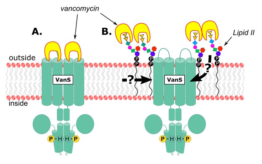
Microorganisms 2021, 9, 2026 2 of 28
Microorganisms 2021, 9, 2026 2 of 26
2. Background
2. Background
2.1. Mechanism of Vancomycin Resistance in VRE
2.1. Mechanism of Vancomycin Resistance in VRE
Vancomycin inhibits cell wall synthesis. It does so by binding the D-alanyl-D-alanine
Vancomycin inhibits cell wall synthesis. It does so by binding the D-alanyl-D-alanine
((D -Ala-D-Ala) residues of the muramyl pentapeptide portion of lipid II, a precursor in
D -Ala- D -Ala) residues of the muramyl pentapeptide portion of lipid II, a precursor in
peptidoglycan synthesis
peptidoglycan synthesis (Figure
(Figure 1)
1) [25–28].
[25–28]. This
This binding
binding event
event interferes
interferes with
with crosslinking
crosslinking
of the pentapeptide and formation of mature peptidoglycan (Figure
of the pentapeptide and formation of mature peptidoglycan (Figure 1) [29], 1) [29], ultimately
ultimately
causing osmotic cell lysis.
causing osmotic cell lysis.
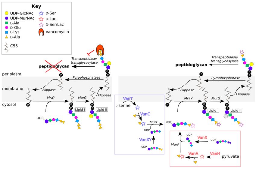
Figure 1.
Figure 1. Vancomycin
Vancomycin resistance
resistance mechanism.
mechanism. Left:
Left:In
Invancomycin-susceptible
vancomycin-susceptibleenterococci,
enterococci,vancomycin
vancomycinbinds the
binds D-Ala-
the D-
D -Ala-
Ala terminus of the muramyl pentapeptide, inhibiting formation of the properly cross-linked peptidoglycan layer
D -Ala terminus of the muramyl pentapeptide, inhibiting formation of the properly cross-linked peptidoglycan layer ofof the
cell wall. Right: In VRE, the D-Ala-D-Ala target is remodeled to either D-Ala-D-Ser or D-Ala-D-Lac, neither of which is
the cell wall. Right: In VRE, the D-Ala-D-Ala target is remodeled to either D-Ala-D-Ser or D-Ala-D-Lac, neither of which is
recognized by vancomycin.
recognized by vancomycin.
Resistant enterococci have acquired a suite of resistance genes. Several of the associ-
ated gene
gene products
productsworkworktogether
togethertotoalter
alterthe
theD-Ala-
D-Ala- D-Ala
D-Ala target
target ofof vancomycin,
vancomycin, prevent-
preventing
ing vancomycin
vancomycin binding
binding (Figure
(Figure 1).different
1). In In different VREVRE strains,
strains, D-Ala-
D -Ala- D-Ala
D -Ala is remodeled
is remodeled to
to ei-
either D-alanyl-
ther D-alanyl- D-lactate
D -lactate (D(-Ala-
D-Ala- D-Lac)
D -Lac) oror D-alanyl-
D -alanyl- D-serine
D -serine (D(-Ala-
D-Ala- D-Ser),
D -Ser), thereby
thereby reduc-
reducing
ing
the the affinity
affinity of vancomycin
of vancomycin forfor
its its ligand
ligand [30–37].This
[30–37]. Thisremodeling
remodelingisis accomplished
accomplished by
three essential enzymes
enzymes encoded
encodedin inthe
thevancomycin-resistance
vancomycin-resistancegene genecluster:
cluster:First,
First,either
eithera
a pyruvatedehydrogenase
pyruvate dehydrogenase(VanH) (VanH)or oraa serine/alanine
serine/alanineracemase
racemase(VanT);
(VanT); second,
second, a ligase
that joins D-lactate or D-serine to D-alanine (the naming convention for these ligases is
described in Section 2.3); and third, a D-Ala-D -Ala dipeptidase
D-Ala dipeptidase (VanX
(VanX or VanXY). VanH
and VanT convert
convert pyruvate to D D-lactate and L-serine to D -serine, respectively [38–40],
which can thenthen be
be coupled
coupledwithwithDD-Ala-Alaby bythetheappropriate
appropriateligase
ligase
toto form
form D -Ala-
D-Ala- D -Lac
D-Lac or or
D-
D -Ala- D -Ser [38,39,41–53]. This dipeptide is added to the UDP-MurNAc-tripeptide
Ala-D-Ser [38,39,41–53]. This dipeptide is added to the UDP-MurNAc-tripeptide by the by
the endogenous
endogenous MurF MurF enzyme,
enzyme, which which
has has sufficiently
sufficiently broad
broad specificity
specificity to accommodate
to accommodate the
the modified
modified substrate
substrate [54].[54]. The resulting
The resulting (D-Lac/(D-Lac/ D -Ser)-UDP-MurNAc
D-Ser)-UDP-MurNAc pentapeptide
pentapeptide is incor-is
incorporated
porated into lipid
into lipid II and II displayed
and displayed on the onexterior
the exterior of cell,
of the the cell, effectively
effectively eliminating
eliminating the
the cell’s
cell’s vulnerability
vulnerability to vancomycin.
to vancomycin. D -Ala-
D-Ala- D-Ala D -Ala dipeptides
dipeptides produced
produced by thebynormal
the normalcell-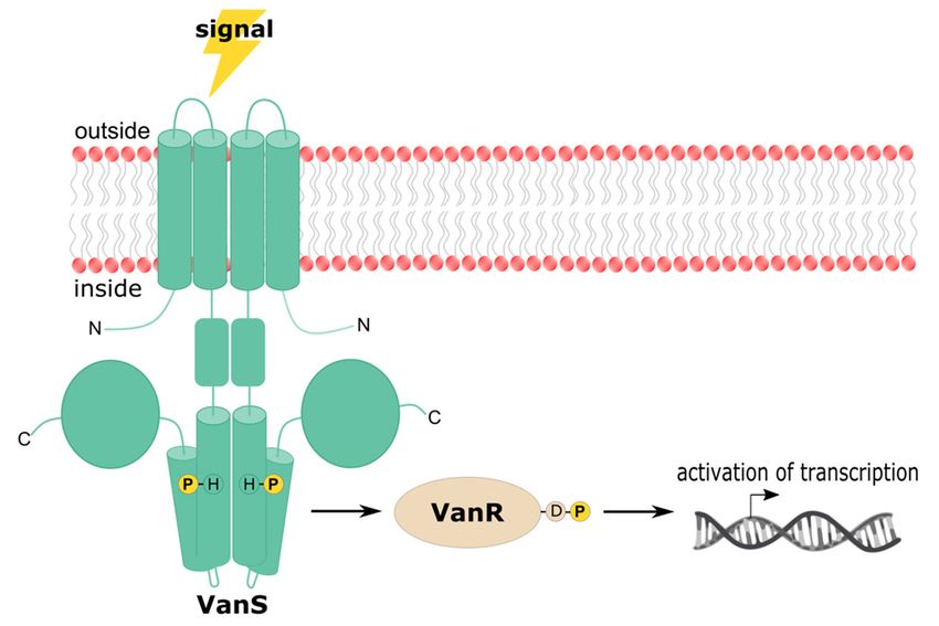
Microorganisms 2021, 9, 2026                                                                                                                 3 of 26

                                        cell-wall biosynthetic machinery are cleaved by VanX/VanXY, preventing them from being
                                        included in the nascent peptidoglycan chain [55–57]. More details about the remodeling
                                        aspects of vancomycin resistance can be found in a number of reviews [58–65].

                                        2.2. Vancomycin Resistance Phenotypes
                                             VRE isolates are assigned to one of nine types, which are genotypically and pheno-
                                        typically distinct. These types are denoted by the letters A–E, G, L, M, and N, and their
                                        characteristics are summarized in Table 1. Collectively, these types are referred to as the
                                        “VRE alphabet” [42,66]. This alphabet should not be considered final, as new resistance
                                        types continue to be discovered [67].

                                  Table 1. Characteristics of vancomycin resistance in the nine types of VRE.

                                              MIC             MIC
 VRE       Terminal                        Vancomycin     Teicoplanin                                Species
 Type      Dipeptide        Inducible                                   Acquired   Transferable                             References
                                            (µg/mL)        (µg/mL)

   A                            Yes         64 to >1000    16 to 512      Yes         Yes            E. faecalis,
          D -Ala- D -Lac                                                                                              [15,16,30,36,38,39,68–73]
                                                                                                     E. faecium
   B                            Yes          4 to 1024       ≤0.5         Yes         Yes            E. faecalis,
          D -Ala- D -Lac                                                                                                  [33,43,68,73–79]
                                                                                                     E. faecium
                                                                                                   E. gallinarum,
   C       D -Ala- D -Ser      Yes/No         2 to 32      ≤0.5 to 1      No           No         E. casseliflavus/     [32–34,46,56,80–86]
                                                                                                     flavescens
   D                            No           16 to 256     0.25 to 64     Yes          No            E. faecalis,
          D -Ala- D -Lac                                                                                                     [45,87–92]
                                                                                                     E. faecium
   E       D -Ala- D -Ser       Yes             16            0.5         Yes          No            E. faecalis           [48,49,93–95]
   G                            Yes             16            0.5         Yes         Yes            E. faecalis,
           D -Ala- D -Ser                                                                                                  [50,51,96–98]
                                                                                                     E. faecium
   L      D -Ala- D -Ser        Yes              8           N/A          Yes         No             E. faecalis                [99]
   M      D -Ala- D -Lac        Yes         128 to 512    0.5 to >256     Yes         Yes            E. faecium             [52,100,101]
   N      D -Ala- D -Ser        No           12 to 16          0.5        Yes         Yes            E. faecium               [53,102]

                                              Most VRE isolated from human infection sites are E. faecalis or E. faecium, with the
                                        latter being the more prevalent species [80,81]. A-type E. faecium are responsible for the
                                        majority of nosocomial VRE infections and are particularly difficult to treat due to their
                                        resistance to all commonly-used glycopeptide antibiotics [103]. B- and C-type VRE are also
                                        clinically significant in humans [81,103–106]. Due to the differing levels of vancomycin
                                        resistance among these types and the various species in which they present, proper typing
                                        of isolates is critical for the treatment of VRE infections. This will become particularly
                                        important in the event that type-specific treatments are developed.
                                              The VRE types can be categorized based on whether they use D-Ala-D-Lac or D-Ala-
                                        D -Ser to achieve vancomycin resistance. VRE types A, B, D, and M fall into the former
                                        group, and types C, E, G, L, and N into the latter. The use of D-Ala-D-Lac versus D-
                                        Ala-D-Ser controls the level of resistance observed. D-Ala-D-Lac is bound 1000-times
                                        less tightly by vancomycin than D-Ala-D-Ala [26,38,74,107], and thus VRE in which the
                                        peptidoglycan precursors contain D-Ala-D-Lac are resistant to high concentrations of
                                        vancomycin [45,52,87,108–110]. D-Ala-D-Lac is also bound more weakly by teicoplanin, ex-
                                        plaining why VRE belonging to types A, D, and M are also teicoplanin-resistant [52,87,108].
                                              D-Ala-D-Ser is also bound less tightly by vancomycin and teicoplanin than D-Ala-D-Ala,
                                        but the difference in affinity is less dramatic than is seen for D-Ala-D-Lac, with the remodeled
                                        precursors being bound ~3- to 8-fold less tightly [37]. Consistent with this modestly reduced
                                        binding, VRE types using D-Ala-D-Ser (types C, E, G, L, and N) exhibit only low-to-moderate
                                        levels of resistance to vancomycin and teicoplanin [32,34,48,50,51,53,64,93,96–99,102,111,112].
                                              Expression of vancomycin resistance genes can be inducible or constitutive. VRE
                                        exhibiting inducible expression include types A, B, C, E, G, L, and M. For these organ-
                                        isms, precursors containing D-Ala-D-Lac or D-Ala-D-Ser are only incorporated into the
                                        cell wall when vancomycin is present; in the absence of the antibiotic, D-Ala-D-Ala is
                                        used [32,33,52,58,60,82,83,99,109,113]. In contrast, VRE expressing the resistance genes
                                        constitutively (types C, D, and N) produce the alternative dipeptides even in the absence
                                        of vancomycin [32,53,58,80,82–84,111,112,114]. The mechanisms regulating inducible and
                                        constitutive expression of resistance will be discussed in Section 3.
Microorganisms 2021, 9, 2026 4 of 26
2.3. Vancomycin Resistance Genotypes
Historically, the sequence of the D-Ala-D-Lac or D-Ala-D-Ser ligase gene has been used
to classify different VRE types [38,39,41–48,50–53,93]. The nomenclature of these genes
parallels that of their respective VRE types: The ligase gene of A-type VRE is referred to
as vanA, that of B-type vanB, and so on. VRE isolates can be typed based on van ligase
sequence using a variety of PCR techniques [66,100,115–117]. Within some VRE types, the
sequences of the van genes differ sufficiently to warrant subtyping. For example, C-type
VRE are subdivided into C1-, C2/3-, and C4-types [44,46,47,80,85]. Other subtyped VRE
include B, D, and G [97,118–123].
Several other genotypic characteristics define the VRE types, including the compo-
sition and organization of the resistance-gene cluster (Figure 2). All VRE contain the
three essential HAX genes that are required for D-Ala-D-Ala remodeling, as discussed
in Section 2.1 [88,124]. In addition to these genes, all operons contain the regulatory
genes vanR and vanS [124], which control how the expression of vancomycin resistance
is induced. Additional “accessory” genes are found in some resistance operons (A, B,
D, G, M), which may contribute to resistance, but are not essential. A common acces-
sory gene is vanY, which encodes a D,D-carboxypeptidase that complements the action
of VanX by removing the terminal D-Ala residue from UDP-MurNAc pentapeptides that
have escaped remodeling [68,125–128]. Some VRE lacking the VanY protein still exhibit
D , D -carboxypeptidase activity, because their VanXY proteins have dual D , D -dipeptidase
and D,D-carboxypeptidase activities [57]. Other accessory genes include vanZ in A-type
Microorganisms 2021, 9, 2026 5 of 28
VRE and vanW in B- and G-type VRE [129,130]. These genes encode proteins of unknown
function, though vanZ seems to play a role in teicoplanin resistance [130–132].
Organization
Figure2.2.Organization
Figure ofof enterococcal
enterococcal vancomycin-resistance
vancomycin-resistance genegene clusters
clusters for resistance
for resistance types types
A- A–N.
Regulatory
N. Regulatorygenes
genesare
areshown
shownin in dark
dark gray,
gray, remodeling genesininwhite,
remodeling genes white,and
and accessory
accessory genes
genes in in gray
gray gradient.
gradient. Arrows
Arrows indicate
indicate thethe approximatepositions
approximate positions of
of promoters.
promoters.
In
Insome
someVRE,
VRE,sequences
sequencesof the regions
of the flanking
regions the resistance
flanking operonoperon
the resistance reveal reveal
that thethat the
operon
operonwas
wasacquired
acquiredenen
bloc by by
bloc transposition. For example,
transposition. the A-type
For example, resistance
the A-type operon operon
resistance
lies
lieswithin
withinthe
thewell-characterized
well-characterized transposon
transposonTn1546 [15,126,133].
Tn1546 Resistance
[15,126,133]. can also
Resistance be be ac-
can also
acquired via conjugation of plasmids harboring the resistance operon [15,43,69–
quired via conjugation of plasmids harboring the resistance operon [15,43,69–72,114,134–139].
72,114,134–139]. The majority of VRE can transfer resistance genes via conjugation, while
types C, D, E, and L-type VRE cannot, suggesting that their resistance genes are chromo-
somally located [43,49,52,53,75,81,87,93,94,96,99].
3. Regulation of the Expression of Vancomycin ResistanceMicroorganisms 2021, 9, 2026 5 of 26
The majority of VRE can transfer resistance genes via conjugation, while types C, D, E,
and L-type VRE cannot, suggesting that their resistance genes are chromosomally lo-
cated [43,49,52,53,75,81,87,93,94,96,99].
3. Regulation of the Expression of Vancomycin Resistance
Many types of VRE express the vancomycin-resistance phenotype only after exposure
to the antibiotic, making the regulation of resistance an intriguing potential target for
treatment of VRE. Specifically, compounds that inhibit the expression of resistance could
function as antibiotic adjuvants [140], enhancing vancomycin’s potency and restoring an-
tibiotic sensitivity to VRE. Developing such compounds requires a detailed understanding
of the regulatory mechanisms governing resistance. This review focuses on these mecha-
nisms; it aims to complement published discussions of this topic, and to provide an update
on a key question in the field: How do VRE sense vancomycin?
Regulation of the resistance phonotype in VRE depends upon the vanRS regulatory
genes, which encode the VanRS two-component system. A two-component system (TCS) is
a type of signaling system found in prokaryotes, archaea, and certain eukaryotes, including
plants and fungi. Notably, they are not found in metazoans [141–145]. These systems
sense and respond to environmental stimuli via a phosphotransfer signaling cascade [146].
Microorganisms 2021, 9, 2026 6 of 28
TCSs consist of a sensor histidine kinase (HK) and a cognate response regulator (RR); in
the VanRS TCS, these proteins are VanS and VanR, respectively. The signal sensed by the
VanRS TCS is vancomycin, and the response is expression of the vancomycin-resistance
by the VanRS TCS is vancomycin, and the response is expression of the vancomycin-re-
genes [147,148]. Upon sensing vancomycin, VanS autophosphorylates on a conserved histi-
sistance genes [147,148]. Upon sensing vancomycin, VanS autophosphorylates on a con-
dine residue (Figure 3). The phosphoryl group is then transferred to VanR [124,149]. When
served histidine residue (Figure 3). The phosphoryl group is then transferred to VanR
phosphorylated, VanR is activated
[124,149]. When phosphorylated, VanRand upregulates
is activated the transcription
and upregulates of the vancomycin-
the transcription of
resistance operon [149–151]. In the absence of a vancomycin signal, VanS
the vancomycin-resistance operon [149–151]. In the absence of a vancomycin signal, dephosphorylates
VanS
VanR, switching VanR,
dephosphorylates off theswitching
resistanceoffpathway [148,152,153].
the resistance pathway This general mechanism
[148,152,153]. This generalappears
to be broadly
mechanism applicable
appears among
to be broadly different
applicable VRE different
among types; however,
VRE types; regulatory details vary
however, regu-
significantly
latory and significantly
details vary are discussed andbelow, beginning
are discussed withbeginning
below, an overview
withofan
the architectures
overview of and
activities
the of theand
architectures VanRS proteins.
activities of the VanRS proteins.
Figure
Figure3.3.
Signal transduction
Signal mechanism
transduction of the VanRS
mechanism of the TCS.
VanRS VanS receives
TCS. VanSa receives
vancomycin signal that signal
a vancomycin
triggers autophosphorylation
that triggers of VanS on
autophosphorylation of aVanS
conserved histidine residue.
on a conserved VanS
histidine transfersVanS
residue. the phos-
transfers the
phoryl group to VanR, activating VanR. VanR then acts as a transcription factor and mediates ex-
phosphoryl group to VanR, activating VanR. VanR then acts as a transcription factor and mediates
pression of resistance genes. In the absence of a vancomycin signal, VanS removes the phosphoryl
expression
group of resistance
from VanR, genes. Inexpression
down-regulating the absenceof of
thearesistance
vancomycin signal, VanS removes the phosphoryl
genes.
group from VanR, down-regulating expression of the resistance genes.
3.1. VanS Architecture and Activity
VanS is a Class-I HK, belonging to the same family as EnvZ [154]. Members of this
family are membrane-bound and homodimeric, and contain a periplasmic domain, a
transmembrane (TM) domain consisting of two transmembrane helices, a linker re-
gion/HAMP domain, a dimerization and histidine phospho-acceptor (DHp) domain, and
a catalytic ATP-binding (CA) domain. These domains participate in signal sensing, signalMicroorganisms 2021, 9, 2026 6 of 26
3.1. VanS Architecture and Activity
VanS is a Class-I HK, belonging to the same family as EnvZ [154]. Members of this
family are membrane-bound and homodimeric, and contain a periplasmic domain, a trans-
membrane (TM) domain consisting of two transmembrane helices, a linker region/HAMP
domain, a dimerization and histidine phospho-acceptor (DHp) domain, and a catalytic
ATP-binding (CA) domain. These domains participate in signal sensing, signal transduc-
tion, and/or the catalytic activity of the HK. To date, no structures have been determined
for any VRE VanS proteins, and the topologies described herein are therefore inferred from
known structures of related HKs and protein prediction software [155,156]. The predicted
domains of VanS are listed for each VRE ortholog in Table 2.
Table 2. SMART-predicted domains of the VRE VanS proteins [155].
Location and Length
VanS Protein Sequence HAMP DHp Histidine CA
Periplasmic Linker
Ortholog Accession a TMH1 b TMH2
Domain Region Domain Domain Phospho-Acceptor Domain
(Residue Number)
A WP_002305818.1 19–41 78–97 42–77 98–153 154–221 164 266–376
[41] (23 aa) (20 aa) (26 aa) (56 aa) (68 aa) (111 aa)
B WP_002368696.1 7–29 133–155 30–132 156–222 157–208 223–289 233 334–445
[42] (23 aa) (23 aa) (103 aa) (67 aa) (52 aa) (67 aa) (112 aa)
C1 WP_063856733.1 1–17 37–56 18–36 57–114 115–182 125 227–337
[44] (17 aa) (20 aa) (23 aa) (58 aa) (68 aa) (111 aa)
WP_016608740.1 4–23 36–58 24–35 59–114 115–182 125 227–337
C2/3 [47] (20 aa) (23 aa) (12 aa) (56 aa) (68 aa) (111 aa)
C4 4–23 36–58 24–35 59–114 115–182 125 227–337
ABX79412.1 (222) (20 aa) (23 aa) (12 aa) (56 aa) (68 aa) (111 aa)
D WP_063856730.1 21–43 76–98 44–75 99–155 156–223 166 268–379
[45] (23 aa) (23 aa) (32 aa) (57 aa) (68 aa) (112 aa)
E WP_063856734.1 13–35 55–77 36–54 78–134 135–205 145 250–357
[49] (23 aa) (23 aa) (19 aa) (57 aa) (71 aa) (108 aa)
G WP_063856732.1 12–34 70–90 35–69 91–144 145–212 155 257–366
[50] (23 aa) (21 aa) (35 aa) (54 aa) (68 aa) (110 aa)
L WP_063856745.1 17–39 66–88 40–65 89–140 141–208 151 253–364
[99] (23 aa) (23 aa) (26 aa) (52 aa) (68 aa) (112 aa)
M WP_063856748.1 12–31 57–79 32–56 80–140 81–133 141–208 151 253–364
[52] (20 aa) (23 aa) (35 aa) (61 aa) (53 aa) (68 aa) (112 aa)
N WP_063856749.1 15–37 61–83 38–60 84–140 141–208 151 253–364
[53] (23 aa) (23 aa) (23 aa) (57 aa) (68 aa) (112 aa)
a Representative VanS protein sequences were chosen because they belong to the first resistance gene cluster of each type to be characterized.
For E-type, the accession listed is that of VanSE from strain N00-410. There is no VanSE protein sequence available from strain BM4405, the
first E-type strain to have its resistance gene cluster characterized. b Numbers in each entry correspond to the range of residue numbers
forming the relevant domain.
3.1.1. Periplasmic Domain
In the EnvZ family of HKs, the periplasmic domain is thought to detect the acti-
vating signal, although in some cases signals may be sensed by other domains (e.g., the
TM domain). The VanS periplasmic domain (together with the TM domain and HAMP
domain/linker region) lies within the N-terminal half of the protein, which displays consid-
erably more sequence variability than the C-terminal half. The size of the VanS periplasmic
domain also differs greatly between the different VRE types, ranging from 12 to 103 residues
in length. This heterogeneity in length and composition suggests that periplasmic domains
from different VanS orthologs may adopt different structures and thus sense vancomycin
differently. Based on the length of the periplasmic domain (Table 2), VanS proteins can be
described as either “intramembrane-sensing” or “periplasmic-sensing” HKs. HKs with
short periplasmic domains (Microorganisms 2021, 9, 2026 7 of 26
domain, this approach alone is not definitive. Possible signal-sensing mechanisms of
different VanS orthologs will be discussed in more detail in Section 3.3.
3.1.2. TM Domain
TM domains contribute to signal sensing in some HKs (including possibly VanS). In
addition, the TM domain transduces the signal to the catalytic domain, thereby bridging
the sensing and catalytic events in the HK [165]. Beginning at the N-terminus, the first
transmembrane α-helix (TMH1) passes the membrane from inside to outside the cell; here,
it is linked via the periplasmic domain to the second transmembrane α-helix (TMH2),
which crosses the membrane again to reenter the cell.
Molecular structures are key contributors to our knowledge of sensing and signal
transduction. However, obtaining structural information for membrane-bound domains
like the TM domain is challenging. Nonetheless, a general idea of how signal transduction
can function through the TM domain has been developed. This model, based on HK
models derived from crystallographic, NMR, and disulfide cross-linking experiments,
suggests that signals are transduced by some combination of rotations, translations, and
scissoring motions of the TM helices [166–169]. However, this conceptual framework
allows for many possible variations, and in the case of VanS, it is unknown precisely
how the TM domain changes conformation in the presence of vancomycin. Indeed, even
though different VanRS TCSs share a common signal (vancomycin) and response (resistance
gene expression), it cannot be concluded that all VanS proteins share a common signal-
transduction mechanism, as underlined by the low sequence identity of the N-terminal
regions of VanS orthologs.
3.1.3. Linker Region/HAMP Domain
All VanS proteins contain a membrane-proximal region connecting TMH2 and the
DHp domain, which is responsible for propagating the signal to the DHp and CA do-
mains; deletion of this region abrogates HK activity [170,171]. This linker region contains
~60 amino acids (Table 2). In most VanS proteins, this region is not annotated as containing
any specific domain, but in VanSB and VanSM this region is predicted to adopt a HAMP-
domain fold [155,172]. HAMP domains are so-named by virtue of their presence in HKs,
adenylyl cyclases, methyl-carrier proteins, and phosphatases [172,173]. HAMP-domain
sequences are not highly conserved, but they share a canonical two-helix coiled-coil struc-
ture [168]; in dimeric HKs, the coiled coils from each protomer associate into a parallel
four-helix bundle [174–176]. While the linker regions of the other VRE VanS orthologs have
not yet been annotated as HAMP domains, they are predicted to be α-helical; thus, given
the lack of sequence conservation within HAMP domains, it is entirely possible one or
more of these VanS proteins will also prove to contain a HAMP domain.
3.1.4. DHp Domain
Following the linker region/HAMP domain is the conserved kinase region of the HK,
consisting of the DHp and CA domains. These domains are ~70 and ~110 amino acids
in length, respectively. The DHp domain earns the first half of its name by contributing
to the dimerization of HK protomers; for example, in the EnvZ family of HKs, the DHp
domain forms a long helical hairpin, with the two α-helices of each protomer dimerizing to
form a four-helix bundle [177,178]. Rearrangements of this helical bundle permit switching
between kinase and phosphatase activities. The first third of the first DHp helix harbors
the conserved histidine (His164 in A-type VanS), which is autophosphorylated upon HK
activation [179,180]. Situated within the aptly-named H box, this histidine residue is
absolutely required for signal transduction [165]. The H-box represents the site at which
the CA domain docks, bringing the CA domain into close proximity to the histidine
phospho-acceptor [165]. In addition to its importance to the autophosphorylation activity
of the HK, the H box is required for phosphotransfer and phosphatase activities [181].
Phosphotransfer from the H-box to the RR is made possible by the binding of the RR to theMicroorganisms 2021, 9, 2026 8 of 26
lower portion of the DHp four-helix bundle [141,182]; this portion of the DHp also contains
the X region, which is important for phosphatase activity [181].
For the A-type VanRS proteins, the aforementioned binding interaction has recently
been quantified. Both full-length, detergent-solubilized VanSA and the cytosolic portion
of VanSA display low micromolar affinity for VanRA , with KD values of 1.9 ± 0.7 µM
and 6.8 ± 1.4 µM, respectively [183]. Perhaps unsurprisingly, these values fall within the
low-micromolar affinity window identified for other HK-RR interactions [184].
3.1.5. CA Domain
In prokaryotic HKs, the CA domain adopts an α/β sandwich topology known as the
Bergerat fold [185,186]. Within this domain, the ATP required for autophosphorylation
is bound in a crevice within two α-helices, partially covered by a mobile loop called the
“ATP lid.” The ATP-binding site and the ATP lid encompass several conserved motifs
known as the N, G1, F, and G2 boxes [154,179,180]. Mutations in these conserved motifs
can have different effects on HK activity; in particular, some abrogate phosphatase activity,
which can cause constitutive expression of the resistance genes. Several such mutations are
discussed in Section 3.4.
3.2. VanR Architecture and Activity
VanR belongs to the OmpR family of RRs [187,188] and is divided into two domains:
an N-terminal receiver domain and a C-terminal effector domain, joined by a flexible
linker [189]. These domains work together to convert the vancomycin signal sensed by
VanS into a transcriptional response. There are no published structures of VanR proteins
from VRE, but structures are known for many other OmpR-family RRs, including VanR
from S. coelicolor [190]. These orthologous structures allow us to make structural inferences
for the VRE VanR proteins.
3.2.1. Receiver Domain
The receiver domain accepts the phosphoryl group from VanS, with phosphorylation
occurring on a conserved aspartate residue (Asp53 for VanRA ). In OmpR-related RRs,
this aspartate is situated at the end of the third β-strand of an α/β sandwich [189]. Once
phosphorylated, the receiver domain undergoes a conformational change, allowing it to
dimerize at a conserved α4-β5-α5 interface [191].
3.2.2. Effector Domain
The effector domain of VanR is a winged-helix DNA-binding domain [187,192], with
helix α8 serving as the recognition helix of the winged-helix motif. Insertion of this helix
into the major groove of the DNA allows VanR to bind to its target promoters, thereby
facilitating expression of the resistance genes, as well as upregulation of the vanRS genes.
VanR targets either one or two promoters, depending on the relative orientations of the
vanRS and vanHAX genes. For resistance operons in which the vanRS genes are located
upstream of the remodeling-enzyme genes (types A, B, D, G, and M; see Figure 2), VanR
recognizes two distinct promoters, one controlling expression of vanHAX and the other
controlling expression of vanRS [150,151,193,194]. However, for operons in which the vanRS
genes lie downstream of the remodeling genes (types C, E, L, and N), only a single promoter
is used [53,83,94,193]. VanRA and VanRB have been shown to bind their DNA targets in
both the phosphorylated and unphosphorylated states; however, transcription of resistance
genes is achieved only when VanR is phosphorylated [129,148–150,152,153]. A plausible
model to explain this observation is that phosphorylation-induced dimerization enhances
DNA binding, either by conformational changes that give rise to optimal orientation of the
effector domains and/or through an avidity effect [150,151,195,196]. This process is then
reversed by dephosphorylation [197–201].
The activities of the VRE VanR proteins will be more clearly understood once structures
are determined for these proteins. Although it is possible to formulate models of VanRMicroorganisms 2021, 9, 2026 9 of 26
Microorganisms 2021, 9, 2026 10 of 28
architecture by examining structures of related RRs, the specific details revealed by true
experimental
true experimentalstructures may
structures mayprovide
provideinsights
insightsinto
into treating VRE. For
treating VRE. Forexample,
example,thera-
therapeutics
that disrupt
peutics the VanR-VanS
that disrupt or VanR-DNA
the VanR-VanS or VanR-DNAinteractions might
interactions restore
might vancomycin
restore sensitivity
vancomycin
sensitivity
to VRE, andto VRE, and are
are thus thusinvestigating.
worth worth investigating.
3.3.VanS
3.3. VanS Sensing
Sensing of Vancomycin
of Vancomycin
The
Theexpression
expressionof vancomycin
of vancomycin resistance is initiated
resistance when when
is initiated VanS detects vancomycin
VanS detects vancomycin
ininthe periplasmic space. The mechanism by which this occurs
the periplasmic space. The mechanism by which this occurs remains oneremains one of theofprinci-
the principal
pal open
open questionsin
questions in the
the field.
field.Addressing
Addressing thisthis
question has proven
question challenging,
has proven at least at least
challenging,
ininpart because VanS is an integral membrane protein, and therefore a
part because VanS is an integral membrane protein, and therefore a difficult difficult subject for subject
biochemical and biophysical analysis. Furthermore, different VanS orthologs
for biochemical and biophysical analysis. Furthermore, different VanS orthologs may may employ
different vancomycin-detection mechanisms, meaning that insights gleaned from one sys-
employ different vancomycin-detection mechanisms, meaning that insights gleaned from
tem cannot necessarily be translated to another.
one system cannot necessarily be translated to another.
Broadly speaking, VanS could detect vancomycin via two distinct mechanisms: It
Broadly speaking, VanS could detect vancomycin via two distinct mechanisms: It
might detect the antibiotic directly, by binding to it (Figure 4A), or indirectly, by sensing
might detect theeffect
some downstream antibiotic directly, activity
of vancomycin by binding(Figureto 4B).
it (Figure
There is4A), or indirectly,
currently little con-by sens-
ing some downstream effect of vancomycin activity
sensus as to which model is correct for any given VRE type. (Figure 4B). There is currently little
consensus as to which model is correct for any given VRE type.
Figure 4. Hypothetical models illustrating vancomycin sensing hypotheses. (A) Direct sensing by
Figure 4. Hypothetical models illustrating vancomycin sensing hypotheses. (A) Direct sensing by
binding
binding ofof vancomycin
vancomycin to the
to the periplasmic
periplasmic domain
domain of VanS.
of VanS. (B) Indirect
(B) Indirect sensingsensing of vancomycin
of vancomycin by by
detection
detection ofof some
some not-yet-determined,
not-yet-determined, downstream
downstream effect effect resulting
resulting from thefrom the vancomycin-
vancomycin- D-Ala-D- D -Ala- D -
Ala
Alabinding
bindingevent. Two
event. Twohypothetical sensing
hypothetical routes
sensing (detecting
routes membrane
(detecting stress orstress
membrane Lipidor
II buildup)
Lipid II buildup)
are
areindicated
indicatedbyby
thethe
heavy black
heavy arrows.
black arrows.
Direct
Direct binding
binding provides the most
provides the conceptually straightforward
most conceptually model. A direct-bind-
straightforward model. A direct-
ing mechanism
binding has beenhas
mechanism most
beenconvincingly demonstrated
most convincingly for the non-VRE
demonstrated species
for the Strep- species
non-VRE
tomyces coelicolor,
Streptomyces which expresses
coelicolor, the VanS ortholog
which expresses the VanSVanS Sc. A direct interaction between
ortholog VanSSc . A direct interaction
vancomycin-VanSSc was deduced using a vancomycin photoaffinity probe, which was
between vancomycin-VanSSc was deduced using a vancomycin photoaffinity probe, which
shown to label native protein in S. coelicolor membranes, as well as recombinant VanSSc in
was shown to label native protein in S. coelicolor membranes, as well as recombinant
E. coli membranes [202]. Unlabeled vancomycin effectively competed with the vancomy-
VanSSc in E. coli membranes [202]. Unlabeled vancomycin effectively competed with
cin photoprobe, arguing for the specificity of this interaction. While this result is compel-
the vancomycin photoprobe, arguing for the specificity of this interaction. While this
ling, it must be noted that these studies employed membrane preparations that contained
result
lipid II; is compelling,
vancomycin it must
binding tobe noted
lipid that these
II would studies
tend to produce employed membrane
a high local preparations
concentration
of the antibiotic, which could give rise to labeling from nonspecific proximity. However, a high
that contained lipid II; vancomycin binding to lipid II would tend to produce
local
this concentration
concern is lessenedofbythe antibiotic,
a recent NMRwhich could
study that givearise
shows to interaction
direct labeling from nonspecific
between
proximity. and
vancomycin However, this
a peptide concern is lessened
corresponding by a recent
to the periplasmic NMRof
domain study
VanSthat shows a direct
Sc [203].
interaction between vancomycin and a peptide corresponding to the periplasmic domain
of VanSSc [203].
In contrast to the direct-binding model, indirect-detection models include any mech-
anisms that do not involve a direct physical interaction between vancomycin and VanS.Microorganisms 2021, 9, 2026 10 of 26
One such indirect model suggests that VanS senses increased lipid II levels resulting from
vancomycin’s inhibition of transglycosylase and transpeptidase enzymes (illustrated in
Figure 4B) [204]. An alternative model posits that VanS senses changes in membrane prop-
erties resulting from vancomycin activity. A precedent for the latter model may be found
in other HKs that are thought to alter conformation within the membrane in response to
changes in temperature, thereby functioning as “molecular thermometers,” such as DesK
in Bacillus subtilis or CorS in Pseudomonas syringae [205,206].
Yet another potential indirect-detection mechanism involves the regulation of VanS by
one or more additional proteins. Many HKs are known to be regulated by other proteins,
which may be upregulated in the presence of the HK stimulus, or may themselves bind the
stimulus [207,208]. For example, the stress-sensing HK LiaS of B. subtilis is regulated by
the small membrane protein LiaF. LiaF inhibits LiaS, turning “off” expression of the LiaS-
regulated genes in the absence of signal [209]. LiaS adopts the same domain architecture
as most VanS orthologs, having two transmembrane helices and a small periplasmic
domain [210]. Hence, the LiaS example suggests that regulation by auxiliary proteins is at
least formally possible for VanS proteins; however, to our knowledge this mechanism has
not been carefully investigated for any VanS orthologs.
3.3.1. VanSA Sensing of Vancomycin
VanSA is the most well-studied of the VRE VanS orthologs, and much evidence is
available that relates to its mechanism of vancomycin sensing. Early work focused on
determining which compounds activate VanSA , with activation being assessed by the
ratio of D-Ala-D-Lac- to D-Ala-D-Ala- in peptidoglycan precursors, the activity of the
D , D -dipeptidase VanX, or the expression levels of the vanHAX genes [29,211–214]. These
experiments revealed that VanSA is activated by a myriad of antimicrobial agents that inter-
fere with cell-wall synthesis and/or compromise the integrity of the cell envelope. These
compounds include glycopeptide antibiotics such as teicoplanin, avoparcin, ristocetin, and
of course vancomycin, as well as structurally unrelated compounds such as bacitracin,
amphomycin, moenomycin, penicillin G, and tunicamycin. The structural heterogeneity of
these different activators would seem to argue against a direct-binding model, since it is
unlikely that a single binding site could recognize such a diverse array of ligands. Because
most of the activating compounds listed interfere with cell-wall biosynthesis, a model in
which VanSA senses vancomycin by detecting lipid II accumulation appears viable [29,212];
however, such indirect sensing mechanisms have not been thoroughly investigated for
VanSA . We note that not all activating compounds need act by the same mechanism. For
example, glycopeptide antibiotics appear to be more potent activators of VanSA than other
agents [212–214]; hence, it is possible that VanSA directly binds vancomycin and other
glycopeptides, whereas other compounds activate the enzyme through different means.
Late-stage intermediates in cell-wall biosynthesis, such as lipid II, are not the only
potential candidates for activating VanSA . This was shown by Ulijasz et al., who devised a
VanSA reporter system in B. subtilis, using the PvanH promoter fused to a lacZ gene [147].
They found that fosfomycin and D-cycloserine (albeit at high concentrations) could activate
their reporter, as well as cell-wall hydrolytic enzymes such as lysozyme and mutanolysin.
These treatments cause the build-up of a wide range of different peptidoglycan precursors
and breakdown products. The structural heterogeneity of these molecules again makes it
unlikely that a single binding site in VanSA directly recognizes them. However, membrane
stress is a common consequence of all of these treatments, and may therefore be a more
credible candidate for the activating signal. Consistent with this idea, the membrane-
perturbing agent chlorhexidine gluconate also activates VanSA , as revealed by RNA-seq
analysis in E. faecium [215]. Control experiments in a ∆vanRS strain showed no increase in
vanHAX transcript abundance, implicating VanS in sensing the chlorhexidine [215].
In addition to the cellular assays for VanSA activation described above, activation
can also be probed in the purified enzyme, by measuring its autophosphorylation, phos-
photransfer, and dephosphorylation activities. If vancomycin directly activates VanSA , itMicroorganisms 2021, 9, 2026 11 of 26
should increase autophosphorylation and phosphotransfer activity, decrease phosphatase
activity, or both. However, detergent-solubilized VanSA displays no change in any of its
activities in the presence of vancomycin [183]. Adverse effects of detergent micelles on
VanSA activity can be ruled out, since when VanSA is reconstituted in either amphipols or
nanodiscs, its autophosphorylation and dephosphorylation activities also do not change in
the presence of vancomycin [183,201]. These in vitro findings argue against a direct-binding
model for VanSA .
Despite this large body of evidence favoring an indirect-detection model for VanSA ,
evidence also exists supporting a direct-binding model [216–221]. A sedimentation-velocity
experiment performed using detergent-solubilized VanSA revealed a shift in the sedimen-
tation coefficient of VanSA in the presence of vancomycin, suggesting that vancomycin in-
duces a conformational change in the protein, presumably via a direct interaction [222,223].
Additionally, vancomycin was found to alter the circular dichroism spectrum of detergent-
solubilized VanSA , which has been interpreted as evidence for direct binding of the antibi-
otic, with a dissociation constant KD of approximately 70 µM [222,224]. Interestingly, this
relatively high KD value is roughly one to two orders of magnitude higher than the antibi-
otic concentrations required to inhibit growth of antibiotic-sensitive Enterococci [216–221],
raising questions about whether this binding is relevant to activation of the resistance
phenotype. An additional caveat is that these results were obtained in the presence of
detergents, which can alter the conformations and activities of many membrane proteins. In
particular, VanSA ’s autophosphorylation activity is highly sensitive to detergents [183,225].
Finally, as we weigh indirect vs. direct sensing mechanisms for VanSA , we note
that models can be conceived that combine elements of both mechanisms. For example,
VanSA activation might entail recognizing a vancomycin-lipid II complex, rather than the
antibiotic alone. Support for this idea comes from the S. coelicolor system, where VanS
activation only occurs when vancomycin binds its D-Ala-Ala target [226], even though
vancomycin has been shown to bind directly to the sensor’s periplasmic domain [203].
In summary, while the preponderance of evidence currently points toward an indirect-
detection mechanism for VanSA , tantalizing data also exist that support a direct-binding
model. Ultimately, this question will not be resolved without further study.
3.3.2. VanSB Sensing of Vancomycin
The vancomycin-sensing mechanism of VanSB is less well-studied than that of VanSA ,
but the cumulative weight of the evidence points to a direct-sensing mechanism. First,
VanSB is activated only by vancomycin [212], in stark contrast with VanSA . In the preceding
section, we noted that it is difficult to conceive of how VanSA ’s small periplasmic domain
would be able to recognize the structurally diverse set of molecules that activate resistance,
providing suggestive support for an indirect-binding mechanism for VanSA . Conversely,
VanSB ’s narrow specificity for its activator makes it plausible that the protein does bind
vancomycin directly.
VanSB ’s ligand preference maps to its periplasmic domain, with mutations in this region
altering ligand specificity and rendering B-type E. faecalis resistant to teicoplanin [227,228].
These mutations will be discussed in more detail in Section 3.4. The VanSB periplasmic
domain does not exhibit a high degree of sequence homology to any domains of known
structure; however, threading experiments predict that it can adopt a PAS-domain fold
(P. Rotsides, unpublished results). This would be consistent with the lack of homology
with other proteins, since PAS domains typically exhibit low pairwise similarities with one
another, and contain no highly conserved residues [157]. A PAS domain in VanSB would
not be unprecedented among HKs, as a number of other sensor kinases possess periplasmic
PAS domains, including CitA, PhoQ, and DcuS [163,164,229]. However, VanSB is the only
enterococcal VanS ortholog for which a periplasmic ligand-binding domain is predicted.
Combined with the specificity of VanSB for vancomycin, this observation suggests that
VanSB may sense vancomycin by direct binding, which could make it an outlier among the
VRE VanS proteins.VanSB would not be unprecedented among HKs, as a number of other sensor kinases pos-
sess periplasmic PAS domains, including CitA, PhoQ, and DcuS [163,164,229]. However,
VanSB is the only enterococcal VanS ortholog for which a periplasmic ligand-binding do-
Microorganisms 2021, 9, 2026 main is predicted. Combined with the specificity of VanSB for vancomycin, this observa-12 of 26
tion suggests that VanSB may sense vancomycin by direct binding, which could make it
an outlier among the VRE VanS proteins.
3.4.
3.4.Inducibility
Inducibilityof of
Vancomycin Resistance
Vancomycin Expression
Resistance Expression
InInthe canonical
the canonical model for VanRS
model function,
for VanRS exposure
function, to vancomycin
exposure leads to expres-
to vancomycin leads to ex-
sion of theof
pression vancomycin-resistance
the vancomycin-resistance genes, via the via
genes, intermediate steps of steps
the intermediate activation and au- and
of activation
tophosphorylation
autophosphorylation of VanS and and
of VanS subsequent phosphotransfer
subsequent phosphotransfer to VanR. However,
to VanR. in prac-
However, in prac-
tice,
tice,vancomycin
vancomycin induces
induces expression
expressionof resistance in only
of resistance a subset
in only of VRE
a subset types:types:
of VRE A, B, A,E, B, E,
G,
G,L,L,M,M,andandsome
some C.C.
InIn
contrast, in other
contrast, in otherVRE types
VRE (D, N,
types (D,and somesome
N, and C) vancomycin-
C) vancomycin-
resistance
resistancegenes
genesareareexpressed
expressedconstitutively,
constitutively,regardless
regardlessofofwhether
whetherthe theantibiotic
antibiotic is is
pre-
present.
sent. By comparing and contrasting the inducible and constitutive systems,
By comparing and contrasting the inducible and constitutive systems, we can gain insights we can gain
insights into mechanisms
into mechanisms of VanRSof VanRS signaling.
signaling.
As noted earlier, VanS possesses
As noted earlier, VanS possesses both both
kinasekinase
activityactivity
(i.e., autophosphorylation
(i.e., autophosphorylationand
phosphotransfer
and phosphotransfer to VanR) and phosphatase activity (dephosphorylation ofin-
to VanR) and phosphatase activity (dephosphorylation of VanR). In VanR).
ducible systems, vancomycin induces expression of resistance by tipping the kinase/phos-
In inducible systems, vancomycin induces expression of resistance by tipping the ki-
phatase balance in favor of the former [148]. However, in the non-inducible VRE types (C,
nase/phosphatase balance in favor of the former [148]. However, in the non-inducible VRE
D, and N), VanR is constitutively phosphorylated. This might result from VanS proteins
types (C, D, and N), VanR is constitutively phosphorylated. This might result from VanS
having constitutively active kinase or defective phosphatase activities, or from the com-
proteins having constitutively active kinase or defective phosphatase activities, or from the
plete loss of VanS (note that in the absence of VanS, VanR can still be phosphorylated by
complete loss of VanS (note that in the absence of VanS, VanR can still be phosphorylated by
small-molecule phosphoryl donors such as acetyl phosphate). Since phosphorylated VanR
small-molecule phosphoryl donors such as acetyl phosphate). Since phosphorylated VanR
has a long half-life (up to 17.6 h), resistance genes can be transcribed for a considerable
has a long half-life (up to 17.6 h), resistance genes can be transcribed for a considerable
amount of time following a phosphorylation event [149].
amount
Muchofoftimeour following
knowledgea about
phosphorylation event
the inducibility of[149].
vancomycin resistance is derived
Much of our knowledge about the inducibility
from analysis of mutant VanS proteins with constitutive kinase of vancomycin resistance
activities/loss is derived
of phospha-
from analysis of mutant VanS proteins with constitutive kinase activities/loss
tase activity [64,193]. A handful of these mutants are shown in Figure 5 and will be dis- of phos-
phatase
cussed in activity [64,193].
the following A handful of these mutants are shown in Figure 5 and will be
sections.
discussed in the following sections.
Domain
Figure5.5.Domain
Figure architecture
architecture of of VanS
VanS showing
showing notable
notable mutations
mutations affecting
affecting inducibility.
inducibility. Domain
Domain
identitiesare
identities arefound
found
in in
thethe key.
key. Conserved
Conserved motifs
motifs (H,N,X,G1,
(H, X, N, G,
G1,andG, G2
andboxes)
G2 boxes)
are inare in yellow.
yellow.
Mutations are identified based on amino acid numbering of their respective VanS ortholog, designated
by superscripts (a corresponds to VanSA , b to VanSB , and c to VanSC1 ).
3.4.1. Mutations Abrogating Inducibility of Resistance
Mutations affecting inducibility of vancomycin resistance were first identified in
B-type VRE grown under teicoplanin selection [227,228,230]. Amino-acid substitutions
leading to the constitutive expression of vancomycin resistance were found in the DHp do-
main, both in the H box (S232F, S232Y, T237K, and T237M) and immediately downstream of
the H box (E247K) [227,228,230]. These mutants are also resistant to teicoplanin, as constant
remodeling of peptidoglycan precursors eliminates the D-Ala-D-Ala target of teicoplanin.Microorganisms 2021, 9, 2026 13 of 26
To our knowledge, the enzymatic consequences of these mutations have not been experi-
mentally tested, but clues about their effects can be found in other HKs, in which similar
substitutions within and near the H box abrogate phosphatase activity [231,232]. Hence,
it appears likely that loss of phosphatase activity explains the constitutive expression
of resistance associated with substitutions in the VanSB H box. Supporting this notion,
a recent mutational study of VanSA showed that substitution of residue T168 (correspond-
ing to residue T237 of VanSB ) decreased VanSA phosphatase activity without affecting
autophosphorylation activity [183].
Loss of all VanS activity should also lead to a constitutively resistant phenotype,
as VanR can still be activated by endogenous small-molecule phosphoryl donors such as
acetyl phosphate. Consistent with this idea, when a stop codon is inserted after codon 30
in VanSB , constitutive resistance to both vancomycin and teicoplanin results [233].
For C-type VRE, some isolates (typically C1) express resistance constitutively, while
others (C2/3 and C4) exhibit inducible resistance. The constitutive phenotype appears to
map to substitutions in the DHp and CA domains. Comparison of VanS sequences from
constitutive and inducible strains revealed several notable substitutions associated with
constitutive behavior: R200L, D312N, D312A, and G320S [83]. Residue 200 is found in the X
region, and mutations to the corresponding region of the DHp domain in EnvZ have been
shown to disrupt phosphatase activity [181]. Hence, R200L appears to provide another
example in which loss of VanS phosphorylation activity causes loss of inducibility.
The VanSC substitutions D312N, D312A, and G320S fall between the F and G2 boxes
of the CA domain. In the related Class-I HK EnvZ, mutations to the F box primarily affect
phosphotransfer, while mutations to the G2 box affect all three enzymatic activities [181].
Thus, the mechanistic basis of these mutations is not yet clear. However, in a B-type
clinical isolate of VRE, a six-residue deletion in the G2 box significantly disrupted only the
phosphatase activity of VanSB [234]. Tentatively, then, we suggest that the D312N, D312A,
and G320S mutants abrogate inducibility by decreasing VanSC phosphatase activity.
3.4.2. Mutations Affecting Resistance to Teicoplanin
Certain point mutations within the sensor region cause VanSB to be activated by te-
icoplanin. For example, an E. faecalis strain selected for growth in the presence of teicoplanin
was found to contain a A30G mutation in its VanSB protein, which conferred teicoplanin
resistance by making the resistance genes inducible by teicoplanin [227]. Residue 30 is
predicted to lie at the beginning of VanSB ’s periplasmic domain, suggesting that the A30G
mutation may alter glycopeptide recognition by the periplasmic domain. Alternatively,
it is possible that wild-type VanSB can bind to teicoplanin, but is unable to transduce
this detection event to the protein’s catalytic region. If this is true, the A30G mutation,
lying as it does at the junction between the protein’s first transmembrane helix and its
periplasmic domain, may enhance the efficiency of signal transduction. Consistent with
this notion, other teicoplanin-resistant VanSB mutations have been found either in the
HAMP domain (D168Y) or between the HAMP and DHp domains (E221G) [227,233]. Be-
cause this region is important for signal transduction, the ability of these mutants to confer
teicoplanin resistance might also reflect more efficient signal transduction in the presence
of teicoplanin.
The response of A-type VRE to teicoplanin can also be altered by substitutions in
the sensor region of VanSA , including L50V, E54Q, and Q69H, all of which fall within the
predicted periplasmic domain [235]. In these variants, transcription of resistance genes
cannot be induced by teicoplanin; however, they retain their inducibility by vancomycin.
This is consistent with a direct-binding model in which VanSA recognizes glycopeptides
via its periplasmic domain, with the ability to sense teicoplanin being specifically lost in
the mutant strains. However, it is difficult to reconcile this model with the observation that
vancomycin does not alter the enzymatic activities of VanSA in vitro [183], suggesting that
more complex models may be required to explain glycopeptide sensing by VanSA .You can also read


