Virulence Factors in Coagulase-Negative Staphylococci - MDPI
←
→
Page content transcription
If your browser does not render page correctly, please read the page content below
pathogens
Review
Virulence Factors in Coagulase-Negative Staphylococci
Angela França *, Vânia Gaio, Nathalie Lopes                      and Luís D. R. Melo *

                                          Laboratory of Research in Biofilms Rosário Oliveira, Centre of Biological Engineering, University of Minho,
                                          4710-057 Braga, Portugal; vaniagaio@ceb.uminho.pt (V.G.); nathalie.lopes@ceb.uminho.pt (N.L.)
                                          * Correspondence: afranca@ceb.uminho.pt (A.F.); lmelo@deb.uminho.pt (L.D.R.M.); Tel.: +351-253-601-968 (A.F.);
                                            +351-253-601-989 (L.D.R.M.)

                                          Abstract: Coagulase-negative staphylococci (CoNS) have emerged as major pathogens in healthcare-
                                          associated facilities, being S. epidermidis, S. haemolyticus and, more recently, S. lugdunensis, the most
                                          clinically relevant species. Despite being less virulent than the well-studied pathogen S. aureus, the
                                          number of CoNS strains sequenced is constantly increasing and, with that, the number of virulence
                                          factors identified in those strains. In this regard, biofilm formation is considered the most important.
                                          Besides virulence factors, the presence of several antibiotic-resistance genes identified in CoNS is
                                          worrisome and makes treatment very challenging. In this review, we analyzed the different aspects
                                          involved in CoNS virulence and their impact on health and food.

                                          Keywords: coagulase-negative staphylococci; biofilms; virulence factors

                                          1. Introduction

                                        Staphylococci are a widespread group of bacteria that belong to human and animals
Citation: França, A.; Gaio, V.; Lopes,
                                          normal microflora [1]. Staphylococcus genus comprises two main groups, the coagulase-
N.; Melo, L.D.R. Virulence Factors in     negative staphylococci (CoNS) and coagulase-positive staphylococci (CoPS), which were de-
Coagulase-Negative Staphylococci.         fined according to their ability to produce the enzyme coagulase [2]. Staphylococcus species
Pathogens 2021, 10, 170.                  were first characterized by Friedrich Rosenbach, who established that yellow/orange
https://doi.org/10.3390/                  colonies corresponded to CoPS species and white colonies to CoNS [3]. Among Staphylo-
pathogens10020170                         cocci, Staphylococcus aureus, belonging to the CoPS group, and Staphylococcus epidermidis,
                                          from the CoNS group, are the most frequently isolated of each group, the reason why
Academic Editor:                          most of the CoNS studies are focused on these species [4]. Nonetheless, CoNS cover a
Wioleta Chajȩcka-Wierzchowska            large and continuously expanding group of bacteria, with more than 50 species described
Received: 30 December 2020                so far, which are currently distributed into 41 main species, divided into more than 20
Accepted: 29 January 2021                 subspecies (reviewed in [5]). Since CoNS are common colonizers of human skin, they have
Published: 4 February 2021                been recurrently considered culture contaminants rather than recognized as the causative
                                          agent of important infections [6,7]. Despite their benign interaction with the host, it is now
Publisher’s Note: MDPI stays neutral      known that these species can cause critical infections, especially in immunocompromised
with regard to jurisdictional claims in
                                          patients, the reason why they are currently acknowledged as opportunistic pathogens and
published maps and institutional affil-
                                          have been gaining increasing importance in the healthcare field (reviewed in [8,9]).
iations.
                                               The increase of CoNS impact on the clinical field was emphasized by the extensive
                                          medical progress, where the use of implantable medical devices and the increasing number
                                          of vulnerable patients have allowed CoNS to cause significant infections in humans [10].
                                          More importantly, these factors have elevated the number of morbid, chronically ill and
Copyright: © 2021 by the authors.         immunocompromised patients, as well as the mortality rates related to CoNS [11,12].
Licensee MDPI, Basel, Switzerland.        Moreover, as CoNS are known to colonize both farm and domestic animals, they may
This article is an open access article
                                          as well establish infections upon opportunity, although to a lesser extent. For instance,
distributed under the terms and
                                          several bovine mastitis infections associated with CoNS have increasingly being reported
conditions of the Creative Commons
                                          over the years [13–15]. Additionally, several studies demonstrated that infections caused
Attribution (CC BY) license (https://
                                          by these species affect debilitated domestic animals as cats with conjunctivitis, upper
creativecommons.org/licenses/by/
                                          respiratory tract and skin or wound infections [16], or dogs with keratitis or urinary
4.0/).

Pathogens 2021, 10, 170. https://doi.org/10.3390/pathogens10020170                                        https://www.mdpi.com/journal/pathogens
Pathogens 2021, 10, 170 2 of 45
tract infections [17,18]. This is especially concerning, since it was already demonstrated
that CoNS may be transferred from pets to their owners [19,20]. Moreover, some farm
animals such as chickens are known to be the main reservoirs of antimicrobial resistance
genes [21]. Importantly, CoNS have also been detected as contaminants of food products.
Contamination with CoNS has been found in ready-to-eat foods of animal origin [22,23],
retailing raw chicken meat [24], and in bulk tank milk or minced meat [25]. Altogether,
the infections caused by CoNS species have become more frequent and harmful to both
humans and animals, and, subsequently, entail an increase in the economic burden [26].
The virulence factors of CoNS have been considered to a lesser extent than CoPS
(e.g., S. aureus) since they are coagulase “free”. Nevertheless, the continuous findings and
updates on species and subspecies have revealed a heterogeneous group, ranging from
nonpathogenic to facultative pathogenic species, with distinct virulence potential levels [27].
Some isolates have become increasingly concerning, as Staphylococcus lugdunensis, which
lately has been recognized as a pathogenic bacterium with a high virulence impact [28].
S. lugdunensis can cause highly acute and destructive events of infective endocarditis (IE),
leading to higher mortality rates than other CoNS species, which generally cause less severe
infections [29]. Despite some similarities with S. aureus, CoNS are generally less pathogenic
and present a smaller array of virulence factors, being less studied than the major CoPS
pathogen (reviewed in [8]). Nevertheless, these species deserve special attention due to
their significant impact on the clinical and food fields, resulting from several virulence
factors. Colonization of surfaces and formation of biofilms by CoNS bacteria has long
been considered their main virulence factor, being known that the heterogeneity of bacteria
within biofilms may contribute to their persistence with emphasis on persister cells, viable
but non-culturable (VBNC) cells and small colony variants (SCVs). Moreover, resistance to
antibiotics and the production of bacteriocins and enterotoxins are aspects also contributing
to their virulence.
In order to enhance the knowledge on CoNS pathogenicity, these virulence factors
and their impact on health and food will be further discussed in this review.
2. Adhesion and Biofilm Formation
In the natural, industrial, and clinical environments, bacteria grow predominantly in
biofilms. Biofilms are multicellular and structured communities of microorganisms adhered
to a substratum and embedded in a matrix of extracellular polymeric substances [30].
These communities provide protection from several external stresses such as antimicrobial
agents [31] and the attacks mounted by the host defenses [32,33] facilitating, thus, the
survival of the cells inside the biofilm. As referred to above, CoNS capacity to form
biofilms is considered a major virulence factor and, thus, the mechanisms underlying
biofilm formation gained special attention in the last decades. Biofilm formation by CoNS
is an intricate and multistep process that can be primarily divided into three phases: (i)
adhesion or attachment to a surface, (ii) maturation into a complex multicellular structure
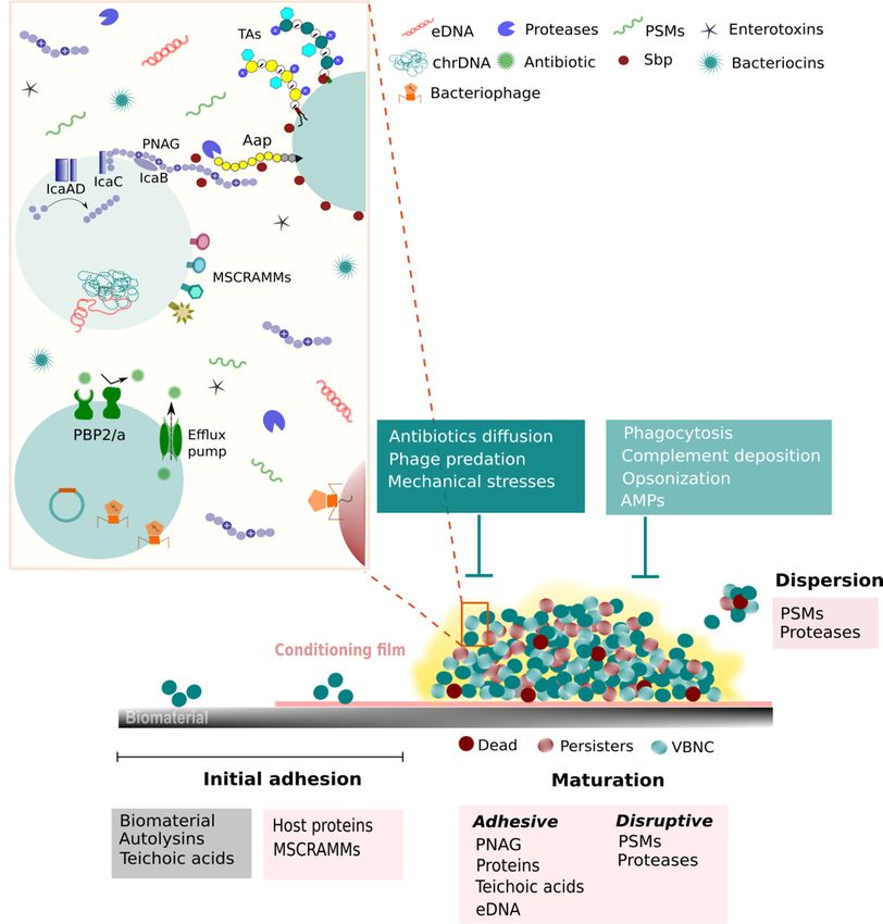
and (iii) dispersion of cells into the surrounding environment [34] (Figure 1).
Surface-associated adhesins have an important role in biofilm formation, both in the
initial adhesion to host proteins and tissues, and biofilm maturation. These adhesins com-
prise covalently and non-covalently anchored proteins, as well as non-proteinaceous factors
(reviewed in [35–38]). Covalently anchored proteins, or also called cell wall-anchored
(CWA) proteins, are characterized by the presence of the LPXTG motif, which is recognized
by the sortase enzyme that, in turn, conducts the process of anchoring the protein to the
peptidoglycan [39]. Briefly, in S. epidermidis, CWA proteins can be divided, based on the
presence of common characteristic domains, into two main families [36]: (i) microbial
surface component recognizing adhesive matrix molecules (MSCRAMM), which integrate
members of the serine-aspartate repeat (Sdr) and S. epidermidis surface (Ses) proteins and
(ii) G5-E repeat proteins family that includes the accumulation associated protein (Aap). A
defining feature of the MSCRAMMs family is the presence of two tandemly linked IgG-like
folded domains, which can engage in ligand binding by the “dock, lock and latch” mecha-Pathogens 2021, 10, 170 3 of 45
nism [40]. This mechanism enables a stable adhesin-ligand complex which is important to
ensure a tight binding under the fluid shear forces that frequently occurs around indwelling
medical devices (IMD) [40]. Moreover, there are also uncategorized CWA proteins puta-
tively involved in biofilm formation as, for instance, biofilm-homolog protein (Bhp) [36].
Within the group of the non-covalently anchored cell wall are the autolysins/adhesins AtlE
and Aae. Lastly, the non-proteinaceous group is composed of teichoic acids (TAs) and the
polysaccharide intercellular adhesion (PIA) [35–38]. The function of these molecules will
be briefly discussed in this section in the context of their contribution to biofilm formation.
2.1. Initial Adhesion
CoNS have the capacity to adhere to several different surfaces including abiotic
(polyethylene, stain steel, rubber, and glass) or biotic surfaces (living tissue or abiotic
surfaces covered with proteins), the former being more relevant in the context of food
processing industry [41] and the last more relevant in clinical settings [42].
2.1.1. Initial Adhesion to Abiotic Surfaces
The adhesion to abiotic surfaces is primarily mediated by non-specific physicochemical
forces such as hydrophobic and electrostatic interactions [43,44]. Nevertheless, specific
bacterial surface molecules can also foster this process.
AtlE, a major autolysin of S. epidermidis, is primarily involved in cell wall turnover and
cell division and lysis [45]. However, it was shown that AtlE-mediated cell lysis resulted in
DNA release (extracellular DNA, eDNA), which, in turn, promoted the adhesion of the
surrounding cells to the polystyrene surface [46]. Thus, AltE seems to mediate adhesion
through the release of DNA rather than acting itself as an adhesin. Another mechanism for
the generation of biofilm eDNA in S. lugdunensis involves the competence protein ComEB,
presumably via active DNA secretion [47]. Homologous autolysins were reported in other
CoNS species, such as S. caprae (AtlC) [48], S. warneri (Atl [49]), S. saprophyticus (Aas) [50],
and in S. lugdunensis (AtlL) [51]. Aae, another autolysin/adhesin found in S. epidermidis, is
as well implicated in the initial adhesion to abiotic surfaces [42,52]. The protein Aap also
participates in the initial adhesion to abiotic surfaces [53,54]. This protein consists of an
N-terminal Domain-A and a C-terminal Domain B, the Domain-A being the one involved
in the adhesion to abiotic surfaces [53]. Other studies have suggested the involvement of
ClpP [55], SdrF [56], and Bhp [57] in this process.
In respect to non-proteinaceous molecules, TAs play an important role in initial
adhesion. TAs are anionic glycopolymers highly abundant in the cell wall that are involved
in several essential cell functions (reviewed in [58,59]). TAs are divided into wall teichoic
acids, which are covalently attached to the peptidoglycan layer, and lipoteichoic acids that
are anchored to the plasma membrane. Both molecules are tailored with D-alanine esters
by a process called D-alanylation [60]. This process balances the charge of both molecules.
It was shown, in S. aureus, that mutants lacking the genes encoding the enzymes necessary
to incorporate D-alanine into TAs resulted in a stronger negative net charge on the bacterial
cell surface [61], thereby attenuating initial attachment to plastic surfaces [62,63]. Although
TAs are less studied in CoNS, in S. epidermidis, the lack of wall teichoic acids resulted in
impaired initial adhesion to polystyrene surfaces [64].
2.1.2. Initial Adhesion to Abiotic Surfaces
When it comes to biotic surfaces such as living tissues or medical devices that are
readily coated by host proteins after implantation, bacterial adhesion is facilitated by a
different set of interactions, mostly by ligand–receptor specific interactions between host
cells or extracellular matrix proteins and bacterial surface-associated adhesins. S. epidermidis
and S. aureus express dozens of MSCRAMMs that bind to human matrix proteins such as
fibrinogen, fibronectin, vitronectin and collagen, and often combine a binding capacity for
several different matrix proteins [65,66]. As such, MSCRAMMs present a key function in
initial adhesion to biotic surfaces, the role of SdrG being the best well-known. SdrG, alsoPathogens 2021, 10, 170 4 of 45
named Fbe, binds to its ligand, fibrinogen, by the “dock, lock, latch” mechanism. Studies
performed to evaluate S. epidermidis SdrG function in the context of bacterial adhesion
showed that this adhesin is important in vitro, not only to mediate bacterial binding
to fibrinogen coated-surfaces [40] but for platelet adhesion and aggregation [67]. More
recently, it has been suggested that SdrG can bind to host cells, such as osteoblasts [68].
SdrG has also shown to be important in vivo for the colonization of implanted material [69].
A homolog to SdrG, the fibrinogen binding protein (Fbl), was found in S. lugdunensis
Pathogens 2021, 10, x FOR PEER REVIEW 3 of 46 [70]
and is likely to be involved in this bacterium initial adhesion to biotic surfaces.
FigureFigure
1. CoNS1. CoNS virulence factors summary illustration. CoNS species are equipped with several strategies to overcome
virulence factors summary illustration. CoNS species are equipped with several strategies to overcome less
less favorable conditions and, thus, to survive in a variety of different environments. Amongst all strategies, the capacity
favorable
to form biofilms and,
conditions is onethus,
of thetomost
survive in a variety
important. Biofilmofformation
different starts
environments. Amongst
with the adhesion all strategies,
of free-floating thetocapacity
cells a surface,to form
biofilms is one of the most important. Biofilm formation starts with the adhesion of free-floating
either abiotic or biotic, and proceeds through the division and aggregation of cells, which creates the characteristic cells to a surface,
multi- either
abioticlayered structure.
or biotic, In addition,
and proceeds an extra-polymeric
through the division protective matrix isofproduced
and aggregation by the
cells, which cells. This
creates is defined as the
the characteristic matu-
multi-layered
ration phase. This phase is mediated by adhesins, but also by molecules with disruptive properties,
structure. In addition, an extra-polymeric protective matrix is produced by the cells. This is defined as the maturation such as PSMs, since
these are necessary to form channels that ensure the flow of nutrients to all biofilm layers. Moreover, as could be expected,
phase.PSMs
This have
phase is mediated by adhesins, but also by molecules with disruptive properties, such as PSMs, since these are
a pivotal role in the final step of the biofilm lifecycle, the dispersion, as it allows biofilm cells to escape and
necessary
colonize otherchannels
to form that
places. It is ensure to
important thestress
flowthat,
of nutrients to all biofilm
in the illustration, only alayers. Moreover,ofassome
brief description could be expected,
of the molecules PSMs
have ainvolved
pivotal in theinseveral
role mechanisms
the final employed
step of the biofilm by CoNS to
lifecycle, respond
the and subsist
dispersion, to external
as it allows biofilmstresses
cellsare depicted.
to escape Aap,
and colonize
other accumulation associated protein;
places. It is important to stressAMPs,
that, Antimicrobial peptides;
in the illustration, onlychrDNA,
a brief chromosomal
description of DNA;
some eDNA,
of theextracellular
molecules DNA;
involved in
MSCRAMMs, Microbial surface components recognizing adhesion matrix molecules; PBP2/a, penicillin-binding protein 2
the several mechanisms employed by CoNS to respond and subsist to external stresses are depicted. Aap, accumulation
and 2a; PNAG, poly-N-acetylglucosamine; PSMs, phenol-soluble modulins; Sbp, Small basic protein; TAs, teichoic acids;
associated
VBNC, protein; AMPs, Antimicrobial
Viable but-non culturable cells. peptides; chrDNA, chromosomal DNA; eDNA, extracellular DNA; MSCRAMMs,
Microbial surface components recognizing adhesion matrix molecules; PBP2/a, penicillin-binding protein 2 and 2a; PNAG,
poly-N-acetylglucosamine; PSMs, phenol-solubleSurface-associated adhesins
modulins; have
Sbp, an important
Small role TAs,
basic protein; in biofilm formation,
teichoic both inViable
acids; VBNC, the
but-non culturable cells. initial adhesion to host proteins and tissues, and biofilm maturation. These adhesins com-
prise covalently and non-covalently anchored proteins, as well as non-proteinaceous fac-
tors (reviewed in [35–38]). Covalently anchored proteins, or also called cell wall-anchored
(CWA) proteins, are characterized by the presence of the LPXTG motif, which is recog-
nized by the sortase enzyme that, in turn, conducts the process of anchoring the proteinPathogens 2021, 10, 170 5 of 45
Also involved in the adhesion of S. epidermidis to biotic surfaces are the proteins
SdrF [71,72], SesC [73] and Embp [74], due to their affinity to, respectively, collagen and
keratin, fibrinogen and fibronectin. In other CoNS additional adhesins with specificity
to bind collagen were found, such as the SrdX in S. capitis [75], and the protein SrdI in
S. saprophyticus that binds to both collagen [76] and fibronectin [77].
Besides the aforementioned role of the autolysins and TAs in the initial adhesion to
abiotic surfaces, these molecules also present an important role in bacterial cells adhesion to
biotic surfaces. The bifunctional autolysins AtlE and Aae, due to their affinity to vitronectin
(AtlE and Aae), fibrinogen (Aae) and fibronectin (Aae) [46,52] and TAs because of their
capacity to bind fibronectin [63,64] and adhere to epithelial and endothelial cells [78,79].
2.2. Maturation
After adhering to the surface, bacterial cells start dividing, forming aggregates and
shaping its distinctive 3D appearance. To maintain a robust structure, cells need not
only to be attached to a surface but also to stick to each other. As such, biofilm cells
are embedded in a matrix composed of self-produced polysaccharides, proteins, lipids,
eDNA and RNA, and TAs [80,81], but can also include molecules of the surrounding
environment [82,83]. This extracellular matrix is fundamental for structural and functional
roles as it provides stability against mechanical forces and creates a unique environment
that is essential for the biofilm lifestyle [82,84]. Importantly, the matrix also plays a part
in protection against disinfectants, antibiotics, immune cells activity, and bacteriophage
(phage) predation [36,85,86]. Nevertheless, to ensure a functional organization, where
nutrients are distributed into the deeper layers of the biofilm, channels need to be molded.
To do so, disruptive forces need to be applied. Thus, during the biofilm maturation process,
there is a thin balance between adhesive and disruptive forces [34].
2.2.1. Intercellular Aggregation Accomplished by Adhesive Forces
In S. epidermidis, the most predominant adhesive molecule is the PIA, also named poly-
N-acetyl glucosamine (PNAG) due to its chemical composition [87]. PNAG is synthesized
by the enzymes codified by the intercellular adhesion (ica) locus, which is composed of the
genes icaA, icaD, icaB, icaC [88], and the regulatory gene icaR, which is located upstream
of the icaADBC and, thus, divergently transcribed [89]. In S. lugdunensis, even though
icaADBC homologs were identified, the locus organization differs substantially from that
of other staphylococci [90]. In addition, the icaR gene is absent even though another ORF
was found in this position [90]. These differences may suggest an evolutionary adaptation
that is likely to confer an advantage to this species [90,91].
Due to its proven fundamental role for biofilm structure, PNAG was for many years
thought to be a requisite for biofilm formation. However, strains that did not harbor
the ica genes were still able to form a biofilm, although less robust [92,93]. Thus, it was
hypothesized that molecules other than PIA were implicated in biofilm maturation. We thus
learned that biofilm formation can be supported or completely mediated by proteins [34–36].
In fact, S. lugdunensis biofilms are mostly composed of proteins rather than PNAG [90,93].
It was found that IsdC, an iron-binding protein, has a pivotal role in S. lugdunensis biofilm
accumulation by promoting cells aggregation through homophilic interactions between
IsdC molecules on neighboring cells [94].
Regarding proteins involvement in biofilm maturation, Aap is one of the best well-
studied proteins in S. epidermidis. Biofilm accumulation by Aap is determined by Domain-B
that becomes active only upon cleavage of the native protein [95,96]. Accordingly, the
matrix of S. epidermidis biofilms is composed of a mixture of fully and partially cleaved pro-
teins [35]. Recently, it was shown that the bacterial metalloprotease SepA is able to cleave
the Domain-A of Aap resulting in enhanced biofilm accumulation in S. epidermidis [97].
Nevertheless, other unknown proteases, from either bacteria or the host, can cleave Aap,
thereby contributing to biofilm accumulation. Aap promotes cell–cell adhesion by forming
twisted rope-like structures through a Zn2+ dependent mechanism [98,99]. In addition,Pathogens 2021, 10, 170 6 of 45
Aap is known to interact with N-acetyl glucosamine moieties potentially binding to PNAG,
forming a protein-polysaccharide biofilm network [100]. Similarly, Embp seems to interact
with PNAG contributing, this way, to the biofilm maturation, as, alone, it seems to be
insufficient to create biofilm aggregation [74].
More recently, Sbp was also found to play an important role in S. epidermidis biofilm
accumulation, having particular importance in the development of the biofilm architec-
ture [101]. Sbp forms amyloid-like fibrils that function as a biofilm scaffold instead of
directly inducing cell aggregation [102]. In addition, it was reported that Sbp interacts,
through the fibrils formed, with the Domain-B of Aap also contributing to biofilm accumu-
lation [102].
Other proteins such as SesC [73,103,104], SesJ [105], and SesI [106] were suggested
to be involved in biofilm maturation. However, more studies are needed to undercover
their relevance and mechanisms of action. Still, within the proteins domain, it is important
to note that MSCRAMMs can also promote biofilm accumulation through homophilic
interactions between MSCRAMMs in neighboring cells [107].
Lastly, as a result of their anionic character, both TAs [62,108] and eDNA originated
from AtlE-mediated autolysis [109–111], can have accessory functions in aggregation by
interacting with other surface polymers, via electrostatic interactions, thereby acting as a
“glue”.
2.2.2. Biofilm Structuring Accomplished by Disruptive Forces
As aforementioned, the disruption of the intercellular interactions is necessary for
the formation of channels that ensure the passage of nutrients and waste in and out
of the biofilm. In staphylococci, proteases [112], nucleases [113], and phenol-soluble
modulins (PSMs) [114,115] have been implicated in this role. However, only PSMs have
been consistently demonstrated to assist in biofilm structuring, both in vitro and in vivo
(reviewed in [116]).
PSMs are amphipathic α-helical molecules with strong surfactant-like properties. As
such, it is thought that PSMs contribute to biofilm structuring by disrupting non-covalent
interactions that occur between biofilm matrix molecules [42]. S. epidermidis produces six
PSM peptides: PSMα, PSMβ1, PSMβ2, PSMδ, PSMε, and PSMγ (δ-toxin) [117], which
are encoded in the chromosome, and the PSM-mec that is encoded in the mobile genetic
element SCCmec [118]. PSMβ peptides have been shown to be a key effector in biofilm
structuring and dispersion both in vitro and in vivo [114]. A deletion mutant of the β-type
PSMs developed a more compact and extended biofilm than the parental strain [114]. As
could be expected, PSMβ also has a role in the biofilm dispersion phase, the last step of
the biofilm lifecycle. It was shown that, depending on the level of production, PSMβ can
lead to either biofilm structuring (medium concentrations) or biofilm dispersion (higher
concentrations) [114]. The production of PSMs is strictly regulated by the accessory gene
regulator (agr) quorum-sensing (QS) system, which will be discussed further in Section 2.4
2.3. Dispersion
As the biofilm grows older, cell clusters may leave the biofilm [119]. This is an
important phase as it contributes to biofilm expansion, bacteria survival, and disease
transmission [119]. While not as explored as the initial adhesion or biofilm maturation,
the dispersion step is a complex process having drawn some attention in past years, in
particular, in oral bacteria and Pseudomonas aeruginosa, with only a few studies performed
in staphylococcal species.
Currently, the dispersion phase is divided into two mechanisms, which are defined
based on the initial trigger: (i) passive dispersion, also called detachment, which includes
processes mediated by external factors, and (ii) active dispersion, which integrates processes
actively employed by bacteria in response to external signals [120]. Passive dispersion can
occur by several different mechanisms such as abrasion (removal of cells due to collision
with particles), grazing (due to the activity of eukaryotic predators), erosion, and sloughingPathogens 2021, 10, 170 7 of 45
(removal of cells or larger pieces of the biofilm by fluid shear) (reviewed in [119–121]).
Also in this category are the techniques developed to induce detachment such as enzymes
with the capacity to degrade biofilm matrix macromolecules (mainly polysaccharides and
proteins) and physical biofilm disruption [119]. One of the best well-known enzymes with
the capacity to disperse S. epidermidis biofilms is Dispersin B [122], a PNAG-degrading
enzyme produced by Actinobacillus actinomycetemcomitans [123]. To what concerns the
active mechanisms of dispersion and the effector molecules associated, as detailed in
Section 2.2.2, PSMs, in particular, β-type PSMs, are the major players.
Even though a lot of research has been focused on the characterization of biofilm
cells phenotype, very little is known about the cells released from the biofilm. Initially, it
was hypothesized that, after leaving the biofilm, cells would immediately revert to their
planktonic phenotype [112]. However, later on, other studies have demonstrated that cells
released from biofilms present a particular phenotype, although transient, that is different
from both planktonic and biofilm cells [124,125]. In S. epidermidis, the cells released from
biofilms present a higher tolerance than biofilm or planktonic cells to some antibiotics [126]
and elicit a more pro-inflammatory response in a murine model of hematogenous dissemi-
nated infection [127]. Nevertheless, more studies are necessary to further understand the
mechanisms behind biofilm dispersion and the role of the cells released in the virulence of
CoNS.
2.4. Regulation of Biofilm Formation
To form the complex structure displayed in biofilms, bacteria have to tightly coordinate
every single step of the process. As such, there are several regulatory systems involved
in biofilm formation by staphylococcal species, the agr QS system being one of the best
characterized (reviewed in [37,128,129]).
Shortly, the agr system is a classical two-component signaling system that is activated
by an autoinducing peptide (AIP) when this reaches a critical concentration, i.e., “quorum”
cells in the population. This signal is sensed by bacteria that synchronize their response.
The agr locus codifies the RNAII and RNAIII transcriptional units that are regulated by
two different promoters, respectively, P2 and P3. The RNAII transcript encodes the genes
agrBDCA and the RNAIII the hld gene that is responsible for the production of the PSMγ
(δ-toxin) [130]. Interestingly, although S. lugdunensis holds an agr-like system, the hld gene is
encoded elsewhere [131,132]. Mechanistically, the signaling cascade starts with agrD, which
is post-translationally modified and exported by AgrB. The extracellular accumulation of
the AIP is detected by the histidine kinase AgrC that, in turn, activates the DNA-binding
regulator AgrA. This activates P2 and P3 promoters [130,133], as well as the ones controlling
the expression of α- and β-type PSMs transcripts [134]. Lastly, RNAIII, the effector of the
agr system, directly controls the upregulation of genes encoding enzymes, toxins, and
PSMs, and it downregulates several genes encoding surface-associated adhesins [135]. This
regulation occurs either by modulating transcription initiation or at the post-translational
level by interacting with the target gene transcript [133].
Probably related to agr system downregulation of adhesins and upregulation of PSM
and other proteases, the dysfunctionality of the agr system in S. epidermidis results in
thicker biofilms with defects in dispersion capacity [32,136]. Although one may think that
the agr negative phenotype is not advantageous as it impairs the bacterium capacity to
disseminate, this phenotype is frequently seen in bacteria isolated from catheter-related
infections. This suggests that mutations in the agr system have an adaptive advantage to
cause IMD-associated infections [32], possibly because a thicker biofilm is likely to confer
fitness advantage in chronic infections [32,137,138]. It was proposed that the naturally
occurring mutations in the agr system are likely to be related to the high metabolic burden
that the maintenance of the agr system poses to the cell [129].
A second QS system molecule influencing biofilm formation in CoNS is the autoinducer-2
(AI-2) that belongs to the LuxS/AI-2 QS system. Due to its wide distribution in many bac-
terial species, this seems to be an interspecies communication system [129]. AI-2 controlsPathogens 2021, 10, 170 8 of 45
biofilm formation by positively regulating the ica operon repressor icaR. In S. epidermidis,
the absence of AI-2 resulted in higher expression of PNAG and, consequently, increased
biofilm formation [139]. In addition, the absence of AI-2 led to increased virulence in
central venous catheter-associated infection model [139]. It is important to mention that
studies performed with other S. epidermidis strains reported a contradictory effect, where
AI-2 leads to icaR negative regulation [140].
Biofilm formation can also be regulated by different factors such as Sigma B (SigB)
and the staphylococcal accessory regulator A (SarA). Sig B is an alternative sigma factor
of RNA polymerase, which leads to global changes in gene expression when activated
by stressful situations. The lack of SigB in S. epidermidis resulted in increased expression
of icaR, which repressed the production of PNAG and, consequently, impaired biofilm
formation [141,142]. In addition, the disruption of SigB production led, in S. epidermidis,
to impaired colonization in a catheter-associated infection model [143]. Lastly, SarA is
a general transcription factor that binds to AT-rich sequences, activating or repressing
the expression of the target genes [129]. Nevertheless, the effect of SarA in S. epidermidis
biofilm formation is highly strain-dependent. While in some strains SarA mutation led to a
biofilm-negative phenotype through the downregulation of ica operon expression by an
IcaR-independent pathway [144], in aap- and ica-negative strains resulted in higher biofilm
formation capacity through the overexpression of the protein Empb and release of eDNA
by a SepA and AtlE-mediated process [145].
Additionally, although not a true regulator, the insertion of the insertion sequence
(IS)256 in ica genes abolishes PNAG production [146].
3. Persistence as a Tolerance Mechanism
Bacteria can quickly respond to unfavorable environmental or stressful conditions by
lowering their metabolic activity, altering their gene expression, or by inducing genetic
changes, entering a state of dormancy [147]. Biofilms per se are an example of a bacterial
stress condition, namely due to nutrients and oxygen deprivation [148]. Biofilm-embedded
communities are characterized by the presence of heterogeneous cells, with distinct physi-
ological states, whose emergence depends on the micro-environmental conditions in its
surroundings [149,150]. Therefore, since the access to nutrients and oxygen at the deeper
biofilm layers is more unfavorable than in the upper layers, variant subpopulations of cells
can emerge [151]. Importantly, CoNS can switch to a different mode of growth and adjust
their gene expression patterns, metabolic activity, and phenotype, to promote their survival
in stressed or limited environmental conditions [152–154]. Recently, two CoNS species,
namely S. epidermidis and S. haemolyticus, exhibited different strategies to overcome the
impact of nutrient depletion [155]. While S. epidermidis managed to survive through the
accumulation of cardiolipin and/or lyso-cardiolipin, S. haemolyticus employed a completely
different strategy, surviving the nutrient depletion created by building an extremely simple
lipidome, made of only diglucosyl-diacylglycerol and phosphatidylglycerol. Additionally,
the authors claimed that bacteria at the stationary phase seemed to have similar behavior as
when exposed to starvation [155]. Considering the entrance of bacteria into dormancy upon
stressful conditions (e.g., starvation), the analysis of bacteria in the stationary phase may
highlight some potential strategies used to survive those environments. Some dormancy
phenotypes have been already found and will be detailed below.
Bacterial Cells Dormant Phenotypes: A Tolerance Mechanism
Several authors have been debating the possible different phenotypic states that
bacteria can undertake, which enable a diminished inflammatory response and higher
tolerance to the antimicrobial therapies applied [156–158]. Persisters, VBNC, and SCVs are
the physiological states currently under debate.
In the 1940s, persister cells were described and defined as a group of cells that exhibit
a drug-tolerant phenotype [159,160]. This small subpopulation of cells, when exposed
to antibiotic pressure, becomes slow-growing by reducing their metabolism, rather thanPathogens 2021, 10, 170 9 of 45
promoting an active response. Once the stress is removed, persister cells can resume
growth, contributing to the antibiotic tolerance observed among biofilms cells [161,162].
The formation of persister cells in culture can be reached through two distinct ways:
triggered or spontaneously. Triggered persisters, also defined as type I persisters, emerge
when cells encounter some stress, such as starvation, and the persistence level may depend
on the type and the intensity of the trigger [161]. Spontaneous persisters (type II persisters)
occur during the stationary phase culture and persist as long as the steady-state growth
is maintained [161,163,164]. The presence of persisters can be found on both planktonic
and biofilm populations, as observed in S. epidermidis cells when exposed to levofloxacin
and vancomycin [165]. Goneau et al. were able to induce the formation of persister cells
in S. saprophyticus using antibiotics from different classes (ciprofloxacin, ampicillin, and
gentamicin), exhibiting a greater antibiotic tolerance during the stationary phase than in the
exponential phase [166]. A recent study on CoNS demonstrated the formation of persister
cells after exposure to various biocides (polymyxin, sodium sulfacetamide, lysing solution).
Independently of the biocide tested, S. epidermidis and S. capitis strains were able to form
persister cells [167].
Later in 1982, the existence of another phenotype, viable but non-culturable cells,
was proposed. These cells were identified by Xu et al., which showed that these bacterial
cells could not grow on routine or selective media [168]. Moreover, VBNC cells were
discriminated from dead cells, since they were similar to live cells, containing an intact
membrane, an active mRNA transcription, metabolic activity, and respiration [169,170].
At least eighty-five bacterial species have been shown to enter a VBNC state, including
foodborne and clinical pathogens [171]. To date, S. epidermidis is the only known CoNS
reported to adopt this survival strategy [172]. Cerca and co-workers have developed a
model where the proportions of VBNC cells in S. epidermidis biofilms can be modulated.
Briefly, the authors demonstrated that the induction of VBNC cells could be achieved
by increasing the glucose concentration in the growth medium and that this induction
could be somehow prevented by the supplementation of the medium with Ca2+ and
Mg2+ [172]. Several methods have been suggested to uncover the existence of VBNC cells.
Assessing viability and culturability is the key to provide an estimation of the number of
these cells [173]. Therefore, numerous approaches have been combined to evaluate cell
viability and culturability, such as the usage of fluorescent microscopy or flow cytometry
and colony forming units (culture methods), respectively [174,175]. Additionally, a study
demonstrated that the combination of LIVE/DEAD staining with quantitative PCR can also
reveal the presence of VBNC cells in CoNS biofilms [175]. Over the years, the similarities
and/or differences between persisters and VBNC cells phenotypes have been a motive for
intense debate since both are employed by bacteria under the same stress conditions. Some
authors suggest that persister cells are indeed VBNC cells [176,177], while others oppose
this interpretation as the similarities hypothesized for both phenotypes were described
in different species [178]. Moreover, it has been suggested that persister cells are more
associated with antibiotic stress and can easily regain growth after antibiotic removal,
whereas the VBNC state seems to be linked to different environmental conditions and,
in some species, the removal of the stress factor is not enough to revert the phenotype,
requiring a more specific condition to revive the cells [178].
SCVs were first described more than 100 years ago [179]. These cells are known as
natural occurring bacterial subpopulations, demonstrating a similar slow growth rate as
the previously described dormant phenotypes and, as the name implies, exhibit a smaller
size than their parental wild-type bacteria, setting a challenge in their identification [180].
Since then, SCVs were found in a wide range of bacterial species such as S. aureus [181] and
CoNS species and are generally correlated to biomaterial-associated infections [180,182].
Several aspects of the pathogenic potential of SCVs have been described, mainly their
enhanced biofilm-forming ability [183], evasion from the immune system response [184],
and their resistance against antimicrobial agents [185,186]. Interestingly, Onyango et al.
revealed that S. epidermidis and S. lugdunensis were capable of developing SCVs pheno-Pathogens 2021, 10, 170 10 of 45
types following the exposure to a wide range of environmental stress conditions, such
as pH alterations (pH5), osmotic stress (0–20% NaCl), low temperature (4 ◦ C), and to the
presence of antimicrobial agents (vancomycin and penicillin G) [187]. Additionally, the
authors found a thicker extracellular matrix in all SCVs populations in comparison to their
corresponding control cells [187]. Therefore, this feature may represent an adaptation in
biofilm formation to provide a stronger defense against antimicrobial agents, as suggested
by other authors [188].
However, the genetic bases underlying these dormant phenotypes are still not well
characterized. Therefore, it is important to recognize the impact of these dormant pheno-
types, as a tolerance mechanism adopted by CoNS, in nowadays clinical infections and
food safety.
4. Antibiotic Resistance
The significance of CoNS species has increased over the years, mainly due to their
multidrug resistance profile [24,189–193] and their ability to grow as biofilms, which
are even more refractory to antibiotics as reported worldwide [31,191,194–197]. Several
mechanisms have been discussed concerning the increase of antimicrobial resistance, from
which (i) the barrier formed by the matrix surrounding the cells within biofilms, whose
thickness and composition can hinder the penetration and/or diffusion of antibiotics [198],
and (ii) the fact that staphylococci biofilms are very prone to mutations that may increase
their resistance towards antibiotics [199,200] stand out. Moreover, the presence of cells with
distinct physiologies, such as persister, SCVs and VBNC cells, also increases tolerance to
antibiotics [201,202] (reviewed in [203–205]). Another possible explanation for the high rate
of antimicrobial resistance is that CoNS share the same niches of colonization with S. aureus,
allowing horizontal gene transfer (HGT) of several genes and mobile elements encoding
for antibiotic resistance [206]. In fact, HGT among staphylococcal species has already
been proven with the detection of many resistant phenotypes related to multiresistant
genes located on mobile genetic elements [25,207,208]. The importance of mobile genetic
elements as virulence factors in CoNS will be further explored in more detail in Section 5.
Fighting these threats has hence become of ultimate importance, with the main strat-
egy being the application of a cocktail of several antibiotics for a prolonged period of
time [24,209–213]. However, the high tolerance of CoNS biofilm cells commonly causes the
failure of antibiotics, even when the most severe therapies are used [214] and, in the cases
associated with the use of IMD, may implicate the removal of the infected device, resulting
in prolonged hospital stays and increased morbidity and mortality rates [42,203,215]. Inter-
estingly, the problematic of IMD-associated infections was emphasized by a recent study
that assessed S. epidermidis in-host evolution in a case of pacemaker-associated endocardi-
tis, which has shown that increased tolerance to antibiotics and capacity to form biofilms
occurred during the course of infection [216]. This helps to explain the often inefficacy of
antibiotics to treat S. epidermidis infections. Besides the impact on human health, CoNS
infections and contamination are also alarming from the veterinary and food production
standpoint, where antimicrobial resistance has correspondingly been reported (reviewed
in [14,24,217,218]). Among CoNS strains, antimicrobial resistant rates have been increasing
over the years, resulting from (i) the incorrect and/or widespread use of antibiotics, (ii) the
use of antibiotics in domestic and farm animals, (iii) the low discovery rate of newer antibi-
otics, and (iv) due to the intrinsic environmental conditions contributing to the adaptation
of bacteria to antimicrobial compounds [219–226].
4.1. Resistance to β-lactams
Some of the more representative species of CoNS are known to present a high resis-
tance rate to methicillin [25,227], as, for instance, S. epidermidis [228], S. haemolyticus [229],
and S. sciuri [230]. This phenotype is not region-specific as studies from Europe to North
America have shown that 60 to 80% of the CoNS species retrieved from bloodstream
infections were resistant to methicillin (MR-CoNS) [231–234]. Not surprisingly, such iso-Pathogens 2021, 10, 170 11 of 45
lates often present increased tolerance to most β-lactam antibiotics, whose structure and
mechanism of action are similar to methicillin [235,236]. The resistance to the action of
β-lactamase was first described as the result of the hydrolysis of the β-lactam ring of such
antibiotics, by penicillinases [237], as determined by the plasmid-mediated staphylococ-
cal β-lactamase bla-Z [238]. Now, it is known that staphylococcal species can produce a
specific penicillin-binding protein (PBP2a), which is responsible to completely inactivate
the activity of most β-lactams, and that this resistant phenotype is complex and related
to the existence of SCCmec, a staphylococcal cassette chromosome containing the mecA
gene, which encodes the PBP2a protein [239–241]. Importantly, already back in the 1980s,
it was found that only about 10–20% of CoNS isolated from nosocomial infections were
penicillin-susceptible, contrary to the 80% of commensal isolates being susceptible to me-
thicillin [242,243]. Thisseems to remain true in the present days, where more than 90% of
CoNS isolated in the hospital settings present increased resistance to penicillin-derived
antibiotics [222,229,244].
4.2. Resistance to Other Antibiotics
Over the years, there has been an increase in the number of CoNS strains resistant to
glycopeptides, which are the antibiotics often used to treat MR-CoNS infections, as well as
the emergence of resistance to newer antibiotics, hindering the current treatment options.
For instance, S. epidermidis was shown to be resistant to up to eight distinct antibiotics with
different mechanisms of action and it is estimated that, among nosocomial isolated strains,
80% of the isolates present resistance to antibiotics beyond methicillin [232,245]. Contrast-
ing with previous studies with a broad range of CoNS, where few isolates and species
presented increased tolerance to antibiotics like vancomycin and teicoplanin [246–248],
the emergence of isolates with reduced susceptibility to glycopeptides has been reported
in several species (Table 1). Surprisingly, resistant isolates of S. epidermidis [249,250] and
S. haemolyticus [251,252] were detected already three decades ago. S. warneri [253–255] and
S. capitis [256,257] have also joined the list with several isolates resistant to vancomycin,
generating outbreaks especially in neonatal units, where S. epidermidis resistant isolates
are also frequently found [258]. Fortunately, vancomycin remains an effective antibiotic
against most of the CoNS isolates [189,222,245,259], being on the top list of antibiotics
used to fight these infections, either alone or in combination with other antibiotics as
cefazolin [209], rifampicin [213,260], and fosfomycin [261], among others. Rifampicin is
also frequently used to treat staphylococcal infections; however, this antibiotic is associ-
ated with the rapid development of resistance when used alone and, as such, it should
be used as part of a combined therapy [262–264]. For instance, the use of vancomycin
or levofloxacin with rifampicin has been proved to be a good combination to treat these
infections [213]. Significant and concerning increases in the resistance to ciprofloxacin,
clindamycin, erythromycin, gentamicin, and tetracycline have been found over the last few
years [22,191,219,265–270]. Resistance to tetracycline is commonly based on the acquisition
of mobile resistance genes that lead to the dissociation of tetracyclines from their ribosomal
binding sites and transportation of the antimicrobial agents out of the cell through drug
efflux pumps [271,272]. Linezolid belongs to a newer class of antibiotics (oxazolidinones)
and appeared as a promising alternative to fight staphylococcal infections with multi-drug
resistance to common antibiotics [273,274], including glycopeptides, to which bacteria have
already developed resistance mechanisms. Nevertheless, resistance to linezolid has already
been reported in staphylococci including CoNS [275,276]. Another antimicrobial belonging
to the next-generation antibiotics is daptomycin, which has proven to be more effective
than vancomycin against MR-CoNS [277]. Despite being a new antibiotic, there are already
reports of isolates resistant to daptomycin [278], hence, the combination therapy with other
antibiotics as rifampicin [279] may be suggested.Pathogens 2021, 10, 170 12 of 45
Table 1. Reports of antimicrobial resistance of the 20 more frequently isolated CoNS against the 20 main antibiotics used in clinical and veterinary settings.
S. capitis S. S. carnosus S. cohnii S. S. S. equorum S. S. hominis
S. lentus
S. urealyticus caprae S. utilis S. urealyticus condimenti epidermidis S. linens haemolyticus S. novobiosepticus
Ampicillin +[280] +[281] +[282] +[280] +[283] +[284] +[280] +[284] +[280] +[283]
Cefazolin +[280] +[280] NF 2 +[280] NF 2 +[191] +[280] +[280] +[280] +[285]
Fosfomycin +[257] +[286] NF 2 +[287] +[288] +[289] NF 2 +[290] +[287] +[287]
inhibitorsn
Cell wall
synthesis
Imipenem +[280] +[291] NF 2 +[280] NF 2 +[291] NF 2 +[280] +[280] NF 2
Methicillin +[256] +[281] +[292] +[293] NF 2 +[294] +[293] +[294] +[294] +[294]
Penicillin +[295] +[296] +[297] +[284] +[283] +[284] +[298] +[284] +[299] +[300]
Oxacillin +[301] +[296] +[282] +[296] NF 2 +[284] +[302] +[299] +[299] +[300]
Teicoplanin +[303] +[304] NF 2 +[305] NF 2 +[298] +[302] +[290] +[302] +[306]
Vancomycin +[256] +[307] +[297] +[308] NF 2 +[303] +[302] +[304] +[304] +[309]
Ciprofloxacin +[310] +[296] +[297] +[296] NF 2 +[310] +[302] +[310] +[300] +[300]
inhibitors
synthesis
NA 1
Levofloxacin +[301] NF 2 +[311] +[191] NF 2 +[300] NF 2 +[301] +[301] +[300]
Rifampicin +[256] +[286] NF 2 +[312] +[313] +[302] +[282] +[284] +[290] +[22]
Clindamycin +[301] +[286] +[311] +[284] NF 2 +[284] +[302] +[284] +[301] +[300]
Erythromycin +[301] +[296] +[297] +[284] +[313] +[299] +[284] +[299] +[299] +[314]
Gentamicin +[301] +[296] +[297] +[296] NF 2 +[310] +[315] +[299] +[299] +[300]
inhibitors
synthesis
Protein
Linezolid +[295] NF 2 +[316] +[317] NF 2 +[318] +[302] +[300] +[300] +[300]
Quinupristin-
+[319] +[311] NF 2 +[284] NF 2 +[284] NF 2 +[284] +[320] +[321]
Dalfopristin
Tetracycline +[284] +[322] +[297] +[284] +[283] +[284] +[284] +[299] +[299] +[300]
Tigecycline NF 2 +[311] +[311] +[311] NF 2 +[284] NF 2 +[323] +[300] +[300]
membrane
Alter. cell
Daptomycin +[301] NF 2 NF 2 +[311] NF 2 +[303] NF 2 +[324] +[324] +[283]Pathogens 2021, 10, 170 13 of 45
Table 1. Cont.
S. S. saprophyticus S. succinus S.
S. lugdunensis S. pasteuri S. sciuri S. vitulinus S. simulans S. warneri
piscifermentans S. bovis S. casei xylosus
Ampicillin +[280] +[218] NF 2 +[280] +[280] +[315] +[280] +[282] +[284] +[284]
Cefazolin +[280] NF 2 NF 2 +[280] +[280] NF 2 +[280] NF 2 +[280] +[280]
Fosfomycin +[325] NF 2 +[326] +[327] +[220] NF 2 NF 2 NF 2 +[289] +[306]
inhibitors
Cell wall
synthesis
Imipenem NF 2 NF 2 NF 2 NF 2 NF 2 NF 2 NF 2 NF 2 +[328] NF 2
Methicillin +[293] +[329] +[330] +[294] +[293] +[331] +[294] +[332] +[294] +[293]
Penicillin +[295] +[284] +[330] +[284] +[284] +[296] +[295] +[333] +[284] +[284]
Oxacillin +[296] +[299] +[330] +[300] +[300] +[299] +[295] +[282] +[299] +[299]
Teicoplanin +[334] NF 2 NF 2 +[305] +[319] NF +[319] NF 2 +[304] +[335]
Vancomycin +[24] NF 2 +[330] +[330] +[330] +[307] +[336] +[316] +[304] +[330]
Ciprofloxacin +[296] NF 2 NF 2 +[300] +[300] +[296] +[295] NF 2 +[300] +[310]
inhibitors
synthesis
NA 1
Levofloxacin +[334] NF 2 NF 2 +[300] +[300] NF 2 +[320] NF 2 +[300] +[311]
Rifampicin +[337] NF 2 +[330] +[338] +[220] +[315] +[22] +[316] +[301] +[22]
Clindamycin +[295] +[270] +[330] +[300] +[284] +[333] +[295] +[270] +[284] +[284]
Erythromycin +[296] +[299] +[312] +[300] +[300] +[299] +[284] +[270] +[299] +[299]
Gentamicin +[296] +[299] NF 2 +[300] +[191] +[299] +[295] +[316] +[299] +[299]
inhibitors
synthesis
Protein
Linezolid +[339] NF 2 NF 2 +[330] +[300] +[340] +[295] NF 2 +[295] +[300]
Quinupristin-
NF 2 NF 2 NF 2 +[319] +[284] NF 2 +[284] NF 2 +[320] +[311]
dalfopristin
Tetracycline +[334] +[299] +[282] +[284] +[333] +[299] +[284] +[314] +[299] +[299]
Tigecycline +[323] NF 2 NF 2 +[300] NF 2 NF 2 +[284] NF 2 +[323] +[22]
membrane
Alter. cell
Daptomycin NF 2 NF 2 NF 2 +[283] +[284] NF 2 NF 2 NF 2 +[301] +[283]
1 NA, nucleic acids; 2 NF—Not found.Pathogens 2021, 10, 170 14 of 45
4.3. Antimicrobial Resistance in the Community
Despite most of the studies being focused on clinical strains, it is known that staphylo-
cocci isolated from healthy individuals may also present increased antimicrobial tolerance.
In fact, several studies report the carriage of distinct CoNS antibiotic-resistant commen-
sal strains by the community [195,341,342], even in remote populations [227]. Although
community strains may present lower resistance rates, as, for instance, only up to 20% of
S. epidermidis commensal strains were found to be resistant to methicillin [343,344], con-
trasting with approximately 80% of resistance found among clinical isolates [245,345,346],
the main existence of commensal strains with antimicrobial resistance colonizing humans
and other mammals is alarming [347]. This is especially concerning in immunocompro-
mised individuals, as CoNS are often considered opportunistic pathogens that may cause
severe infections, whose treatment would be hindered by the existence of isolates with
antimicrobial resistance, as reviewed by Heilmann et al. [8]. The presence of CoNS with
increased tolerance or resistance to antibiotics in animals and food is also worrying. Several
studies report the isolation of CoNS with multidrug resistance recovered from bovine masti-
tis [14,348], retailing chicken meat [349], livestock, bulk tank milk, and minced meat [25], as
well as from ready-to-eat foods [23]. Undoubtedly, the presence of antimicrobial-resistant
strains in animals represents a challenge to the animal hosts, as infections become harder
to treat, but may also be problematic to human hosts upon transmission of resistant strains
resulting from close contacts between people and companion or farm animals [208,220,221].
5. Mobile Genetic Elements
As mentioned before, CoNS infections are associated with the establishment of
biofilms. It is actually in these complex structures that HGT phenomena are favored
due to high cell density, high genetic competence, and availability of mobile genetic el-
ements [350]. HGT is a highly important force driving bacterial evolution. Bacterial
adaptation to new niches and environments frequently occurs through the acquisition of
new genes by HGT processes. There are three different mechanisms to which HGT can
occur: (i) transformation, (ii) transduction, and (iii) conjugation. Transformation occurs
when a DNA fragment from a dead or compromised cell enters a competent bacterial
cell. Transduction consists of the transfer of DNA between bacterial cells through a phage.
Although some lytic phages can transduce, generally temperate phages are more frequently
associated with this HGT mechanism. Under the lysogenic cycle, the viral genome is
integrated into the bacterial chromosome establishing a prophage. Under certain stimuli,
phage genomes are excised from the bacterial genome, and, occasionally, they exchange a
small piece of bacterial DNA for a piece of the phage genome. The newly formed phages
are then composed of these DNA regions that can be further inserted in new bacterial
cells upon a new cycle of infection. Conjugation is the transfer of DNA directly from
one cell to another through cell–cell contact. This process usually involves the transfer of
plasmids. High genetic relatedness has shown to be a key factor influencing HGT because,
as phylogenetic distance increases, HGT phenomena diminish [351]. As phage propagation
depends on host genetic similarity, transduction usually just occurs throughout the same
species or genus [352]. In opposition, plasmids and integrative and conjugative elements
can cross the interspecies barrier [353].
The HGT mechanism also influences the size of the nucleotide sequence that is trans-
ferred. While on phage-mediated transduction up to 45 kb chromosomal DNA or plasmids
(small- or middle-sized) are transferred, larger plasmids are transferred through conju-
gation [354,355]. Moreover, in 2016, Haaber et al. discovered a mechanism named auto-
transduction in which phages are spontaneously released from the bacterial chromosome,
and, after infecting a susceptible cell, they transfer DNA from this cell to the lysogenic
population [356].
In the last decade, it was evidenced that CoNS might act as reservoirs of genes
that can be transferred between different staphylococci, having the potential to increase
the virulence of several species, namely S. aureus [357,358]. Indeed, genes conferringYou can also read