Depression and Cardiovascular Disease: The Viewpoint of Platelets - MDPI
←
→
Page content transcription
If your browser does not render page correctly, please read the page content below
International Journal of
            Molecular Sciences

Review
Depression and Cardiovascular Disease:
The Viewpoint of Platelets
Patrizia Amadio 1, * , Marta Zarà 1 , Leonardo Sandrini 1 , Alessandro Ieraci 2                 and
Silvia Stella Barbieri 1, *
 1   Unit of Brain-Heart Axis: Cellular and Molecular Mechanism, Centro Cardiologico Monzino IRCCS,
     20138 Milan, Italy; marta.zara@ccfm.it (M.Z.); leonardo.sandrini@ccfm.it (L.S.)
 2   Laboratory of Neuropsychopharmacology and Functional Neurogenomics, Department of Pharmaceutical
     Sciences, University of Milan, 20133 Milan, Italy; alessandro.ieraci@unimi.it
 *   Correspondence: patrizia.amadio@ccfm.it (P.A.); silvia.barbieri@ccfm.it (S.S.B.); Tel.: +39-02-58002 (P.A.);
     +39-02-58002021 (S.S.B.)

 Received: 18 September 2020; Accepted: 9 October 2020; Published: 13 October 2020

 Abstract: Depression is a major cause of morbidity and low quality of life among patients with
 cardiovascular disease (CVD), and it is now considered as an independent risk factor for major
 adverse cardiovascular events. Increasing evidence indicates not only that depression worsens
 the prognosis of cardiac events, but also that a cross-vulnerability between the two conditions occurs.
 Among the several mechanisms proposed to explain this interplay, platelet activation is the more
 attractive, seeing platelets as potential mirror of the brain function. In this review, we dissected
 the mechanisms linking depression and CVD highlighting the critical role of platelet behavior during
 depression as trigger of cardiovascular complication. In particular, we will discuss the relationship
 between depression and molecules involved in the CVD (e.g., catecholamines, adipokines, lipids,
 reactive oxygen species, and chemokines), emphasizing their impact on platelet activation and
 related mechanisms.

 Keywords: platelets; depression; catecholamines; adipokines; low density lipoproteins; reactive
 oxygen species; chemokines

1. Introduction
     Cardiovascular disease (CVD), still the most common cause of death worldwide [WHO, The Top
10 Causes of Death, https://www.who.int/news-room/fact-sheets/detail/the-top-10-causes-of-death,
Accessed date: 20 June 2019], remains the major target for public health efforts. The association
between psychosocial factors and CVDs has long been recognized, and a recent meta-analysis of
prospective epidemiological studies found that psychological factors predict cardiovascular morbidity
and mortality [1,2]. Specifically, depression has been associated with coronary heart disease (CHD) [3,4],
and with atrial fibrillation [5,6]. After acute coronary syndrome (ACS), depression is a risk factor for all
cause and cardiac mortality, as well as for composite outcomes including mortality or non-fatal cardiac
events [7]. However, the relationship between depression and CVD is multifaceted and bidirectional:
not only depression may increase the risk of CVD, but also cardiovascular events may increase the risk
of depression [8]. The vast majority of studies are not able to determine whether this association is
causative or temporally related, raising the eternal chicken-and-egg dilemma.
     Behavioral factors, autonomic dysregulation, activation of the hypothalamic–pituitary–adrenal
(HPA)-axis, inflammatory response [3], oxidative stress [9], serotoninergic and neurotrophins pathway
dysregulation [10–12], endothelial dysfunction and platelet activation [3] are the proposed mechanisms
underlying this relationship.

Int. J. Mol. Sci. 2020, 21, 7560; doi:10.3390/ijms21207560                             www.mdpi.com/journal/ijms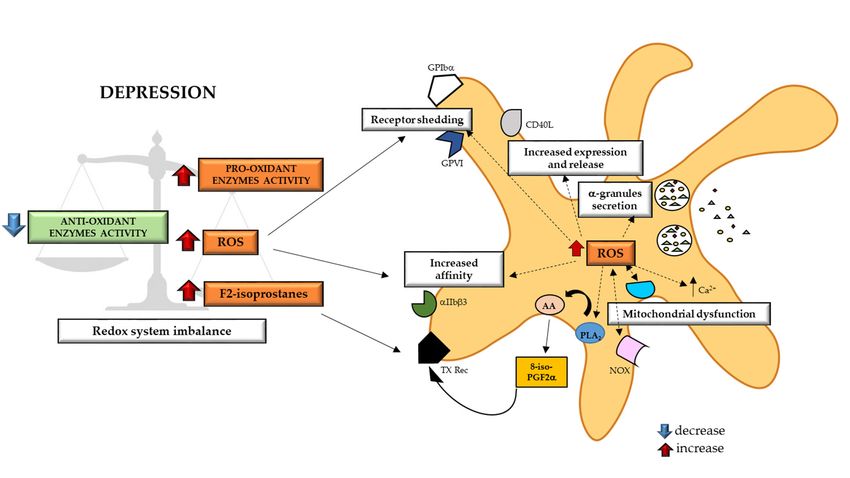
pathway dysregulation [10–12], endothelial dysfunction and platelet activation [3] are the proposed
mechanisms underlying this relationship.
      Among these mechanisms, the platelet activation is one of the most attractive, seeing platelets
as a potential mirror of the brain (dys)-function [13]. Indeed, even though platelet and neurons are
basically
       Int. J.different   cells,
               Mol. Sci. 2020, 21,they
                                   7560 share common characteristics in subcellular organization [14] and in 2 of 33
protein composition [15–21], representing as consequence an alternative tool to investigate neuronal
dysfunction as well as a peripheral tracer of the onset and progression of brain-related pathologies.
               Among these mechanisms, the platelet activation is one of the most attractive, seeing platelets
      Moreover, the platelet hyper-reactivity could at least partially explain the increased
       as a potential mirror of the brain (dys)-function [13]. Indeed, even though platelet and neurons are
vulnerability of depressed patients to acute thrombotic event and ischemic heart disease [22], as well
       basically different cells, they share common characteristics in subcellular organization [14] and in
as their increased mortality post-myocardial infarction [23].
       protein composition [15–21], representing as consequence an alternative tool to investigate neuronal
      Of note, platelets of depressed patients display a greater exposure of anionic phosphatidylserine
       dysfunction as well as a peripheral tracer of the onset and progression of brain-related pathologies.
determinants, an increased activation of glycoprotein (GP) IIb/IIIa [22], a greater granules secretion
               Moreover, the platelet hyper-reactivity could at least partially explain the increased vulnerability
[24], a higher expression of P-selectin and GPIb [25,26], and an enhanced aggregation in response to
       of depressed patients to acute thrombotic event and ischemic heart disease [22], as well as their
collagen and thrombin compared to control subjects [22,27], whereas platelet aggregation is
       increased mortality post-myocardial infarction [23].
unchanged when Adenosine Diphosphate (ADP) and TRAP1-6 were used [22,26,27] (Figure 1). An
               Of note, platelets of depressed patients display a greater exposure of anionic phosphatidylserine
extensively investigated molecule in the relation between depression and platelets activation is the
       determinants, an increased activation of glycoprotein (GP) IIb/IIIa [22], a greater granules secretion [24],
serotonin (5-HT) [28,29]. Platelets share with serotoninergic neurons several similarities in 5-HT
       a higher expression of P-selectin and GPIb [25,26], and an enhanced aggregation in response to
uptake, storage, metabolism and release mechanisms, representing a good surrogate to study
       collagen and thrombin compared to control subjects [22,27], whereas platelet aggregation is unchanged
neuropsychiatric research [30]. Of note, platelets from depressed patients show a greater aggregation
       when Adenosine Diphosphate (ADP) and TRAP1-6 were used [22,26,27] (Figure 1). An extensively
in response to 5-HT [31–33], display enhanced platelet serotonin uptake [30], that favors platelet
       investigated molecule in the relation between depression and platelets activation is the serotonin
response to ADP [34], and an increased platelet 5-HT2 receptor binding and density [35,36] (Figure 1).
       (5-HT) [28,29]. Platelets share with serotoninergic neurons several similarities in 5-HT uptake,
Despite the already proved importance of 5-HT in this contest also other circulating molecules may
       storage, metabolism and release mechanisms, representing a good surrogate to study neuropsychiatric
be involved.
       research [30]. Of note, platelets from depressed patients show a greater aggregation in response to
      This review will be focused on the prothrombotic state of patients affected by depressive
       5-HT [31–33], display enhanced platelet serotonin uptake [30], that favors platelet response to ADP [34],
disorders. In particular, it will analyze the impact of catecholamines, adipokines, lipids, reactive
       and an increased platelet 5-HT receptor binding and density [35,36] (Figure 1). Despite the already
oxygen species and chemokines, in2 the pathophysiological link between depression and CVD,
       proved importance of 5-HT in this contest also other circulating molecules may be involved.
emphasizing the critical role of platelet activation and the related molecular mechanisms.

     FigureFigure
            1. Platelets of depressed
                    1. Platelets        patients show
                                  of depressed           hyper-reactivity.
                                                    patients                Patients withPatients
                                                              show hyper-reactivity.       depression,withshow   hyper- show
                                                                                                            depression,
     reactive  platelets. Indeed,
            hyper-reactive         platelets
                             platelets.  Indeed,of platelets
                                                    depressed   patients display
                                                             of depressed  patientsadisplay
                                                                                      greatera exposure      of anionic
                                                                                                greater exposure     of anionic
     phosphatidylserine    determinants,
            phosphatidylserine              an increased
                                  determinants,            activation
                                                    an increased      of Glycoprotein
                                                                  activation            IIb/IIIa,IIb/IIIa,
                                                                             of Glycoprotein       a higher  expression
                                                                                                           a higher expression
     of P-selectin  and Glycoprotein
            of P-selectin and Glycoprotein Ib, Ib,
                                               an an
                                                   increased
                                                      increasedexpression
                                                                 expressionand   sensitivityofof5HT
                                                                            and sensitivity         5HT  2 receptor, an
                                                                                                       2 receptor, an enhanced
     enhanced   activity
            activity      of SERT
                     of SERT  and anand    an enhanced
                                        enhanced           aggregation
                                                     aggregation         and sensitivity
                                                                   and sensitivity          in response
                                                                                   in response              to collagen
                                                                                                   to collagen  thrombin, and
     thrombin,   and serotonin.
            serotonin.   Platelet Platelet   hyper-reactivity
                                  hyper-reactivity              is also confirmed
                                                       is also confirmed            by a platelet
                                                                           by a greater    greater granules
                                                                                                      platelet granules
                                                                                                                secretion. GP:
            Glycoprotein; vWF: von Willebrand Factor; 5-HT: 5-Hydroxytriptamine; 5-HT2 : 5-HT receptor; SERT:
            plasma membrane serotonin transporter. ↑: increased compared to control.

           This review will be focused on the prothrombotic state of patients affected by depressive disorders.
      In particular, it will analyze the impact of catecholamines, adipokines, lipids, reactive oxygen species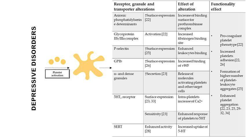
Int. J. Mol. Sci. 2020, 21, 7560                                                                    3 of 33

and chemokines, in the pathophysiological link between depression and CVD, emphasizing the critical
role of platelet activation and the related molecular mechanisms.

2. Catecholamines
     The catecholamines are adaptive and maladaptive stress hormones; they activate behavioral
and physiological processes facilitating the overcoming of stress [37]. Endogenous catecholamines
include dopamine (DA), noradrenaline (norepinephrine/NE), and adrenaline (epinephrine/EPI) [37].
Catecholamines, produced and released by the sympathetic system, brain and adrenal medulla [37],
exert their effects on multiple organs/compartments [38]. Although catecholamines are essential
constituents of physiologic cardiovascular regulation, their effects are greatly emphasized by abnormal
conditions [39].
     The response to EPI and NE is mediated by a set of G protein-coupled adrenergic receptors
(ARs), α and/or β-adrenergic receptors, that are targets for several cardiovascular drugs [37,40]. DA
receptors are all members of the G protein-coupled receptor family and they are divided into two
subtypes: D1-like receptors coupled with Gs alpha subunit (Gs) (D1 and D5) and D2-like receptors
coupled with Gi alpha subunit (Gi) (D2, D3, D4). NE, EPI and DA have a prominent position in
the pathogenic mechanisms of several cardiovascular disorders, such as angina pectoris, heart failure,
arterial hypertension, atherosclerosis and thrombosis [39,41].

2.1. Catecholamines in Depression
     The discovery, in the 1960s, that the inhibition of neuronal uptake of NE, the primary target for
tricyclic antidepressants, reduced depressive symptoms, led to hypothesize and then to show that
a deficit in catecholamine transmission could account for the depression [42]. On the other hand,
the contribution of DA was largely neglected until few years ago.
     Beyond alterations in adrenergic and dopaminergic receptors availability, and the consequent
modification in the downstream pathways in the brain [43–46], depressive disorders have been also
associated with changing in peripheral levels of catecholamines.
     In spite of the scarce and outdated studies, plasma levels of EPI and NE result increased in
depressed patients and their levels correlate with the severity of the pathology [47,48]. In agreement
with previous data, more recent studies showed that patients suffering from depression and other
major affective disorders have increased urinary levels of EPI, NE and DA [49], and the existence of
a positive association between urinary EPI or NE and depressive symptoms [50] (Table 1).
     All these findings provide the evidence that alterations in peripheral catecholamines levels may
be relevant also in depression and not only in stress response, and pave the way to the potential link
among catecholamines, depression and CVDs.

2.2. Catecholamines and Platelet Function
     Since human platelets express both adrenergic and dopaminergic receptors [51–53], the high
catecholamine levels may easier explain the association between depression and CVD. Basically,
through platelet α2-adrenergic or dopaminergic receptors, they modulate thrombopoiesis [54,55], and
platelet function [56–58]. Low concentrations of catecholamines and dopamine potentiate the effects of
other agonists (e.g., ADP, collagen, and thrombin) enhancing platelet aggregation, whereas at high
concentrations are sufficient alone to induce human platelet aggregation, granule secretion, and release
of platelet markers (e.g., Platelet Factor 4 (PF4) and β-thromboglobulin (BTG) [59–61] (Table 1).
     Specifically, the effects of EPI on human platelet activation has been extensively investigated
in vitro providing the evidence that in platelet α2-adrenergic receptors are selectively coupled to Gz
family members but not to Gq or G12 family members [62]. The activation of Gz mediated by EPI,
inhibits cyclic Adenosine Monophosphate (cAMP) formation and promotes the activation of Rap1B and
PI 3-kinase [63], enhancing the effects of other agonists. Interestingly, EPI, not affecting Phospholipase
C (PLC), is unable to cause platelet shape change [64].
Int. J. Mol. Sci. 2020, 21, 7560 4 of 33
Remarkable, EPI infusion induces a threefold increase of platelet thromboxane (TX) production [65],
and enhances platelet fibrinogen binding and platelet aggregation induced by thrombin [66].
In vivo infusion or in vitro exposure to EPI, enhances ADP-induced platelet aggregation and clot
formation both in healthy subjects treated with ticagrelor and in ACS patients under acetylsalicylic
acid and ticagrelor therapy [67,68] (Table 1).
Table 1. Catecholamines levels in MDD and effect on megakaryocytes and platelets.
Catecholamines in Depression and Platelets
DEPRESSION EFFECT ON MEGAKARYOCYTES
Stimulus Levels Stimulus Receptor Effect
Megakaryocyte adhesion and
EPI Increased circulating and EPI α-2-
migration [55]
NE urinary levels [47–49] NE adrenoceptor
Pro-platelets formation [55]
Increased urinary levels
DA DA D1/D2 Megakaryocytes differentiation [54]
[49]
EFFECT ON PLATELETS
Stimulus Levels Stimulus Receptor Effect
Low concentrations:
EPI α-2-
Increase the sensitivity to collagen,
NE adrenoceptor
thrombin and ADP [52,56,57,60]
EPI Increased circulating and
NE urinary levels [47–49] High concentrations:
Induce aggregation alone [57,66]
α-2-
EPI Increase TX production [65]
adrenoceptor
Enhance fibrinogen binding [66]
Induce clot formation [65–67]
Low concentrations:
D2 (?)
Increase sensitivity to ADP [61]
DA
Induce platelet microaggregation
Increased urinary levels D2-like
DA [58]
[49] receptor
Induce platelet adhesion [58]
High concentrations:
D2 (?)
Induce the release of a-granules [61]
EPI: Epinephrine; NE: Norepinephrine; DA: Dopamine; D1/D2: Dopamine Receptors; ADP: Adenosine diphosphate;
PF: Platelet Factor 4. ?: still under debate.
Despite NE induces platelet activation by binding, like EPI, α2-adrenergic receptors, its action is
two or three times less effective than EPI [52,60] (Table 1).
Finally, dopamine potentiates platelet microaggregate formation and adhesion to collagen under
low shear flow induced by ADP via D2-like receptor [58] (Table 1), however dopamine infusion
in hypertensive and normotensive men do not influence platelet count, platelet size and plasma
concentration of β-thromboglobulin [69].
Overall these data suggest that the inappropriate activation of the sympathoadrenal axis
occurring under depression may increase the sensitivity of circulating platelets to agonists with
severe consequences on CVD outcome.
3. Adipokines
Neuroendocrine regulators of energy metabolism are crucial in determining cardiovascular
risk [70], and are associated with depression disorders [71]. In this contest, adipose tissue plays an
endocrine role by synthesizing and secreting bioactive compounds named adipokines, whose secretion
is essential to energy and metabolic homeostasis [72]. The most studied adipokines are leptin and
adiponectin, whose alteration is reflected on both neuronal [73] and cardiovascular alterations [74,75].Int. J. Mol. Sci. 2020, 21, 7560 5 of 33
Of note, among classical adipokines, also non-conventional metabolic regulators, like neurotrophins,
could play a pivotal role in influencing both these pathology [76].
3.1. Leptin
Leptin is a hormone mainly secreted by adipocytes, it is involved in the control of food intake [77]
and its increased levels are associated to obesity [78]. The peripheral actions of leptin include stimulation
of inflammatory reaction, oxidative stress, atherosclerosis and thrombosis, thus promoting endothelial
dysfunction, arterial stiffness, development and vulnerability of atherosclerotic plaques [79]. Moreover,
it has been reported that CHD patients have higher leptin levels compared to controls [80,81]. Its serum
concentrations are increased after myocardial infarction (MI) [82], and its high levels are associated
with an increased risk of cardiac death, ACS, non-fatal MI, stroke and hospitalization for congestive
heart failure [83,84].
3.1.1. Leptin in Depression
Modifications of leptin metabolism and its gene expression, as well as its receptor, have been
reported among patients with mental health disorders, including depression [85], independently of
drug treatment [86]. However, the relationship between circulating leptin levels and depression is
under debate. Some authors stated that depression is associated with low circulating and brain leptin
levels [87–90], suggesting a correlation between leptin levels and the depressive mood. This data are
supported by the observation that administration of leptin exerts an antidepressant-like effect [91],
through dopaminergic neurotransmission regulation in mesolimbic areas [92]. In particular, leptin
reduces symptoms of depression and has an anxiolytic effect affecting the HPA [93,94], and stimulating
brain-derived neurotrophic factor (BDNF) production and function [95–97]. In addition, the deletion
of leptin receptor (LepRb) and its downregulation are associated with depression-like behavioral
impairments, indicating that leptin-lepRb signaling is involved in the molecular mechanism of leptin
antidepressant action [98,99].
On the other hand, some studies did not find any difference in the leptin levels between depressed
patients and control group [100–103], or measured higher levels of leptin in depressed patients [104–109].
In addition, a positive association between circulating leptin levels and depressive symptoms [110],
mild/moderate but not severe depression [104,111], and with self-reported depressive symptoms,
especially in women [109], was recently identified. A marked sexual difference in leptin levels has
been consistently reported, usually both healthy and depressed women have higher leptin levels than
men [88,104,112]. Several reasons of these sex-discrepancies have been hypothesized, including: (a)
the greater amount of subcutaneous and intra-abdominal adipose tissue in women (b) the difference in
male and female eating behavior or upregulated leptin mRNA in proportionally larger adipocytes of
females and (c) the testosterone levels, that inversely correlates with leptin levels [113].
In general, confounders such as time of blood sampling, age, Body Mass Index (BMI),
gender-associated metabolic disturbances, medication history and clinical type and features of
depressive disorders, might impact peripheral leptin levels, and thereby justify inconsistent results
obtained [101,104,107,114,115].
The presence of atypical major depressive disorders (MDD) may be an additional explanation of
this contradictory results. High concentrations of leptin are specifically associated with atypical MDD
and with symptoms that represent the core features of the atypical subtype, whereas no association
was found for the typical subtype or when considering the general diagnosis of MDD [106]. This
finding is consistent with the hypothesis of a leptin resistance process which blunts leptin central action,
despite increasing peripheral concentrations, and leads to hyper-leptinemia in obese subjects [106].
Data from a large international consortium identified that 15% of patients with atypical depression
carried a higher number of genetic risk variants for increased BMI, leptin and C-reactive protein
(CRP), meaning that atypical depression and obesity-related traits may be the two faces of the same
syndrome [108].Int. J. Mol. Sci. 2020, 21, 7560 6 of 33
Taken together, all these data indicate the necessity of further investigation about circulating leptin
levels in depressive disorders to understand its real impact on cardiovascular risk and thrombosis.
3.1.2. Leptin and Platelet Function
Among other receptors, platelets express on their surface also receptors for peptide hormones,
including the long form of leptin receptor (LEPRL) [116], suggesting that alteration of leptin
levels occurring in depressive disorders may alter platelet response contributing to cardiovascular
complications. Indeed, leptin promotes arterial thrombosis, potentiates platelet aggregation in
mouse [117,118], increases platelets adhesion and potentiates ADP- and thrombin-induced aggregation
in human [119,120], even if such effect was not observed in all subjects [116–118,121–123].
Furtherly, it has been shown that leptin induces platelet activation through almost two
different signaling cascade mechanisms. The first includes the activation of Janus kinase 2 (JAK2),
phosphatidylinositol 3-kinase (PI3K), protein kinase B (PKB), insulin receptor substrate-1 (IRS-1), and
phosphodiesterase 3A (PDE3A), with a consequent increase of PDE3A and a decrease of cAMP [119,120].
The second one leads to GPIIb/IIIa activation, increase of Ca2+ and TX production through the activation
of phospholipase C γ2 (PLCγ2), protein kinase C (PKC), and phospholipase A2 (PLA2 ) pathway [124].
3.2. Adiponectin
Adiponectin is an anti-inflammatory adipokine and contributes to increase insulin sensitivity
protecting, therefore, against diabetes, atherosclerosis and thrombosis. Accordingly, high concentrations
of adiponectin have been associated with a reduction in the risk of CVD [125–127] and an increase
in endothelial nitric oxide production [128]. Conversely, patients with CVD, with increased carotid
intima–media thickness and with obesity, exhibit low plasma adiponectin levels [129].
3.2.1. Adiponectin in Depression
The relationship and the modulation of adiponectin in depressive disorders have been extensively
studied. Specifically, several studies and a recent meta-analysis showed that MDD patients have
low adiponectin levels [105,130–134], and that successfully antidepressant treatment increases its
levels [135]. Interestingly, an inverse correlation between adiponectin levels and Hamilton Depression
Rating Scale (HAM-D), indicating the depression symptoms severity at admission [130,132,136], or
cumulative duration of depression [137] was identified. However, other studies were unable to confirm
this observation [103,138–140]. Again, several confounding factors have to be taken into consideration.
It is well known that (a) ethnic difference [140,141] between Asians and Europeans due to different
body composition and metabolic profile [142], (b) matrix in which adiponectin is measured (plasma
or serum) [134], (c) sex-dependent adiponectin levels [132,143], (d) body weight [137], (e) presence
of metabolic syndrome [144], (f) onset of depressive event [107], and (g) the subtypes of depressive
disorders [133,145–147] influence adiponectin levels.
However, when the modification of adiponectin levels are presents, they may contribute to platelet
activation and then to thrombosis.
3.2.2. Adiponectin and Platelet Function
Concerning the impact of adiponectin on platelet function, both mice and human platelets
express adiponectin receptors AdipoR1 and AdipoR2 [124]. Adiponectin alone does not affect
platelet adhesion/aggregation in human [120], nevertheless its deletion in a mouse model increases
agonist-induced platelet aggregation and enhances thrombus formation after photochemically-induced
arterial injury [148]. The antithrombotic effect of adiponectin may be related to its ability to
influence leukocytes behavior, to reduce polymorphonuclear (PMN) leukocyte- and monocyte-platelet
aggregates [149], and to inhibit macrophage-related Tissue Factor (TF) expression and activity with
the consequent impairment in the coagulation cascade [150].Int. J. Mol. Sci. 2020, 21, 7560 7 of 33
3.3. Neurothrophins
The neurotrophin (NT) family consists of Nerve Growth Factor (NGF), BDNF, NT-3 and NT-4 (also
named NT-4/5). NTs not only are stimulators of nerve growth, survival and differentiation [151] but
they also exert effects on immune cells [152–154], blood vessels/angiogenesis [155,156], wound healing
and tissue repair [156,157], and most importantly on glucose, lipid and energy dynamics [158–161].
They are considered key regulators of metabolism [162], and for their pleiotropic functions, NTs have,
as consequence, a huge importance in both depression [76], and CVD [76].
BDNF influences endothelial function [163], monocyte activation [154], and thrombus dimension
and stability [164]. It takes part to cardiovascular development [165] but also to the onset of
cardiovascular alterations and disease [166], including hypertension [167,168], atherosclerosis [169,170]
and thrombosis. Reduced BDNF plasma levels has been found in metabolic syndrome [170], ACS [171,
172], and type 2 diabetes [173], suggesting that alterations in its circulating levels may be associated to
pathological conditions.
NGF and NT-3, similarly to BDNF, are involved in the cardiac development [174] and
regeneration [175], angiogenetic process [165,176], hypertension onset [76,177], and atherosclerotic
lesions formation [170,172,178].
The role of NT-4 and related pathways in CVD are not well characterized; however, binding
the same receptor of BDNF (Tropomyosin receptor kinase B-TrkB), it might have similar functions of
BDNF in controlling blood pressure [168].
Interestingly, several NT polymorphisms, including BDNF rs6265 (Val66Met) [179–183], NGF
rs11102930 [184] and rs78701042 [185], have been associated to increased risk of ACS and
adverse outcome.
3.3.1. Neurotrophins in Depression
By their nature NTs play a key role in preventing depressive disorders [186], and data obtained
in rodent models of depression indicate that administration of BDNF and NGF have significant
antidepressant effects [187,188]. NTs do not control directly mood but, they are fundamental in
the activity-dependent modulation of networks and changes in plasticity [11]. Low NT levels
have been associated with several affective disorders including bipolar depression (BD) [189,190],
depression [12,191], mania [192], and obsessive compulsive disorders [193]. In particular, reductions
in serum and plasma BDNF have been found in patients affected by depression [194–196], and in those
who committed suicide [197,198]. Despite a large cohort study [199] and meta-analyses [12,200,201]
confirmed these results, there are a studies that have not found any relationship between BDNF and
depression [202], or that provided evidence of positive correlation between BDNF and higher scores
of scales for assessing depression in specific subgroups of patients [203]. Interestingly, genetic and
epigenetic modifications in the BDNF gene have been associated to depressive disorders [204].
Similarly, alteration in circulating NGF levels have been detected in patients with depression.
Clinical studies showed its reduced levels in patients with MDD compared to healthy control, and that
NGF is negatively associated with depressive symptoms [205,206]. Nevertheless, in elderly patients
this association seems to be attenuated [207].
As regard to NT-3 and 4/5 results are still controversial. Some studies found increased circulating
levels of NT-3 [208,209], and NT-4/5 [209,210] in BD patients, while other studies demonstrated
reduced [189,211] or unchanged levels [212,213].
3.3.2. Neurotrophins and Platelets Function
The impact of NTs on platelet function has not been well-investigated yet. To our knowledge, it is
only known that NGF can bind platelet surface and induce aggregation [214], while no information
is available on the effect of BDNF on platelet function, even though BDNF can bind a specific site
on platelets surface with subsequent internalization [215]. Platelets contain both NGF and BDNF,conceivable that NGF and BDNF are not stored in the same granules and that their release is mediated
by different mechanisms, even though these mechanisms are still poorly characterized [219].
In particular, NGF is released within 10 min under calcium-free conditions, while after 60 min
in presence of calcium his release is significantly reduced [219]. This event is faster when platelets are
co-incubated with ibuprofen, while indomethacin do not affect NGF release [219].
Int. J. Mol. Sci. 2020, 21, 7560 8 of 33
On the other hand, only a low amount BDNF, stored in the α-granules [215], is released under
calcium-free conditions, while calcium markedly increases its release. Ibuprofen and clopidogrel but
not
thatindomethacin
are spontaneously and released
aspirin upon
decrease its release
platelet activation[219,220].
[215–219]. Interestingly, when in
From data present platelets
literature areis
stimulated
conceivablewith thrombin,
that NGF the mechanism
and BDNF are not stored ofinBDNF
the samerelease is consequent
granules and that theirto release
the activation
is mediated of
Protease-Activated receptor-1 (PAR-1). PAR-1 peptide induces a biphasic
by different mechanisms, even though these mechanisms are still poorly characterized [219]. BDNF release, where only
the first phase is NGF
In particular, calcium mobilization-dependent,
is released within 10 min underascalcium-free
provided by its inhibition
conditions, mediated
while after 60 minbyin
Prostaglandin-1
presence of calcium (PGE-1) pretreatment
his release [217]. Inreduced
is significantly this contest,
[219]. authors
This event demonstrated
is faster when that BDNF are
platelets is
released
co-incubated by thewith
fusion of α-granules
ibuprofen, with the Opendo
while indomethacin Canalicular
not affect NGFSystem (OCS),
release forming the swollen
[219].
OCS [217].On the other hand, only a low amount BDNF, stored in the α-granules [215], is released under
Interestingly,
calcium-free it has while
conditions, been shown
calciumthat the amount
markedly of serum
increases BDNF
its release. well reflect
Ibuprofen and the amount but
clopidogrel of
BDNF found in platelets
not indomethacin and aspirin[221].decrease
In agreement with
its release the decreased
[219,220]. levels when
Interestingly, of BDNF under
platelets aredepression
stimulated
conditions
with thrombin, [222,223], the plateletofBDNF
the mechanism BDNFcontent
releaseisis significantly
consequent to reduced in patients
the activation with depression
of Protease-Activated
compared
receptor-1to(PAR-1).control PAR-1
subjectspeptide
[224], and the antidepressant
induces a biphasic BDNF pharmacotherapy
release, where only normalize
the firstitsphase
levels.is
Then,
calcium it ismobilization-dependent,
possible to speculate that as the plateletbypre-activation
provided state of MDD
its inhibition mediated patients leads to
by Prostaglandin-1 their
(PGE-1)
BDNF reservoir[217].
pretreatment depletion
In this[224], influencing
contest, authorsnegatively
demonstratedendothelial
that BDNF function and thrombus
is released by the growing
fusion of
and stabilitywith
α-granules [154,164,166]
the Open(Figure 2). System (OCS), forming the swollen OCS [217].
Canalicular
The genetic knock-in
Interestingly, it has beenmouse showncarrying
that BDNFVal66Met
the amount of serum polymorphisms,
BDNF wellthat recapitulates
reflect the amount theof
phenotypic hallmarks of human disease (e.g., depression and CVD)
BDNF found in platelets [221]. In agreement with the decreased levels of BDNF under depression [179] has been a helpful model
toconditions
investigate the relationship
[222,223], the plateletbetween
BDNF this polymorphism
content andreduced
is significantly plateletin function.
patientsSpecifically,
with depression the
presence
compared of to
this polymorphism
control subjects [224],predisposes
and the to platelet hyper-activated
antidepressant pharmacotherapy phenotype, enhancing
normalize P-
its levels.
selectin expression, GPIIb/IIIa receptor activity, ability to bind leukocytes
Then, it is possible to speculate that the platelet pre-activation state of MDD patients leads to their and fibrinogen, and
aggregation
BDNF reservoir [179,225].
depletion [224], influencing negatively endothelial function and thrombus growing
and stability [154,164,166] (Figure 2).
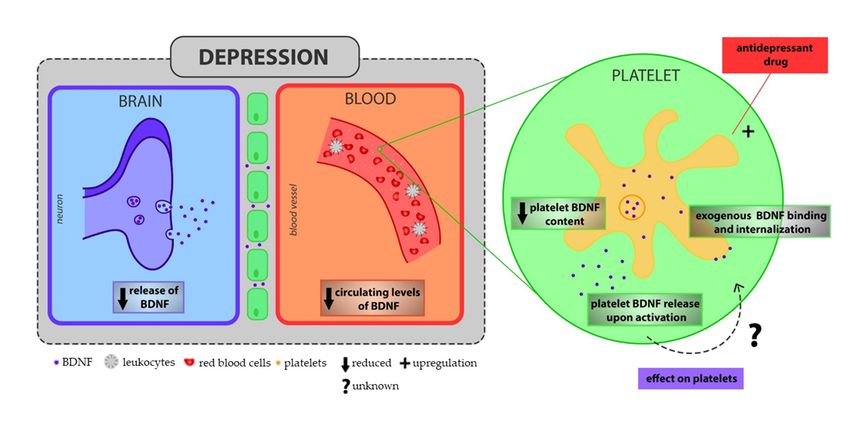
Figure 2. The controversial role of BDNF on platelet function. Depressed patients are characterized
by the2.reduction
Figure of BDNFrole
The controversial levels in the on
of BDNF brain, thatfunction.
platelet is well reflected
Depressed by patients
its decreased plasma and
are characterized
serum levels [221–223]. Remarkably, platelet BDNF content is reduced in depressed patients
by the reduction of BDNF levels in the brain, that is well reflected by its decreased plasma and serum [224],
but antidepressant
levels treatment isplatelet
[221–223]. Remarkably, able toBDNF
restorecontent
their levels [224], indicating
is reduced that apatients
in depressed pre-activated
[224], state
but
may promote treatment
antidepressant platelet emptying of theirtheir
is able to restore BDNF reservoir.
levels Platelets,that
[224], indicating when activated, can
a pre-activated staterelease
may
BDNF [154,215,217,218],
promote platelet emptying andof extracellular BDNF canPlatelets,
their BDNF reservoir. bind a specific site on platelets
when activated, surface
can release BDNFwith
subsequent internalization [215]. However, there are no information about its possible effect
[154,215,217,218], and extracellular BDNF can bind a specific site on platelets surface with subsequenton platelets.
BDNF: Brain-derived Neurotrophic Factor.
The genetic knock-in mouse carrying BDNFVal66Met polymorphisms, that recapitulates
the phenotypic hallmarks of human disease (e.g., depression and CVD) [179] has been a helpful
model to investigate the relationship between this polymorphism and platelet function. Specifically,
the presence of this polymorphism predisposes to platelet hyper-activated phenotype, enhancing
P-selectin expression, GPIIb/IIIa receptor activity, ability to bind leukocytes and fibrinogen, and
aggregation [179,225].Int. J. Mol. Sci. 2020, 21, 7560 9 of 33
4. Lipid Molecules and Lipoproteins
Lipids are essential structural components of cell membranes and play a crucial role in different
metabolic pathways and cellular functions [226]. Lipids alteration is associated with cardiovascular
risk [70]. A rise in cholesterol concentration increases the risk for death by CVD [227], and low-density
lipoprotein (LDL), contributing to the development of atherosclerotic plaques, are included among
the traditional risk factors for thrombosis. LDL may be oxidized, entrapped in macrophages inducing
their differentiation in foam cells, and they may also bind to proteoglycans in the arterial intima. In
addition to the critical role of LDL, a reduction of high-density lipoproteins (HDL) may cooperate to
promote atherosclerosis. HDL inhibits oxidation of LDL, removes cholesterol from foam cells and
reduces inflammation [228,229].
4.1. Lipid, Low Density Lipoprotein and Lipids Peroxidation in Depression
A large body of literature has provided knowledge on the relation between lipid status and
psychotic disorders [230–232], whereas very few data is available on depression.
Nevertheless, a relation between depression and lipid disturbances has been recently
demonstrated [233–240].
Actually, high total cholesterol (TC) and LDL are significantly associated with depressive symptoms
and severity, with depression prospective course [236], and with metabolic syndrome in MDD [237].
In support of the relation between the severity of depression and cholesterol alteration, Enko et
al. showed that, although depressed patients display only a slight increase in TC and LDL, there
is a positive correlation between BDI-II depression score and triglycerides (TG), TC and LDL, and
a negative, even though not significant, correlation with HDL [238]. In particular, an increase in
LDL/HDL ratio has been observed in MDD patients [235,239,240]. By contrast, other studies showed
that lower concentrations of TC and LDL were associated with MDD [241,242], and with incidence
of depression [243–246], and that during first episodes of MDD higher TG levels and low HDL but
similar LDL levels were found [247]. In addition, emerging data from a meta-analysis showed that
depression was inversely associated with TC levels, and directly related to HDL levels, especially
in women [235], whereas a U-shaped relationship with LDL was found [234]. The cross sectional
nature of most studies, the different categorization of age, sampling and dissimilar tools of evaluation
of depression applied in the studies, as well as the considerations of confounder factors (e.g., BMI,
drug treatments) might explain the controversial findings about depression and circulating lipids and
lipoproteins. Moreover, several studies analyzed the association of metabolic alterations with MDD,
without carrying information about the inclusion of the BD individuals or without specifying which
episode they were experiencing [237], all factors that could affect the conclusions.
Additionally to lipid molecules, the potential relationship with lipid peroxidation products as
well as with oxidized LDL (oxLDL) and depression have to be considered [248].
The lipid peroxidation marker malondialdehyde (MDA) is increased in MDD patients compared to
healthy control [249–252], and correlates with depression severity [253,254]. Similarly, the metabolites
of F2 isoprostanes, an additional marker of lipid peroxidation, are greater in urine, plasma and serum
of patients with depressive disturbances or MDD. This difference is particularly marked in elderly
men [255], and is sex- and age-independent [256]. Only one study did not find significant difference
between depressed and control subjects; however, this conclusion may be a result of the small sample
size used [257].
Depressed patients have also higher levels of serum oxLDL antibodies than normal control [258],
and a positive correlation emerged between serum oxLDL/LDL ratio with both Centre Epidemiological
Studies Depression Scale (CES-D) score and perceived stress in a Japanese population [259]. Interestingly,
depressed patients with great serum oxLDL antibodies are at high risk for atherosclerosis or have
atherosclerotic lesions [9,258], suggesting a relationship among depression, oxLDL and CVD.Int. J. Mol. Sci. 2020, 21, 7560 10 of 33
Overall, the positive association found between LDL or oxLDL and BMI [260,261], and between
BMI and depressive symptoms [71], suggests that depression, directly or indirectly, leading to weight
gain and to metabolic syndrome, could predispose to lipid alteration and to CVD.
4.2. Low Density Lipoprotein, Lipids Peroxidation and Platelet Function
Disorders in lipid status, above described, in patients with depression and CVD may contribute
to platelets activation leading to acute thrombotic events.
LDL increases the sensitivity of blood platelets to agonist stimulation, making their response
faster and more extensive [228]. Individuals with high plasma-LDL have hyper-reactive platelets
and greater plasma levels of platelet activation markers, including BTG and soluble CD40 ligand
(CD40L) [228]. In vitro, platelets exposed to LDL display hyper-aggregability, increased fibrinogen
binding and surface-expression of P-selectin, and increased production of TX, and generation of
Reactive Oxygen Species (ROS) [228,262].
Of note, oxLDL can influence platelet activation, apoptosis, and association with
monocytes/macrophages [228]. In particular, by binding scavenger receptors on platelets (e.g.,
CD36) [228,263], oxLDL enhances NADPH oxidase-2 (NOX-2)-mediated generation of ROS and
platelet hyper-reactivity [264], including platelet degranulation, GPaIIb3-integrin activation, apoptosis,
thrombin generation, and shape change. Finally, LDL–oxLDL enhances platelet release of CXCL12
(Stromal Cell-derived Factor-1-SDF-1), that in turn prompts LDL–oxLDL uptake and synergistically
augments the LDL–oxLDL-induced pro-oxidative and thrombogenic impact on platelet function [265].
5. Reactive Oxygen Species
ROS (i.e., superoxide anion (O•2 -), hydrogen peroxide (H2 O2 ), hydroxyl radical (•OH), hydroxyl
ion (OH−) consist of radical and non-radical oxygen species formed by the partial reduction of
oxygen [266]. ROS modulate several physiological processes, however when their excessive production
is not counteracted by antioxidant capacity of human physiology there is imbalance in the redox system,
resulting in tissue damage and in the development and progression of several diseases. The implication
of ROS in the pathogenesis of CVDs and thrombosis has been well described [267]. Abnormal ROS
increase has been observed in atherosclerosis [268], and in coronary artery disease (CAD) patients and
associated with future CVD events [269]. The most well-known sources of ROS in the cardiovascular
system are NOX family of enzymes, uncoupled endothelial nitric oxide synthase (eNOS), mitochondria
and xanthine oxidase (XO), whose function is critical in determining the onset and progression of
CVD [270].
5.1. Reactive Oxygen Species in Depression
Clinical studies have reviewed the possible impact of oxidative stress in the pathophysiology
of depression [271–274], focusing on ROS iper-production or on the activation of enzymes relevant
in pro/antioxidant processes [e.g., NOX, XO, superoxide dismutase (SOD) and catalase (CAT)] [274],
and experimental models have established that the enhanced ROS production favors depression-like
phenotype [275].
Several peripheral markers of oxidative stress and mechanisms implicated in redox balance are
altered in MDD, nevertheless, there is a significant heterogeneity across the studies [276], specifically
as regard to ROS alteration [277], hence importance of careful phenotyping of the depressed and
control subjects.
Decreased SOD activity [253,278] in red blood cells (RBC) of depressed patients have been
measured in first [253], recurrent [251], and bipolar episodes [279] suggesting also a connection with
different subtypes of depressive manifestations.
The reduction of SOD activity associated with increased XO activity [278] and unchanged CAT
activity [253,276] well explain the increased ROS generation detected in patients with depression [252,
276]. However, other studies showed increased RBC SOD activity [249,250,252], that is potentiallyInt. J. Mol. Sci. 2020, 21, x FOR PEER REVIEW 11 of 34
Int. J. Mol. Sci. 2020, 21, 7560 11 of 33
potentially explained as a protective mechanism induced by organism to counterbalance oxidative
stress that occurs during depressive disorders (Figure 3).
explained as a protective mechanism induced by organism to counterbalance oxidative stress that
A recent meta-analysis indicated that depression is associated with enhanced oxidative damage,
occurs during depressive disorders (Figure 3).
as provided by increased urinary and serum/plasma levels of 8-hidroxy-2′-deoxyguanosine (8-
A recent meta-analysis indicated that depression is associated with enhanced oxidative damage,
OHdG) and F2-isoprostanes [273] (Figure 3).
as provided by increased urinary and serum/plasma levels of 8-hidroxy-20 -deoxyguanosine (8-OHdG)
Noteworthy is that mitochondrial dysfunction is considered the preferential mechanism as
and F2-isoprostanes [273] (Figure 3).
source of ROS in MDD [280,281]; indeed, reduced mitochondrial function is implicated in depression
Noteworthy is that mitochondrial dysfunction is considered the preferential mechanism as source
onset and progression [281]. This finding is relevant since the brain is a highly active organ with high
of ROS in MDD [280,281]; indeed, reduced mitochondrial function is implicated in depression onset
energy consumption, and then it is more susceptible to the deleterious effect of excessive ROS
and progression [281]. This finding is relevant since the brain is a highly active organ with high energy
production related to mitochondrial dysfunctions.
consumption, and then it is more susceptible to the deleterious effect of excessive ROS production
Overall, the imbalance of redox system, with an increased production of ROS, that characterizes
related to mitochondrial dysfunctions.
depression disorders may cover a key role in promoting platelets activation.
Overall, the imbalance of redox system, with an increased production of ROS, that characterizes
depression
5.2. Reactivedisorders may cover
Oxygen Species a key role
and Platelet in promoting platelets activation.
Function
5.2. Reactive OxygenROS
Extracellular Species and Platelet
promote Function of GPIIb/IIIa interacting with thiol groups in the
the activation
extracellular domain, and the shedding of GPVI and GPIbα through a mechanism mediated by A
Extracellular ROS promote the activation of GPIIb/IIIa interacting with thiol groups in
Disintegrin and Metalloproteases (ADAM) [282,283]. These events have been recently associated with
the extracellular domain, and the shedding of GPVI and GPIbα through a mechanism mediated
increased coagulation factor binding and enhanced thrombin and fibrin generation, favoring a pro-
by A Disintegrin and Metalloproteases (ADAM) [282,283]. These events have been recently associated
coagulant phenotype of platelets [284]. Activated platelets, via NOX, cyclooxygenases, eNOS, XO,
with increased coagulation factor binding and enhanced thrombin and fibrin generation, favoring
and mitochondrial respiration [285,286], are able to generate per se ROS, that in turn re-activate
a pro-coagulant phenotype of platelets [284]. Activated platelets, via NOX, cyclooxygenases, eNOS,
platelets [287,288], especially in older patients [289]. As consequence, intra-platelets ROS support
XO, and mitochondrial respiration [285,286], are able to generate per se ROS, that in turn re-activate
platelet activation promoting α-granule exocytosis [290], increasing the sensitivity of platelets
platelets [287,288], especially in older patients [289]. As consequence, intra-platelets ROS support
receptor like GPIIb/IIIa, GPIbα and GPVI [291,292]. When platelets are exposed to thrombin-or
platelet activation promoting α-granule exocytosis [290], increasing the sensitivity of platelets receptor
collagen ROS act as second messenger [293], inducing calcium mobilization [293], upregulating
like GPIIb/IIIa, GPIbα and GPVI [291,292]. When platelets are exposed to thrombin-or collagen ROS
CD40L surface expression and release [294], and generating isoprostanes including 8-iso-
act as second messenger [293], inducing calcium mobilization [293], upregulating CD40L surface
prostaglandin F2α (PGF2α) that can promote platelet aggregation via TX receptor in the presence of
expression and release [294], and generating isoprostanes including 8-iso-prostaglandin F2α (PGF2α)
low concentrations of other agonists [295–297] (Figure 3).
that can promote platelet aggregation via TX receptor in the presence of low concentrations of other
Interestingly, alteration in platelets mitochondrial bioenergetics has been detected in MDD
agonists [295–297] (Figure 3).
patients compared to a matched control subjects [298,299], supporting the hypothesis of interplay
Interestingly, alteration in platelets mitochondrial bioenergetics has been detected in MDD patients
among ROS, platelet activation and depression.
compared to a matched control subjects [298,299], supporting the hypothesis of interplay among ROS,
platelet activation and depression.
Figure 3. Increased oxidative stress that occurs during major depressive disorders (MDD), can activate
Figure 3. Increased oxidative stress that occurs during major depressive disorders (MDD), can
platelets. Depressed patients are characterized by an imbalance in redox system with increased
activate platelets.
pro-oxidant enzymeDepressed
activity notpatients are characterized
counterbalanced by anenzyme
by anti-oxidant imbalance in redox system This
[251–253,276,278,279]. with
increased pro-oxidant enzyme activity not counterbalanced by anti-oxidant enzyme
imbalance promotes an excessive production of ROS and an increase in F2 isoprostanes circulating [251–Int. J. Mol. Sci. 2020, 21, 7560 12 of 33
levels. Extracellular ROS is then able to activate platelets increasing GPIIb/IIIa receptor affinity,
and inducing GPIα and GPVI receptors shedding with consequent activation of downstream
pathways [282,283]. In the same time, the activation of platelets due to pro-oxidant environment favors
intra-platelets production of ROS. Intra-platelets ROS support platelet activation promoting α-granule
exocytosis, increasing the sensitivity of platelet receptors, acting as second messenger in thrombin- or
collagen-activated platelets, inducing calcium mobilization, upregulating CD40L surface expression
and release and generating isoprostanes, including 8-iso-PGF2α [activating AA metabolism] [290–294].
PGF2α can in turn activates TX receptor beyond supporting platelet activation [295–297]. Finally,
the redox imbalance is furtherly fed by a vicious cycle of ROS production due to activation of NOX
enzyme [285–289] and to alteration of platelet mitochondrial function [298,299]. ROS: Reactive Oxygen
Species; GP: glycoprotein; CD40L: CD40 Ligand; Ca2+ : Calcium; NOX: NADPH oxidase; PLA2 :
Phospholipase A2 ; AA: Arachidonic Acid; TX: Thromboxane.
6. Inflammatory Factors
Inflammatory process has been extensively described in worsening CVD prognosis, and platelets
are recognized as active mediators of this mechanisms [300]. The pro-inflammatory status has been
also associated with MDD and is exhaustively described in literature. In particular, pro-inflammatory
cytokines like interleukin-2 (IL-2), IL-6, soluble IL-6 receptor, tumor necrosis factor-α (TNF-α) and
interferon-γ (IFN-γ) are increased while anti-inflammatory cytokines like IL-4 and IL-10 are decreased
during depressive disorders [301]. More recent studies demonstrated that also chemokines contribute
to neurobiological processes relevant to psychiatric disorders [302], suggesting another point of
connection between depression and cardiovascular disease.
6.1. Chemokines
Chemokines are small (8–12 kDa) chemotactic cytokines, which have an important role in directing
the migration of blood cells to target tissues. Chemokines are classified into 4 groups, with the CC- and
CXC-types being the most common [303]. Alteration in circulating levels of chemokines like CXCL8
(IL-8), CCL2 (Monocyte Chemoattractant Protein-1 (MCP-1), CCL26 (Eosinophil Chemotactic Protein-3
(Eotaxin-3), CCL5 (Regulated on Activation of Normal T cell-expressed and secreted (RANTES)
entities) CXCL10 (γ-Interferon-inducible Protein-10 (IP-10) or of chemokines receptors (e.g., fractalkine
receptor-CX3 CR1) are associated with subclinical or proclaimed cardiovascular pathology (e.g., MI,
CAD, atherosclerosis) or with cardiovascular death [304–306]. In addition, it has been showed that
the fractalkine receptor CX3 CR1 plays a key role in atherosclerosis [305], and that a polymorphism in
its gene is associated with a reduced risk for CAD [306].
Overall, chemokines represent a promising therapeutic target in cardiovascular disease [303], and
recently have been also related to depression disorders [307].
6.1.1. Chemokines in Depression
Several studies highlighted the link between depressive symptoms and elevated circulating levels
of chemokines, and the main results obtained are summarized in a recent review [307].
Higher blood levels of CCL11 (Eotaxin-1), RANTES, SDF-1 and CXC3 L1 (fractalkine) were found
in depressed patients compared to controls [308–311] (Table 2).
In particular, Ogłodek et al. showed that RANTES and CXCL12 levels were significantly increased
in both women and men with depressive disorders and that a relation between circulating levels
of these chemokines and severity of depressive symptoms exists [309]. By contrast, Leighton et
al., in a meta-analysis including 7 studies, did not find significant difference in circulating levels of
RANTES and CCL3 (Macrophage Inflammatory Protein-1α (MIP-1α) between MDD patients and
control group [307] (Table 2). However, need to be mentioned that the conclusion of Leighton et al. may
be incorrect due to the inclusion of studies that evaluated indiscriminately the RANTES levels in plasma,
serum and whole blood of depressed patients (Table 2). Indeed, it has been reported that depressedInt. J. Mol. Sci. 2020, 21, 7560 13 of 33
patients have reduced serum levels of RANTES [308] and antidepressant treatment can restore its
levels [311]. Nevertheless, reduced RANTES serum levels in depressed patients may be related to
the activated platelet phenotype, that leads to the emptying of RANTES platelet reservoir [312].
Increased plasma levels of fractalkine in moderate-severe depressed patients compared to control
subjects was found [313] and this result was confirmed by other studies, in which a correlation between
fractalkine plasma levels and the severity of depressive symptoms was identified [314,315] (Table 2).
However, considering the scarcity of information about circulating levels of fractalkine in association
with depression, further studies are needed, also taking into account the possible confounding factors
(e.g., age, BMI) and the heterogeneity of MDD.
Instead, CCL4 (MIP-1β) has been found lower in the serum of depressed patients compared to
not depressed ones [307].
Finally, MCP-1 levels are usually higher in depressed patients compared to controls [302,310,
311,316,317], with the exception of some works [131,318] (Table 2). The careful analyses of these
works highlighted that patient groups are highly heterogeneous, including different subgroups of
depressed patients, e.g., patients with bipolar disturbs [316], obsessive-convulsive disorders [319],
exhaustion [320], and nocturnal disturbs [321], leading to find no difference, almost in all cases, in
circulating levels of MCP-1. Indeed, it is showed that MDD patients with suicidal ideation have
surprisingly reduced levels of this chemokine [308].
In conclusion, the studies investigating the relationship between chemokines and depression are
still few and often controversial, more research is needed to define whether their circulating levels are
really enhanced in depression.
6.1.2. Chemokines and Platelet Function
The presence of chemokine receptors on the surface of platelets has been controversial for a long
time, but now accumulated evidence show that CXCR4, CCR4, CX3 CR1 and lower but still functional
amounts of CCR1 and CCR3 are present on platelets [322–325].
As regard of chemokines whose levels result altered in MDD, only few were investigated in relation
to platelets function. Nevertheless, some of them (e.g., MCP-1, Eotaxin-1, SDF-1 and fractalkine) are
promising mediators of the link between depression and platelets activation.
It has been demonstrated that the activation of CCR1, 3 and 4 receptors by specific chemokines,
including MCP-1, MIP-1α, Eotaxin, and RANTES, induces platelet aggregation and release of platelet
granule contents [325] (Table 2).
In addition, fractalkine, through the activation of CX3 CR1, promotes adhesion of platelets on
fibrinogen and collagen [326], and induces not only monocyte recruitment but also platelet accumulation
at sites of arterial injury [327]. It has been recently demonstrated that also GPIb can acts as a fractalkine
receptor [328], thus suggesting another possible target in regulating platelets activation. Interestingly,
a positive correlation between levels of fractalkine and platelet activation in patients with CVD was
identified [326] (Table 2).
Finally, SDF-1, expressed on both megakaryocytes and platelets [322], modulates megakaryocytes
maturation and megakaryocytopoiesis [329], induces platelets activation, enhancing platelet
aggregation and intra-platelet Ca2+ flux, and it modulates the expression of CXCR4-CXCR7 receptors
on platelets surface [330]. Of note, the expression of CXCR4-CXCR7 receptors on platelets have
a prognostic value in CVD [330] (Table 2).You can also read