Mechanisms by Which Kinesin-5 Motors Perform Their Multiple Intracellular Functions - MDPI
←
→
Page content transcription
If your browser does not render page correctly, please read the page content below
International Journal of
               Molecular Sciences

Review
Mechanisms by Which Kinesin-5 Motors Perform Their
Multiple Intracellular Functions
Himanshu Pandey                   , Mary Popov, Alina Goldstein-Levitin and Larisa Gheber *

                                             Department of Chemistry and Ilse Katz Institute for Nanoscale Science and Technology, Ben-Gurion University of
                                             the Negev, P.O. Box 653, Beer-Sheva 84105, Israel; himanshu@post.bgu.ac.il (H.P.); popovma@bgu.ac.il (M.P.);
                                             alinag@post.bgu.ac.il (A.G.-L.)
                                             * Correspondence: lgheber@bgu.ac.il; Tel.: +972-74-7795283

                                             Abstract: Bipolar kinesin-5 motor proteins perform multiple intracellular functions, mainly during
                                             mitotic cell division. Their specialized structural characteristics enable these motors to perform
                                             their essential functions by crosslinking and sliding apart antiparallel microtubules (MTs). In this
                                             review, we discuss the specialized structural features of kinesin-5 motors, and the mechanisms by
                                             which these features relate to kinesin-5 functions and motile properties. In addition, we discuss
                                             the multiple roles of the kinesin-5 motors in dividing as well as in non-dividing cells, and examine
                                             their roles in pathogenetic conditions. We describe the recently discovered bidirectional motility in
                                             fungi kinesin-5 motors, and discuss its possible physiological relevance. Finally, we also focus on the
                                             multiple mechanisms of regulation of these unique motor proteins.

                                             Keywords: kinesin-5; microtubules; mitosis; cell-cycle regulation of kinesin activity


Citation: Pandey, H.; Popov, M.;
Goldstein-Levitin, A.; Gheber, L.            1. Historical Outlook
Mechanisms by Which Kinesin-5
                                                   Following the discovery of kinesin in 1985 [1], the first evidence indicating that kinesin-
Motors Perform Their Multiple
                                             related motor proteins play crucial roles in mitotic chromosome segregation emerged soon
Intracellular Functions. Int. J. Mol. Sci.
                                             after in the early 1990s. These kinesin-related motor proteins were found to interfere
2021, 22, 6420. https://doi.org/
                                             with the dynamics of the mitotic spindle, a microtubule (MT)-based bipolar structure that
10.3390/ijms22126420
                                             facilitates chromosome segregation during mitosis (Figure 1). The first kinesin-5 family
                                             member that was discovered, based on its homology to the Drosophila melanogaster kinesin
Academic Editor: Anne Houdusse
                                             heavy chain (DmKHC, kinesin-1), was BimC from the filamentous fungus Aspergillus
Received: 11 May 2021
                                             nidulans. BimC and DmKHC share 42% similarity in an N-terminal ~400 residue-long
Accepted: 7 June 2021                        globular region now known as the motor domain, while showing very low similarity in
Published: 15 June 2021                      the stalk and tail domains. Mutations in the bimC gene prevented spindle pole body (SPB)
                                             separation yet had no effect on organelle distribution in the cell [2]. Shortly after, two other
Publisher’s Note: MDPI stays neutral         members of the kinesin-5 family were identified by sequence homology. Schizosaccharomyces
with regard to jurisdictional claims in      pombe Cut7 was found to be essential for SPB separation during spindle formation [3],
published maps and institutional affil-      while Xenopus laevis Eg5 (XlEg5), which is only expressed during first rapid replication
iations.                                     following egg fertilization [4], was found to be crucial for mitotic spindle formation by
                                             moving towards the plus-end of MTs [3,5]. These three motors share higher homology
                                             relative to their homology with DmKHC [3,4] and, therefore, it was suggested that BimC,
                                             Cut7, and XlEg5 correspond to members of a separate sub-family of kinesin motors, termed
Copyright: © 2021 by the authors.
                                             the BimC sub-family [6]. The physiological roles of members of this sub-family appeared
Licensee MDPI, Basel, Switzerland.           to differ from those of kinesin-1 motors. For instance, proteins of the BimC sub-family
This article is an open access article       had no effect on organelle localization yet were found to be critical for centromere or SPB
distributed under the terms and              separation during mitotic spindle assembly [6]. Indeed, members of the BimC sub-family
conditions of the Creative Commons           were identified on the basis of this function, such as Saccharomyces cerevisiae Cin8 and
Attribution (CC BY) license (https://        Kip1, which perform partially overlapping essential roles in spindle formation and SPB
creativecommons.org/licenses/by/             separation [7–9].
4.0/).

Int. J. Mol. Sci. 2021, 22, 6420. https://doi.org/10.3390/ijms22126420                                              https://www.mdpi.com/journal/ijms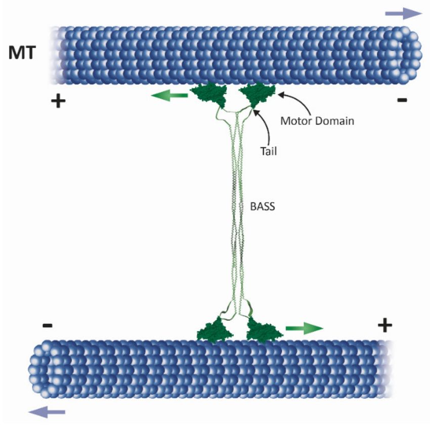
Int. J. Mol. Sci. 2021, 22, 6420                                                                                                2 of 25

      Figure 1. Major roles of kinesin-5 motors in mitotic spindle dynamics (adapted from [10]). (A) Separation of spindle poles
      during spindle assembly in closed (top) and open (bottom) mitosis [11]. Kinesin-5 motors mediate spindle pole-separation by
      crosslinking and sliding apart anti-parallel spindle MTs. Dynein, the minus-end-directed motor (yellow shape) contributes
      to spindle assembly in open mitosis [12,13]. Green and purple arrows indicate the direction of movement of the kinesin-5
      motors and the spindle poles, respectively. (B) Metaphase: chromosome congression takes place at the middle of the bipolar
      spindle. Different types of MTs are indicated: kinetochore MTs (kMTs), astral or cytoplasmic MTs (cMTs) and interpolar
      MTs (iMTs). At metaphase, the chromosomes contain pairs of sister chromatids that are attached via their kinetochores to
      kMTs. Kinesin-5 motors stabilize the spindle by crosslinking anti-parallel iMTs of the mid-zone (indicated at the bottom).
      In metazoans, kinesin-5 motors maintain spindle bipolarity and drive poleward flux [14–16]. (C) Anaphase A. The onset
      of anaphase A. When cohesion between sister chromatids is lost, the sister chromatids are separated and migrate to the
      opposite spindle poles. In parallel, poleward flux-based depolymerization of kMTs takes place. (D) During anaphase B
      spindle elongation, the two spindle poles are further separated by an elongating spindle. Such elongation is mediated by
      cortical forces generated, for example, by dynein motors attached to the cortex which translocate along cMTs. Additional
      force is provided by kinesin-5-motors by sliding apart anti-parallel iMTs at the spindle mid-zone.

                                        Later, other kinesin-5 motors were discovered using antibodies specific for kinesin-
                                   related proteins (KRPs), such as D. melanogaster Klp61F, which was shown to be a ho-
                                   motetrameric ~130 kDa protein essential for mitosis [17] and human Eg5 (HsEg5), whose
                                   localization is regulated by p34CDC2 -mediated phosphorylation [18]. As the kinesin family
                                   grew, individual members were assigned to different sub-families based on function and
                                   sequence, including the BimC sub-family, now referred to as the kinesin-5 sub-family [19].
                                   Kinesin-5 motors also share a homotetrameric structure that consists of four identical
                                   subunits that co-assemble with two pairs of catalytic motor domains on each side of a rod-
                                   shaped stalk (Figure 2) [20–22]. XlEg5, HsEg5, and Klp61F were shown to move towards
                                   the plus-ends of MTs in vitro [5,20,23]. In addition, it was shown that motile properties of
                                   kinesin-5 motors are important for spindle formation since a mutant of XlEg5 with reduced
                                   motile activity resulted in slower spindle assembly [24]. Interestingly, the small molecule
                                   monastrol was found to specifically inhibit XlEg5 motility in vitro, which resulted in inhi-
                                   bition of bipolar spindle formation and mitotic arrest [23,25,26]. This discovery prompted
                                   development of new kinesin-5-specific inhibitors for use in anti-cancer treatments [27–30].
                                   The first evidence that kinesin-5 motors are able to crosslink MTs was demonstrated by
                                   immune electron microscopy, when D. melanogaster Klp61F was detected between anti-
                                   parallel spindle MTs [31]. Later it was demonstrated that in vitro, XlEg5 can walk towards
                                   the plus-ends of the two MTs it crosslinks (Figure 2) [32]. For twenty years, it was be-
                                   lieved that kinesin-5 motors are strictly plus-end-directed, but surprisingly, three kinesin-5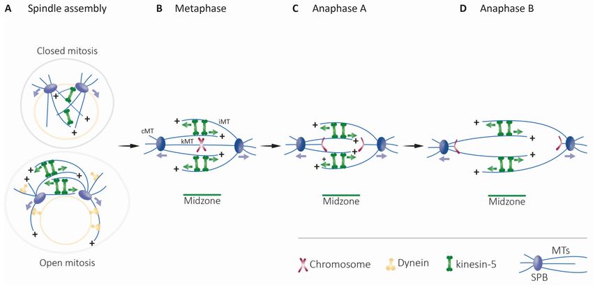
Int. J. Mol. Sci. 2021, 22, 6420                                                                                                  3 of 25

                                   motors, namely S. cerevisiae Cin8 and Kip1 and S. pombe Cut7, were shown to be bidirec-
                                   tional in vitro [33–36]. The fact that three kinesin-5 motors from fungi cells can switch
                                   directionality suggests that such bidirectional motility is of physiological importance.

                                   Figure 2. Schematic presentation of how a full-length kinesin-5 tetramer (green) crosslinks and slides
                                   apart antiparallel spindle MTs (blue). The globular motor domains and tail domains are located at the
                                   ends of the bipolar structure, connected through the central stalk which includes the bipolar-assembly
                                   (BASS) domain in the middle region [37,38]. The pair of motor domains at each end interacts with one
                                   of the two anti-parallel MTs and hence crosslinks and slides them apart. Green arrows indicate the
                                   plus-end direction of kinesin-5 motor domain movement on the MTs, while purple arrows indicate
                                   the direction of MT movement during anti-parallel sliding, with minus ends leading. Within the
                                   spindle, MT minus ends are concentrated near the spindle poles. Thus, kinesin-5 induced antiparallel
                                   MT sliding separates the spindle poles apart.

                                   2. Structural Features
                                         Kinesin-5 motors are structurally adapted to mediate anti-parallel MT sliding, thus
                                   serving their essential function of separating the spindle poles. These motors are unique in
                                   that they act as homotetramers, with pairs of catalytic motor domains located on opposite
                                   sides of a 60 nm-long rod-like minifilament [21,22,37,38] (Figure 2). Kinesin-5 motor
                                   domains share high sequence homology to other members of the kinesin superfamily and
                                   contain ATP- and MT-binding sites [39–44]. As discussed below, the catalytic domains
                                   contain kinesin-5-specific regions and are followed by a flexible 14–18 amino acid-long
                                   neck linker that contains family-specific features and which is believed to interact directly
                                   with the catalytic domain and influence processivity and directionality. In addition, the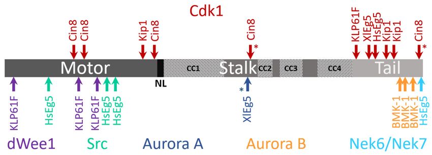
Int. J. Mol. Sci. 2021, 22, 6420                                                                                             4 of 25

                                   non-motor N-terminal region is considerably longer in kinesin-5 than in kinesin-1 motors,
                                   while the unique loop 5 within the catalytic domains presents structural features specific
                                   to the kinesin-5 family that are likely responsible for the unique mechanistic properties of
                                   these proteins.

                                   2.1. The Homotetrameric Kinesin-5 Complex
                                         The homotetrameric bipolar structure of kinesin-5 motors is believed to be essential
                                   for the crosslinking and sliding apart of anti-parallel MTs during mitosis [32,45] since
                                   monomeric and dimeric kinesin-5 variants that do not form tetramers in vivo are non-
                                   functional [22,46]. It has also been demonstrated that the tetrameric structure is not
                                   sufficient for kinesin-5 mitotic functions in spindle dynamics, as tetrameric kinesin-5
                                   chimeras containing the catalytic domains of kinesin-1 or chromokinesin exhibited MT-
                                   crosslinking activity but were non-functional in spindle dynamics [47]. The kinesin-5
                                   stalk contains four regions of heptad repeat sequences that form an α-helical coiled-coil,
                                   responsible for their multimerization. Deletion studies on the S. cerevisiae kinesin-5 Cin8
                                   and comparisons with other kinesin-5 proteins suggest that the coiled-coil region located
                                   immediately after the neck region (coil 1, 100–200 amino acids; Figure 3) is essential for
                                   self-interaction and sufficient for dimer Cin8 formation [46]. The central bipolar assembly
                                   (BASS, Figure 2) domain that spans ~200 residues in the central part of the stalk is essential
                                   for kinesin-5 activity and cell viability [46,48], indicating the importance of this domain in
                                   the organization of the bipolar homotetrameric kinesin-5 complex. In recent studies, the
                                   crystal structure of the D. melanogaster kinesin-5 Klp61F BASS domain revealed that the
                                   BASS domain consists of two anti-parallel coiled-coils at its center, stabilized by alternating
                                   hydrophobic and ionic four-helical interfaces [38]. The helixes emerge from the central part
                                   of the domain towards the N-terminal, where they bend, swap partners, and form parallel
                                   coiled-coils offset by 90◦ . Based on this structure, it has been proposed that the central
                                   BASS domain plays a role in transmitting forces between motors situated at the opposite
                                   ends of the molecule [38,49].

                                   2.2. The C-Terminal Tail Domain
                                         The tail domain of kinesin-1 has been reported to inhibit motility [50,51], apparently
                                   via a mechanism involving crosslinking of the two catalytic domains by the tail in the active
                                   kinesin dimer [52]. Direct evidence for an interaction between the motor and tail domains in
                                   kinesin-5 motors has recently been provided, through cryo-electron microscopy (CryoEM),
                                   for the D. melanogaster kinesin-5 Klp61F [53]. It has been further demonstrated that, for the
                                   human homolog HsEg5, the tails stabilize motor domains in the microtubule-bound state
                                   by slowing ATP-binding, resulting in high-force production at both homotetramer ends [53].
                                   The tail domains of kinesin-5 motors from X. laevis and D. melanogaster have also been
                                   shown to be involved in MT crosslinking [45,54]. The tail domain of S. cerevisiae Cin8 was
                                   also found to be essential for the MT crosslinking in vitro [55]. However, the tail domain
                                   alone was found to be insufficient for the intracellular functions of this protein [46]. Finally,
                                   kinesin-5 homologs carry an important Cdk1 (p34/Cdc2) kinase phosphorylation site in
                                   the tail domain (Figure 3). In higher eukaryotes, this site is located within a conserved
                                   “BimC box” that is reportedly phosphorylated during mitosis [18] and see below. Phospho-
                                   deficient mutants of human, D. melanogaster, and X. laevis kinesin-5 homologs in which this
                                   site is perturbed did not localize to the spindle apparatus [18,56,57]. In fungal cells, the
                                   role of Cdk1 phosphorylation in the tail is unclear since mutations of S. cerevisiae and S.
                                   pombe kinesin-5 homologs at this site produced no obvious phenotype [58–60].
                                         The influence of the tail domain on kinesin-5 directionality has been addressed in
                                   several studies. In the case of the S. cerevisiae kinesin-5 Cin8, which is minus-end-directed in
                                   single molecule assays under high ionic strength conditions [33], directionality preference
                                   was abolished in truncated Cin8 lacking the tail domain [55]. The motility of single
                                   tail-less Cin8 molecules was slow, processive, and bidirectional, in striking contrast to
                                   the behavior of the wild type protein. Moreover, tail-less Cin8 was unable to properly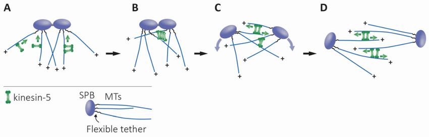
Int. J. Mol. Sci. 2021, 22, 6420                                                                                                 5 of 25

                                   cross-link MTs in vitro or support viability of the cell when present as the sole kinesin-5
                                   motor [55]. On the other hand, deletion of the tail domain of S. pombe Cut7 did not affect
                                   minus-end directionality in a multi-motor MT gliding assay, although single molecule
                                   motility of the tail-less Cut7 variant was not examined [36]. Since in the tetrameric complex
                                   the C-terminal tail domains of kinesin-5 motors are located near the N-terminal motor
                                   domains [37,38] (Figure 2) and since it has been recently demonstrated that the C-terminal
                                   tail modulates the ATPase rate of the human kinesin-5 [53], it is tempting to speculate that
                                   specific interactions between the tails and motor domains regulate the directionality of
                                   bidirectional kinesin-5 motors.

      Figure 3. Schematic presentation of phosphorylation sites within kinesin-5 motor sequences. Motor domain, neck linker
      (NL), the stalk containing putative coiled-coil (CC) regions (dotted patterns), and the tail are indicated by shades of
      gray. Asterisks indicate an unidentified function for the phosphorylation site. For the different phosphorylation sites, the
      corresponding kinases (color coded) are indicated at the top (Cdk1) or bottom (other kinases). Location of the different
      phosphorylation sites within the amino-acid sequence is scaled according to the length of a particular kinesin-5 motor
      protein. Top: Phosphorylation sites for Cdk1 [17,18,31,56–58,61–66]; Bottom: phosphorylation sites for dWee1 [67], Src [68],
      Aurora A [69], Aurora B [64,70], and Nek6/Nek7 [71–73].

                                   2.3. Neck Linker, Neck Cover Bundle and the N-Terminal Non-Motor Extension
                                         The flexible 14–18 amino acid-long neck linker that immediately follows the C-
                                   terminus of the catalytic domain was shown to undergo ATP- and microtubule-dependent
                                   docking onto the catalytic domain. Such docking is believed to coordinate the hand-over-
                                   hand movement of the two motor domains in the kinesin dimer and control the directional
                                   motility and processivity of kinesin motors [74–79]. Studies on monodirectional kinesin
                                   motors have shown that interactions between the neck linker and the catalytic motor
                                   domain are critical directionality determinants [75,76,80–85]. Plus-end stepping direction-
                                   ality was maintained by chimeric kinesin motors having a catalytic motor domain from
                                   a minus-end directed kinesin-14 and neck regions from a plus-end directed kinesin-1. In
                                   addition, chimeric kinesin motors with the catalytic motor domain from plus-end directed
                                   kinesin-1 and neck regions from minus-end directed kinesin-14 persisted with minus-end
                                   stepping directionality, suggesting that regions outside the catalytic motor domain dictate
                                   directionality [76,84,86].
                                         The kinesin-5 neck linker is longer than that of kinesin-1, a trait that was suggested to
                                   contribute to the relatively reduced processivity of kinesin-5 motors [81,87]. In contrast to
                                   kinesin-1, where prior to ATP binding the neck linker is disordered [77,88,89], the kinesin-5
                                   neck linker is ordered and points to the minus-end of the MTs [79,90–93]. These differences
                                   may account for the different kinetics of catalytic cycle stages reported for kinesin-1 and
                                   kinesin-5 motors [39,40,42].
                                         Recent studies have indicated that additional interactions stabilize the motor domain
                                   docking of the neck linker. Molecular dynamics simulations identified a nine residue-
Int. J. Mol. Sci. 2021, 22, 6420 6 of 25
long N-terminal region in kinesin-1 motors (termed the neck linker cover strand (CS) or
β0) responsible for a conformational change of the neck linker that is essential for force
generation [94,95]. Upon ATP binding, this region contributes to the formation of a β-sheet
with the neck linker, the structure that is believed to contribute to stabilization of the
motor domain-docked conformation of the neck linker, which is important for plus-end-
directed motility [94–97]. The N-terminal non-motor regions of kinesin-5 motors contain
longer extensions, as compared to kinesin-1 motors [10,40]. However, Cryo EM and kinetic
experiments indicated that the longer N-terminal region of the plus-end-directed kinesin-5
Eg5 is docked onto the motor domain with the neck linker under several nucleotide-
based conditions [79,93], suggesting that although their lengths are different, the neck
linker CS performs similar functions in terms of stabilizing docked conformations of the
neck linker in kinesin-1 and kinesin-5 motors. Moreover, it was recently reported that
the neck linker of the plus-end-directed kinesin-5 Eg5 assumes different conformations,
as compared to kinesin-1, in some nucleotide-bound states [98]. Stabilization of these
conformations in the Eg5 motor may rely on sequences in the neck linker and CS specific
to kinesin-5 motors. The bidirectional kinesin-5 motors contain longer and divergent
non-motor N-terminal extensions, as compared to kinesin-1 and the plus-end-directed
kinesin-5 motors [10]. Formation of a CS bundle in the MT- and ATP-like bound state
had recently been shown for a monomeric construct of the bidirectional S. pombe kinesin-5
Cut7 containing six N-terminal non-motor residues [99]. It is, however, possible that the
additional N-terminal sequences present in the N-terminus of the bidirectional kinesin-
5 motors, including Cut7, stabilize the conformations of the neck linker in a manner
compatible with bidirectional motility.
2.4. Loop 5
In all kinesin motors, the alpha 2 helix breaks into a short loop, termed loop 5, which
is located near the nucleotide-binding pocket. The sequence and length of loop 5 varies
between different kinesin families, ranging from 7–10 residues in kinesin-1 to ~18 residues
in kinesin-5 motors [100]. The conformation of the kinesin-5 loop 5 changes between
“open” and “closed” conformations, affecting the ATPase cycle and MT affinity of kinesin-5
motors and mediating allosteric communication with the nucleotide- and MT-binding
sites [92,100–102]. Coordinated conformational changes in loop 5, the nucleotide-binding
site, and the neck linker during the ATPase cycle have been observed in solution [92,103]
and were abolished upon deletion of a portion of loop 5 [92]. Specific point mutations
affecting proline residues within the loop decreased both MT and nucleotide affinity and
slowed the structural loop 5-dependent rearrangements that control neck linker dock-
ing [100]. In addition, deletion of loop 5 decreased the rate of MT-stimulated ADP release
by kinesin-5 monomers and dimers [101]. Finally, loop 5 could synchronize the actions of
kinesin-5 dimers, thus enabling initiation of dimer stepping from a two-head MT-bound
state, thought to be unique to kinesin-5 motors [101,104]. Recently obtained CryoEM data
for MT-bound kinesin-5 in different nucleotide-bound states support the role of loop 5
as a central coordinator of intramolecular rearrangements during the catalytical ATPase
cycle [79,93].
Loop 5 is also the binding site for small molecule inhibitors specific to vertebrate
kinesin-5 motors [105] that allosterically inhibit ATPase activity and directional kinesin-5
motility [23,106,107], as well as its intracellular functions [25,108,109]. Because of the clin-
ical importance of loop 5 in binding specific kinesin-5 inhibitors, extensive mutagenesis
studies have been performed with the aim of understanding the mechanism of inhibi-
tion [110–113]. The majority of mutants examined maintained activity, albeit with variably
reduced sensitivity to the inhibitors. The set of mutants examined further demonstrated
the inhibitor binding energy to be distributed around the pocket and that the binding
pocket is able to accommodate different types of inhibitory molecules [110]. The extensive
mutagenesis studies and structural data suggest a mechanism whereby allosteric inhibitors
bind to a specific conformation of loop 5 and by stabilizing this conformation prevent anyInt. J. Mol. Sci. 2021, 22, 6420 7 of 25
subsequent rearrangement of the motor domain required for the catalytical cycle. Loop 5
of the D. melanogaster kinesin-5 Klp61F, which is insensitive to allosteric inhibitors such as
monastrol [112], assumes a different conformation than do the vertebrate kinesin-5 motors
when bound to MTs in the presence of AMP-PNP [79,114]. This supports the notion that
kinesin-5 allosteric inhibitor binding is dependent on a vertebrate-specific conformation of
loop 5.
2.5. Loop 8
The flexible loop 8 breaks the beta strand 5 of the kinesin motor domain into two
sections [115]. In the MT-bound state of the motor, loop 8 faces the MT lattice and thus
is thought to be part of the MT-binding domain [116,117]. It has also been shown that
in the dimeric structure of kinesin-1, loop 8 of one motor domain interacts with loop
10 of the other. This interaction was proposed to serve as an inter-subunit connection,
allowing communication between the motor domains in an active dimer [116]. The amino
acid sequence of S. cerevisiae Cin8 includes a large (99 amino acid) insertion in loop 8,
which is not essential for intracellular function [7]. Loop 8 of Cin8 was found to regulate
the directionality of the motor, since replacement of this insert with the short loop 8
of the S. cerevisiae Kip1 induced bias to the minus end-directed movement of Cin8 in a
single-molecule motility assay [33]. It had been suggested that loop 8 can regulate the
directionality of Cin8 through its noncanonical binding to MTs [118]. However, it was
recently demonstrated that, in the monomeric form of Cin8, loop 8 had no effect on the
directionality of Cin8 in a multi-motor MT gliding assay [119]. In contrast, a study from
our laboratory demonstrates that loop 8 of Cin8 directly interacts with the MTs and induces
bidirectional motility in both full-length and dimeric forms of Cin8 (Himanshu et al., in
preparation). These results may indicate that in order for loop 8 to regulate the directionality
of Cin8, two motor domains have to interact with the same MT, as in the scenario of native
tetrameric and dimeric forms of Cin8.
3. Motor Activity
Kinesin-5 motors perform essential functions in spindle dynamics by binding to and
moving along MTs. Because of their bipolar structure, these motors can crosslink and slide
apart anti-parallel spindle MTs (Figures 1 and 2), thus providing the outwardly-directed
force that separates the spindle poles.
3.1. Velocity, Processivity and Anti-Parallel MT Sliding
Since their initial discovery, the mechano-chemical cycle of kinesin-5 motors has
been extensively studied using monomeric, dimeric, and full-length variants [39,90,93,100,
102,106,120]. Such studies revealed that the MT-stimulated ATPase rate of monomeric
kinesin-5 is slower than that of kinesin-1 (~7/s vs. ~50/s) [90,120,121], with Pi release
being the rate-limiting step [106]. Kinesin-5 motors, moreover, exhibit unique motile prop-
erties [39,40,42,122,123], as seen below. Since kinesin-5 motors carry their catalytic domain
at the N-terminus, they were initially thought to be exclusively plus-end-directed. Indeed,
in multi-motor MT gliding assays, where in vitro-polymerized MTs are translocated by
surface-bound motors, monomeric and dimeric human, mouse, and Xenopus laevis kinesin-5
variants were shown to move MTs with the minus-ends leading, consistent with a plus-end
directionality of the motors [81,124–126]. In contrast to kinesin-1, the velocity of MT gliding
driven by kinesin-5 motors was similar for monomers and dimers, suggesting that in order
to translocate MTs, coordination between the two motor domains within a dimer is not
essential, such that single motor domains can mediate motility so long as the MT is main-
tained near the surface via the binding of neighboring motors [124,126]. Plus-end-directed
MT gliding of full-length kinesin-5 proteins from human, D. melanogaster, X. laevis, and
S. cerevisiae was also demonstrated [20,32,34,35,56,81,127,128]. However, the velocity of
kinesin-5-mediated MT gliding was considerably slower (10–70 nm/s) [39,123] than that of
kinesin-1 motors (~500–1000 nm/s) [1,129–131], indicating differences in the mechanochem-Int. J. Mol. Sci. 2021, 22, 6420 8 of 25
ical cycle [42]. Finally, in single molecule fluorescence motility assays, kinesin-5 motors
from vertebrates were also shown to be slow and plus-end-directed [23,81,87,128,132],
although the velocity and directionality of single molecules from other members of the
kinesin-5 sub-family differed significantly (see below).
Processivity, defined as the number of consecutive steps taken by a motor before
detaching from MT tracks, varies among the different kinesin sub-families [39]. Although
kinesin-5 motors were shown to be processive, such processivity is lower than that of
kinesin-1 motors. Dimeric human kinesin-5 variant takes ~8 steps on average before de-
taching [133], while the dimeric kinesin-1 motors take several hundreds of steps [134].
Kinesin-5 processivity was shown to be force-dependent in a bead-based optical trap as-
say [133,135]. The processivity of full-length kinesin-5 motors is higher as compared to that
of dimeric constructs, yet is still lower than that of kinesin-1 motors [23]. Finally, it was
suggested that the longer neck-linker region (see below) is associated with the reduced pro-
cessivity of kinesin-5 motors [81,87]. The low processivity of kinesin-5 motor could reflect
adaptation to their intracellular function, where ensembles of kinesin-5 motors crosslink
interpolar spindle MTs (see below), reducing the requirement for high processivity.
MT architecture within the mitotic spindle is such that the plus-ends are pointing
towards the midzone (Figure 1). Therefore, to separate the spindle poles, the bipolar
kinesin-5 motors must crosslink and slide anti-parallel interpolar MTs of the midzone
in a plus-end-directed manner. Indeed, crosslinking and anti-parallel sliding of MTs
was demonstrated for kinesin-5 homologs from yeast to vertebrates [33,34,45,127,128].
X. laevis XlEg5 was shown to walk in a plus-end-directed manner along the two MTs it
crosslinks [32], with force production during anti-parallel sliding being correlated with the
length of the zone of MT overlap [136]. Under physiological salt conditions, crosslinking
two MTs was shown to activate X. laevis Eg5. On a single MT, this motor exhibited diffusive
bidirectional motility, which may be attributed to an additional MT-binding domain in
the Eg5 tail [54]. This diffusive mode switches to plus-end-directed processive motility
upon the binding of a second MT to produce anti-parallel MT sliding [128]. These findings
demonstrate the adaptation of kinesin-5 motors to perform anti-parallel sliding only when
bound between two anti-parallel MTs.
Mitotic spindle motors function in a crowded environment, in the presence of other
MT-binding motor and non-motor proteins. Several studies have addressed the function-
ality of kinesin-5 motors in the presence of other motors. X. laevis Eg5 was shown to
antagonize and slow MT motility driven by the fast kinesin-1 in gliding assays [137], while
the D. melanogaster kinesin-5 Klp61F was shown to antagonize the minus-end-directed
kinesin-14 Ncd [48]. These findings may correspond to the “braking” activity of kinesin-5
motors within the spindle [136,138–140] and reflect their ability to balance spindle forces
during mitosis.
3.2. Bidirectional Motility of Fungal Kinesin-5 Motors
Although the plus-end-directed motility of kinesin-5 motors is essential for separating
spindle poles during spindle assembly and anaphase B, two earlier studies reported that S.
cerevisiae kinesin-5 Cin8 is minus-end-directed when moving as a single molecule in high
ionic strength conditions, and switches directionality in several other experimental condi-
tions [33,34]. Following the first reports on Cin8 bidirectionality, the kinesin-5 homologs
S. cerevisiae Kip1, and S. pombe Cut7 were also reported to be bidirectional and exhibit
switchable directionality under certain in vitro experimental conditions [35,36]. Subsequent
work revealed intramolecular domains that affect the directionality of these three kinesin-5
motors [10], such as the N-terminal non-motor extension [141,142], the C-terminal tail
domain [55], and the large insert in the loop 8 within the catalytic domain [33], which was
also demonstrated to induce non-canonical binding of Cin8 to MTs [118]. Phosphorylation
of three cyclin-dependent kinase 1 (Cdk1) sites in the catalytic domain of Cin8, which
regulates its intracellular activity [58,60,61,143], with two of these sites being located in
large loop 8, was also shown to affect Cin8 directionality [144].Int. J. Mol. Sci. 2021, 22, 6420 9 of 25
Intermolecular interactions between bidirectional kinesin-5 motors or between these
motors and non-motor proteins also affect motor directionality. Based on multi-motor
MT gliding and anti-parallel MT-sliding assays, it was suggested that the switch from
fast minus-end- to slow plus-end-directed motility results from the coupling of S. cere-
visiae Cin8 and Kip1 motors through the MT with which they both interact [34,35,145].
This model, which was recently supported by a theoretical study [146], does not apply
to S. pombe Cut7, which was shown to be minus-end-directed in both single molecule
and multi-motor MT gliding assays [36]. It has also been demonstrated that crowding
of Cut7 by motor and non-motor proteins can drive directional switching from minus-
to plus-end-directed motility, with it being suggested that such crowding reflects a steric
blockage mechanism [142]. Finally, it was reported that accumulation of Cin8 into clusters
slowed motility and induced a switch from minus- to plus-end-directed motility [62]. This
study further proposed that since the ability to move in both directions is an intrinsic
property of Cin8 tetramers [33,55,147], specific intermolecular interactions between Cin8
tetramers in a cluster control the directionality of this motor [62]. Based on the characteri-
zation of single-molecule and cluster motility, a recently developed model predicts that
directionality switching of the bidirectional Cin8 is caused by an asymmetric response of
its active motion to opposing forces, referred to as drag [148]. The model showed excellent
quantitative agreement with experimental data obtained under high and low ionic strength
conditions. This recent analysis identified a robust and general mechanism that explains
why bidirectional motor proteins reverse direction in response to experimental conditions,
including changes in motor density and molecular crowding and in multimotor motility
assays [148].
3.3. Interaction with MT Ends
Several lines of evidence suggest that in addition to moving along MTs, kinesin-5
motors also interact with and control the dynamics of MT ends. S. cerevisiae Kip1 was
shown to bind to MT plus-ends in cells and to follow the plus-end of growing and shrinking
MTs, which is likely related to Kip1-mediated MT stabilization at the end of anaphase
B [35]. Plus-end accumulation and stabilizing activity in vitro were also demonstrated for
a dimeric chimera comprising the X. laevis Eg5 catalytic domain and a kinesin-1 stalk [132].
Protection of MT depolymerization by this chimera was inhibited by specific kinesin-5
inhibitors [149]. On the other hand, deletion of S. cerevisiae Cin8 and inactivation of the
D. melanogaster kinesin-5 Klp61F led to longer and more stable MTs [150–152], indicating
that these motors destabilize MTs. Finally, Cin8 was shown to accumulate and track the
minus-ends of dynamic MTs in vitro [62]. The physiological significance of such behavior
remains to be elucidated.
4. Intracellular Function
4.1. Roles in Dividing Cells
4.1.1. Structure and Dynamics of the Mitotic Spindle
In eukaryotic cells, separation of duplicated chromosomes during mitosis is medi-
ated by the MT-based mitotic spindle [153]. MTs of the spindle are arranged with their
minus-ends concentrated at the centrosomes or SPBs, and are divided into three classes
based on their function [154,155] (Figure 1). The plus-ends of the kinetochore MTs (kMTs)
capture chromosomes at protein structures termed kinetochores and facilitate chromosome
movement within the spindle [156,157]. Cytoplasmic or astral MTs (cMTs) are captured at
sites on the cell cortex and mediate spindle positioning [158–160]. Interpolar MTs (iMTs)
overlap in an anti-parallel array in the middle region of the spindle, the midzone [161–163],
and are focused in parallel arrays near the poles [155]. The mitotic bipolar spindle as-
sembly is established by the separation of the two spindle poles into a bipolar structure
where the two spindle poles are located at the opposite ends of the overlapping iMT ar-
ray (Figure 1A) [164–166]. This separation is primarily accomplished by pushing forces
applied from within the spindle on iMTs [7,8,25,63,167]. In higher eukaryotes that divideInt. J. Mol. Sci. 2021, 22, 6420 10 of 25
via open mitosis accompanied by nuclear envelope breakdown, [166,168–170], additional
pulling forces that contribute to centrosome separation are applied by nuclear envelope-
or cortex-bound dynein motors on cMTs (Figure 1A) [171–176]. Following mitotic spindle
assembly, chromosomes are captured at the kinetochores by the plus-ends of MTs ema-
nating from the spindle poles, ultimately leading to their congression to the middle of
the spindle in metaphase (Figure 1B). Attachment of all chromosomes to kMTs emanating
from both poles and consequent inactivation of the spindle assembly checkpoint leads to
separation of the sister chromatids by degradation of the cohesion complex [177–179] and
their movement to the opposing poles via depolymerization of the kMTs at their plus-ends
(anaphase A, Figure 1C) [180,181]. Following anaphase A, the spindle elongates to further
separate the two groups of chromatids (anaphase B). This event is largely realized by
pushing forces from within the spindle and by polymerization of iMTs at their plus-ends
(Figure 1D) [16,182].
4.1.2. Bipolar Spindle Assembly, Maintenance, and Elongation
Members of the kinesin-5 sub-family have been shown to perform essential roles
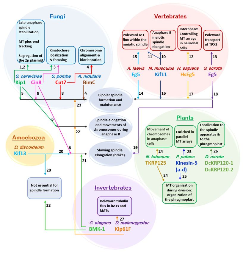
related to mitotic spindle dynamics in eukaryotic cells (Figure 4) [15,40,42,122,123]. The
key roles of kinesin-5 motors, namely the establishment and maintenance of a mitotic
spindle (Figure 4), relies on their ability to crosslink and slide anti-parallel MTs apart, due
their unique bipolar structure (Figure 2). These functions are highly conserved in eukary-
otes, with numerous studies indicating that a loss of kinesin-5 function leads to mitosis
failure due to a lack of spindle pole separation prior to spindle assembly, or as a result of
spindle collapse after establishment of the bipolar spindle [5,7,17,18,26,183–185]. However,
it remains to be fully established whether or not anti-parallel MT sliding is essential for
spindle assembly, or if MT crosslinking by kinesin-5 motors is sufficient [186]. To date,
only two organisms, the nematode Caenorhabditis elegans and the slime mold Dictyostelium
discoideum have been reported as being able to form a functional bipolar spindle in the
absence of kinesin-5 motors (Figure 4) [70,187]. The moss Physcomitrella patens can be partly
considered a third exception as although kinesin-5 is not essential for mitotic spindle forma-
tion in this organism, it is still required for post-anaphase spindle/phragmoplast integrity
and chromosome segregation (Figure 4). [188]. The role of kinesin-5 motors in anaphase
B spindle elongation, however, remains controversial. An anaphase-facilitating function
was demonstrated in S. cerevisiae [189–192] and insect cells [193] (Figure 4). Moreover,
the S. cerevisiae kinesin-5 motors Cin8 and Kip1 were shown to be partially destabilized
so as to prevent anaphase onset in cells carrying damaged DNA [194]. In other cases, it
was suggested that kinesin-5 motors slow spindle elongation rates by applying “brakes”
involving the crosslinking of spindle MTs [139,140,187,195]. (Figure 4). Interestingly, some
features of kinesin-5 homologs are characteristic to specific taxonomic groups, such as the
bidirectional motility of kinesin-5 motors in fungi, the abundance of kinesin-5 motors in
plants or the poleward transport of kinesin-5 motors by TPX2 and dynein in vertebrates
(Figure 4).Int. J. Mol. Sci. 2021, 22, 6420 11 of 25
Figure 4. Physiological roles of kinesin-5 motors. Functions common to at least two kinesin-5 motors are indicated in blue
ellipses. Functions identified for a single kinesin-5 motor are indicated in framed rectangles. Colored spheres indicate
different groups of species. Arrows point to specific intracellular functions. Numbers indicate the related references:
(1) [35]; (2) [196–198]; (3) [151,199–202]; (4) [203]; (5) [7–9,204]; (6) [189–192]; (7) [139]; (8) [3,205]; (9) [2]; (10) [206]; (11) [207];
(12) [208,209]; (13) [210,211]; In [211], human Eg5 was expressed in Sus scrofa cells. (14) [4,5,56]; (15) [212,213]; (16) [184];
(17) [18,214]; (18) [210]; (19) [195]; (20) [187]; (21) [140,215]; (22) [31,216]; (23) [17]; (24) [217]; (25) [188]; (26) [218]; (27) [14,216];
(28) [70].
4.1.3. Models for Maintaining Spindle Bipolarity
Biophysical models describing the role of kinesin-5 motors in maintaining the bipolar
spindle structure have been proposed in different organisms, based on the force-balanced
model suggested for S. cerevisiae spindles [204,219]. According to this model, bipolarity of
the spindle is maintained by a balance of inwardly- and outwardly-directed forces exerted
by the minus-end-directed kinesin-14 Kar3 and the kinesin-5 motors Cin8 and Kip1, whichInt. J. Mol. Sci. 2021, 22, 6420 12 of 25
are plus-end-directed between anti-parallel MTs [33–35]. In support of this notion, it has
been demonstrated that in S. pombe, spindles could been assembled when kinesin-5 and
kinesin-14 motors were simultaneously deleted [167]. Normally, the onset of anaphase B is
triggered by a shift in the balance of these forces in favor of spindle elongation. Similar
mechanisms were suggested for D. melanogaster embryos [220] and X. laevis [221,222].
4.1.4. Effects on MT Turnover and Dynamics
In higher eukaryotic cells, kinesin-5 motors were shown to affect the poleward
turnover of tubulin (poleward flux) in kMTs and iMTs [212,213,223], which contributes to
chromosome congression and separation, respectively [14,31,216]. Poleward flux is not
observed in yeast cells [224,225], however, kinesin-5 motors have been shown to affect MT
dynamics in S. cerevisiae cells [35,150,151]. In addition, kinesin-5 motors have been shown
to bind to kinetochores, focus kinetochore clusters, and limit the length of kMTs in S. cere-
visiae [151,199–202]. Finally, the S. cerevisiae kinesin-5 Kip1 is required for segregation of the
2-micron plasmid [196–198], the function of which can be related to the minus-end-directed
motility of Kip1 (Figure 4) [35].
4.1.5. Functions at the Spindle Poles
Several reports suggest that localization near the spindle poles is important for kinesin-
5 function. First, in a number of organisms, kinesin-5 motors were found to be enriched
near centrosomes or SPBs [5,56,188,226–228]. Second, it had been demonstrated that in
higher eukaryotes undergoing open mitosis, kinesin-5 motors are actively transported
towards the poles by the dynein–dynactin complex [226,227,229]. The Ran-regulated
spindle-pole localizing factor TPX2 [230] was found to recruit kinesin-5 to the poles in
Xenopus spindles [231], possibly via connecting the kinesin-5 to the dynein complex, as
demonstrated in mammalian cells [232]. Dynein-dependent poleward kinesin-5 delivery is
absent in yeast that undergo closed mitosis. However, kinesin-5 motors also co-localize with
spindle poles prior to spindle assembly in yeast cells [3,62]. The discovery of switchable
kinesin-5 motor directionality in budding and fission yeast [33–36] suggests possible
physiological roles for this ability in closed mitosis (Figure 5). Bidirectionality may be
the evolutionary adaptation that allows these motors to localize to spindle poles prior to
spindle assembly by minus-end directed motility on nuclear MTs, without the function
of the minus-end-directed dynein [10,62]. At this location, kinesin-5 motors can capture
anti-parallel MTs emanating from the neighboring pole and mediate their anti-parallel
sliding by plus-end directed motility, thus promoting spindle assembly (Figure 5) [62].
In support of this hypothesis, a recent computational model identified a set of functions
essential for the generation and stability of spindle bipolarity in fission yeast, showing that
the bidirectionality of kinesin-5 motors enables proper localization to the spindle apparatus,
which is necessary for spindle assembly [233].
Building a stable bipolar spindle requires enough antiparallel MT interactions for
the separation of the poles by plus-end-directed kinesin-5 motor motility. An interesting
question is whether the MTs emanating from the unseparated neighboring poles are flexible
enough to be captured and aligned by the kinesin-5 motors accumulated at the poles. In
recent years, a few studies in yeast have shed light on this question. It has been shown in
S. pombe that random rotational pivoting helps microtubules from the neighboring poles
to come into contact prior to spindle assembly [234]. Recently, by engineering a flexible
tethering protein, Spc110, which links microtubule minus ends to the SPB core, Fong
et al. demonstrated that thermal microtubule pivoting is specifically important during
early spindle assembly of S. cerevisiae, and that this pivoting motion is sufficient to bring
microtubules from neighboring poles into contact [235]. In support of this notion, in high-
voltage electron tomography of early mitotic spindles in S. cerevisiae it has been shown
that, after SPB duplication, while the SPBs are still connected by a bridge, the MTs from
each SPB interdigitate at sharp angles [236]. According to our model, such overlap of MTs
emanating from the neighboring poles, induced by their flexible pivoting, is sufficient forInt. J. Mol. Sci. 2021, 22, 6420 13 of 25
the fungi kinesin-5 motors located at the poles, to capture these MTs and mediate spindle
assembly via plus-end directed antiparallel MT sliding (Figure 5).
Figure 5. Proposed model for the physiological role of the bidirectional motility of fungi kinesin-5 motor proteins, adapted
from [62]. (A) Prior to spindle assembly, the minus-end directed motility of bidirectional kinesin-5 motors causes their
accumulation at the minus-ends of the MTs, near the SPBs. (B) At this stage, MTs from duplicated spindle poles come
into antiparallel contact due to thermal pivoting, facilitated by flexible tether linking MT minus-ends and the SPB [235].
Kinesin-5 motors, accumulated in clusters near the SPBs, capture these antiparallel-oriented MTs, crosslink, and slide them
apart via their plus-end directed motility. This plus-end directed motility is achieved by the clustering of kinesin-5 motors
near the spindle poles and the binding to the two antiparallel MTs [62,148]. (C) The initial separation of the spindle poles,
caused by the antiparallel sliding of MTs, generates additional MT overlap—which creates additional sites for the kinesin-5
motors to crosslink and slide apart antiparallel MTs. (D) Formation of the final bipolar spindle structure is achieved. The
curved purple arrows indicate the direction of SPB movement during separation. Green arrows indicate the direction of
motor motility.
4.2. Roles in Non-Dividing Cells
Although the majority of studies addressing the physiological roles of kinesin-5
motors have focused on their roles in mitosis, several studies have also revealed roles
for these proteins in interphase, including regulation of the biogenesis and function of
Ago1-complexes [237], as well as Golgi organization and polypeptide synthesis, in human
cells [238,239]. The most extensively studied function of kinesin-5 motors in interphase is in
neuronal cells, due to the clinical relevance of this system. Kinesin-5 motors are expressed
in post-mitotic neurons [240], where they may play a role in the proper development
of mammalian neuronal processes, including axon growth cone guidance, elongation
and branching [208,209,241] and modulation of neuronal growth and migration [242].
In dendrites, inhibition of kinesin-5 motors causes changes in cell morphology and MT
organization. Taken together, these observations suggest that kinesin-5 regulates axon
and dendrite growth, acting as a brake on MT bundle assembly [243]. It is likely that
cytoplasmic dynein is the principal motor whose forces are attenuated by the braking
function of kinesin-5 [243], acting via two other kinesin motors, kinesin-6 and kinesin-12,
that may also participate in co-regulation of MT patterns in axons and dendrites [240].
The research on kinesin-5 functions in plant cells is considerably less developed than
in fungi and metazoans. However, several studies have revealed that kinesin-5 motors play
non-canonical roles in plant cells [244]. For example, in Nicotiana tabacum, kinesin-5 was
reported to be involved in separating anti-parallel microtubules in the phragmoplast, a
MT-based structure formed during late cytokinesis (Figure 4) [217]. In Arabidopsis thaliana,
disruption of kinesin-5 activity leads to disorganization of intracellular microtubules
during interphase, as well as spindle formation [183]. Additional studies are needed to
fully characterize the multiple functions of kinesin-5 motors in plant cells.Int. J. Mol. Sci. 2021, 22, 6420 14 of 25
4.3. Phosphoregulation
In the eukaryotic cell cycle, mitotic events are coordinated via phosphorylation by
specific kinases [245–250]. The balance between phosphorylation and dephosphorylation
was demonstrated to govern the localization of these kinases within the mitotic spindle and
regulate their functions. The amino acid sequences of kinesin-5 motors contain multiple
phosphorylation sites located within and outside the motor domain (Figure 3), a large
number of which have been demonstrated to regulate kinesin-5 [42,122,123].
It has been reported for a number of eukaryotes that a conserved sequence termed
the “BimC box”, located in the C-terminal tail, possesses a conserved phosphorylation
site for cyclin-dependent kinase 1 (Cdk1; Cdc28 in yeast) (Figure 3). For human Eg5,
phosphorylation at this site regulates spindle targeting, association, and localization, with
characteristic concentration of the kinase at the poles [18]. Moreover, Cdk1 is required for
interaction with dynein through the p150glued subunit of dynactin [63]. Consistently, X.
laevis mutants phosphodeficient in the Eg5 BimC box showed disrupted kinase localization
to the mitotic spindle [56], while in X. laevis spindles assembled from an egg extract,
phosphorylation by Cdk1 increased the binding of Eg5 to MTs [64]. In higher plants,
such as Arabidopsis thaliana, tobacco, and carrot, kinesin-5 motor activity in stabilization
and sliding apart anti-parallel MTs is Cdk1-dependent [218,251–253], and is regulated
by phosphorylation of the BimC box. In D. melanogaster embryonic mitotic spindles,
Klp61F motors, phosphorylated at a Cdk1/cyclin B consensus domain within the BimC
box, concentrate in the mid-zone region of interpolar MT bundles [31]. In addition, it
was shown that Klp61F is phosphorylated both within the BimC box and in the motor
domain [67,228]. This leads to dynamic localization of Klp61F throughout the spindle
and cross-bridging between both parallel and anti-parallel MTs [31,254]. These findings
hint at coordination with another important cluster of phosphorylation sites located in the
N-terminal motor domain, in close vicinity to the ATP-binding pocket.
In yeast, Cdk1 also phosphorylates kinesin-5 motors although the mechanism of
phosphoregulation appears to be different than employed in higher eukaryotes. In fission
yeast, Cdk1 phosphorylation at the C-terminal of the kinesin-5 Cut7 is not required for
association with microtubules [59]. Both S. cerevisiae Cin8 and Kip1 lack the BimC box
motif, although Cdk1 phosphorylation consensus sequences are present in their C-terminal
tails [61]. Phosphorylation in the Kip1 motor domain by Cdk1 was shown to be required for
spindle pole separation [61]. Cin8 was, furthermore, demonstrated as being differentially
phosphorylated during anaphase at three Cdk1 sites located in its motor domain [58].
Such phosphorylation induces Cin8 detachment from spindles and reduces the spindle
elongation rate [58], with each of the three sites playing unique roles in phosphoregulation
of Cin8 [60,255]. In vitro experiments with Cin8 phospho-mutants suggest that phospho-
rylation of the three sites within the motor domain provides fine tuning of motor activity.
Accordingly, phospho-mimetic mutants possess weaker MT-motor interactions, increased
motor velocity, and minus-end-directionality [144].
Aside from Cdk1, other kinases have been shown to regulate kinesin-5 motors
(Figure 3). For example, the tail domain of HsEg5 contains a phosphorylation site rec-
ognized by Nek6/Nek7 kinases. This site contributes to the accumulation of Eg5 at the
spindle poles and is necessary for subsequent spindle pole separation [71,72]. Phosphoryla-
tion of the HsEg5 motor domain by Src kinase tunes motor activity so as to obtain optimal
spindle morphology [68]. The D. melanogaster kinesin-5 Klp61F is phosphorylated at the
motor domain by the Wee1 tyrosine kinase, which may thus affect Loop 5 conformation
and motor function [67]. Finally, regulation by the Aurora kinases was also reported for
some kinesin-5 motors. C. elegans kinesin-5 was reported to be phosphorylated at the tail
domain by the Aurora B kinase, which regulates its spindle localization [64,70]. The X.
laevis kinesin-5 Eg5 was shown to be phosphorylated by the Aurora A kinase at the stalk
domain [69], although such phosphorylation was found to be non-essential for spindle
formation [64]. Work in the coming years is likely to reveal the complex mechanism by
which phosphorylation by different kinases regulates kinesin-5 motor functions.Int. J. Mol. Sci. 2021, 22, 6420 15 of 25
4.4. Other Post-Translation Modifications
It had recently been reported that the motor domain of the human kinesin-5 Eg5
undergoes acetylation at a specific lysine residue (K146). Using the acetylation-mimic
K146Q mutant, it was further shown that such acetylation disrupts a salt-bridge formed
between K139 and D91 in a neighboring α-helix [256]. This disruption increased the
coupling of the neck linker to the catalytic domain, enhanced motor performance under
load, and increased binding to MTs [256]. While this study clearly revealed that acetylation
can regulate the functions of kinesin-5 motors, additional studies are needed to expand on
the generality of this finding.
5. Kinesin-5 Motors and Pathological Conditions
Since mitosis is a fundamental process in proliferating cells, the mitotic spindle offers
a prime target for cancer therapy [257–259]. Inhibitors that act by “shutting down” key
components of kinesin-5 motors may thus cause mitotic arrest of proliferative tissues.
Historically, chemotherapeutic drugs designed to interfere with the cell cycle specifically
targeted microtubules. Such drugs include the clinically used taxanes and vinca alka-
loids [260,261]. The development of resistance to microtubule-targeted drugs and severe
side effects raised interest in alternative targets. The human kinesin-5 Eg5, which is in-
volved in the formation of a bipolar spindle, meets the criteria for an appropriate target in
cancer. Eg5 is over-expressed in hematological malignancies and many solid tumors, such
as breast, ovarian, bladder, and pancreatic cancers, yet is found at negligible levels in non-
proliferative tissues [262–265]. In breast cancer patients, Eg5 over-expression is associated
with poor prognosis. Accordingly, Eg5 is considered a typical oncoprotein and has been
proposed as a potential prognostic biomarker and target for therapeutic agents in oral and
breast cancer treatment [266,267]. Eg5 possesses unique structural features that selectively
dispose it to small molecule inhibitors. Within the Eg5 motor domain, loop 5 presents a
prolonged structure, forming a druggable allosteric pocket located between helix α3 and
loop 5, [91]. Moreover, loop 5 is flexible [268] and, as mentioned in the “structural informa-
tion” section, undergoes an “open” to “closed” structural transition that correlates with
rearrangements at the active site during the ATPase cycle [92,100–102]. Since the discovery
of monastrol, a specific vertebrate kinesin-5 inhibitor that binds to loop 5 [25,26], a variety
of Eg5-specific inhibitors have been developed [269–271]. These inhibitors arrest cells in
mitosis, producing characteristic monoastral spindles [108,109,149], and lead to apoptosis
in proliferative tissues either by binding to loop 5 or other allosteric sites and inhibiting
ATPase activity [30,102,103,111,149,272]. To develop and test promising Eg5 inhibitors,
different approaches are used, including in silico drug-design predictive tools that seek to
bridge computational modeling with drug design and cancer therapy [259,273,274]. Based
on these studies, several kinesin-5 inhibitors are currently in clinical trial [29,275–277].
Several reviews shed light on the state of the art in terms of kinesin-5 inhibitors serving
as anti-tumor drugs, reporting the results of clinical trials of the most potent candidates
and offering reasons for their poor clinical outcome [40,259,278–280]. To summarize, more
than 40 Phase I and II clinical trials assessing inhibitors of the kinesin-5 Eg5, starting with
the first generation drug ispinesib [29] and followed by filanesib [276], were suspended or
discontinued. When used as monotherapy, Eg5-targeting agents have only been moderately
successful, instead causing common adverse effects, such as neutropenia [278]. Therefore,
a multi-drug combination is likely to improve efficacy [281]. It has also been demonstrated
that the kinesin-12 motor Kif15 can functionally replace inhibited Eg5 and cause drug
resistance [282]. To overcome Kif15-mediated cancer drug resistance, a strategy involving
a combination drug therapy employing both inhibitors has been suggested [283].
Author Contributions: Conceptualization, L.G.; writing—original draft preparation, review, and
editing, H.P., M.P., A.G.-L., and L.G.; supervision and funding acquisition, L.G. All authors have read
and agreed to the published version of the manuscript.You can also read