The molecular motor kinesin: From single-molecule mechanisms to joint action - Dissertation der Fakultät für Biologie der ...
←
→
Page content transcription
If your browser does not render page correctly, please read the page content below
The molecular motor kinesin:
From single-molecule mechanisms
         to joint action

                  Dissertation

          der Fakultät für Biologie der

         Ludwig-Maximilian-Universität

                   München

                 vorgelegt von

                Bettina Ebbing

              aus Wolfratshausen

                  08.01.2008
Erstgutachter:                PD Dr. Günther Woehlke

Zweitgutachter:               Prof. Manfred Schliwa

eingereicht:                  08.01.2008

Tag der mündlichen Prüfung:   04.04.2008

                                   II
Ehrenwörtliche Erklärung:

Hiermit erkläre ich, dass die vorliegende Dissertation von mir selbständig und ohne
unerlaubte Hilfsmittel angefertigt wurde und ich keine anderen als die angegebenen
Quellen und Hilfsmittel verwendet habe.

__________________

Bettina Ebbing

                                          III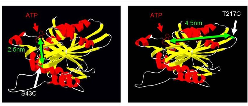
Table of Contents
1         Introduction ..................................................................................................... - 1 -

    1.1       STRUCTURE OF THIS THESIS ................................................................................................... - 1 -
    1.2       INTRACELLULAR TRANSPORT ................................................................................................ - 2 -
          1.2.1     Axonal transport............................................................................................................- 2 -
          1.2.2     Molecular motors ..........................................................................................................- 3 -
          1.2.3     Kinesin...........................................................................................................................- 4 -
          1.2.4     The chemo-mechanical cycle.........................................................................................- 6 -
    1.3       OPTICAL TECHNIQUES ........................................................................................................... - 8 -
          1.3.1     TIRF microscopy ...........................................................................................................- 9 -
          1.3.2     Confocal microscopy...................................................................................................- 11 -
          1.3.3     FRET ...........................................................................................................................- 12 -
          1.3.4     Optical tweezers ..........................................................................................................- 14 -
    1.4       OUTLINE OF THE PRESENT THESIS........................................................................................ - 16 -
    1.5       REFERENCES ....................................................................................................................... - 17 -

2         Molecular determinants of processivity in kinesin......................................... - 20 -

    2.1       ABSTRACT ........................................................................................................................... - 20 -
    2.2       INTRODUCTION.................................................................................................................... - 21 -
    2.3       RESULTS.............................................................................................................................. - 22 -
          2.3.1     Design of chimaeras....................................................................................................- 22 -
          2.3.2     Co-operative gliding behaviour ..................................................................................- 22 -
          2.3.3     Single-molecule behaviour ..........................................................................................- 24 -
    2.4       DISCUSSION......................................................................................................................... - 26 -
    2.5       METHODS ............................................................................................................................ - 27 -
          2.5.1     Cloning, protein expression and purification..............................................................- 27 -
          2.5.2     Gliding assays .............................................................................................................- 28 -
          2.5.3     Single-molecule motility assays...................................................................................- 28 -
    2.6       REFERENCES ....................................................................................................................... - 29 -

3         Kinesin-1’s neck-linker docking .................................................................... - 31 -

    3.1       ABSTRACT ........................................................................................................................... - 31 -
    3.2       INTRODUCTION.................................................................................................................... - 32 -
    3.3       RESULTS.............................................................................................................................. - 34 -
          3.3.1     Mutant design and proof of expected disulfide bonds .................................................- 34 -
          3.3.2     Motility behaviour .......................................................................................................- 35 -

                                                                          IV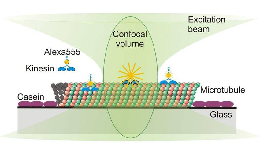
3.3.3    Microtubule affinities ..................................................................................................- 37 -
    3.4      DISCUSSION......................................................................................................................... - 39 -
    3.5      MATERIAL AND METHODS .................................................................................................. - 40 -
          3.5.1    Protein expression and purification ............................................................................- 40 -
          3.5.2    NcKin protein backgrounds.........................................................................................- 41 -
          3.5.3    Introduction of disulfide bridges .................................................................................- 41 -
          3.5.4    Gliding assay...............................................................................................................- 41 -
          3.5.5    Microtubule Co-sedimentation assay ..........................................................................- 42 -
          3.5.6    Statistical analysis of experimental results .................................................................- 42 -
    3.6      REFERENCES ....................................................................................................................... - 43 -

4         Time-resolved FRET between Kinesin-1 and its substrate ATP................... - 45 -

    4.1      ABSTRACT ........................................................................................................................... - 45 -
    4.2      INTRODUCTION.................................................................................................................... - 46 -
    4.3      RESULTS.............................................................................................................................. - 47 -
          4.3.1    Design of FRET constructs..........................................................................................- 47 -
          4.3.2    Spectroscopic bulk assay.............................................................................................- 48 -
          4.3.3    Single-molecule TIRF-assay........................................................................................- 50 -
          4.3.4    Confocal assay ............................................................................................................- 51 -
          4.3.5    Measuring single ATP events ......................................................................................- 53 -
    4.4      DISCUSSION AND FUTURE PROSPECTS ................................................................................. - 57 -
    4.5      MATERIAL AND METHODS .................................................................................................. - 58 -
          4.5.1    Cloning, protein expression and purification..............................................................- 58 -
          4.5.2    Protein labelling and spectroscopic assay ..................................................................- 59 -
          4.5.3    TIRF assay ..................................................................................................................- 59 -
          4.5.4    Confocal assay ............................................................................................................- 60 -
    4.6      REFERENCES ....................................................................................................................... - 61 -

5         Effect of spastic paraplegia mutations in KIF5A kinesin on transport activity- 63 -

    5.1      ABSTRACT ........................................................................................................................... - 63 -
    5.2      INTRODUCTION.................................................................................................................... - 64 -
    5.3      RESULTS.............................................................................................................................. - 66 -
          5.3.1    Motility and processivity .............................................................................................- 66 -
          5.3.2    Enzymatic activity .......................................................................................................- 68 -
          5.3.3    Heterozygous patients .................................................................................................- 69 -
          5.3.4    Cargo transport assays ...............................................................................................- 70 -
    5.4      DISCUSSION......................................................................................................................... - 76 -
    5.5      MATERIALS AND METHODS ................................................................................................. - 79 -

                                                                         V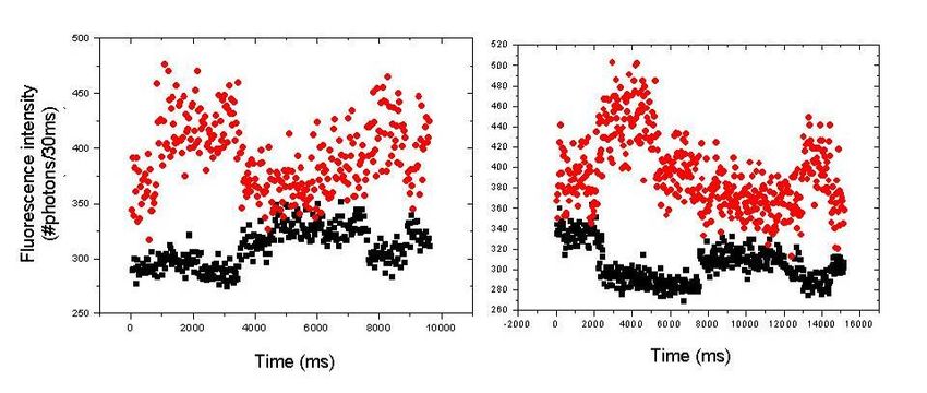
5.5.1     Cloning, protein expression and purification..............................................................- 79 -
        5.5.2     ATPase assay...............................................................................................................- 79 -
        5.5.3     Gliding assays .............................................................................................................- 80 -
        5.5.4     Laser Trapping assay ..................................................................................................- 80 -
        5.5.5     Quantum dot assays ....................................................................................................- 81 -
        5.5.6     Data analysis...............................................................................................................- 81 -
  5.6       REFERENCES ....................................................................................................................... - 82 -
Summary ............................................................................................................... - 85 -

Publications ........................................................................................................... - 88 -

Meeting Abstracts .................................................................................................. - 89 -

Curriculum Vitae .................................................................................................... - 90 -

Acknowledgements… ............................................................................................ - 92 -

                                                                       VI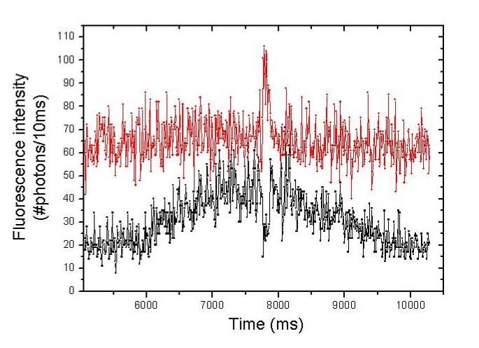
Chapter 1: Introduction

1 Introduction

1.1 Structure of this thesis

The present thesis studies the mechanism of the microtubule motor protein kinesin
using different biochemical and biophysical approaches. It is divided into five parts,
each presenting one publication-style chapter focussing on one specific approach.
The first chapter gives a general introduction into the kinesin-family of motor proteins,
as well as on the techniques used in this thesis. Chapter 2 compares a processive
and a non-processive kinesin, chapter 3 elucidates the primary mechanical event
leading to motility in conventional kinesins (Kinesin-1 subfamily). Chapter 4 presents
a biophysical study aimed at measuring the ATP turnover with microscopic methods,
chapter 5 investigates the basis of defective kinesins found in spastic paraplegia
patients. These studies thus address the mechanism of kinesin at a molecular level,
using wildtype and mutant motor proteins, as well as in vitro assays for kinesin’s
gliding and enzymatic activity.

                                          -1-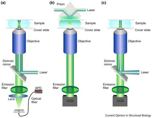
Chapter 1: Introduction

1.2 Intracellular transport

Cell motility is one of the major achievements in evolution. Primitive cells were
probably immobile, floating in the primordial soup. The innovation of directed
intracellular motion allowed cell motility. The possibility to alter the cell shape and
subcellular structures like cilia enabled the cell to direct its movement. In multicellular
organisms, migration of cells during development and in search of foreign organisms
to the defend the host against infection are required for the organisms’ health. On the
other hand, uncontrolled cell migration is a property of a malignant cancer cells.

Not only migrating cells, but also stationary cells exhibit dramatic changes in their
morphology. Striking examples are the contraction of muscle cells, the elongation of
nerve axons or the constriction of a dividing cell during mitosis. But also more subtle
movements within the cell are essential elements in growth and differentiation of cells
– active separation of chromosomes, cytoplasmic streaming and transport of
membrane vesicles. These internal movements are carefully controlled by the cell to
take place at specific times and in particular locations.

Many types of motility are based on ATP-hydrolysing enzymes that convert chemical
energy into mechanical work. This conversion is accomplished by a special class of
enzymes, so-called motor proteins. Together with the cytoskeleton, a cytoplasmic
system of fibres, they are essential for intracellular transport and thus for cell motility.
Three types of cytosolic fibres build the cytoskeleton: actin filaments (7 to 9 nm in
diameter), intermediate filaments (10 nm in diameter) and microtubules (24 nm in
diameter). These cytoskeletal fibres are well-ordered polymers built from small protein
subunits held together by noncovalent bonds. (Lodish et al., 2000). The cytoskeleton
plays a structural role by supporting the cell membrane and providing tracks along
which organelles and other elements transported by molecular motors move. Due to
their regulated, polar arrangements, cytoskeletal fibres can also produce movement
by themselves without associated molecular motors.

1.2.1 Axonal transport

A highly sophisticated form of intracellular transport is axonal transport. In the human
body axons can be up to one meter in length and numerous proteins, mRNA or even
whole organelles, need to be brought from the neuronal cell body to the synapse

                                            -2-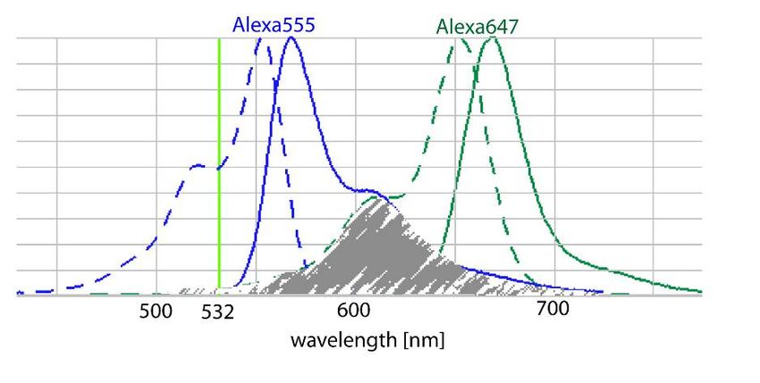
Chapter 1: Introduction

(Kandel et al., 1995). Insufficient supply of the synapse leads to neurodegeneration
representing the origin of severe neuronal diseases.

Transport in axons occurs in anterograde and in retrograde direction, and has two
major components of distinct velocity, termed fast and slow components of axonal
transport (Lasek, 1967). Anterograde transport going towards the cell periphery is
driven by different kinesin motors (1.2.3), retrograde transport by dynein. Anterograde
transport is responsible for supply of the synapse and maintenance of the structure,
whereas retrograde transport collects metabolites and pieces of membranes for
recycling in the cell body and delivers chemical messages. It is thought that vesicular
cargoes are mainly delivered by fast axonal transport, slow axonal transport is
associated with cytoskeletal and cytosolic proteins (Kandel et al., 1995). Recent
findings suggest that slow and fast components of axonal transport are driven by the
same molecular motors and that the difference in velocity is due to more frequent
stops (Brown et al., 2005).

1.2.2 Molecular motors

Not only axonal transport, but also muscle contraction, meiosis and mitosis, organelle
transport and almost every biological movement is driven by protein machines called
molecular motors. These specialized proteins convert chemical energy or ion
potentials into mechanical work. Despite this common feature they vary in their
function and structure. Figure 1.1 illustrates the structure of three different
representatives of cytoskeletal motors.

                                           -3-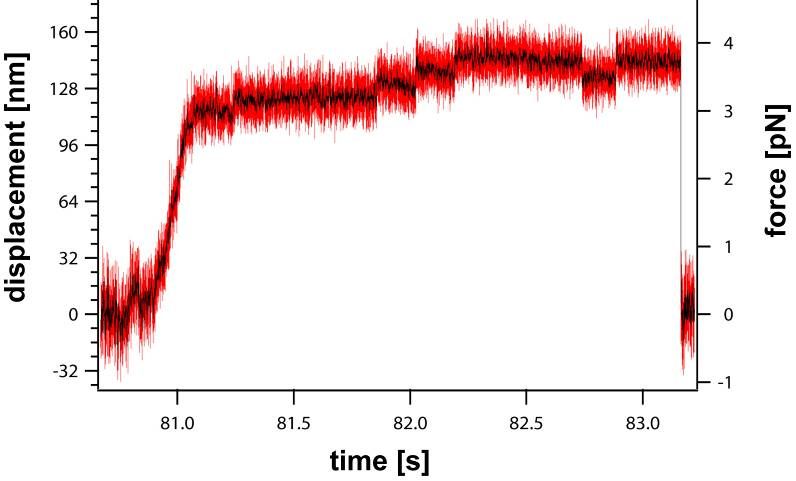
Chapter 1: Introduction

 Figure 1.1    Representatives of cytoskeletal motors.
 Kinesin-1 and cytoplasmic dynein are shown here as representatives of microtubule
 binding motors. Myosin II represents an actin-based motor. All three motors have
 dimerised heavy chains. In the case of myosin and kinesin they are joined by an
 extended coiled-coil – the stalk, shown in blue. The motor domains contain the
 catalytic sites and are shown in yellow. The associated light chains are shown in
 purple. (from Woehlke and Schliwa, 2000)

The three classes of motor proteins, namely myosins, kinesins and dyneins, use two
types of cytoskeletal filaments as tracks. Myosins interact with actin filaments,
whereas kinesins and dyneins interact with microtubules. Typically, these motors
have a globular motor domain, also referred to as “head” domain. This catalytic head
domain contains two crucial properties of a molecular motor: a site for ATP hydrolysis
and a nucleotide-dependent binding site to the track. In many cases, the globular
head is followed by an extended stalk, which dimerises via a coiled-coil structure to
yield a double-headed molecular motor (Figure 1.1). The associated polypeptides
(intermediate and light chains) differ largely for all three motor classes indicating a
broad variety of functions (Woehlke and Schliwa, 2000).

1.2.3 Kinesin

After myosin and dynein, a third force-generating ATPase, which is involved in
intracellular transport, was identified (Brady, 1985; Vale et al., 1985). Named after the
Greek word kinein (to move), kinesin was found to transport axoplasmic organelles on
microtubules. These first identified kinesins from brain tissues and from squid giant
axons belong to the Kinesin-1 family (also conventional kinesin). In some higher
vertebrates, one member of this family was found exclusively in neurons and is
therefore named neuronal kinesin heavy chain (nKHC or KIF5A) (Niclas et al., 1994).

                                           -4-
Chapter 1: Introduction
All conventional and unconventional kinesins share a high degree of sequence
similarity in their motor domains, whereas all other parts are diverse (Vale and
Fletterick, 1997). The motor domain is defined as the force-generating element of the
protein and can be C- or N-terminal. In several kinesin families, two globular motor
domains form dimers via a coiled-coil, usually termed neck. In addition to these two
domains, many kinesin proteins contain a long coiled-coil domain termed stalk.
Finally, there is often a globular domain at the C-terminus, termed tail domain. It
usually has a regulatory function and binds to cargos or light chains.
Figure 1.2 Domain organisation of conventional kinesin.
The motor domain and the neck (yellow) are overlaid by the crystal structure. The
stalk consists of coiled-coil structures and flexible regions, whereas the tail region is
globular and binds to the cargo or light chains. (from Woehlke and Schliwa, 2000)
The two heavy chains of conventional kinesin are twice as heavy as the two
associated light chains (110-140 kD versus 60-80 kD) (Bloom et al., 1988; Kuznetsov
et al., 1988). Only the heavy chains are required for kinesin´s motility, whereas the
light chains have regulatory and cargo-binding function (Stenoien and Brady, 1997;
Verhey et al., 1998). The domain organisation of heavy chains is shown schematically
in Figure 1.2. At the N-terminus, ~320 amino acids form the motor domain. It contains
the microtubule and nucleotide binding sites and its three-dimensional structure has
been solved (Kull et al., 1996; Woehlke et al., 1997). The neck-linker joins the N-
terminal motor domain with the C-terminal coiled-coil neck. Depending on the bound
nucleotide, the neck-linker adopts different positions relative to the catalytic core
(Rice et al., 1999). The neck-domain and the approximately 50 nm long coiled-coil
stalk domain are joined by a flexible hinge domain. The stalk is interrupted by a
second flexible region, called the kink. The kink allows the molecule to bend in a way
-5-Chapter 1: Introduction
that the C-terminally globular tail domain comes close to the motor domain and
inhibits its ATPase activity. If the tail binds a cargo directly or via light chains the
inhibition is repealed (Adio et al., 2006; Seiler et al., 2000).
1.2.4 The chemo-mechanical cycle
The first identified kinesin (conventional kinesin, KIF5B, Kinesin-1 or uKHC) is the
best studied member of the kinesin superfamily and is considered prototypic for the
entire superfamily. Today, the chemo-mechanical cycle of kinesin is resolved in great
detail and some key questions are still to be solved. Figure 1.3 shows a consensus
kinetic model of the chemo-mechanical cycle (Cross, 2004; Valentine and Gilbert,
2007).
Conventional kinesin can walk distances up to 1 µm along microtubules without
detaching (Howard et al., 1989). This phenomenon is called processivity and as a
consequence, at least one head of the dimeric motor always remains attached to the
microtubule. Each step is coupled to the hydrolysis of one ATP molecule (Hua et al.,
1997; Schnitzer and Block, 1997). With each step the center of the molecule is
displaced 8 nm along the microtubule, representing the distance between adjacent
tubulin dimers (Svoboda et al., 1993). A prerequisite for processive movement is the
precise coordination of the chemo-mechanical cycles of both heads. A commonly
accepted model is the “hand-over-hand” model (Asbury et al., 2003; Kaseda et al.,
2003; Schief et al., 2004; Yildiz et al., 2004) (Figure 1.3). Here, one head passes the
other and binds to the next microtubule binding site. Therefore, one head takes a 16
nm step, but the centre of mass is displaced 8 nm per step. During one cycle the
heads switch between strong and weak microtubule binding states in a nucleotide-
dependent manner (Gilbert et al., 1998; Hackney, 1994; Ma and Taylor, 1997).
In solution, a kinesin dimer contains one ADP per head. Upon microtubule binding,
only one head locks onto the microtubule and loses its ADP. If ATP is bound to this
head, the neck-linker docks (Rice et al., 1999) and brings the second head into a
favourable position to bind the next microtubule binding site. Microtubule binding
takes place after the ADP release (Sablin and Fletterick, 2001). In this intermediate
state, both heads are bound to adjacent tubulin binding sites. The rear head
hydrolyses the nucleotide and after the release of inorganic phosphate it detaches
from the microtubule while the other head holds on. At this point, the heads have
exchanged their roles and a new chemo-mechanical cycle starts.
-6-Chapter 1: Introduction
Figure 1.3 Chemo-mechanical cycle of Kinesin-1
This sequence of events summarises the chemo-mechanical cycle of one kinesin
head (see text for details). Kinesin is shown in black/grey; microtubules in blue;
abbreviations for nucleotides bound to kinesin heads: ATP (T); ADP (D); phosphate
(P); no nucleotide (NN)
This is a somewhat oversimplified model, however, as many aspects like force, strain
or conformational changes are not taken into account. Most of the data is obtained
from pre-steady state kinetics and bulk experiments, where the values are averaged
over many molecules without evaluating the quality of single ones (e.g. dead motors).
Recently developed techniques allow to measure speeds, forces and kinetics of
single molecules. A variety of these techniques was employed in this thesis and will
be described in the next section.
-7-Chapter 1: Introduction
1.3 Optical techniques
To overcome the boundaries of temporal and spatial resolution is the major driving
force for the development of highly sophisticated optical techniques. The limit of
spatial resolution of a light microscope is set by the wavelength of visible light. The
best resolution one can get with a conventional light microscope is to distinguish
objects, which are 0.2 µm apart. Due to this fact it is hard to image subcellular
structures and various approaches like electron microscopy have been developed to
overcome this restriction.
Until today, we are not at the limit of all four dimensions (time and space), although it
is possible to image single molecules with a sub-millisecond time resolution
(Verbrugge et al., 2007). A big step towards higher spatial resolution was the
invention of fluorescent dyes (Coons and Kaplan, 1950). Here, it is possible to
illuminate only the subject of interest by labelling it with a fluorescent dye, which is
then purposely exited and detected with a microscope. Depending on the
requirements different fluorescent microscopes can be used (Figure 1.4). The basic
epifluorescence microscope illuminates the whole sample and reduces the
background only by using an emission filter.
Figure 1.4 Different types of fluorescent microscopes.
Based on an epifluorescent microscope (c), the resolution can be improved by
minimising the excitation volume. The most popular one is the confocal microscope
(a), where the excitation volume is reduced to a single spot, which allows scanning the
sample in three dimensions and rebuilding the pictures digitally. Another approach is
called “Total Internal Reflection Fluorescence” or TIRF microscopy (b), where the
excitation volume is only a thin layer. The reduction of the excitation volume leads to
less unspecific background fluorescence and therefore to a better resolution of the
specific fluorescent signal. (from Haustein and Schwille, 2004)
-8-Chapter 1: Introduction
1.3.1 TIRF microscopy
TIRF microscopy uses the emerging evanescent wave due to the reflection of a laser
beam at the glass-water boundary.
The incident laser beam is directed in a supercritical angle to the glass surface, where
it is reflected (Figure 1.5). For the reflection it is necessary that the immersion oil has
a similar refractive index as the glass and that the medium of the sample has a lower
refractive index than the glass. With these two prerequisites the only possible area of
reflection is the glass-water boundary. Due to the reflection an evanescent wave
emerges and penetrates the sample, but its intensity decays exponentially with the
distance to the glass-water boundary. Therefore the evanescent wave can only excite
fluorophores at close proximity to the glass surface. Depending on the angle, the
refractive indices and the wavelength of the laser beam, the excited layer is between
70 and 300 nm deep and usually 150-200 nm (Schneckenburger, 2005).
Figure 1.5 Principle of TIRF microscopy.
The incident laser is directed in a supercritical angel to the glass-water boundary,
where it is reflected. The intensity of the emerging evanescent wave decays
exponentially with the depth. The refractive index of the culture medium n2 has to be
smaller than the refractive index of the glass and immersion oil n1. (from Sako and
Uyemura, 2002)
This exciting layer is fixed to the surface and can not be moved in the third dimension,
but allows 2D imaging at high time resolution and low background. These properties
are used to image specifically adhesion structures of cells or anything else occurring
only at the glass surface.
-9-Chapter 1: Introduction
TIRF microscopes are usually available as a prism-type TIRF or an objective-type
TIRF. They differ in the way how the incident laser beam is brought to the
supercritical angel. The prism-type TIRF directs the laser through a prism, where it is
broken in the right angle to reflect at the glass surface. In this microscope the
excitation and emission path is separated and there is no access to the sample during
imaging.
The objective-type TIRF directs the incident laser through an objective with a
numerical aperture higher than 1.38 N.A. (Sako and Uyemura, 2002). This is
important, because the laser passes the objective off-axis and is refracted by the lens
of the objective. The higher the numerical aperture, the stronger the refraction and
only a strong refraction can bring the laser beam into a supercritical angel (Figure
1.6).
Figure 1.6 Objective-type TIRF microscope.
The incident laser beam is directed through the objective off-axis. Therefore it is
refracted by the high numerical aperture lens and brought into a supercritical angel to
the glass-water boundary. The reflected laser beams back through the objective to the
dichroic mirror, which blocks the detection part from excitation light. The evanescent
wave emerges from the glass water-boundary and excites a thin layer of the sample.
(from Sako and Uyemura, 2002)
The advantage of this type of TIRF microscope is that the excitation and emission
path is on the same side of the microscope and the sample is accessible during
imaging, e.g. for changing the medium or adding reagents.
In this thesis an objective-type TIRF was used to reduce the background in
microscopic assays and allowed to image single kinesins, site specific labelled with a
fluorophore.
- 10 -Chapter 1: Introduction
1.3.2 Confocal microscopy
Another approach to enhance resolution involves a confocal microscope (Egger and
Petran, 1967). Here, the excitation volume is reduced to a small spot, which
originates from a focussed laser beam. In a conventional confocal microscope a
scanning device can position this spot in three dimensions. A photomultiplier detects
the intensity of fluorescence in the spot and after scanning a special region in the
sample the image can be assembled digitally. In front of the photomultiplier a pinhole
cuts off emission light from regions out of focus. The special confocal microscope
used in this thesis was build by Sander Verbrugge from the Vrije Universiteit in
Amsterdam ((Verbrugge et al., 2007);Figure 1.7). Here, the scanning device does not
move the confocal spot, but the sample with a scanning table. Therefore, the spot is
very stable and well defined. This setup was built to measure the change of
fluorescence intensity in one spot over time rather than to scan whole images.
The emission light is detected by a highly sensitive single-photon-counting avalanche
photodiode. Such a detector counts the time between photon arrivals with
nanosecond accuracy. The number of photons arrived within a defined time (binning
time) is summed up and displayed on the screen. Thus, change of intensity in the
spot is detected while the measurement ongoing.
- 11 -Chapter 1: Introduction
Figure 1.7 Confocal setup at the VU Amsterdam.
This confocal setup was build by Sander Verbrugge (Verbrugge et al., 2007) at the
Vrije Universiteit Amsterdam. The excitation part (red circle) is open and can be fine-
tuned during measurements. The emission part (green circle) is shielded from light.
The sample holder and scanner are constructed into a Nikon microscope corpus.
In the present thesis this techniques was used to measure intensity fluctuations of a
fluorophore-labelled kinesin stepping through the spot. The intensity fluctuations were
due to “Förster-Resonance-Energy-Transfer” or FRET, which is explained in more
detail in the next subsection.
1.3.3 FRET
FRET is a technique used in fluorescent microscopy or spectroscopy to elucidate the
interaction of two molecules. The prerequisites for FRET are a donor fluorophore with
an emission spectrum that overlaps with the absorption spectrum of an acceptor
fluorophore, and a close proximity of donor and acceptor dye ((Lakowicz, 1999);
Figure 1.8).
- 12 -Chapter 1: Introduction
Figure 1.8 Overlapping spectra of donor and acceptor fluorophores.
The excitation (dashed lines) and emission (solid lines) spectra of Alexa555 and
Alexa647 fluorophores as an example of a FRET pair. The overlap of the emission
spectra of the donor (Alexa555, blue) and the excitation spectra of the acceptor
(Alexa647, green) is hatched in grey. The integral of this overlap is deciding for the
Förster radius R0. (Spectra from Invitrogen SpectraViewer)
The efficiency of the energy transfer from one donor to the acceptor is described by
the Förster equations, which is dependent on the Förster radius (R0) (Förster, 1948)
equation(1)).
(1)
The Förster radius is the distance at which the efficiency is 50% and is usually 1 to 10
nm. Depending on the overlapping integral of the two spectras the Förster radius is
specific for every FRET pair. Common FRET pairs are Cy2 and Cy3, YFP and GFP or
GFP and rhodamine.
The protein of interest can be tagged N- or C-terminally, and co-expressed with the
fluorescent proteins (YFP, GFP etc.). This makes labelling procedures unnecessary
and ensures a one to one labelling stoichiometry. The disadvantages are the poor
spectroscopic properties of these fluorescent proteins, in particular low quantum yield
and blinking. Therefore it is recommended to use organic dyes that are more stable
and have high quantum yields. This is especially important for single molecule
applications.
- 13 -Chapter 1: Introduction
For the detection of FRET always the donor is excited and the emission of the donor,
the acceptor or both can be measured. In case of donor emission, the fluorescent
intensity decays if FRET occurs. On the other hand, if the acceptor emission is
measured it is enhanced as soon as the energy is transferred from the donor (Figure
1.9).
Figure 1.9 Principle of FRET
The protein is labelled with the donor fluorophore (green) and the nucleotide is
labelled with the acceptor fluorophore (red). The donor is excited directly with a laser
beam. If the nucleotide binds to the protein, the two fluorophores come in close
proximity to each other and the acceptor is excited via FRET.
In this thesis FRET was used to measure the binding time of fluorescent ATP to the
site specific labelled kinesin. The energy transfer was measured in bulk in a
fluorimeter and on single molecules in a confocal setup.
1.3.4 Optical tweezers
Optical tweezers, also known as optical traps, are instruments that are usually
implemented in a conventional light microscope. This instrument allows manipulating
small colloidal particles with a strongly focussed laser beam. Since its invention by
Arthur Ashkin (1986), optical traps are broadly used in medicine and science. They
allow the direct manipulation of viruses and cells as well as cell compartments in
living cells (Ashkin and Dziedzic, 1987; Sheetz and Kuo, 1993) without destroying the
examined biological system. With optical tweezers it was possible to measure the
force producing properties of single molecular motors for the first time by adsorbing
them at low densities to dielectric microspheres, which were then manipulated by the
tweezer (Block et al., 1990),Figure 1.10).
- 14 -Chapter 1: Introduction
Figure 1.10 Kinesin in an optical trap.
The polystyrene bead (grey) with attached kinesin molecule (black) is trapped in the
focussed laser beam (red). Brought in contact with the microtubule (blue) the kinesin
starts to walk along its track and pulls the bead out of the trap. The back pulling force
is directly proportional to the displacement of the bead.
The possibility to measure forces between one and several hundred piconewton and
to detect movements on sub-nanometer scales makes optical tweezers a perfect
instrument for investigating force generating proteins, like kinesin. The optical
tweezers used in this thesis were built by Dr. Anabel Clemen and later on supervised
by Dr. Johann Jaud from the Physik Department, Lehrstuhl E22 für Biophysik,
Technische Universität München. The motor protein is adsorbed to a polystyrol bead,
which can be trapped and manipulated with optical tweezers. After trapping, the bead
is brought to surface-attached microtubules, where kinesins can bind and start to walk
(Figure 1.10.) The microtubules are labelled with fluorophores and can be detected
with a coupled fluorescent microscope.
- 15 -Chapter 1: Introduction
1.4 Outline of the present thesis
This thesis consists of a collection of published and submitted research articles. They
all have one common theme: the biochemical and biophysical properties of kinesin
motors. The methods used in this present work vary from kinetic ensemble to single-
molecule measurements and focus on fundamental properties of human and fungal,
processive and non-processive kinesins.
Chapter 2 deals with questions about processivity. What makes a motor
processive? What are the structural determinants? And is it possible to transfer them
to an unprocessive motor? To investigate these questions the dimeric but
unprocessive motor NKin3 from the filamentous fungus Neurospora crassa was
compared with its processive counterpart NcKin. The processivity of chimaeras with
swapped motor domains were examined in various in vitro assays. A meaningful
assay to determine the processivity is the filament-gliding assay. But the average
runlength can only be determined in single-molecule assays, which were performed in
a TIRF microscope.
Chapter 3 addresses the consequences of neck-linker docking in the chemo-
mechanical cycle. The neck-linker docking is essential for the functionality of kinesin
(Rice et al, 1999). But how does the neck-linker promote the stepping of the motor?
This question was investigated with NcKin mutants possessing an artificial disulfide
bridge between the neck-linker and the motor core. This disulfide bridge can be
switched on or off reversibly by oxidation and reduction. These mutants have different
properties depending on the particular position where the disulfide bridge formed.
Apart from their filament-gliding activity, their affinity to microtubules is altered. This
was investigated with a microtubule co-sedimentation assay.
Chapter 4 describes the most challenging project of this thesis. The aim was to
measure the binding times of single ATP molecules to a single kinesin while it is
walking along a microtubule. The method of choice was FRET. Specially designed
human kinesin constructs were labelled with a donor fluorophore next to the ATP
binding site. ATP was labelled with an acceptor fluorophore. The occurrence of FRET
was shown in a spectroscopic bulk experiment, measurements on a single molecule
level were performed with a special confocal microscope in collaboration with the
Vrije Universiteit Amsterdam. This setup is highly sensitive and measures with sub-
millisecond accuracy.
- 16 -Chapter 1: Introduction
Chapter 5 characterises the defects of human neuronal kinesin (KIF5A) mutants
linked to the neurodegenerative disease hereditary spastic paraplegia (HSP).
Which properties are altered in these mutants in comparison to the wildtype protein?
Why do patients develop the disease, although they still have one wildtype allel of the
gene? To answer this question a novel cargo transportation assay was developed,
where mixtures of heterodimeric and homodimeric kinesins work in concert. This
mimics the axonal transport in heterozygous patients and gives an insight why this
disease emerges.
1.5 References
Adio S, Reth J, Bathe F, Woehlke G. 2006. Review: regulation mechanisms of
Kinesin-1. J Muscle Res Cell Motil:1-8.
Asbury CL, Fehr AN, Block SM. 2003. Kinesin moves by an asymmetric hand-over-
hand mechanism. Science 302(5653):2130-2134.
Ashkin A, Dziedzic JM. 1987. Optical trapping and manipulation of viruses and
bacteria. Science 235(4795):1517-1520.
Ashkin A, Dziedzic JM, Bjorkholm JE, Chu S. 1986. Observation of a single-beam
gradient force optical trap for dielectric particles. Optics Letters 11:288-290.
Block SM, Goldstein LS, Schnapp BJ. 1990. Bead movement by single kinesin
molecules studied with optical tweezers [see comments]. Nature
348(6299):348-352.
Bloom GS, Wagner MC, Pfister KK, Brady ST. 1988. Native structure and physical
properties of bovine brain kinesin and identification of the ATP-binding subunit
polypeptide. Biochemistry 27(9):3409-3416.
Brady ST. 1985. A novel brain ATPase with properties expected for the fast axonal
transport motor. Nature 317(6032):73-75.
Brown A, Wang L, Jung P. 2005. Stochastic Simulation of Neurofilament Transport in
Axons: The "Stop-and-Go" Hypothesis. Mol Biol Cell 16(9):4243-4255.
Coons AH, Kaplan MH. 1950. Localization of antigen in tissue cells; improvements in
a method for the detection of antigen by means of fluorescent antibody. J Exp
Med 91(1):1-13.
Cross RA. 2004. The kinetic mechanism of kinesin. Trends Biochem Sci 29(6):301-
309.
Egger MD, Petran M. 1967. New reflected-light microscope for viewing unstained
brain and ganglion cells. Science 157(786):305-307.
Förster T. 1948. Zwischenmolekulare Energiewanderung und Fluoreszenz. Annalen
der Physik 437(1):55-75.
- 17 -Chapter 1: Introduction
Gilbert SP, Moyer ML, Johnson KA. 1998. Alternating site mechanism of the kinesin
ATPase. Biochemistry 37(3):792-799.
Hackney DD. 1994. The rate-limiting step in microtubule-stimulated ATP hydrolysis by
dimeric kinesin head domains occurs while bound to the microtubule. J Biol
Chem 269(23):16508-16511.
Haustein E, Schwille P. 2004. Single-molecule spectroscopic methods. Curr Opin
Struct Biol 14(5):531-540.
Howard J, Hudspeth AJ, Vale R. 1989. Movement of microtubules by single kinesin
molecules. Nature 342(Nov. 9):154-158.
Hua W, Young EC, Fleming ML, Gelles J. 1997. Coupling of kinesin steps to ATP
hydrolysis. Nature 388(6640):390-393.
Kandel ER, Schwartz JH, Jessell TM. 1995. Neurowissenschaften.
Kaseda K, Higuchi H, Hirose K. 2003. Alternate fast and slow stepping of a
heterodimeric kinesin molecule. Nat Cell Biol 5(12):1079-1082.
Kull FJ, Sablin EP, Lau R, Fletterick RJ, Vale RD. 1996. Crystal structure of the
kinesin motor domain reveals a structural similarity to myosin [see comments].
Nature 380(6574):550-555.
Kuznetsov SA, Vaisberg EA, Shanina NA, Magretova NN, Chernyak VY, Gelfand VI.
1988. The quaternary structure of bovine brain kinesin. Embo J 7(2):353-356.
Lakowicz J. 1999. Principles of Fluorescence Spectroscopy.
Lasek RJ. 1967. Bidirectional transport of radioactively labelled axoplasmic
components. Nature 216(5121):1212-1214.
Lodish H, Berk A, Zipursky SL, Matsudaira P, Baltimore D, Darnell JE. 2000.
Molecular Cell Biology. 4th ed.
Ma YZ, Taylor EW. 1997. Interacting head mechanism of microtubule-kinesin
ATPase. J Biol Chem 272(2):724-730.
Niclas J, Navone F, Hom-Booher N, Vale RD. 1994. Cloning and localization of a
conventional kinesin motor expressed exclusively in neurons. Neuron
12(5):1059-1072.
Rice S, Lin AW, Safer D, Hart CL, Naber N, Carragher BO, Cain SM, Pechatnikova E,
Wilson-Kubalek EM, Whittaker M, Pate E, Cooke R, Taylor EW, Milligan RA,
Vale RD. 1999. A structural change in the kinesin motor protein that drives
motility. Nature 402(6763):778-784.
Sablin EP, Fletterick RJ. 2001. Nucleotide switches in molecular motors: structural
analysis of kinesins and myosins. Curr Opin Struct Biol 11(6):716-724.
Sako Y, Uyemura T. 2002. Total internal reflection fluorescence microscopy for
single-molecule imaging in living cells. Cell Struct Funct 27(5):357-365.
- 18 -Chapter 1: Introduction
Schief WR, Clark RH, Crevenna AH, Howard J. 2004. Inhibition of kinesin motility by
ADP and phosphate supports a hand-over-hand mechanism. Proc Natl Acad
Sci U S A.
Schneckenburger H. 2005. Total internal reflection fluorescence microscopy:
technical innovations and novel applications. Curr Opin Biotechnol 16(1):13-
18.
Schnitzer MJ, Block SM. 1997. Kinesin hydrolyses one ATP per 8-nm step. Nature
388(6640):386-390.
Seiler S, Kirchner J, Horn C, Kallipolitou A, Woehlke G, Schliwa M. 2000. Cargo
binding and regulatory sites in the tail of fungal conventional kinesin. Nat Cell
Biol 2(6):333-338.
Sheetz MP, Kuo SC. 1993. Tracking nanometer movements of single motor
molecules. Methods Cell Biol 39:129-136.
Stenoien DL, Brady ST. 1997. Immunochemical analysis of kinesin light chain
function. Mol Biol Cell 8(4):675-689.
Svoboda K, Schmidt CF, Schnapp BJ, Block SM. 1993. Direct observation of kinesin
stepping by optical trapping interferometry [see comments]. Nature
365(6448):721-727.
Vale RD, Fletterick RJ. 1997. The design plan of kinesin motors. Ann Rev Cell Dev
Biol 13:745-777.
Vale RD, Reese TS, Sheetz MP. 1985. Identification of a novel force-generating
protein, kinesin, involved in microtubule-based motility. Cell 42(1):39-50.
Valentine MT, Gilbert SP. 2007. To step or not to step? How biochemistry and
mechanics influence processivity in Kinesin and Eg5. Curr Opin Cell Biol
19(1):75-81.
Verbrugge S, Kapitein LC, Peterman EJ. 2007. Kinesin moving through the spotlight:
single-motor fluorescence microscopy with submillisecond time resolution.
Biophys J 92(7):2536-2545.
Verhey KJ, Lizotte DL, Abramson T, Barenboim L, Schnapp BJ, Rapoport TA. 1998.
Light chain-dependent regulation of Kinesin's interaction with microtubules. J-
Cell-Biol 143(4):1053-1066 issn: 0021-9525.
Woehlke G, Ruby AK, Hart CL, Ly B, Hom-Booher N, Vale RD. 1997. Microtubule
interaction site of the kinesin motor. Cell 90(2):207-216.
Woehlke G, Schliwa M. 2000. Walking on two heads: the many talents of kinesin. Nat
Rev Mol Cell Biol 1(1):50-58.
Yildiz A, Tomishige M, Vale RD, Selvin PR. 2004. Kinesin walks hand-over-hand.
Science 303(5658):676-678.
- 19 -Chapter 2: Molecular determinants of processivity in kinesin
2 Molecular determinants of processivity in
kinesin
2.1 Abstract
The protein family of kinesins can be divided in processive and non-processive
motors. Processive motors have the ability to move along microtubules in a stepwise
fashion without detaching. Non-processive motors detach after every cycle of ATP
hydrolysis. To find out which parts of kinesin are required for coupling of kinesin’s two
motor heads, chimaeras with swapped domains of the processive Neurospora crassa
kinesin (NcKin) and its non-processive counterpart NcKin3 were tested for their motile
properties. The chimaera with the NcKin motor domain and the NcKin3 neck/stalk
portion moved processively along microtubules although with a significantly
decreased average runlength, suggesting a perturbing effect of the non-processive
neck. The reverse chimaera containing motor domains of the non-processive kinesin
NcKin3 was unable to perform processive movement, despite the presence of the
Kinesin-1 neck coiled-coil. These observations suggest that determinants of
processivity are in the motor core and that this processive movement is enhanced by
the neck. The present thesis contributed the characterisation of multiple and single-
molecule motility assays to the study.
- 20 -Chapter 2: Molecular determinants of processivity in kinesin
2.2 Introduction
Conventional kinesin is the founding member of the kinesin superfamily and belongs
to the class of Kinesin-1 motors (Lawrence et al., 2004; Vale et al., 1985). Its
stepwise movement over long distances without detachment from the filament is one
of its astonishing properties. This processive behaviour can be explained by the
hand-over-hand model, which is now in principle commonly accepted (Hackney,
1995; Ma and Taylor, 1997; Yildiz et al., 2004). Here, the movement is based on the
alternated binding of the two motor heads to the following microtubule binding site.
The rear head does not detach from the microtubule before the front head has bound
tightly. This mechanism requires precisely coordinated microtubule affinities of the
two motor domains. Otherwise, the motor would dissociate from the microtubule in
between steps, or stick to the filaments when both motor domains are in a strong
microtubule binding state (Crevel et al., 2004; Cross, 2004; Hackney, 1994; Howard
et al., 1989).
This remarkably fine-tuned coordination is supported by several structural and kinetic
properties. Some models predict that intramolecular strain between leading and
trailing head builds up and controls coordination (Guydosh and Block, 2006; Hancock
and Howard, 1999; Rosenfeld et al., 2003). However, the size of this force is unclear
and hard to measure, and thus the precise mechanism how strain is linked to motor
kinetics is unknown.
To locate structural determinants involved in processive head-head coordination, we
made use of chimaeric kinesins of a processive, conventional Kinesin-1 motor, and a
recently discovered dimeric Kinesin-3 motor that is not processive but also moves to
the microtubule plus-end (Adio et al., 2006). This is in contrast to previous studies
which investigated chimaeras with the non-processive, minus-end directed kinesin
Ncd. These studies found, that the directionality is not determined by the motor
domain, but it influences the processivity of the motor (Case et al., 1997; Henningsen
and Schliwa, 1997; Hirose et al., 2000). The present question is, whether the
processivity is also influenced if the motor domains have the same directionality and
whether it is possible to transfer processivitv to a non-processive motor.
- 21 -Chapter 2: Molecular determinants of processivity in kinesin
2.3 Results
2.3.1 Design of chimaeras
Chimaeras with swapped motor domains of the fungal kinesins NcKin and NcKin3
served as experimental models for the present study (Figure 2.1). NcKin is a
‘conventional’ processive Kinesin-1, whereas NcKin3 is a non-processive Kinesin-3
(Adio et al., 2006; Jaud et al., 2006; Lakamper et al., 2003). One construct contained
NcKin’s motor head attached to the neck of NcKin3 (Head1/Neck3). The reverse
chimaera contained the motor head of NcKin3 joined with the neck of NcKin
(Head3/Neck1). The fusion points were chosen between the neck-linker and the neck
coiled-coil. We then asked which part of the motor was necessary and sufficient for
processive movement.
Figure 2.1 Design of chimeric kinesins.
The motor domain and neck-linker of the processive Kinesin-1 motor (amino acids 1-
342) was connected to the coiled-coil and tail domain of the non-processive Kinesin-3
motor (amino acids 428 to 558). This construct was named Head1/Neck3 chimaera.
Vice versa, the Head3/Neck1 chimaera was generated by fusing the Kinesin-3 motor
domain and linker region (amino acids 1-429) to the coiled coil of the NcKin protein
(amino acids 340-433).
2.3.2 Co-operative gliding behaviour
In multi-motor gliding assays, motors were attached to the coverslip, and fluorescently
labelled microtubules were transported over the surface. The velocity of microtubule
transport is an indirect measurement for the velocity of the motors. In general, the
coating density of motors on a coverslip influences the microtubule gliding velocity,
and only in the special case of processive motors the velocity is independent of the
motor density (Howard et al., 1989). While non-processive kinesins work in a
cooperative manner, a single processive kinesin displaces microtubules at the same
- 22 -Chapter 2: Molecular determinants of processivity in kinesin
speed as multiple kinesins. Thus, the dependence on the coating density is the first
hint if a motor is processive or not. The Head1/Neck3 chimaera showed similar
gliding velocities at high and low coating density to wildtype NcKin (1.48 ± 0.01 µm/s
at 1.6 µM and 1.61 ± 0.03 µm/s at 0.3 µM). In contrast, gliding velocity of the NcKin3
reference construct was accelerated at higher motor densities (Adio et al., 2006). The
reverse chimaera Head3/Neck1 was not accelerated by higher motor coating
densities. In fact, gliding velocity decreased from 0.58 ± 0.04 µm/s at low densities to
0.45 ± 0.01 µm/s at high densities, indicating mutual hindrance of the motors.
To further investigate the processivity of the kinesin mutants, we observed
microtubule transport by a single motor. Therefore, the motor density was decreased
to the point, where a microtubule can only be bound by one kinesin. Under these
conditions the microtubule can pivot around its anchoring point during displacement
and is never displaced further than its own length (Figure 2.2 A). The observation of
this special behaviour provides strong evidence for processivity.
Figure 2.2 Pivoting in the conventional gliding assay.
(A) Attachment points of kinesins are indicated in red, moving direction of the
microtubules with red arrows. If only one kinesin binds the microtubule, the direction of
movement pivots around this point. (B) Gliding assay with Head1/Neck3 chimaera
shows pivoting around one point. The microtublule is only displaced its own length.
Scale bar 5 µm.
- 23 -Chapter 2: Molecular determinants of processivity in kinesin
Both NcKin and the Head1/Neck3 chimaera showed characteristic pivoting behaviour
(Figure 2.2 B), suggesting that they are processive enzymes. Pivoting at low motor
densities was never observed for NcKin3 and the Head3/Neck1 chimaera.
Additionally, microtubules moved further than their own length, indicating, that more
than one kinesin is necessary to displace a microtubule. It is therefore most likely that
NcKin3 and the Head3/Neck1 chimaera are non-processive motors.
2.3.3 Single-molecule behaviour
To directly prove processivity of the Head1/Neck3 chimaera, we tested the mutants
along with wildtype references in single-molecule motility assays. Fluorescently
labelled kinesin motors were observed over time in a TIRF-microscope to detect
processive runs. Similar to the NcKin wildtype reference, the Head1/Neck3 mutant
moved continuously along microtubules (Figure 2.3) at a velocity of 1.40 ± 0.03 µm/s
(mean ± S.E.) and over distances of up to 3 µm (Table I, Figure 2.3).
Figure 2.3 Single-molecule properties of the Head1/Neck3 chimaera.
(A) Kymographs show displacements of a single fluorophore-labelled Head1/Neck3
motors along the microtubule. (B) Wildtype NcKin was used as a control. Isolated
chimeric motors performed processive runs on a microtubule with a velocity
essentially identical to the average velocity under multiple motor conditions. Motors
were observed for 5 sec with an integration time of 200 ms at an ATP concentration of
20 µM.
To quantify the extend of processivity, the runlength of the chimeric kinesins was
measured. For that purpose the histogram containing the runlength of 74 individual
motors was analysed by single exponential curve fitting. The runlength of the
- 24 -Chapter 2: Molecular determinants of processivity in kinesin
Head1/Neck3 chimaera was 0.67 ± 0.13 µm which is 2-3-fold shorter than for wildtype
NcKin (~1.8 µm, (Lakamper et al., 2003)) (Table I,Figure 2.4).
Figure 2.4 Runlenght of the Head1/Neck3 chimeara.
The runlenghts of the Head1/Neck3 chimaera was plotted in a histogram and fitted
with an exponential decay (0.67 ± 0.13 µm). Short runlengths are probably
underestimated due to limited time resolution (200 ms integration time).
The reverse Head3/Neck1 chimaera never showed any processive runs under single
molecule conditions. Together with the observations from multi-motor gliding assays
we can state that this motor is non-processive. Our data show that the motor domain
of processive NcKin is able to induce processive behaviour in otherwise non-
processive NcKin3. Conversely, the results show that the neck of NcKin3 is capable
to join two kinesin motor domains in a way that promotes processive motility, albeit to
reduced extent compared to the NcKin wildtype control.
- 25 -Chapter 2: Molecular determinants of processivity in kinesin
Table I: Gliding velocities and runlengths of chimeric and wild type
motors
Velocity [µm/s] Velocity [µm/s] Runlength [µm]
Construct
(multi-motor) (single-molecule) (single-molecule)
NcKin3
0.52 ± 0.04 n/a n/a
(Adio et al., 2006)
0.58 ± 0.04
Head3/Neck1 n/a n/a
n=90
1.75 ± 0.09
2.29 ± 0.01 2.38 ± 0.01 (Lakamper and
NcKin
n=60 n=16
Meyhofer, 2005)
1.60 ± 0.02 1.40 ± 0.03 µm/s 0.67 ± 0.13 µm
Head1/Neck3
n=120 n=80 n=74
n/a: not applicable
2.4 Discussion
The chimaeras showed that on one hand, the motor domain determines the
processivity and on the other hand, that the stalk/neck domain can influence the
grade of processivity. The Head1/Neck3 had some structural elements which were
sufficient to transfer the ability to step continuously into a non-processive background.
These structural elements are obviously absent in the reverse construct,
Head3/Neck1, which is unable to move processively, despite its Kinesin-1 neck
domain. These findings are in good agreement with data obtained from chimaeras
with the non-processive Ncd (Case et al., 1997). Transferring the motor domain of
Ncd to a processive motor also yielded in a non-processive motor. Taken together, it
does not matter which directionality or processivity the motor domain had as a
wildtype protein, the structural elements for processivity lie within the motor domain.
In addition to the information on motor domain, our study also reveals important
features of kinesin’s neck. In agreement with previous studies, the Head1/Neck3
construct (lacking the conventional neck-domain) shows severely altered processivity.
Mutational analysis of the conventional kinesin neck suggested that passive
mechanical features (stiffness, in particular) affect the runlengths of processive
kinesins, and is optimised in wildtype (Jaud et al., 2006). The Head1/Neck3 chimaera
showed a slower velocity and diminished runlength, indicating that the likelihood of
- 26 -You can also read