In Human T Cells Dissecting the Tio-NF-κB Signalosome
←
→
Page content transcription
If your browser does not render page correctly, please read the page content below
Dissecting the Tio-NF-κB Signalosome
           in Human T Cells

    Aufklärung des Tio-NF-κB Signalosoms
                in humanen T-Zellen

            Der Naturwissenschaftlichen Fakultät
    der Friedrich-Alexander-Universität Erlangen-Nürnberg
                             zur
          Erlangung des Doktorgrades Dr. rer. nat.

                        vorgelegt von
                    Sarah Jill de Jong
                    aus Seeheim-Jugenheim
Als Dissertation genehmigt von der Naturwissenschaftlichen Fakultät
der Universität Erlangen-Nürnberg.

Tag der mündlichen Prüfung:               11. August 2011

Vorsitzender der Prüfungskommission:      Prof. Dr. Rainer Fink

Erstberichterstatter:                     Prof. Dr. Lars Nitschke

Zweitberichterstatter:                    Prof. Dr. Bernhard Fleckenstein
Man muss sich Beeilen, Wenn man noch etwas Sehen will . . .
Alles Verschwindet!

                                      Paul Cézanne (1839-1906)
Contents
Summary . . . . . . . . . . . . . . . . . . . . . . . . . . . . . . . . . . . . . . . . . . . . vi
Zusammenfassung . . . . . . . . . . . . . . . . . . . . . . . . . . . . . . . . . . . . . . . vii
1 Introduction 1
1.1 Nuclear Factor of KappaB (NF-κB) . . . . . . . . . . . . . . . . . . . . . . . . . . 1
1.1.1 Activation of NF-κB . . . . . . . . . . . . . . . . . . . . . . . . . . . . . . . 2
1.1.2 Targets and Cross-Talk of NF-κB . . . . . . . . . . . . . . . . . . . . . . . . 6
1.1.3 Corruption of NF-κB Signaling in Lymphoid Malignancies . . . . . . . . . . 7
1.2 Simian Rhadinoviruses . . . . . . . . . . . . . . . . . . . . . . . . . . . . . . . . . . 8
1.2.1 Herpesvirus saimiri and its Oncoproteins StpC and Tip . . . . . . . . . . . 8
1.2.2 Herpesvirus ateles and its Oncoprotein Tio . . . . . . . . . . . . . . . . . . 10
2 Rationale 15
3 Results 17
3.1 Tio-induced Canonical NF-κB Signaling . . . . . . . . . . . . . . . . . . . . . . . . 18
3.1.1 Tio Induces Canonical NF-κB Activity . . . . . . . . . . . . . . . . . . . . . 18
3.1.2 Tio-induced Canonical NF-κB Activity Depends on NEMO . . . . . . . . . 19
3.1.3 Tio-induced Canonical NF-κB Activity Depends on IKKβ Activity . . . . . 23
3.1.4 Tio-induced Canonical NF-κB Activity Targets p100 and RelB Expression . 24
3.1.5 Tio-induced NF-κB Activity Maintains Survival of Virus-transformed Human
T Cells . . . . . . . . . . . . . . . . . . . . . . . . . . . . . . . . . . . . . . 26
3.2 Tio-induced Non-canonical NF-κB Activity . . . . . . . . . . . . . . . . . . . . . . 28
3.2.1 Tio Induces Activation and Nuclear Translocation of p52 and RelB . . . . . 28
3.2.2 Tio Induces Stabilization of NIK . . . . . . . . . . . . . . . . . . . . . . . . 29
3.2.3 Tio Is Complexed with TRAF3 and TRAF2 . . . . . . . . . . . . . . . . . . 30
3.2.4 Tio-induced Non-canonical NF-κB Activity Maps to its N-terminus . . . . . 32
3.2.5 Tio-induced Non-canonical NF-κB Activity Maps to Amino Acids 210-217 . 34
3.2.6 Tio Induces Decay of TRAF3 . . . . . . . . . . . . . . . . . . . . . . . . . . 36
4 Discussion 39
4.1 Tio-Induced Canonical NF-κB Signaling . . . . . . . . . . . . . . . . . . . . . . . . 39
4.2 Tio-Induced Non-canonical NF-κB Signaling . . . . . . . . . . . . . . . . . . . . . . 43
4.3 Cross-Talk of NF-κB Signaling . . . . . . . . . . . . . . . . . . . . . . . . . . . . . 46
5 Materials and Methods 49
5.1 Materials . . . . . . . . . . . . . . . . . . . . . . . . . . . . . . . . . . . . . . . . . 49
5.2 Methods . . . . . . . . . . . . . . . . . . . . . . . . . . . . . . . . . . . . . . . . . . 57
Abbreviations 65
Bibliography 67
iiiList of Figures
1.1 NF-κB Signaling Pathways . . . . . . . . . . . . . . . . . . . . . . . . . . . . . . . . . . 3
1.2 Model of NEMO . . . . . . . . . . . . . . . . . . . . . . . . . . . . . . . . . . . . . . . . 4
1.3 Regulation of NIK Stability in Non-Canonical NF-κB Activation . . . . . . . . . . . . . 5
1.4 Model of the Tio Oncoprotein . . . . . . . . . . . . . . . . . . . . . . . . . . . . . . . . 12
3.1 NF-κB Activity Induced by Tio . . . . . . . . . . . . . . . . . . . . . . . . . . . . . . . 18
3.2 NF-κB Activity Induced by Tio in the Presence of NEMO . . . . . . . . . . . . . . . . 19
3.3 cIAP2 Expression Induced by Tio in the Presence of NEMO . . . . . . . . . . . . . . . 20
3.4 NF-κB Activity Induced by Tio in the Presence of NEMO Deletion Mutants . . . . . . 21
3.5 NF-κB Activity Induced by Tio in the Presence of Mutant NEMO L329P . . . . . . . . 22
3.6 Effects of IKKβ Inhibition on Tio-induced NF-κB Activity . . . . . . . . . . . . . . . . 24
3.7 Processing of p100 and Expression of RelB in the Presence of Tio . . . . . . . . . . . . 25
3.8 Survival of Virus-transformed Human T Cells in the Presence of IKKβ Inhibitor . . . . 26
3.9 Expression of p100/p52, RelB, and cIAP2 in Virus-transformed Human T Cells . . . . 27
3.10 DNA Binding of Tio-induced Nuclear p52 and RelB . . . . . . . . . . . . . . . . . . . 28
3.11 Stabilization of NIK in Jurkat T Cells and Virus-transformed Human T Cells . . . . . 29
3.12 Tio Complexes with TRAF3 and TRAF2 in Virus-transformed Human T Cells . . . . 31
3.13 Processing of p100 in the Presence of Tio Deletion Mutants . . . . . . . . . . . . . . . 33
3.14 Processing of p100 in the Presence of Tio Point Mutants . . . . . . . . . . . . . . . . . 34
3.15 Processing of p100 and Expression of RelB in the Presence of mMotif1 Mutants . . . . 35
3.16 TRAF3 Decay in the Presence of Tio and mMotif1 Point Mutants . . . . . . . . . . . 36
4.1 Model of the Tio-NFκB Signalosome . . . . . . . . . . . . . . . . . . . . . . . . . . . . . 41
vSummary NF-κB transcription factors are pleiotropic regulators of lymphoid proliferation. Deregula- tion of NF-κB signaling is associated with malignant lymphoproliferative diseases. Whereas ample evidence has accumulated for oncogenic NF-κB signaling in tumors of the B cell lin- eage, only little is known about its role in T-lymphoid malignancies. Herpesvirus ateles is a T-lymphotropic γ-herpesvirus that induces fulminant T-cell leukemias and lymphomas in New World primates. Its oncogenic phenotype is mediated by the oncoprotein Tio, which is membrane-anchored and mimics a constitutively active, ligand-independent receptor interfering with host-cellular signaling. Infection of human T cells with Tio-expressing herpesviruses induces in vitro growth transformation and leads to the outgrowth of stable cell lines. These T-cell lines can serve as a model system to better understand T-cellular signaling pathways involved in oncogenesis. This work discloses Tio as a potent activator of canonical and non-canonical NF-κB signal- ing. Induction of the canonical NF-κB route premises recruitment of the ubiquitin ligase TRAF6 to the N-terminus of Tio. Signal transmission via the IKK complex proceeds through ubiquitin-mediated activation of NEMO and IKKβ activity. Tio-induced cano- nical NF-κB activity targets the cellular proto-oncogenes cIAP2, IL-8, p100 and RelB. Notably, the inhibition of constitutive canonical signaling in virus-transformed human T-cell lines leads to downregulation of cIAP2, p100 and RelB expression and results in cell death. Thus, Tio-induced canonical NF-κB activity is required for the maintenance of a transformed phenotype, i.e. sustained survival and proliferation. Activation of the non-canonical NF-κB route is induced independently of TRAF6, NEMO and IKKβ. Tio triggers processing of the p100 precursor to p52 and leads to subsequent nuclear transloca- tion and DNA binding activity of p52 together with its dimeric partner RelB. These effects are accompanied by stabilization of the central non-canonical regulator NIK. Moreover, the essential modulators of NIK destabilization, TRAF3 and TRAF2, are complexed with Tio and thereby likely sequestered from NIK to foster its stabilization. In addition, Tio causes a decrease of TRAF3 protein levels. This depletion of cellular TRAF3 pools may further augment stabilization of NIK. The induction of non-canonical NF-κB activity by Tio was mapped to a C-terminal proline-anchored seven-amino-acid motif. The integrity of this motif is dispensable for canonical NF-κB activity, but is required for induction of p100 processing as well as TRAF3 decay. Taken together, Tio triggers each NF-κB pathway by a distinct, spatially separable mech- anism without any apparent signs of cross-talk. Therewith, Tio provides a tool to dissect oncogenic NF-κB signaling pathways in human T cells. Identification of pathway-specific target genes might allow to define novel therapeutic approaches to malignant T-lymphoid diseases associated with aberrant NF-κB activation. vi
Zusammenfassung

NF-κB Transkriptionsfaktoren sind pleiotrope Regulatoren lymphoider Proliferation. Ihre
Deregulation ist mit malignen, lymphoproliferativen Erkrankungen assoziiert. Zahlreiche
Hinweise sprechen für eine onkogene Aktivität von NF-κB in Tumoren der B-Zell-Reihe,
wogegen im Hinblick auf T-lymphoide Tumore nur wenig bekannt ist. Herpesvirus ateles
ist ein T-lymphotropes γ-Herpesvirus, das T-Zell-Leukämien und Lymphome in Neuwelt-
Primaten induziert. Sein onkogener Phänotyp wird dem Onkoprotein Tio zugeschrieben,
welches membrangebunden vorliegt und einen konstitutiv-aktiven, liganden-unabhängigen
Rezeptor imitiert. Die Infektion mit Tio-exprimierenden Herpesviren transformiert hu-
mane T-Zellen zu permanentem Wachstum in vitro. Die resultierenden T-Zell-Linien die-
nen als Modellsysteme zur Aufklärung onkogener, T-zellulärer Signalwege.
Diese Arbeit beschreibt Tio als Aktivator des kanonischen und nicht-kanonischen NF-κB-
Signalwegs. Die Induktion des kanonischen NF-κB Signalwegs basiert auf der Rekrutier-
ung der Ubiquitin-Ligase TRAF6 an den N-Terminus von Tio. Die Signaltransmission
über den IKK-Komplex erfordert eine Ubiquitin-vermittelte Aktivierung von NEMO und
die Kinase-Aktivität von IKKβ. Auf diesem Weg induziert Tio die zellulären Proto-
Onkogene cIAP2, IL-8, p100 und RelB. Die Inhibition des konstitutiven, kanonischen
Signals in Virus-transformierten humanen T-Zellen führt entsprechend zu einer Herunter-
regulation der cIAP2-, p100- und RelB-Expression und bemerkenswerterweise auch zum
Zelltod. Folglich ist die Tio-vermittelte, kanonische NF-κB Aktivität für die Aufrechter-
haltung des transformierten Phänotyps notwendig, also für das Überleben und die Pro-
liferation der Zellen. Die Aktivierung des nicht-kanonischen NF-κB Signalwegs durch
Tio ist unabhängig von TRAF6, NEMO und IKKβ. Typischerweise induziert Tio die
Prozessierung des p100 Vorläuferproteins zu p52, sowie die nukleäre Translokation und
DNA-Bindungsaktivität von p52 und RelB. Diese Effekte gehen mit einer Stabilisierung
des zentralen, nicht-kanonischen Regulators NIK einher. Des Weiteren komplexiert Tio
die essentiellen Modulatoren der NIK-Stabilität, TRAF3 und TRAF2. Dadurch wird NIK
wahrscheinlich von diesen Modulatoren getrennt und stabilisiert. Zusätzlich führt Tio zu
einer Reduktion des TRAF3-Proteingehalts, was die NIK-Stabilisierung weiter verstärken
könnte. Die Induktion der nicht-kanonischen NF-κB-Aktivität durch Tio konnte auf ein
C-terminales, Prolin-verankertes, sieben Aminosäuren langes Motiv eingegrenzt werden.
Die Integrität dieses Motives ist verzichtbar für die kanonische NF-κB-Aktivität, aber un-
abdingbar für die p100-Prozessierung und den TRAF3-Abbau.
Zusammenfassend kann gesagt werden, dass Tio die beiden NF-κB-Signalwege unabhängig
voneinander durch unterschiedliche, räumlich separierbare Mechanismen auslöst. Damit
stellt sich Tio als Instrument zur Aufklärung onkogener NF-κB-Signalwege in humanen
T-Zellen dar. Die Identifikation Signalweg-spezifischer Zielgene kann neue therapeutische
Ansätze für maligne T-lymphoide Erkrankungen eröffnen, die mit abnormer NF-κB-Akti-
vierung einhergehen.

                                                                                       vii
1 Introduction
1.1 Nuclear Factor of KappaB (NF-κB)
NF-κB was initially identified as a factor binding to the intronic enhancer of the im-
munoglobulin kappa light chain in activated B cells (Sen and Baltimore, 1986) and has
emerged as one of the most pleiotropic mammalian transcriptional regulators over the last
25 years. NF-κB-controlled events range from proliferation, apoptosis, cell adhesion and
tissue remodeling to inflammation. Exacerbated activation of NF-κB is associated with a
broad panel of diseases including lymphoid malignancies and solid tumors (Ghosh, 2007).
Most intensively, NF-κB has been studied in lymphoid B cells and T cells. T lymphocytes
constitute a major part of the adaptive immune system and take a central role in regu-
lating and coordinating humoral and cellular immune responses. NF-κB plays a versatile
role throughout the T-cellular life cycle. It guides T-cell development, proliferation and
survival and thereby controls the T-cellular immune response. Excessive NF-κB activity
can, however, lead to malignant T-cell transformation (Vallabhapurapu and Karin, 2009;
Staudt, 2010).
The Mammalian NF-κB Family NF-κB is not a single transcription factor, but rather
a family comprising five members: p65 (RelA), RelB, c-Rel, p50 (with its precursor p105,
NF-κB1) and p52 (with its precursor p100, NF-κB2). These structurally related proteins
are characterized by sequence homologies within their N-terminal Rel homology domain
(RHD) that enables dimerization, translocation into the nucleus and binding to specific
decameric DNA consensus sequences, called κB-sites. A C-terminal transcriptional acti-
vation domain (TAD), which enables direct transactivation of gene transcription, is only
present in p65, RelB and c-Rel. p50 and p52 can only act as positive transcriptional reg-
ulators upon dimerization with the TAD-containing subunits. While p65 preferentially
interacts with c-Rel and p50, RelB tends to form heterodimers with NF-κB2, either with
the precursor p100 or its processed form p52 (Hayden and Ghosh, 2008).
Inhibitors of NF-κB (IκB) In unstimulated cells, NF-κB dimers are retained in the cyto-
plasm bound to inhibitors of the IκB family. IκBs function by masking a conserved nuclear
localization signal (NLS) within the RHD. There are three typical IκBs (IκBα, IκBβ and
IκB) that all feature multiple ankyrin repeats as a structural hallmark (Kanarek et al.,
2010). The p105 and p100 precursor molecules also harbor ankyrin repeats and can thus
11 Introduction function as IκB-like proteins, to retain their NF-κB subunit dimerization partners in the cytoplasm (Liou et al., 1992; Dobrzanski et al., 1995). Upon stimulation, NF-κB dimers are liberated from IκB inhibition, thereby exposing their NLS, to shuttle into the nucleus. In the case of p105 and p100, partial proteasomal proteolysis of the ankyrin repeat-containing C-terminal halfs will generate p50 and p52, thereby relieving these molecules from cyto- plasmic retention (Savinova et al., 2009). While p105 undergoes constitutive processing in a co-translational mechanism to generate high p50 levels even in unstimulated cells (Lin et al., 1998), p100 processing is inducible and stimulus dependent (Chang et al., 1994). 1.1.1 Activation of NF-κB Commonly, two distinct routes to NF-κB activation are distinguished: the canonical (or classical) and the non-canonical (or alternative) pathway (Fig. 1.1). However, a gen- eral principle in NF-κB activation is a stimulus-inducible cascade of phosphorylation and ubiquitination events, which culminates in nuclear translocation of NF-κB dimers. Af- ter ligation and oligomerization of NF-κB-inducing receptors, signaling intermediates are recruited to activate inhibitor of κB-kinases (IKKs), which subsequently phosphorylate and thereby mark the IκBs or IκB-like molecules for proteasomal degradation (DiDonato et al., 1997; Mercurio et al., 1997). Thereby, the preformed NF-κB dimers are released to translocate into the nucleus and to bind to κB-sites, which comply to the consensus sequence 5’ GGGRNWYYCC3’ (N - any base, R - purine, W - A or T, Y - pyrimidine) (Hayden and Ghosh, 2008). Canonical NF-κB Signaling Induction of canonical NF-κB signaling is observed in re- sponse to a wide variety of stimuli. In lymphocytes, these are most prominently antigen- receptor engagement, pro-inflammatory cytokines (e.g. TNF - tumor necrosis factor, IL-1 - interleukin-1) and Toll-like receptor ligands (Gerondakis and Siebenlist, 2010). Various signaling intermediates and kinases are utilized to activate the IKK complex, the central point of convergence in canonical NF-κB signaling. This tripartite complex consists of two serine/threonine kinases, IKKα and IKKβ, as well as a regulatory subunit termed NEMO (NF-κB essential modulator or IKKγ). NEMO serves as an integrator of upstream signaling events. Mutual to all stimuli is the induction of NEMO oligomerization, which provides the platform for enzymatic activation of the IKK kinases, especially IKKβ. The major substrates of this kinase complex are the IκBs, most prominently IκBα (Gautheron and Courtois, 2010; Israël, 2010). Phosphorylated IκB is destined for Lys48 -linked polyubi- quitination and therewith for proteasomal degradation. This releases NF-κB heterodimers from cytoplasmic retention and allows transport into the nucleus. Canonical NF-κB ac- tivation induces NF-κB complexes containing p65, p50 or c-Rel, with p65:p50 being the predominant dimer subtype (Fig. 1.1, Vallabhapurapu and Karin, 2009). 2
1.1 Nuclear Factor of KappaB (NF-κB)

                            Canonical                                      Non-Canonical
                             Pathway                                          Pathway
                       pro-inflammatory stimuli                            TNF-family members

                                                                                                phosphorylation
                                                                                  NIK
                                      TRAF6

                              NEMO
                         IKKα         IKKβ            proteasome
                                                                           IKKα         IKKα

                               IκBα           IκBα   degradation    p100         p100
                                      p50                                               RelB
                               p65

                                      p50                                           p52
                                p65                                          RelB

Fig. 1.1 NF-κB Signaling Pathways.
   Overview of the pathways that lead to NF-κB activation. The canonical pathway is triggered by
   various pro-inflammatory stimuli. TRAF6 mediates an IKK complex-dependent phosphorylation of
   IκBα, subsequent proteasomal degradation releases the prototypical p65:p50 dimers. The non-canonical
   pathway is triggered by a subset of TNF-family members. NIK kinase activity stimulates an IKKα dimer
   to induce phosphorylation and processing of the p100 precursor to generate p52. RelB:p52 complexes
   then translocate into the nucleus to regulate target gene transcription.

NEMO-mediated IKK complex activation In order to become active, the IKKs need to
be serine-phosphorylated within their kinase activation loop (Delhase et al., 1999). This
modification is thought to induce a conformational change, which results in kinase activity.
The precise molecular events of IKK complex activation remain, however, enigmatic and
might be variable depending on the stimulus (Vallabhapurapu and Karin, 2009). Auto- and
trans-phosphorylation events of the IKKs can be observed in response to various stimuli
(Rothwarf and Karin, 1999). Furthermore, several upstream kinases have been proposed
for the IKKs; these include MEKK3 (MAPK kinase kinase) (Yang et al., 2001) and the
TAK kinase complex (composed of the TAK1 kinase (TGFβ-associated kinase), TAB2
and TAB3 (TAK binding protein)) (Wang et al., 2001). Although IKKα is required as an
integral part of the IKK complex, its phosphorylation is dispensable for canonical NF-κB
induction (Delhase et al., 1999; Li et al., 1999). Indisputable, however, is the fact that IKK

                                                                                                                  3
1 Introduction
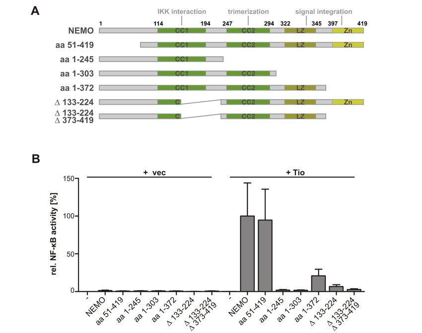
Fig. 1.2 Model of NEMO.
Overview of the NEMO signaling domains. The molecule spans 419 amino acids. CC1 mediates inter-
action with IKK. CC2 mediates oligomerization. LZ and Zn integrate upstream signals. CC, coiled-coil;
LZ, leucine zipper; Zn, zinc finger.
complex activation is mechanistically linked to polyubiquitin-mediated oligomerization of
NEMO. NEMO is the regulatory subunit of the IKK complex and serves as an integra-
tor of upstream signaling events. It displays a helical structure and is composed of two
N-terminally located coiled-coil domains (CC1 and CC2), which mediate interaction with
the IKKs and allow oligomerization of NEMO. The C-terminal part of NEMO contains a
leucine zipper and a zinc finger, which serve to integrate incoming signals (see Fig. 1.2;
Gautheron and Courtois, 2010). To this end, NEMO senses Lys63 -linked polyubiquitin
chains through non-covalent binding (Wu et al., 2006). Thus, NEMO recruits its IKK
complex partners to polyubiquitinated receptor-bound signaling intermediates like mem-
bers of the TRAF family (tumor necrosis factor receptor-associated factor) as well as the
TAK1 kinase.
TRAFs are E3-ubiquitin ligases that conjugate Lys63 -linked chains onto substrates (Ha
et al., 2009). In contrast to Lys48 -linked chains, Lys63 -connected polyubiquitin chains are
non-degradative and serve an important role in signal transduction cascades by transmit-
ting activating signals to downstream partners (Yang et al., 2010). The TRAFs that are
most prominently involved in canonical NF-κB signaling are TRAF2 and TRAF6. On the
one hand, both molecules serve as adaptors bridging the IKK complex with cytoplasmic
tails of ligated receptors. On the other hand, their ubiquitin ligase activity contributes to
full NF-κB activation by inducing auto-ubiquitination (Megas et al., 2011). Ubiquitinated
TRAFs oligomerize and thereby facilitate binding to NEMO (Gautheron and Courtois,
2010). Additionally, NEMO itself can be covalently modified by the attachment of Lys63 -
linked polyubiquitin, which fosters its oligomerization (Tang et al., 2003). The importance
of NEMO:ubiquitin interaction is highlighted by the findings that NEMO mutants deficient
for ubiquitin-binding are associated with human immunodeficiency pathologies (Vinolo
et al., 2006). Further complexity is added to the regulation of NEMO by recent reports of
NEMO conjugation with linear ubiquitin chains. The pathophysiological relevance of this
interaction, nevertheless, has not been unraveled (Tokunaga et al., 2009).
Non-canonical NF-κB Signaling Induction of non-canonical NF-κB signaling is observed
in response to stimulation of a subset of TNFR-family members including CD40, BAFFR
(B-cell activating factor receptor), and LTβR (lymphotoxin-β receptor). Activation of
41.1 Nuclear Factor of KappaB (NF-κB)
non-canonical NF-κB signaling centers around regulation of the NF-κB-inducing kinase
(NIK). NIK preferentially induces phosphorylation of a homodimeric IKKα complex (Sen-
ftleben et al., 2001; Xiao et al., 2001b). In addition, NIK promotes the interaction of
IKKα with its primary substrate p100 (Xiao et al., 2004). IKKα-phosphorylated p100 is
marked for Lys48 -linked ubiquitination and subsequent partial proteasomal degradation.
This processing generates the active subunit p52. As a result of the elimination of the p100
inhibitory ankyrin-repeats during processing, precomplexed RelB and p52 translocate into
the nucleus (Fig 1.1, right panel; Sun, 2011). The key event in non-canonical NF-κB ac-
tivation is the regulation of NIK stability (Liao et al., 2004). NIK displays constitutive
kinase activity. Therefore, its abundance needs to be kept low to ensure quiescence of the
non-canonical pathway in resting cells. To this end, NIK protein is subject to an unusual
regulatory mechanism: constitutive protein synthesis is followed by immediate proteaso-
mal degradation. This turnover is induced by a Lys48 -ubiquitin ligase complex consisting
of TRAF2, TRAF3 and cIAP1/2 (cellular inhibitor of apopotosis). TRAF2 is complexed
with the ubiquitin-ligases cIAP1/2, while TRAF3 binds to both, NIK and TRAF2, and
thereby serves as an adaptor bridging NIK with the cIAP1/2 E3-ligase module (Fig. 1.3,
left panel). The close vicinity within this complex allows cIAP1/2-mediated Lys48 -linked
ubiquitination of NIK, thereby provoking its proteasomal degradation (Vallabhapurapu
unstimulated stimulated
CD40/ BAFFR/ LTβR CD40/ BAFFR/ LTβR
TRAF2
E3-ligase cIAP1/2 TRAF3
complex
TRAF2 adaptor K48
cIAP1/2 TRAF3
NIK
proteasome
K48
NIK
IKKα IKKα
K48-Poly-Ubi phosphorylation
Fig. 1.3 Regulation of NIK Stability in Non-Canonical NF-κB Activation.
Turnover of the constitutively active kinase NIK is regulated by an E3-ubiquitin ligase complex contain-
ing TRAF2/3 and cIAP1/2. In unstimulated cells, NIK is directly bound by the adaptor TRAF3, which
heterodimerizes with TRAF2 to recruit cIAP1 and -2. These E3-ubiquitin ligases execute Lys(K)48 -
linked ubiquitination of NIK, thereby inducing its proteasomal degradation. Upon stimulation and
multimerization of CD40, BAFFR or LTβR, TRAF2 and TRAF3 are recruited to the cytoplasmic
chains of these receptors. cIAP1 and -2 switch their substrate specificity to induce ubiquitination
and subsequent degradation of receptor-bound TRAF2 and -3. Thereby, cytosolic NIK is stabilized to
phosphorylate and activate IKKα dimers.
51 Introduction et al., 2008; Zarnegar et al., 2008b). Occupation of non-canonical NF-κB-inducing recep- tors will halt the constitutive turnover of NIK by sequestering the ubiquitin ligase complex that mediates NIK degradation. Interaction of the complex with NIK is disrupted by the recruitment of TRAF3 and TRAF2 to the cytoplasmic chains of these receptors. TRAF3 interacts with the receptors and NIK via mutually exclusive binding interfaces. Thus, TRAF3:receptor interaction prohibits TRAF3:NIK interaction (Sanjo et al., 2010). In addition, the receptors are membrane-bound, whereas NIK resides in the cytoplasm, re- sulting in a further spatial separation of NIK from its disintegrating complex. As a result, NIK is stabilized and signaling proceeds. Additionally, cIAP1/2 are recruited to the recep- tors through their complexation with TRAF2. A switch of substrate specificity succeeds these events and leads to cIAP1/2-mediated ubiquitination of receptor-bound TRAF3 and TRAF2 inducing their degradation (Fig. 1.3, right panel; Vallabhapurapu et al., 2008; Zar- negar et al., 2008b). This depletion of cellular TRAF3 and TRAF2 pools further stabilizes NIK to induce a sustained non-canonical NF-κB signal. Thus, TRAF3 serves as a negative regulator in non-canonical NF-κB signaling, as opposed to all other TRAFs in canonical signaling, and must be obliterated to achieve non-canonical NF-κB induction (Liao et al., 2004). The outcome of non-canonical NF-κB signaling is the inducible processing of p100 with successive nuclear translocation of p52 and RelB. 1.1.2 Targets and Cross-Talk of NF-κB The two NF-κB pathways are generally assigned to different biological functions. Whereas canonical signaling involves pro-inflammatory and anti-apoptotic gene regulation, the non- canonical pathway is thought to govern cellular differentiation and organ development. A plentitude of NF-κB-regulated genes have been identified including pro-inflammatory factors like TNF and IFN-γ (Shakhov et al., 1990; Sica et al., 1997), modulators of innate immunity like the chemokine IL-8 (Kunsch and Rosen, 1993) and the adhesion molecule ICAM-1 (Parry and Mackman, 1994) as well as anti-apoptotic genes like cIAP2 (Hong et al., 2000). However, almost all of the genes described thus far are transactivated by canonical NF-κB dimers. Target genes and binding sites of non-canonical dimers have hardly been identified. Analysis of pathway-specific gene targeting is complicated by the fact that receptors triggering NF-κB signaling usually activate both, the canonical and the non-canonical pathway, and simultaneously engage other transcription factors (Basak and Hoffmann, 2008). The restricted accessibility of this subject is based on the multitude of potential dimers, their interactions with divergent κB-sites and other transcriptional regulators, and the dynamic temporal control of NF-κB signaling (Hoffmann et al., 2006). Further complexity is added by cross-talk mechanisms that interconnect the canonical and the non-canonical pathway. Research on NIK has focused on an exclusive kinase- substrate relationship among NIK, IKKα and p100. However, initial and recent reports imply NIK critically contributing to canonical NF-κB signaling as well. For instance, IκBα degradation and p65 nuclear translocation have been proven defective in the absence of 6
1.1 Nuclear Factor of KappaB (NF-κB)

NIK (Ramakrishnan et al., 2004). Moreover, accumulation of NIK after LTβR stimulation
has been shown to amplify canonical NF-κB signaling via the IKK complex and to lead to
enhanced IκBα phosphorylation, nuclear translocation and DNA-binding activity of p65
and c-Rel (Zarnegar et al., 2008a). However, a role for NIK in the canonical pathway
might be cell-type and ligand specific (Thu and Richmond, 2010). Another example for
the interdependence of the pathways is the transcriptional induction of the non-canonical
effectors p100 and RelB through canonical NF-κB dimers (Lombardi et al., 1995; Bren
et al., 2001). Thus, addressing any of the biological NF-κB functions requires dissection of
both, the canonical and the non-canonical pathways.

1.1.3 Corruption of NF-κB Signaling in Lymphoid Malignancies

The pleiotropic role of NF-κB signaling in lymphocyte development and function implies
that these cells rely on a precisely tuned balance between activating and inhibiting signals
and are hence extremely vulnerable to pertubations of NF-κB signaling. Aberrant NF-κB
activation is detrimental and can lead to oncogenic lymphocyte transformation (Staudt,
2010). Therefore, it is not surprising that constitutive NF-κB signaling has been found as
key pathological feature in various lymphoid malignancies. These include virally-induced
tumors like Hodgkin’s lymphoma and adult T-cell leukemia, as well as non-virally-induced
tumors like acute lymphocyte leukemia, multiple myeloma and chronic myelogenous leu-
kemia (Damania, 2004; de Oliveira et al., 2010; Staudt, 2010). Lymphotropic oncogenic
viruses have evolved numerous strategies to corrupt host-cellular NF-κB signaling to sup-
port viral replication as well as oncogenic transformation. Viral oncoproteins can thus
serve as excellent tools to study deregulation of NF-κB signaling; indeed, the first link
between NF-κB and lymphoid malignancies came from studies of the viral oncoprotein
v-Rel (Gilmore, 1999). The best studied examples of oncoviral NF-κB inducers are lym-
photropic herpesviruses like EBV (Epstein-Barr virus) with its oncoprotein LMP1 (latent
membrane protein 1) and KSHV (Kaposi’s sarcoma-associated herpesvirus) with one of its
oncoproteins vFLIP as well as the retrovirus HTLV-1 (human T-cell leukemia virus) with
its oncoprotein Tax (Jost and Ruland, 2007; Jeang, 2010).

Epstein-Barr Virus (EBV) establishes latency in B cells and their immortalization causes
virally-induced lymphomagenesis. LMP1 of EBV is a membrane-bound protein that mim-
ics a constitutive, ligand-independent CD40 receptor, which utilizes the intracellular CD40
signaling machinery including TRAF adaptor molecules to activate NF-κB pathways. Ac-
cordingly, LMP1 is an activator of both, canonical and non-canonical NF-κB signaling.
LMP1 comprises two CTAR (C-terminal activating region) domains. CTAR1 directly re-
cruits cellular TRAF1, TRAF2, TRAF3, and TRAF5, thereby initiating the non-canonical
route of NF-κB. CTAR2, through indirect recruitment of TRAF6, activates the canonical
arm of NF-κB signaling in an IKK complex-dependent manner (Soni et al., 2007).

                                                                                          7
1 Introduction
Kaposi’s Sarcoma Associated Herpesvirus (KSHV) is the etiological agent of the pri-
mary effusion lymphoma (PEL). It encodes multiple oncoproteins that activate NF-κB,
the best studied being vFLIP/K13. Through interaction with NEMO, vFLIP constitu-
tively activates the IKK complex (Liu et al., 2002), whereas vFLIP-induced non-canonical
NF-κB activity requires IKKα, but is independent of NIK (Matta and Chaudhary, 2004).
KSHV-induced NF-κB activity and in particular vFLIP expression is necessary for the
continuous proliferation of PEL cells (Keller et al., 2000; Matta and Chaudhary, 2004).
Human T-Cell Leukemia Virus (HTLV-1) causes adult T-cell lymphoma/ leukemia
(ATL) after long periods of latency. The Tax oncoprotein of HTLV-1 localizes to the
cytoplasm and directly binds to NEMO, thereby activating the IKK complex and cano-
nical signaling. An unconventional activation of non-canonical NF-κB is achieved by Tax
through direct binding of p100 leading to an IKKα- and NEMO-dependent, but NIK-
independent p100 processing (Peloponese et al., 2006). Tax-induced non-canonical NF-κB
signaling seems to be essential for its transforming activity, as the sequence motif in Tax
that induces p100 processing confers the transforming activity of HTLV-1 (Shoji et al.,
2009).
The fact that multiple virus families have evolved strategies to dually address canonical
and non-canonical NF-κB signaling implies a non-redundant pathological role for both
pathways.
1.2 Simian Rhadinoviruses
Herpesviruses are prevalent in many species including human and non-human primates.
The Herpesviridae are classified into three subfamilies - α, β and γ - according to their
genome structure and biological properties. The lymphotropic Gammaherpesvirinae are
further subdivided into two genera. The γ1- or lymphocryptoviruses include the pro-
totypical member EBV. The γ2- or rhadinoviruses are represented by the prototypical
Herpesvirus (H.) saimiri (Saimiriine herpesvirus 2) and its close relative H. ateles (Ate-
line herpesvirus) (Albrecht et al., 1992; Albrecht, 2000). Both, H. saimiri and H. ateles,
are simian T-lymphotropic viruses endemic to New World primates. Within their natural
hosts, the squirrel monkey (Saimiri sciureus) for H. saimiri and the spider monkey (Ateles
spp.) for H. ateles, both viruses establish a lifelong persistence without any apparent signs
of disease (Meléndez et al., 1968, 1972a; Falk et al., 1972). Experimental cross-species
transmission into New World primate species to which these viruses are not endemic, like
Callithrix jacchus and Saguinus spp., induces a very rapid development of fulminant lym-
phatic leukemias and peripheral T-cell lymphomas (Meléndez et al., 1969, 1972b; Hunt
et al., 1972). Moreover, both viruses are capable of transforming simian T lymphocytes
to sustained, antigen-independent growth in cell culture (Fickenscher and Fleckenstein,
2001).
81.2 Simian Rhadinoviruses
1.2.1 Herpesvirus saimiri and its Oncoproteins StpC and Tip
H. saimiri possesses a double-stranded linear DNA genome of approximately 155 kilo-
base pairs (kbp). The genome splits into the coding AT-rich L-DNA (low density DNA),
which is flanked by the tandem repetitions of non-coding GC-rich H-DNA (high density
DNA) (Bornkamm et al., 1976). The viral genome carries up to 77 open reading frames
(ORF) (Albrecht et al., 1992) including gene blocks that are highly conserved among
all herpesvirus families. Unique to H. saimiri is a region located at the far left end of
its L-DNA. It has been found to be highly variable among different strains and led to
the classification of H. saimiri into three subgroups - A, B and C (Medveczky et al.,
1984). The subgroups strongly differ in their oncogenic potential. While all subgroups are
capable of inducing the in vitro growth transformation of phytohemaglutinin-activated,
IL-2-expanded peripheral blood lymphocytes of Callithrix jacchus, only certain strains of
subgroup C (e.g. H. saimiri C488) can transform human T cells to a stable, antigen-
independent growth in culture (Biesinger et al., 1992). Growth-transformed cell lines can
be generated from peripheral blood mononuclear cells, cord blood lymphocytes, as well
as established T-cell clones. Depending on the parental population, outgrowth of CD4-
and/or CD8-positive T-cell lines can be observed (Biesinger et al., 1992). The phenotype
of H. saimiri-transformed human T cells resembles that of activated mature T cells and
retains features of the primary parental cells, e.g. antigen-specific triggering of tyrosine
phosphorylation and intracellular calcium mobilization (Bröker et al., 1993; Mittrücker
et al., 1993). Furthermore, the cell lines show no signs of karyotypic alteration (Troidl
et al., 1994). The secreted cytokine pattern is consistent with that of Th1 cells (e.g. IL-2
and IFN-γ) (Carli et al., 1993). The sequence variability at the left-terminal region of the
L-DNA as well as the differential oncogenic phenotype of various virus isolates led to the
discovery of open reading frames essential for H. saimiri-mediated T-cell transformation
(Murthy et al., 1989). In subgroup A and B strains, these ORFs encode the StpA (saimiri
transformation-associated protein) and StpB proteins, respectively (Hör et al., 2001). The
corresponding region of H. saimiri group C strain genomes encodes two oncoproteins, StpC
and Tip (tyrosine kinase interacting protein), which are derived from a single bicistronic
transcript (Biesinger et al., 1990; Geck et al., 1990). Both proteins of H. saimiri group
C strains are absolutely required for the oncogenic phenotype of lymphoma induction
in vivo and transformation in vitro. However, they are dispensable for viral replication
and persistence (Duboise et al., 1998). The genome of H. saimiri episomally persists in
high copy numbers in transformed human T cells (Biesinger et al., 1992). As opposed to
transformed simian lymphocytes, human T cells do not support lytic growth of the virus;
thus, release of infectious viral particles can not be observed. The majority of viral genes
remain silent during episomal persistence. Nevertheless, translation of stpC/tip transcripts
is detectable (Fickenscher et al., 1996).
91 Introduction StpC Oncoprotein The oncogenic properties of StpC, insinuated through its requirement for viral T-cell transformation and its expression in the transformed state, were confirmed by retroviral delivery to cultured rodent fibroblasts and implantation of these cells into nude mice. Expression of StpC alone led to the phenotypic signs of growth transformation. Transfer of these cells into nude mice resulted in the growth of invasive tumors (Jung et al., 1991). The role of stpC as an oncogene was confirmed in a transgenic mouse model, in which expression of StpC led to tumor formation without the cooperation of any other viral gene product. Surprisingly, the mice developed tumors of epithelial origin, thus differing from the lymphotropic malignancies induced in simian hosts (Murphy et al., 1994). On the molecular level, StpC is a small, 102 amino acid protein that localizes to membranous compartments via its C-terminal hydrophobic region. Its central part consists of 18 con- secutive collagen-like tripeptides ((Gly-X-Y)n , where X and/or Y represent a proline) that are thought to mediate trimerization of the protein. Disruption of the collagen-like re- peats suppresses the transforming activity of StpC (Jung and Desrosiers, 1994; Choi et al., 2000). Signal transduction events initiated by StpC are thus confined to the N-terminal, mostly charged 17 amino acids. StpC has been described to interact with the cellular Ras protein, thereby activating MAP-kinase signaling, yet these findings remain controversial (Jung and Desrosiers, 1995; Glanz, 2008). Nevertheless, oncogenic Ras could substitute for StpC in viral T-cell transformation, both in vitro and in vivo (Guo et al., 1998). The ma- jor function of StpC is the activation of NF-κB via an N-terminal, proline-anchored motif (10 PIEET14 ), a known binding site for TRAF1, -2, -3 (Lee et al., 1999). The main effects of StpC on NF-κB signaling are attributed to the TRAF2 interaction. In addition, StpC was described to bind to TRAF6; the crucial residue for this interaction and subsequent NF-κB activation is Glu12 (Chung et al., 2007). The StpC:TRAF interactions result in an NF-κB activating pathway that seems to involve NIK and the IKK kinases to target IκBα for degradation and p65:p50 for nuclear translocation (Sorokina et al., 2004). Only few targets of StpC-induced NF-κB activity have been described, amongst them T-cell proli- feration enhancers like IL-2 and IFN-γ (Merlo and Tsygankov, 2001; Glanz et al., 2008) and proteins involved in cell migration and adhesion like IL-8 and ICAM1 (Chung et al., 2007). Transformation of rodent fibroblasts by StpC mainly depends on the Pro residue within the PIEET motif (Lee et al., 1999). However, the precise contributions of canonical and non-canonical NF-κB pathways as well as the impact of T-cell-specific factors on the transforming functions of StpC remain elusive and need to be further investigated. Tip Oncoprotein As opposed to StpC, Tip by itself is not capable of transforming rodent fibroblasts. The constitutive expression of Tip in transgenic mice proved to be embry- onically lethal. When Tip, however, was expressed as an inducible transgene, formation of T-cell lymphomas in mice could be detected. These findings point toward its role in mediating the T-cell specificity of H. saimiri-induced transformation events (Wehner et al., 2001). Tip is a 256 amino acid protein and, like StpC, anchors to the membrane via its 10
1.2 Simian Rhadinoviruses
C-terminal region. The N-terminal part of Tip harbors two serine-rich regions followed
by four tyrosine residues in its central part (Tyr94 , Tyr114 , Tyr127 , and Tyr155 ) (Biesinger
et al., 1990; Lund et al., 1995, 1996). The major cellular interaction partner of Tip is
the non-receptor tyrosine kinase Lck, which belongs to the Src family of kinases. Tip re-
cruits Lck via two binding interfaces, the CSKH- (C-terminal Src kinase homology) and
the proline-rich SH3b- (Src homology 3 binding) motifs (Biesinger et al., 1995; Jung et al.,
1995). This interaction will activate Lck to phosphorylate mainly Tyr127 (Heck et al.,
2006). Mutant viruses that either lack the CSKH- and/or the SH3b-motif lose their abil-
ity to growth transform human cord blood lymphocytes in vitro. Mutation of the major
Lck-phosphorylation site Tyr127 rendered viral T-cell transformation strictly dependent on
exogenous IL-2 (Heck et al., 2006). Downstream of Lck, the phosphorylated residue Tyr114
of Tip serves as a docking site for the SH2-domains of STAT1 and STAT3 (signal trans-
ducers of transcription) leading to the induction of their transcriptional activity (Lund
et al., 1997, 1999). Nevertheless, mutant viruses that lack Tyr114 are still competent in
transforming human T cells, indicating that STAT1 and STAT3 activation is dispensable
for viral growth transformation by Tip (Hartley and Cooper, 2000; Heck et al., 2005).
1.2.2 Herpesvirus ateles and its Oncoprotein Tio
Discovery of H. ateles and H. saimiri dates to the same period. Nevertheless, H. saimiri
was the predominant subject of investigation because of its growth properties in cell cul-
ture. H. ateles replicates strictly cell-associated. Therefore, the supernatants of these
cultures display very low and unstable virus titers (Fickenscher and Fleckenstein, 2001).
Accordingly, little is known about H. ateles biology. Whereas induction of lymphoprolif-
erative pathology and in vitro transformation of simian T lymphocytes is comparable for
H. saimiri and H. ateles (Hunt et al., 1972; Falk et al., 1978), transformation of human
T cells could not yet be demonstrated for H. ateles. These differences might be due to viral
growth properties or usage of a receptor that is not present on human lymphocytes. The
H. ateles strain 73 is most commonly investigated and was isolated from lymphocytes of a
Colombian spider monkey (Ateles paniscus) (Falk et al., 1974). The double stranded DNA
genome of H. ateles, structurally identical to that of H. saimiri, is composed of a coding
L-DNA flanked by a non-coding repetitive H-DNA (Fleckenstein et al., 1978). Its primary
sequence was resolved in 2000 (Albrecht, 2000). The 108,409 base pair L-DNA segment
contains 73 ORFs, all of which have corresponding homologues in H. saimiri (Albrecht,
2000). The variable left end of the L-DNA, that confers the oncogenicity of the simian
rhadinoviruses, is transcribed into a single mRNA containing two exons. The spliced tran-
script encodes a protein which was termed Tio (two in one). Eponymous to the two in one
oncoprotein were its structural and functional homologies with StpC and Tip of H. saimiri
(Albrecht et al., 1999; Albrecht, 2000). The N-terminal part of Tio displays homologies
to StpC, while the C-terminal part resembles Tip (Fig. 1.4). This overt homology and
detection of Tio transcripts in in vitro and in vivo transformed simian cells gave rise to
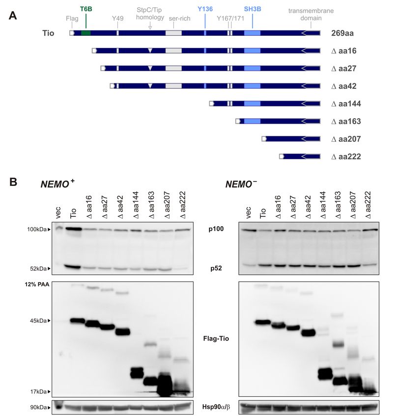
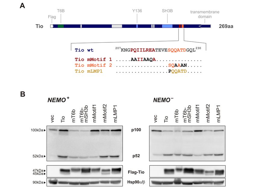
111 Introduction the question whether Tio mediates the oncogenic phenotype of H. ateles. This issue was addressed by recombinantly expressing Tio from a H. saimiri C488 background. In these viruses, the bicistronic stpC/tip coding sequence was deleted and replaced for the tio cod- ing sequence. Tio-recombinant viruses were capable of transforming human T cells and even conferred enhanced growth properties compared with the reconstituted StpC/Tip- expressing viruses. Thus, Tio can functionally substitute for the StpC/Tip oncogenes, thereby establishing its role as an oncoprotein (Albrecht et al., 2004). Tio Oncoprotein The spliced Tio transcript encodes for a polypeptide of 269 amino acids, which migrates as a doublet of 43 and 46 kDa in SDS gels. The double band is thought to be related to post-transcriptional modifications, which so far have not been further defined. A N-terminally Flag-tagged version of Tio shifts to migrate at 45 and 47 kDa. Homodimerization and multimerization of Tio can be observed (Albrecht et al., 1999). The homology of Tio with StpC and Tip is not restricted to sequence similarities, but directly translates onto the molecular mechanisms of signal transduction. Tio anchors to membranous compartments via its C-terminal, hydrophobic region like StpC and Tip. The Tip-homologous part is confined to the membrane-proximal C-terminal half of Tio. Here, the motif 189 PQLPPR194 perfectly matches with the SH3-binding consensus PxxPxR, a polyproline helix followed by an arginine residue (Fig. 1.4, light blue). Tio-SH3b peptides bind to GST-purified SH3 domains of Lyn, Hck, Lck, Src, Fyn and Yes with decreasing affinity (Albrecht et al., 1999). Interaction of Tio with full-length Lck, Src, and Fyn is confirmed, but the Src-family kinases contributing to viral T-cell transformation remain Fig. 1.4 Model of the Tio Oncoprotein. Motifs implied in Tio-induced signal transduction are depicted above the scheme. The corresponding amino acid stretches of Tio are magnified, anchor residues are highlighted in green or light blue. Tio localizes to membranes through its hydrophobic C-terminus. Src-family kinases (SFK) are bound and activated via the SH3b (Src kinase homology 3 binding) motif 189 PQLPPR194 (light blue), subsequently leading to the phosphorylation of tyrosine residue 136 (Y136) of Tio (light blue). Through its N- terminal 5 PQEHEE10 T6b (TRAF6 binding) motif, Tio recruits the E3-ubiquitin ligase TRAF6 to induce canonical NF-κB activation (green). Tio contains four tyrosine residues (Y49, Y136, Y167, Y171) as well as a serine-rich region in its middle part. The boundary of the StpC-homologous N-terminus and the Tip-homologous C-terminus is indicated by the gray arrow. N-terminally Flag-tagged Tio was used throughout this thesis. 12
1.2 Simian Rhadinoviruses

to be identified (Albrecht et al., 1999, 2005). Engagement of their SH3 domains is con-
sidered to activate Src kinases to phosphorylate their substrates on Tyr residues (Moarefi
et al., 1997). Tio harbors four tyrosine residues - Tyr48 , Tyr136 , Tyr167 , and Tyr171 . The
sequences surrounding Tyr136 show similarities to both Tyr114 and Tyr127 of Tip-C488.
In Tio, Tyr136 is the only residue phosphorylated by the bound kinase (Fig. 1.4, light
blue). Growth transformation induced by Tio-expressing viruses strictly depends on both
the integrity of the SH3b motif and the presence of Tyr136 (Albrecht et al., 1999, 2005).
Similar to Tip, Tio induces expression and phosphorylation of STAT1, STAT3 and STAT5
through its SH2-domain-binding pTyr136 . These events rely on an intact SH3b-motif and
Tyr136 (J.-C. Albrecht, unpublished observations).
  Homologies between Tio and StpC are confined to the N-terminal half of Tio. The con-
tinuous collagen-like-repeat structure of StpC (see 1.2.1) can not be found in Tio. Instead,
six of these collagen-like motifs are interlaced into proline-rich sequences throughout the
N-terminal half of Tio. As opposed to StpC, the collagen-like tripeptides are not believed
to contribute to multimerization of Tio (Albrecht et al., 1999). Functional homologies
between Tio and StpC restrict to NF-κB induction, whereas effects of Tio onto the Ras-
MAPK pathway have not been reported. Tio harbors a glutamate-rich, proline-anchored
motif 5 PQEHEE10 at its N-terminus (Fig. 1.4, green). Its sequence is consistent with the
TRAF6 binding consensus PxExxAr/Ac with x representing any amino acid and Ar/Ac
representing aromatic or acidic residues. Dependent on this motif, Tio can directly re-
cruit TRAF6, an E3-ubiquitin ligase involved in NF-κB activation. This interaction was
shown to mediate induction of NF-κB reporter activity in transiently transfected Jurkat
T cells and explains TRAF6 membrane recruitment in virus-transformed human T cells
(Heinemann et al., 2006). Yet, the contribution of Tio-induced NF-κB signaling to trans-
formation remains unclear. Furthermore, the precise nature and co-factor requirements of
Tio-induced NF-κB signals are ambiguous.
  In summary, StpC/Tip and Tio are viral oncoproteins that persistently activate mul-
tiple cellular signaling pathways by mimicking constitutively active, ligand-independent
receptors. These characteristics are reminiscent of other viral oncoproteins like LMP1 of
EBV. Studying human T-cell lines transformed by StpC/Tip- or Tio-expressing viruses
offers the advantage to analyze growth-regulatory pathways in the absence of lytic viral
replication or release of infectious particles. Thus, StpC/Tip and Tio-mediated oncogenic
T-cell transformation can serve as model systems for the induction and maintenance of
T-cell leukemias and lymphomas. This may allow to further clarify signaling pathways
related to these malignancies.

                                                                                           13
2 Rationale
NF-κB is a pleiotropic transcription factor that pivotally contributes to T-cell develop-
ment, proliferation and differentiation. A causative role of aberrant NF-κB activation in
various virally-induced lymphoid malignancies has become manifest. Respective tumor
viruses include human Epstein-Barr virus (EBV), Kaposi’s sarcoma associated herpesvirus
(KSHV), and simian Herpesvirus (H.) saimiri. The oncogenic properties of these viruses
are mediated by viral oncoproteins that all have the ability to potently activate canonical
and non-canonical NF-κB pathways.
The oncoprotein Tio of H. ateles, which is closely related to H. saimiri, essentially
contributes to T-cell transformation in vitro. Tio was previously reported to activate
NF-κB signaling in response to TRAF6 interaction. The mechanisms and targets of this
Tio:TRAF6-mediated NF-κB activation as well as its contribution to viral T-cell transfor-
mation remained, however, elusive. The objective of this project was the evaluation of the
Tio:NF-κB signalosome in human T cells.
• Recruitment of TRAF6 is indicative for activation of the canonical NF-κB pathway.
Therefore, the role of NEMO and IKKβ as co-factors of Tio:TRAF6-induced NF-κB
had to be investigated. Furthermore, the expression of canonical NF-κB target genes
(cIAP2, IL-8, RelB and p100) should be analyzed.
• For LMP-1 of EBV, vFLIP of KSHV, and StpC of H. saimiri, deregulation of NF-κB
signaling has been described to essentially account for viral tumor induction and
maintenance. If Tio employs similar mechanisms as its herpesviral counterparts,
NF-κB activation should contribute to transformation. This hypothesis ought to be
tested by the inhibition of constitutive NF-κB signaling in human T cells transformed
with Tio-expressing viruses.
• Induction of RelB and p100 may result in non-canonical NF-κB activation, which
is typically observed for NF-κB-inducing viral oncoproteins including LMP1, vFLIP
and StpC. To this end, the influence of Tio on the non-canonical NF-κB pathway
should be assessed by evaluating expression of the central kinase NIK, subcellular lo-
calization and activity of the non-canonical subunits p52 and RelB as well as co-factor
requirements (TRAF3, TRAF2 and cIAP2) for non-canonical NF-κB induction.
153 Results
The herpesviral oncoprotein Tio has previously been shown to interfere with host-cellular
signaling pathways in multiple ways. On the one hand, Tio interacts with Src family
kinases (SFK) via a C-terminally-located SH3 binding motif (SH3b). This interaction
results in an activation of the kinase with subsequent phosphorylation of Tyr136 of Tio.
Both, the SH3b motif and Tyr136 , are required for the growth-transformation of human
T cells by Tio-expressing viruses (Albrecht et al., 1999, 2005) and upregulation of the
expression and phosphorylation of STAT1, STAT3, and STAT5. On the other hand, Tio
harbors a TRAF6 binding motif (T6b) at its N-terminus, through which it recruits the
ubiquitin ligase TRAF6, thereby inducing NF-κB activity (Heinemann et al., 2006). Two
model systems are available to study the effects of Tio on cellular signaling pathways:
transient expression of Tio in transfected Jurkat T cells and stable expression of Tio from
a Herpesvirus saimiri C488 background in primary human peripheral blood lymphocytes
(PBLs), which are immortalized by viral infection. From previous studies, Tio variants
carrying mutations within the individual interaction motifs were available within a pEF1-
plasmid background. These are:
• mT6b (mutant TRAF6 binding motif) - substitution Glu7,9,10 Gln within the TRAF6-
binding motif (5 PQEHEE10 ) - this mutant is unable to interact with TRAF6, leading
to a loss of NF-κB induction
• Y136F - mutation Tyr136 Phe - this mutant cannot be phosphorylated, although SFK
binding is intact, leading to a loss of STAT1, STAT3, and STAT5 upregulation and
phosphorylation
• mSH3b (mutant SH3 binding motif) - substitution Pro292 Ala and Arg294 Gly within
the SH3-binding motif 289 PQLPPR294 - this mutant is unable to recruit SFK and
shows the same phenotype as Y136F
To be able to assess cooperative signaling effects of Src family kinase and TRAF6 binding, a
double mutant - Tio-mT6b-mSH3b - was generated. Throughout this thesis, Tio and these
mutants (mT6b, Y136F, mSH3b and mT6b-mSH3b) were used to delineate the effects of
Tio on cellular NF-κB signaling.
173 Results 3.1 Tio-induced Canonical NF-κB Signaling 3.1.1 Tio Induces Canonical NF-κB Activity Tio has been shown to activate NF-κB dependent on TRAF6 interaction, but indepen- dent of SFK binding or Tyr136 phosphorylation (Heinemann et al., 2006). This previous study was conducted with Tio being expressed from a pcDNA3.1 background, in which the transcription unit is driven by the immediate early promoter of HCMV (human cy- tomegalovirus). This promoter is frequently used in eukaryotic expression plasmids and ensures strong expression. However, in analyzing NF-κB signaling its applicability is re- stricted by its transactivation through NF-κB factors (Sambucetti et al., 1989; Prösch et al., 1995). To confirm the NF-κB-inducing capacity of Tio expressed from an NF-κB- independent pEF1-background, Jurkat T cells were co-transfected with Tio and an NF-κB- responsive luciferase reporter (Fig. 3.1 A). Wild-type Tio was able to strongly induce NF-κB-reporter activity, as were mutants Y136F and mSH3b. However, mutation of the TRAF6-binding site alone or in combination with the SH3b motif rendered these mutants completely inactive, reconfirming the necessity of the TRAF6-binding site and the inde- pendance of SFK interaction and Tyr-phosphorylation for NF-κB induction. Binding of Tio to TRAF6 is indicative for the induction of the canonical NF-κB signaling pathway, as TRAF6 has been described to be an adaptor in IL-1- and Toll-like-receptor-induced ca- nonical signaling. Hence, analyses were extended beyond activation of a synthetic reporter construct to evaluate expression of the endogenous NF-κB target genes cIAP2 and IL-8. Both genes have been described to be transactivated by canonical NF-κB p65:p50 dimers in response to cellular and viral stimuli (Mukaida et al., 1994; Mori et al., 1998; Hong Fig. 3.1 NF-κB Activity Induced by pEF1-Tio. Jurkat T cells were electroporated with expression plasmids coding for Flag-tagged Tio or Tio mutants (mT6b, Y136F, mSH3b, mT6b-mSH3b) and were harvested 48 h after transfection. Transfection of pEF1 vector DNA (vec) served as a negative control. The average of at least three independent ex- periments is shown. A Co-transfection of an NF-κB luciferase reporter. NF-κB activity is depicted as relative response ratio with reference to the mean of Tio-transfected cells. B Immunoblot analysis of whole cell lysates for endogenous cIAP2 protein expression. Protein expression of Tio was verified with a Flag antibody. An unspecific band served as loading control (lc). C Endogenous IL-8 in supernatants of transfected cells detected by ELISA. 18
You can also read


