Influence of Age on Skeletal Muscle Hypertrophy and Atrophy Signaling: Established Paradigms and Unexpected Links - MDPI
←
→
Page content transcription
If your browser does not render page correctly, please read the page content below
G C A T
T A C G
G C A T
               genes
Review
Influence of Age on Skeletal Muscle Hypertrophy and Atrophy
Signaling: Established Paradigms and Unexpected Links
Eun-Joo Lee and Ronald L. Neppl *

                                          Department of Orthopaedic Surgery, Brigham and Women’s Hospital, Harvard Medical School,
                                          Boston, MA 02115, USA; elee59@bwh.harvard.edu
                                          * Correspondence: rneppl@bwh.harvard.edu

                                          Abstract: Skeletal muscle atrophy in an inevitable occurrence with advancing age, and a consequence
                                          of disease including cancer. Muscle atrophy in the elderly is managed by a regimen of resistance
                                          exercise and increased protein intake. Understanding the signaling that regulates muscle mass may
                                          identify potential therapeutic targets for the prevention and reversal of muscle atrophy in metabolic
                                          and neuromuscular diseases. This review covers the major anabolic and catabolic pathways that
                                          regulate skeletal muscle mass, with a focus on recent progress and potential new players.

                                          Keywords: Akt signaling; muscle atrophy; muscle hypertrophy; IGF-1/PI3K signaling; aging muscle;
                                          RNA-binding proteins

                                1. Introduction

                                                 Biological aging is broadly defined as the time-dependent loss of functionality and
Citation: Lee, E.-J.; Neppl, R.L.         robustness, and is associated with increased rates of cancer [1,2], cardiovascular and
Influence of Age on Skeletal Muscle       neurodegenerative disease [2–4], diabetes or dysglycemia [5], insulin and anabolic resis-
Hypertrophy and Atrophy Signaling:        tance [6–8]. Many of these disorders and diseases are often accompanied by, and contribute
Established Paradigms and                 to, muscle weakness and sarcopenia, exacerbating the slow gradual loss of muscle mass
Unexpected Links. Genes 2021, 12,
                                          and strength that typically begins in the fourth decade of life [9–11]. In healthy young
688. https://doi.org/10.3390/
                                          individuals, lean muscle accounts for 38–54% and 28–39% of total body mass, in men and
genes12050688
                                          women, respectively. These ranges are quite broad and are dependent upon multiple fac-
                                          tors including physical activity level, overall health, genetic makeup, and nutritional input.
Academic Editor: Karen Mather
                                          Skeletal muscle is comprised of a heterogenous mix of specialized myofibers that differ in
                                          their physiological, metabolic, and biochemical attributes. On the two ends of this spectrum
Received: 2 April 2021
Accepted: 27 April 2021
                                          are slow oxidative fibers, comprised of type I myosin heavy chain proteins (MHC), and
Published: 3 May 2021
                                          the fast-glycolytic fibers, comprised of types IIa, IIx, and IIb (in rodents) MHC proteins.
                                          With advancing age, skeletal muscle loses its responsiveness to anabolic signals, which are
Publisher’s Note: MDPI stays neutral
                                          secreted in ever diminishing levels [8,12–14]. Importantly, fast-glycolytic fibers atrophy at
with regard to jurisdictional claims in
                                          a greater rate than oxidative fibers with advancing age [15–17], contributing significantly
published maps and institutional affil-   to the loss of muscle power that begins its decline from approximately age 40 onwards [18].
iations.                                  It is this loss of muscle power that is related to the increased number of falls among the
                                          elderly [18,19].
                                                 The cellular and molecular mechanisms linking biological aging with the loss of
                                          muscle mass, strength, and functionality are complex. In its simplest form, the maintenance
Copyright: © 2021 by the authors.
                                          of muscle mass, and strength, is due to the balance of anabolic and catabolic processes.
Licensee MDPI, Basel, Switzerland.
                                          Stem cell exhaustion, cellular senescence, altered intercellular communication, losses of
This article is an open access article
                                          proteostasis and genome stability, telomere attrition, mitochondrial dysfunction, epigenetic
distributed under the terms and           alterations, and dysregulation of gene expression and alternative splicing are hallmarks
conditions of the Creative Commons        of, and causal for, the age-associated loss of cellular and biological robustness [20–23]. In
Attribution (CC BY) license (https://     this review, we identify and discuss the cellular and molecular mechanisms responsible
creativecommons.org/licenses/by/          for modulating skeletal muscle mass and strength, and its influence on the non-myocyte
4.0/).                                    contributions to skeletal muscle physiology that become dysregulated with age. Herein

Genes 2021, 12, 688. https://doi.org/10.3390/genes12050688                                                 https://www.mdpi.com/journal/genes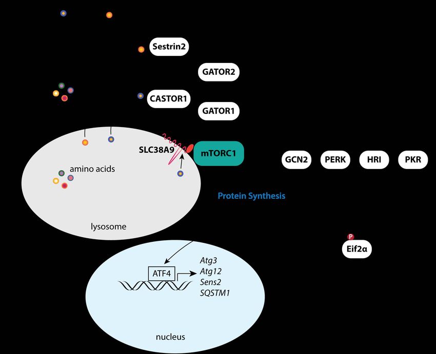
Genes 2021, 12, 688                                                                                           2 of 26

                      we give high priority to in vivo and clinical studies identifying the effects of age on the
                      linkages between gene expression and signaling pathways with muscle function. Finally,
                      we would like to sincerely apologize to our many outstanding colleagues, whose work may
                      have been inadvertently overlooked or not thoroughly discussed due to space constraints.

                      2. PI3K/Akt/mTOR Signaling in Muscle
                           Insulin, and insulin-like growth factor 1 (IGF-1) signaling (IIS) is ubiquitous to multi-
                      cellular animals, connecting nutrient availability to development, longevity, and growth
                      in response to hypothalamic-pituitary growth signals [24]. Activation of the insulin/IGF-
                      1 receptors leads to the phospho-activation of phosphoinositide 3-kinase (PI3K), and
                      the subsequent activation of Akt (Figure 1), a key integrator of intra- and intercellular
                      signals known to promote cell survival, glucose metabolism, and anabolic growth pro-
                      cesses [25–32]. Importantly, IIS has evolutionarily conserved pleiotropic effects that are
                      age/developmental stage and gender dependent. Ames, Snell, and GHRH (lit/lit) dwarf
                      mice have reduced growth hormone (GH) signaling and reduced serum IGF-1 levels, but
                      have up to a 70% extension of their lifespans [33–38]. Heterozygous deletion of the IGF-1
                      receptor gene (Igf1r) have a modest ~6–8% reduction in bodyweight by 7 weeks of age,
                      but live up to 25% longer [39,40]. However, this lifespan extension was limited to females
                      and modulated by genetic background [39,41,42]. Consistent with the influence of gender,
                      18-month-old female mice treated with a selective IGF-1R antagonist for 6 months, but not
                      male mice, showed significant improvements in exercise tolerance, grip strength, and motor
                      coordination [43]. In contrast, transgenic expression of GH in mice induces accelerated
                      growth, hyperinsulinemia despite euglycemia, increased adult body weight and plasma
                      IGF-1 levels, and a nearly 50% reduction in lifespan [44–47].
                           PI3K/Akt/mTOR signaling is a key regulator of glycolytic muscle homeostasis, hy-
                      pertrophic growth, and metabolism through its activation of mTOR-dependent anabolic
                      processes while simultaneously inhibiting both autophagy and Foxo1/3 catabolic pro-
                      cesses [48,49]. Skeletal muscle-specific transgenic overexpression of Akt selectively pro-
                      motes muscle hypertrophy [50]. Glycolytic muscle specific transgenic overexpression of
                      Akt similarly induces glycolytic muscle hypertrophy, promoting weight loss and insulin
                      sensitivity in obese [51] and aged mice [52]. Functional overload [53], IGF-1 stimula-
                      tion [54,55], and skeletal muscle specific transgenic overexpression of IGF-1 [56] positively
                      regulate PI3K/Akt/mTOR signaling in muscle and promote hypertrophic growth. The
                      phospho-activation of Akt is initiated by a series of intracellular signaling events arising
                      from receptor tyrosine kinases (RTKs) including the IGF-1 (IGF-1R) and insulin (Insr)
                      receptors, and G-protein-coupled receptors (GPCR), which signal through scaffolding
                      proteins (e.g., IRS1/2, Grb2/10) to activate PI3K. Active PI3K produces phosphatidyli-
                      nositol (3,4,5)-trisphosphate (PIP3), leading to the phospho-activation of Akt at T308 by
                      PDK1. Maximal activation of Akt requires phosphorylation at the S473 site, which is
                      directly phosphorylated by mTORC2 [26,29]. Comprised of core components mTor, Rictor,
                      SIN1, and mLST8, mTORC2 functions primarily as an effector of insulin/PI3K activity [57].
                      Though the regulation of mTORC2 is uncertain, a growing body of evidence would suggest
                      that mTORC2 activity is positively modulated through its association with ribosomes, the
                      mitochondrial-associated ER membrane, and lysosomes [58–66]. Physiological regulation
                      of Akt activation is achieved, in part, by the PIP3 phosphatase PTEN and members of
                      inositol polyphosphate family of Type IA phosphatases (e.g., Inpp4a, Inpp5a) [57,67,68].
                      Though critical in limiting Akt activation, PTEN is now being recognized for its role in
                      modulating insulin sensitivity and energy expenditure [69]; mice with chronic diabetes or
                      insulin resistance have increased PTEN protein expression [70], while PTEN transgenic
                      mice have decreased muscle mass coincident with increased energy expenditures [71].
Genes 2021, 12, 688 3 of 26
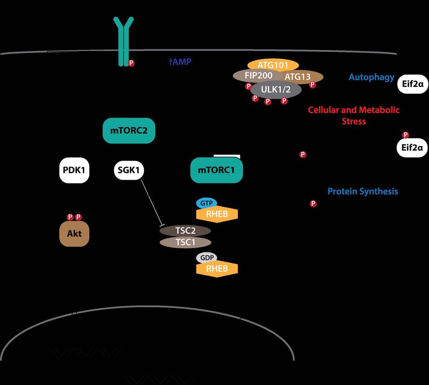
Figure 1. Signaling pathways downstream of insulin/IGF-1 receptor activation and crosstalk with catabolic signaling
pathways. Signaling pathways observed in multiple cells lines and tissues in vivo (black lines). Signaling pathways
observed in cancer cells resistant to PI3K inhibitors (grey lines).
Activated Akt acts directly on multiple targets to modulate cellular growth and
metabolic processes (reviewed in [29]). Akt phosphorylates TSC2 to repress the GAP
activity of the TSC1/TSC2 complex toward RHEB (Figure 1), maintaining RHEB in the
GTP-bound state (RHEB•GTP) to activate mTORC1 [72–74]. In cancer cells resistant to
PI3K inhibitors, SGK1 similarly phosphorylates TSC2 (Figure 1) to activate mTORC1 [75] in
response to direct phosphorylation at S422 by mTORC2 [76,77] or PDK1 [78,79]. In addition,
direct phosphorylation of PRAS40 at T246 by Akt blocks its inhibition of mTORC1 [80–82].
Although PRAS40 can inhibit RHEB•GTP activation of mTORC1 [80], insulin signaling
in Tsc2 null MEFs expressing a phospho-deficient form of TSC2 failed to induce mTORC1
signaling despite the phosphorylation of PRAS40 at T246 [83], suggesting that TSC-RHEB
regulation of mTORC1 activation predominates. GSK3 is a constitutively active Ser/Thr
protein kinase with a large set of functionally diverse direct targets, the majority of which
are functionally inhibited or degraded following GSK3-directred phosphorylation [84,85].
FoxO transcription factors are associated with longevity in humans [86], in part, through
modulating the expression of genes involved in apoptosis, glucose metabolism, cell cycle,Genes 2021, 12, 688 4 of 26
DNA damage repair, and proteolysis [87]. FoxO proteins are exported from the nucleus
and sequestered in the cytoplasm when phosphorylated at their N-terminus and NLS
sequences (T24 and S256 on FoxO1) [88]. Akt phosphorylates both GSK3 [89,90] and FoxO
transcription factors [91,92] to inhibit their respective kinase and transactivating activities.
The mTOR-containing complex 1 (mTORC1) is a major effector of activated Akt,
stimulating the synthesis of proteins, lipids, and nucleotides, while inhibiting protein
degradation pathways [57]. Recruitment of the small ribosomal subunit to mRNA is depen-
dent upon binding of the mRNA 50 cap structure by the eukaryotic translation initiation
factor 4F (eIF4F) complex. Comprised of the initiation factors eIF4E, eIF4G, and eiF4A,
the eIF4F complex is critical for cap-dependent protein synthesis [93,94]. mTORC1 di-
rectly phosphorylates the eIF4E inhibitor 4E-BP1 at T37/46 to promote assembly of the
eIF4F complex on the 50 cap of mRNA [57,95,96]. In addition, mTORC1 phosphorylates
S6K1 at T389 [97,98], activating its kinase activity toward the 40S ribosomal protein S6
(rpS6) [99], eIF4B [100], and the eIF4B inhibitor PDCD4 [101] to stimulate protein synthe-
sis. Phosphorylation of eIF4B at S422 by S6K1/2 stimulates eIF4F activity by potentially
promoting the ATPase, RNA-binding, and RNA-helicase activities of eIF4A [93,100,102].
Akt/mTORC1 signaling promotes de novo lipid synthesis through promoting the nuclear
accumulation of SREBP to initiate the transcription of metabolic genes involved in fatty acid
synthesis [103,104]. Further, pyrimidine synthesis is stimulated through S6K1 phospho-
rylation of CAD [105], while mTORC1-dependent purine synthesis is through activation
of the mitochondrial tetrahydrofolate cycle, in part, through induction of MTHDF2 by
eIF2α-independent activation of ATF4 [106]. In response to cellular stress (e.g., amino acid
insufficiency, unfolded protein response of the endoplasmic reticulum) Eif2α is phospho-
rylated at S51 (Figure 1) to both inhibit cap-dependent mRNA translation and to initiate
the transcription of stress response genes including FGF21, SQSTM1, ATG3, ATG12, and
Sestrin2 (Sesn2) via ATF4 [107–109]. Lastly, autophagy is an essential process for the main-
tenance of cellular homeostasis that may be further induced in response to cellular stress
to reduce organelles and macromolecules into amino acids, nucleosides, fatty acids and
sugars in support of cellular growth and survival [110]. Under growth conditions, active
mTORC1 negatively regulates autophagy (Figure 1) through its direct phosphorylation of
ULK1 at S757 [111] and Atg13 at S258 [112,113].
Nutrient and energy availability is a critical regulator of mTORC1. Increases and
decreases in amino acids leucine and arginine positively and negatively modulate mTORC1
kinase activity via CASTOR1, Sestrin2, and SLC38A9 [114–118], thus providing a critical
feedback mechanism to modulate mTORC1 activity in response to nutrient availability.
Similarly, under low ATP conditions, AMPK may negatively modulate mTORC1 kinase
activity (Figure 1) through phosphorylation of the mTORC1-specific constituent scaffolding
protein Raptor [119] or through the phospho-activation of TSC2 [120,121]. Further, AMPK
phosphorylates mTOR at S1261 within mTORC2, while AICAR-induced AMPK activation
both increases and decreases mTOR autophosphorylation at S2481 when associated with
Rictor and Raptor, respectively [62]. Although the mTOR S1261 phosphorylation was found
to be dispensable for the AMPK-dependent increase in mTORC2 catalytic activity, AMPK-
mTORC2 signaling was found to promote cell survival following glucose withdrawal.
Multiple human studies have indicated significantly reduced rates of mixed, myofib-
rillar and/or mitochondrial protein synthesis in the muscles of elderly subjects [122–129].
However, other more recent human studies show little or no difference in basal muscle
protein synthesis rates between the young and elderly [8,130–136]. While differences in
physical activity level, genetic makeup, and nutritional input clearly contribute to these
earlier observations, the previously reported >30% reductions in basal muscle protein
synthesis rates [122–129] may not be reflective of normal physiological aging. Differences
in muscle biopsy locations, analysis methods accounting for myofibrillar and total protein
synthesis rates, and/or subject screening for average physical activity levels, nutritional
intake, and overall health may similarly contribute. Additional studies with larger co-Genes 2021, 12, 688 5 of 26
horts utilizing methods with improved sensitivity to protein synthesis while incorporating
measures of protein degradation (e.g., proteosomal and autophagic flux) are warranted.
Resistance training increases muscle protein synthesis in both young and elderly men and
women [122,124,126,128,137–140]. Exogenous and post-prandial amino acids increase muscle
protein synthesis in both young and elderly men and women at baseline [8,133,141–143], while
providing an additive effect post exercise [144]. However, the efficacy of both exercise and
amino acids on stimulating muscle protein synthesis is blunted in the elderly [8,139,145],
a phenomenon referred to as anabolic resistance. This occurs despite increased plasma
concentrations of leucine in elderly subjects, as compared to young, following both exercise
and ingestion of essential amino acids [8]. Further, elderly individuals, as compared
to young controls, have blunted inductions of Akt phosphorylation at T308 and S473,
S6K1 phosphorylation at T389, and 4E-BP1 phosphorylation at T37/46 after both exercise
alone, and amino acid supplementation after exercise [145,146], strongly suggesting that
PI3K/Akt/mTOR signaling dysregulation mechanistically contributes to anabolic resistance.
In vivo mechanistic studies in animal models add further support to this notion. Re-
ductions in Akt phosphorylation at S473 and S6K1 phosphorylation at T389 are observed in
the gastrocnemius of old mice as compared to the young controls [147]. This study further
employed targeted transcriptomic and metabolomic analyses, revealing dysregulation of
fatty acid and glucose metabolic processes in old mice. In contrast, significant increases
in basal Akt phosphorylation at S473 [148] and S6K1 phosphorylation at T389 [149], are
observed in the muscles of 33- and 24-month old rats, respectively. RpS6 phosphorylation
at S240/244 is similarly increased in the 24-month old rats [149], indicating activation of
a subset of anabolic signaling branchpoints. It should be noted that the increase in Akt
S473 phosphorylation at 33-months was observed in the slow oxidative soleus, rather than
fast glycolytic muscles predominantly examined for Akt signaling. Interestingly, the slow
oxidative myofibers of the soleus, similar to fast glycolytic myofibers of the gastrocnemius
of rats, positively modulate Akt phosphorylation at S473 following sciatic nerve stimu-
lation [150] and 30 min of treadmill exercise [151]. Importantly, glycogen content in the
soleus (65% reduction) was reduced to a greater extent than in the red (47% reduction)
and white (23% reduction) gastrocnemius following treadmill exercise [151], suggesting
that insulin-stimulated PI3K/Akt-regulated glucose uptake and glycogen storage is simi-
larly activated in oxidative muscles in response to exercise. However, the extent to which
Akt signaling modulates oxidative muscle metabolism through its influence on PGC-1α
transcriptional activity in vivo [152], is uncertain and warrants future investigation.
The percentages of histologically abnormal myofibers, and myofibers staining positive
for both phosphorylated rpS6 and active caspase-3 are increased in the muscles of both
old mice and humans [153]. Counterintuitively, muscle-specific activation of mTORC1
(Tsc1 knockout) in mice induces progressive myofiber atrophy, phenocopying many of
the histological features of aged muscles including increases in both myofiber vacuolation
and the number of caspase-3 positive myofibers [153,154]. Similarly, pharmacological
inhibition of mTORC1 reduced myofiber atrophy and the expression of both Atrogin-1 and
MuRF1 in a mouse model of cancer cachexia [155]. In contrast, short-term activation of
mTORC1 (TSC2 knockdown) specifically in skeletal muscle induced myofiber hypertrophy
and greatly increased hypertrophy on recovery from transient denervation [156]. Con-
sistent with these observations, muscle-specific inhibition of mTORC1 (Raptor knockout)
in mice induced a leftward shift in the myofiber size distribution curve and histological
features including centralized nuclei and vacuoles, and myofiber degeneration [157]. These
apparently discordant observations with respect to myofiber size may more accurately be
rationalized through the effects of Akt/mTORC1 signaling on metabolism, rather than
simply the balance of protein synthesis and protein degradation. Short-term activation of
Akt induces myofiber hypertrophy [51,52] in parallel with a metabolic shift from glycolysis
and oxidative phosphorylation toward the accumulation of branched chain amino acids
and enhancement of the pentose phosphate pathway (PPP) [158]. Importantly, PPP pro-
duces ribose 5-phosphate and NADPH which are critical for the production of nucleotides,Genes 2021, 12, 688 6 of 26
fatty acids, sterols, non-essential amino acids, and cellular antioxidant defenses via re-
duced glutathione [159]. These observations would suggest that the hypertrophic effects
of short-term Akt/mTORC1 signaling become deleterious when prolonged, prioritizing
the synthesis of anabolic precursors rather than energy production, and leading to cellu-
lar and metabolic stress. Increased expression of pro-inflammatory cytokines (e.g., Ccl2,
Ccl3, Il10) and metabolic stress indicators (e.g., FGF21, GDF3) in the hypertrophic muscles
of Akt transgenic mice [158], and in atrophied muscles in which Akt is observed to be
constitutively active [160], further support this notion.
Treatment with rapamycin both extends the lifespan of mice and fruit flies [161–163]
and blocks resistance exercise-mediated increases in protein synthesis in humans [164,165].
Akt phosphorylation at both T308 and S473 are increased in muscle following a 16-h fast
in 24- and 30-month old male mice with respect to 6-month old controls [166]. Male mice
in this study also had significant increases in S6 phosphorylation at S240/244, whereas in
contrast, aged female mice only showed an increase in Akt S473 phosphorylation without
the increase in S6 phosphorylation after fasting [166]. Though suggestive of positive
regulation of Akt via mTORC2 following a fast in aged mice, interpretation of these
observations may be confounded by multiple variables including fasting duration, species,
gender, systemic and muscle metabolic flux, as well as housing and dietary conditions
throughout the lifespan of experimental animals. It is important to note that fasting in
young healthy mice, in which systemic control of blood sugar and insulin is physiologically
normal, induces the positive regulation of proteolytic processes through increased FoxO
transcriptional activity [167] and AMPK induction of autophagy through phosphorylation
of ULK1 at Serines 317, 555, and/or 777 (Figure 1) [111,168,169] while inhibiting protein
synthesis [49,170]. Consistent with this, pharmacological inhibition of mTORC1 restored
muscle histological and functional parameters in Tsc1 mutant mice [154,171] and in aged
rats [149], in part, through restoration of autophagy.
3. Muscle Proteolytic Processes and Negative Regulation of Anabolic Processes
Aging and aging-related diseases are linked to impaired proteome integrity or pro-
teostasis [172,173]. Acute and gradual increases in oxidative damage negatively impact
protein stability and enzymatic function [174–177]; 40–50% of all proteins in old organisms
are estimated to have some form of oxidative damage [178]. Similarly, the accumulation of
misfolded proteins is known to contribute to the development of age-associated patholo-
gies including Alzheimer’s disease, Huntington’s disease, and sporadic inclusion-body
myopathy [172,173,179,180].
Cells have evolved multiple protein degradation processes to maintain proteome
integrity or proteostasis in response to endogenous and exogenous stresses that negatively
impact the protein functionality due to damage or misfolding. Approximately 60–80% of
protein degradation in growing mammalian cells occurs through 26S proteasome cataly-
sis [181–184]. Specificity is achieved through the selective polyubiquitylation of substrates
by specific E3 ubiquitin ligases. While the proteasome can degrade some non-ubiquitinated
proteins, ubiquitination may function independently of the proteasome by selectively
directing proteins (typically membrane proteins) to the lysosome for proteolysis [184].
Similarly, autophagy reduces organelles and macromolecules into amino acids, nucleo-
sides, fatty acids, and sugars in support of cellular growth and survival [110]. The core
autophagy-related proteins essential for formation of the autophagosome formation and
delivery of autophagic cargo to the lysosome may be grouped by functionally: (1) initiation
and phagophore nucleation proteins ULK1, ULK2, ATG1, FIP200, ATG13, VPS34, ATG9,
Beclin, (2) phagophore expansion proteins ATG3, ATG4, ATG7, ATG9, ATG10, ATG12, (3)
cargo sequestration and membrane sealing proteins ubiquitin, cardiolipin, p62/SQSTM1,
PE-conjugated LC3s and GABARAP, and (4) autophagosome maturation and lysosome
fusion proteins ATG4, ATG14, VCP, and PE-conjugated LC3s and GABARAP [185,186].
In humans and animal models, these proteolytic processes become dysregulated with
pathologies associated with muscle atrophy including cancer cachexia, chronic renal fail-Genes 2021, 12, 688 7 of 26
ure, diabetes, denervation, and age [187–189]. Atrophying muscles in old [190] and in
young thyroidectomized and tumor bearing rats [191] have increased levels of protein
polyubiquitylation. Consistent with the experimental models of disuse muscle atrophy in
mice [192], increased levels of protein polyubiquitylation are observed in the muscles of
young human volunteers following an extended 20-day bed rest [193]. Gene expression
analyses during the early stages of muscle wasting due to denervation, disuse, diabetes,
cachexia, and fasting have identified a common set of genes, referred to as atrogenes, whose
expression is coordinately induced or repressed under conditions of muscle atrophy or
growth. Atrogenes whose expression is increased in atrophying muscles include ubiquitin,
core components of the 26S proteosome, eIF4E inhibitor 4E-BP1 (Eif4ebp1), transcription
factors Atf4, Foxo1, and Foxo3 [194,195], the muscle specific E3 ubiquitin ligases MurF1
(Trim63) and Atrogin-1/MAFbx (Fbxo32) [196,197], and autophagosome components LC3
(Map1lc3a) and Gabarap [194,198,199]. Importantly, animal models indicate that both insuf-
ficient [200–203] and excessive [198,199,204] proteolysis, both proteasomal and autophagic,
negatively impact the muscle mass and functionality, suggesting an inflection point be-
tween a level of proteolysis that supports muscle homeostasis and that which promotes
muscle wasting.
Autophagy and ATF4 are both positively regulated in response to cellular stress
(Figure 2) and major effectors of the integrated stress response (ISR). The ISR is a multi-
faceted signaling pathway that concurrently represses cap-dependent protein synthesis
via Eif2α while inducing the translation of select mRNAs which contain short inhibitory
upstream open reading frames (uORFs) in their 50 -untranslated regions including ATF4,
DDIT3 (CHOP), and PPP1R15A (GADD34) [205]. The Eif2α kinases PERK, PKR, and
HRI converge to phosphorylate Eif2α at S51 in response to the unfolded protein response
of the endoplasmic reticulum (UPRER ) [206,207], dsRNA [208,209], and heme depriva-
tion [210,211], respectively. In addition, amino acid sensing plays a key role in both
activating the ISR as well as inhibiting mTORC1 (Figure 2). Reduced levels of amino
acids lead to both an increase in the levels of uncharged tRNAs thereby activating GCN2
kinase activity toward Eif2α [212], and a concurrent reduction in mTORC1 activity via
Sestrin2 [115], CASTOR1 [213] and SLC38A9 [214–216].
FoxO transcription factors are positive regulators of Atrogin-1, MuRF1, LC3, GABARAPs,
ULK2, ATG12, and Beclin [198,199,217,218], thus indicating their central role in promoting
proteolytic processes. Transgenic overexpression of Foxo1 specifically in skeletal muscle
results in mice with decreases in muscle mass, functionality, and sarcomeric protein ex-
pression [219], while muscle specific Foxo1 knockout mice were largely protected from
nephrectomy and dexamethasone-induced muscle atrophy [220]. Muscle-specific genetic
deletion of all three muscle Foxo transcription factors (Foxo1, Foxo3, and Foxo4) abolished
both fasting [221] and insulin-deficient diabetes [222] induced muscle loss and weak-
ness. Consistent with these observations, heterozygous deletion of either Atrogin-1 or
MuRF1 results in mice that are partially protected from denervation-associated muscle
atrophy [196]. Similarly, overexpression of a constitutively active form of FoxO3 in tibialis
anterior muscles markedly increased the autophagosome formation and myofiber atrophy
while siRNAs targeting Foxo3 and overexpression of a dominant negative form of FoxO3
blocked fasting-induced autophagy [198].Genes 2021, 12, 688 8 of 26
Figure 2. Nutrient sensing pathways modulate protein synthesis. Cells respond to cellular stress signals to both inhibit
protein synthesis and increase the expression of genes to alleviate cellular stress.
Proteins may also be cleaved by members of the caspase family of proteases which
are known to play key roles in regulating apoptosis, necrosis, and inflammation [223,224].
Perhaps best known for its role in the activation of IL-1β [224], caspase-1 enzymatic activity
is observed to be significantly increased in the muscles of 24-month old mice as compared
to 10-month old controls [225]. Interestingly, genetic deletion of Nlrp3 blunted both the
age-associated changes in muscle mass and caspase-1 activity, respectively [225], thus
demonstrating a mechanistic link between the NLRP3 inflammasome and age-related
muscle atrophy. The muscles of 24-month old rats show no statistical differences in either
cytosolic cytochrome c levels or cytosolic caspase-3 activity from 6-month old controls [226],
suggesting that mitochondrial-mediated caspase activation is not present at this timepoint.
Further, unlike in liver homogenates, cytochrome c stimulation of muscle homogenates
from 6- and 24-month old rats was unable to increase caspase-3 enzymatic activity, sug-
gesting that additional factors may be necessary for caspase-3 activation in muscle. Acute
insulinopenia stimulates non-lysosomal, ATP-, and proteasome-dependent proteolysis in
rats [227]. Subsequent studies identified caspase-3 as the protease likely responsible for
proteolyzing actomyosin into the 14-kDa actin fragments observed in the atrophying mus-
cles of diabetic rats [228]. Subsequent studies identify caspase-3 as the protease responsible
for proteolyzing actomyosin into the 14-kDa actin fragments observed in the atrophying
muscles of diabetic rats [228] and following denervation injury in mice [229]. Importantly,
the enzymatic activities of caspases-3, -6, -8, and -9 were found to be significantly increased
in the gastrocnemius of cachexic (MAC16 tumor-bearing) as compared to non-cachexicGenes 2021, 12, 688 9 of 26
(MAC13 tumor-bearing) mice [230]. Collectively, these studies suggest that increased
caspase activity may play a causal role in the muscle atrophy associated with systemic
metabolic dysfunction.
Sporadic inclusion body myopathy (sIBM) is the most common degenerative muscle
disease in patients aged 50 years or more, and is often referred to as sporadic inclusion-
body myositis due to high levels of immune cells, including cytotoxic CD8+ T cells, in the
endomysium [231]. sIBM patients as a group respond poorly to anti-dysimmune thera-
pies [232] suggesting that immune system dysfunction is likely not causal for sIBM, but
rather exacerbates sIBM severity and/or progression. Muscle degeneration in sIBM is
characterized by vacuolization and intra-myofiber accumulation of misfolded and polyu-
biquitinated protein aggregates including amyloid-β (Aβ), SQSTM1, LC3, TDP-43, HSP70,
and proteasomal subunits [179,233–237]. In addition, biochemical analyses reveal an in-
crease in LC3-II and SQSTM1 expression along with a decrease in the enzymatic activity of
lysosomal enzymes Cathespin B and D despite their increased expression [236]. However,
though the increase in LC3-II is indicative of mature autophagosomes, the increase in
SQSTM1 would rather suggest a decrease in autophagic flux [238]. Consistent with these
observations, transgenic mice overexpressing R155H or A232E VCP mutants commonly
observed in humans with IBM associated with Paget’s disease recapitulate the progressive
functional and morphological deficits observed in sIBM patients [239]. Treatment of VCP
A232E transgenic mice with arimoclomol restored grip strength and maximal EDL tetanic
force to near WT levels and markedly reduced ubiquitin and TDP-43 aggregates [240],
indicating that dysregulated proteolysis plays a significant role in the pathogenesis of sIBM.
4. Unexpected Links
RNA-binding proteins (RBPs) play essential roles in nearly every aspect of cell biology
through their direct influence on post-transcriptional gene expression [241–243], transla-
tional control [244], RNA quality and stability [245], and RNA splicing [246]. A census
of human RBPs revealed that ~8% of protein coding genes are directly involved in RNA
metabolism (the collective events in the life cycle of RNA molecules including synthesis,
secondary structure modulation, base modification/editing, processing, and degradation),
either through direct interaction or as essential components of ribonucleoprotein (RNP)
complexes [247]. Importantly, alterations in RNA metabolic processes are increasingly
being recognized for its contributions to physiological aging and age-associated disease.
The roles of RBPs in skeletal muscle typically have been studied in the context of develop-
mental and/or disease processes. Identifying mechanistic links between the activities of
RBPs and the cellular processes modulating skeletal muscle physiology in the context of
aging is an ongoing pursuit. Here, we will highlight recently identified, and potentially
new links, between the activity of RBPs, biological aging, and skeletal muscle.
Altered post-transcriptional gene expression regulation, in part through changes in
miRNA expression, play causal roles in the cellular and tissue morphological changes in
the cardiovascular systems of the elderly [248–250]. SNPs in A-to-I RNA editing genes
ADARB1 and ADARB2 are associated with longevity in both humans and Caenorhabditis
elegans [251], and alterations in the editing of the transcriptome is implicated in the etiology
of disease [252,253]. In mice, genetic deletion of Adar induced lethality around ED11.5
due to severe defects in hematopoiesis and disintegration of the liver [254]. Similarly,
mice in which Adarb2 is genetically deleted develop normally, but die between P0 and
P20, becoming progressively seizure prone by P12 [255]. In addition, proteome analyses
of 7- and 30-month old rat gastrocnemius muscles indicates a significant increase in the
expression of the C-to-U RNA editing enzyme APOBEC2 [256], suggesting that RNA
editing in general, and specific subsets in particular (e.g., A-to-I, C-to-U) are temporally
regulated in a cell type and context-dependent manner. Interestingly, such regulation
may be in response to IIS signaling. ADAR2 expression in pancreatic islets, as well as
ADAR2-targeted editing of GluR-B RNA, is positively regulated in obese insulin resistant
mice with hyperinsulinemia [257]. A recent study identified ADAR1 T738 (Adar) andGenes 2021, 12, 688 10 of 26
ADAR2 T553 (Adarb2) as being direct targets of Akt kinase activity in cultured human cell
lines [258]. Akt inhibition increased, whereas phospho-mimetic mutants of the ADAR1
and ADAR2 Akt sites reduced the RNA editing of select transcripts including NEIL1, CCNI,
AZIN1, and CYFIP2. Interestingly, millions of RNA editing sites have been identified in
human cells [259,260]. However, the vast majority of editing sites in humans and primates
are observed in the ubiquitous inverted repeat Alu elements largely found in noncoding
regions of the genome [260]. Further, and in-depth cDNA sequencing analysis of the brain
of a 67-year old male concluded that less than 119 of the transcript sequence variants
identified from 541,777 nt of exon sequences analyzed are likely due to post-transcriptional
RNA editing [261]. Although these observations would suggest that RNA editing plays
a negligible role in introducing post-transcriptional amino acid substitutions in humans,
additional studies in young and old individuals are necessary to determine whether RNA
editing plays an extensive role in biological aging.
Alterations in RNA processing steps including splicing, poly-adenylation, and 50 cap-
ping) are associated with both physiological aging and age-associated disease [20,262–265].
It is estimated that >95% of human multi-exon genes are subject to alternative splicing [266].
Brain, heart, and skeletal muscle have the highest levels of evolutionarily conserved al-
ternatively spliced transcripts [267], and age-associated changes in splicing are observed
in the transcriptomes of muscles from aged humans [20,268,269] and mice [270]. Impor-
tantly, changes in alternative splicing may drive cellular dysfunction through changes in
the binding properties, enzymatic activities, and intracellular localization of a large per-
centage of the proteome [20,262,271], and is implicated as a causal mechanism of cellular
aging through its impact on both metabolic processes and DNA repair [272]. In mammals,
~40 proteins and 5 small nuclear RNAs form the core spliceosomal complex [273,274],
which is dynamically regulated by the activities of multiple RBPs, including members of
the SR protein [275] and heterogeneous nuclear ribonucleoprotein (hnRNP) [276] families.
Individual transcript splicing is modulated, in part, by cis-acting elements within the
nascent transcript (e.g., exonic splicing enhancers/silencers—ESE/ESS, intronic splicing
enhancers/silencers—ISE/ISS) and secondary structure [277]. Importantly, tissue-specific
and ubiquitously expressed RBPs cooperatively interact with the regulatory elements both
within the nascent transcript and of the spliceosome for context- and tissue-specific mRNA
splicing [246,278,279].
Genetic mutations in the splicing factor gene RBFOX1 have been observed in patients
with neurological disorders [280] and in an autistic individual with muscle weakness [281].
Mice in which Rbfox1 has been specifically deleted in skeletal muscle have decreases in
muscle mass and maximal force generation [282], thus establishing a role for Rbfox1 in
muscle development and physiology. In addition, mice in which both Rbfox1 and Rbfox2
are specifically deleted in adult mice, experienced rapid losses of muscle mass, myofiber
cross sectional area, and strength, due in part, to reductions in autophagy and the aberrant
expression of calpain-3 [283]. Further, siRNAs targeting Rbfox1 and/or Rbfox2 significantly
impaired myogenesis in the C2C12 model system through its direct role in promoting
the muscle-specific splicing of Mef2d [284]. Collectively, these studies establish Rbfox1/2
as essential for muscle developmental processes and the maintenance of muscle mass
and strength in adulthood. Whether changes in the expression or activity of these factors
contribute to age-associated muscle atrophy is uncertain.
Amyotrophic lateral sclerosis (ALS) is a progressive age-related neurodegenerative
disease in which motor neurons are selectively degenerated. Both reductions in nu-
clear/cytoplasmic ratio of the splicing factor SFPQ, and the aberrant retention of SFPQ
introns, are observed in motor neurons that were differentiated from ALS patient-derived
iPSCs [285], thus suggesting a causal role for SFPQ in the pathogenesis of ALS. Neuron-
specific deletion of Sfpq in mice led to impaired transcriptional elongation, an effect
most pronounced on long genes (>100 kb), and gross developmental abnormalities in
the brain [286]. Skeletal muscle-specific deletion of Sfpq similarly induced long gene tran-
scriptopathy and premature death starting around P30 [287]. Between P14 and P30, theseGenes 2021, 12, 688 11 of 26
mice exhibited significant skeletal muscle growth defects, attributable to the combination
of an increase in the percentage of oxidative myofibers, accumulation of excess glycogen,
and a decrease in the abundance of OXPHOS complexes I, II, and IV. Although reductions
in mitochondrial oxidative capacity are observed in aged and sarcopenic muscles [288–290],
it is unknown whether dysregulation of SFPQ expression or activity plays a causal role.
A rare cohort of patients have been identified in which heterozygous mutations in HN-
RNPU are associated with intellectual disability and muscle weakness [291–294]. hnRNP-U
is a multifunctional RBP, directly involved in regulating alternative splicing [295–297],
genome architecture [298], X-chromosome inactivation [299], and is known to promote
the DNA base repair through its interaction with NEIL1 [300]. In mice, hnRNP-U protein
expression decreases with advancing age in both the heart [301] and skeletal muscle [160],
suggesting that loss of hnRNP-U may contribute to age-associated pathologies in these
tissues. Mice in which Hnrnpu is deleted in striated muscles (Ckm-Cre) die around P14 due
to a dilated cardiomyopathy (DCM) phenotype, whereas mice in which Hnrnpu is deleted
specifically within cardiomyocytes (Myh6-Cre) die around P10 due to a similar DCM pheno-
type [301]. Mice in which Hnrnpu is deleted specifically in skeletal muscle (ACTA1-Cre) are
born phenotypically normal, but by 3-months of age develop a myopathy-like phenotype
characterized by selective muscle atrophy of glycolytic muscles and histopathological ob-
servations of central nuclei, myofiber degeneration, intracellular inclusions, and extensive
fibrosis [160]. Changes in the expression and splicing of genes are observed in both cardiac
(Ckm-Cre) and skeletal muscles (ACTA1-Cre). Interestingly, the atrophied skeletal muscles
(ACTA1-Cre) exhibit signs of metabolic stress. Akt is observed to be constitutive active
with its phosphorylation at S473 being desensitized to IGF-1 stimulation, SQSTM1 and
Ulk1 phosphorylation at S757 are significantly increased, and multiple genes involved in
the transport/import and biosynthesis of amino acids, as well as the repression of oxida-
tive metabolism are significantly reduced in expression [160]. Though high levels of Akt
S473 [149] and S6K1 T389 [149] phosphorylation are similarly observed in the atrophied
muscles of aged rats, it is unclear whether this is a compensatory effect, or a mechanistic
contributor, likely only at specific stages of muscle atrophy.
Splicing of the insulin receptor gene INSR is regulated in a cell-type specific context
that is dependent upon the development stage or disease condition. Alternative splicing of
exon 11 gives rise to protein isoforms INSR-A (exclusion of exon 11) and INSR-B (inclusion
of exon 11). INRS-A, which is prevalent during fetal development and in the nervous
system, preferentially binds IGF-2 over IGF-1 and insulin, while INRS-B, which is prevalent
in adulthood and highly expressed in adipose tissue, liver and skeletal muscle, is more
sensitive to insulin [302–304]. The splicing of INSR is regulated by multiple RBPs including
CELF1, SRSF3, MBNL1, and hnRNPs A1, F, and H [305–308]. Patients with myotonic dys-
trophy (DM) frequently acquire muscle insulin resistance. Expansion of CTG (DM type 1)
and CCTG (DM type 2) repeats in DMPK [309] and ZNF9 [310], respectively, leads to the
dysregulation of alternative splicing through MBNL1 loss-of-function and CELF1 gain-
of-function [311]. Molecular analyses have identified hnRNP-H, MBNL1, and CELF1 as
modulating the increase in the INSR-A:INSR-B ratio observed in DM patients [308,312–314].
Although insulin resistance in skeletal muscle is the primary defect in type 2 diabetes [315],
and dysregulation of INSR splicing occurs in the insulin target tissues of patients with
insulin resistance [312,313], the role of INSR splicing in the pathogenesis of type 2 diabetes
is unclear [303]. Conflicting observations in which the transcript ratio of INSR-A:INSR-B
splice variants is either decreased [316] or unchanged [317,318] are reported. Similarly, it
has been reported that the INSR-A transcript is exclusively expressed in non-diabetics with
the induction of the INSR-B transcript observed in the muscles of type 2 diabetics [319]. To
the best of our knowledge, age-associated changes in muscle INSR protein isoform expres-
sion, and whether such isoform expression changes are correlated with the development of
type 2 diabetes in the elderly is unknown.
Treatment of Ewing sarcoma cells with the ATP-competitive PI3K and mTOR inhibitor
Dactolisib induced significant reductions in both cell growth and proliferation [320]. GenesGenes 2021, 12, 688 12 of 26
for RBPs with known roles in splicing modulation including FUS, HNRNPM, SFPQ, and
SRSF2 were significantly increased, while coimmunoprecipitation and cosedimentation
of hnRNP-M with core spliceosomal proteins were significantly enhanced following treat-
ment. Importantly, siRNA knockdown of HNRNPM blocked a subset of Dactolisib induced
changes in cassette exon splicing for exons containing proximal hnRNP-M consensus
motifs [320]. Interestingly, hnRNP-M expression is positively corelated with SGK1 phos-
phorylation, and its overexpression rescues the impaired differentiation of C2C12 myoblasts
treated with siRNA targeting Rictor [321]. Though hnRNP-M physically interacts with
Rictor, and siRNAs targeting either Rictor or HNRNPM repressed insulin-stimulated Akt
S473 and SGK1 S422 phosphorylation to a similar degree, the molecular mechanism under-
lying these observations is unknown. hnRNP-E2 (PCBP2) was identified in a two-hybrid
screen of a human bone marrow cDNA library with SIN1 (MAPKAP1), while siRNAs
targeting either SIN1 (MAPKAP1) or hnRNP-E2 (PCBP2) similarly potentiate an increase
in the percentage of apoptotic cells treated with either H2 O2 or TNFα [322]. Though en-
dogenous SIN1 and hnRNP-E2 coimmunoprecipitated in cell lysates and combined siRNA
treatments had an additive effect on both H2 O2 and TNFα induced apoptosis, suggesting
their participation in a common signaling pathway, the molecular mechanism, presumably
activating mTORC2, is unknown. SR protein SF2/ASF (SFRS1) positively modulates pro-
tein translation through an Akt-independent mechanism [323]. MEF and NIH 3T3 cells
overexpressing SF2/ASF had significantly increased S6K1 and 4E-BP1 phosphorylation
without increases is Akt phosphorylation at S473. Interestingly, SF2/ASF physically binds
to both mTOR and the catalytic subunit of the PP2A phosphatase [324] suggesting that
it may facilitate mTORC1-mediated phosphorylation of 4E-BP1 by either recruiting it to
the mRNP complex via direct binding of mRNA ESE or perhaps by indirect inhibition of
PP2A thereby biasing the phosphorylation-dephosphorylation kinetics of 4E-BP1 toward
phosphorylation. Intriguingly, these observations suggest that RBPs involved in nuclear-
cytoplasmic shuttling of RNA may positively regulate mTOR-dependent cell survival
and growth processes, perhaps as part of a feed forward mechanism to insure selective
translation of cell survival and growth transcriptional programs.
5. Conclusions
Worldwide, the number of individuals aged 65 years or older is expected to more than
double by the year 2050 [325]. In the U.S. alone, it is estimated that approximately 45% of
adults greater than 60 years of age have some degree of sarcopenia [326], contributing to
the impaired ability to perform activities of daily living and the loss of independence in
the elderly population. In contrast with other well-studied diseases, and despite the urgent
need by an aging population, muscle atrophy and weakness are frequently considered signs
of “normal aging.” Though it is managed by a regimen of resistance training and increased
protein intake, only a subset of sarcopenic individuals see improvements in muscle mass,
strength, and functionality [11,327]. In the nearly 30 years since the identification of
mTOR [328–330], the complex and often interrelated signaling pathways regulating skeletal
muscle mass, strength, and functionality have begun to be more clearly defined. New
mechanisms and linkages continue to be discovered, defining new regulatory circuits while
further refining the established pathways. Clearly, there has been much exciting progress in
our understanding of these pathways and their contribution to muscle wasting disorders,
but much remains to be done.
Author Contributions: Writing—original draft preparation, E.-J.L. and R.L.N.; writing—review
and editing, E.-J.L. and R.L.N. All authors have read and agreed to the published version of
the manuscript.
Funding: This research received no external funding. The APC was funded by Brigham and Women’s
Hospital Department of Orthopaedic Surgery.
Institutional Review Board Statement: Not applicable.
Informed Consent Statement: Not applicable.Genes 2021, 12, 688 13 of 26
Data Availability Statement: Not applicable.
Acknowledgments: The authors would like to thank Julia Charles for helpful discussions and feedback.
Conflicts of Interest: The authors declare no conflict of interest.
Abbreviations
Abbreviation Full name
Aβ amyloid-β
ADARB1 adenosine deaminase RNA specific B1
ADARB2 adenosine deaminase RNA specific B2
ADAR adenosine deaminase RNA specific
APOBEC2 apolipoprotein B mRNA editing enzyme catalytic subunit 2
CAD carbamoyl-phosphate synthetase 2, aspartate transcarbamoylase, dihydroorotase
FoxO1 Forkhead boxO transcription factor 1
FoxO3 Forkhead boxO transcription factor 3
GAP GTPase activating protein
GCN2 General control nonderepressible 2 kinase
GH Growth hormone
GPCR G-protein-coupled receptor
GSK3 glycogen synthase kinase 3
HRI Heme-regulated inhibitor kinase
IGF-1 Insulin like growth factor 1
IIS Insulin/IGF-1 signaling
miRNA microRNA
MTHFD2 methylenetetrahydrofolate dehydrogenase 2
mTORC1 mTOR containing complex 1
mTORC2 mTOR containing complex 2
PERK PKR-like ER kinase
PI3K phosphoinositide 3-kinase
PKR Protein kinase RNA-activated
PRAS40 proline-rich AKT substrate of 40 kDa
RHEB Ras homolog enriched in brain
rpS6 ribosomal protein S6
RTK Receptor tyrosine kinases
S6K1 ribosomal protein S6 kinase 1
sIBM sporadic inclusion body myopathy/myositis
SNP single nucleotide polymorphism
ULK1 unc-51 like autophagy activating kinase 1
ULK2 unc-51 like autophagy activating kinase 2
References
1. U.S. Cancer Statistics Working Group. US Cancer Statistics: 1999–2009 Incidence and Mortality Web-Based Report; USDHHS, CDC:
Atlanta, GA, USA, 2013.
2. Niccoli, T.; Partridge, L. Ageing as a risk factor for disease. Curr. Biol. 2012, 22, R741–R752. [CrossRef] [PubMed]
3. Centers for Disease Control and Prevention. The State of Aging and Health in America 2013; Centers for Disease Control and
Prevention, US Dept of Health and Human Services: Washington, DC, USA, 2013.
4. Heron, M. Deaths: Leading causes for 2010. In National Vital Statistics Reports: From the Centers for Disease Control and Prevention;
National Center for Health Statistics, National Vital Statistics System: Hyattsville, ML, USA, 2013; Volume 62, pp. 1–97.
5. Caspersen, C.J.; Thomas, G.D.; Boseman, L.A.; Beckles, G.L.; Albright, A.L. Aging, diabetes, and the public health system in the
United States. Am. J. Public Health 2012, 102, 1482–1497. [CrossRef]
6. Facchini, F.S.; Hua, N.; Abbasi, F.; Reaven, G.M. Insulin resistance as a predictor of age-related diseases. J. Clin. Endocrinol. Metab.
2001, 86, 3574–3578. [CrossRef] [PubMed]
7. Rasmussen, B.B.; Fujita, S.; Wolfe, R.R.; Mittendorfer, B.; Roy, M.; Rowe, V.L.; Volpi, E. Insulin resistance of muscle protein
metabolism in aging. FASEB J. 2006, 20, 768–769. [CrossRef]
8. Cuthbertson, D.; Smith, K.; Babraj, J.; Leese, G.; Waddell, T.; Atherton, P.; Wackerhage, H.; Taylor, P.M.; Rennie, M.J. Anabolic
signaling deficits underlie amino acid resistance of wasting, aging muscle. FASEB J. 2005, 19, 422–424. [CrossRef]
9. Thompson, L.V. Age-related muscle dysfunction. Exp. Gerontol. 2009, 44, 106–111. [CrossRef]Genes 2021, 12, 688 14 of 26
10. Doherty, T.J. Invited review: Aging and sarcopenia. J. Appl. Physiol. (1985) 2003, 95, 1717–1727. [CrossRef]
11. Walston, J.D. Sarcopenia in older adults. Curr. Opin. Rheumatol. 2012, 24, 623–627. [CrossRef]
12. Keller, K.; Engelhardt, M. Strength and muscle mass loss with aging process. Age and strength loss. Muscles Ligaments Tendons J.
2013, 3, 346–350. [CrossRef]
13. Breen, L.; Phillips, S.M. Skeletal muscle protein metabolism in the elderly: Interventions to counteract the ‘anabolic resistance’ of
ageing. Nutr. Metab. 2011, 8, 68. [CrossRef]
14. Burd, N.A.; Gorissen, S.H.; van Loon, L.J. Anabolic resistance of muscle protein synthesis with aging. Exerc. Sport Sci. Rev. 2013,
41, 169–173. [CrossRef] [PubMed]
15. LeBrasseur, N.K.; Walsh, K.; Arany, Z. Metabolic benefits of resistance training and fast glycolytic skeletal muscle. Am. J. Physiol.
Endocrinol. Metab. 2011, 300, E3–E10. [CrossRef]
16. Lang, T.; Streeper, T.; Cawthon, P.; Baldwin, K.; Taaffe, D.R.; Harris, T.B. Sarcopenia: Etiology, clinical consequences, intervention,
and assessment. Osteoporos. Int. 2010, 21, 543–559. [CrossRef]
17. Ciciliot, S.; Rossi, A.C.; Dyar, K.A.; Blaauw, B.; Schiaffino, S. Muscle type and fiber type specificity in muscle wasting. Int. J.
Biochem. Cell Biol. 2013, 45, 2191–2199. [CrossRef]
18. Deschenes, M.R. Effects of aging on muscle fibre type and size. Sports Med. 2004, 34, 809–824. [CrossRef]
19. Skelton, D.A.; Kennedy, J.; Rutherford, O.M. Explosive power and asymmetry in leg muscle function in frequent fallers and
non-fallers aged over 65. Age Ageing 2002, 31, 119–125. [CrossRef] [PubMed]
20. Harries, L.W.; Hernandez, D.; Henley, W.; Wood, A.R.; Holly, A.C.; Bradley-Smith, R.M.; Yaghootkar, H.; Dutta, A.; Murray,
A.; Frayling, T.M.; et al. Human aging is characterized by focused changes in gene expression and deregulation of alternative
splicing. Aging Cell 2011, 10, 868–878. [CrossRef]
21. Zhang, R.; Chen, H.Z.; Liu, D.P. The Four Layers of Aging. Cell Syst. 2015, 1, 180–186. [CrossRef]
22. Lopez-Otin, C.; Blasco, M.A.; Partridge, L.; Serrano, M.; Kroemer, G. The hallmarks of aging. Cell 2013, 153, 1194–1217. [CrossRef]
23. Booth, L.N.; Brunet, A. The Aging Epigenome. Mol. Cell 2016, 62, 728–744. [CrossRef]
24. Barbieri, M.; Bonafe, M.; Franceschi, C.; Paolisso, G. Insulin/IGF-I-signaling pathway: An evolutionarily conserved mechanism
of longevity from yeast to humans. Am. J. Physiol. Endocrinol. Metab. 2003, 285, E1064–E1071. [CrossRef]
25. Song, G.; Ouyang, G.; Bao, S. The activation of Akt/PKB signaling pathway and cell survival. J. Cell Mol. Med. 2005, 9, 59–71.
[CrossRef] [PubMed]
26. Manning, B.D.; Cantley, L.C. AKT/PKB signaling: Navigating downstream. Cell 2007, 129, 1261–1274. [CrossRef] [PubMed]
27. Hemmings, B.A.; Restuccia, D.F. PI3K-PKB/Akt pathway. Cold Spring Harb. Perspect. Biol. 2012, 4, a011189. [CrossRef]
28. Franke, T.F.; Kaplan, D.R.; Cantley, L.C. PI3K: Downstream AKTion blocks apoptosis. Cell 1997, 88, 435–437. [CrossRef]
29. Manning, B.D.; Toker, A. AKT/PKB Signaling: Navigating the Network. Cell 2017, 169, 381–405. [CrossRef]
30. White, M.F. Insulin signaling in health and disease. Science 2003, 302, 1710–1711. [CrossRef]
31. Shaw, R.J.; Cantley, L.C. Ras, PI(3)K and mTOR signalling controls tumour cell growth. Nature 2006, 441, 424–430. [CrossRef]
32. Cantley, L.C. The phosphoinositide 3-kinase pathway. Science 2002, 296, 1655–1657. [CrossRef]
33. Brown-Borg, H.M. Hormonal control of aging in rodents: The somatotropic axis. Mol. Cell Endocrinol. 2009, 299, 64–71. [CrossRef]
34. Al-Regaiey, K.A.; Masternak, M.M.; Bonkowski, M.; Sun, L.; Bartke, A. Long-lived growth hormone receptor knockout mice:
Interaction of reduced insulin-like growth factor i/insulin signaling and caloric restriction. Endocrinology 2005, 146, 851–860.
[CrossRef]
35. Bartke, A.; Brown-Borg, H. Life extension in the dwarf mouse. Curr. Top. Dev. Biol. 2004, 63, 189–225. [CrossRef]
36. Brown-Borg, H.M.; Borg, K.E.; Meliska, C.J.; Bartke, A. Dwarf mice and the ageing process. Nature 1996, 384, 33. [CrossRef]
[PubMed]
37. Bartke, A.; Wright, J.C.; Mattison, J.A.; Ingram, D.K.; Miller, R.A.; Roth, G.S. Extending the lifespan of long-lived mice. Nature
2001, 414, 412. [CrossRef]
38. Ikeno, Y.; Bronson, R.T.; Hubbard, G.B.; Lee, S.; Bartke, A. Delayed occurrence of fatal neoplastic diseases in ames dwarf mice:
Correlation to extended longevity. J. Gerontol. A Biol. Sci. Med. Sci. 2003, 58, 291–296. [CrossRef]
39. Holzenberger, M.; Dupont, J.; Ducos, B.; Leneuve, P.; Geloen, A.; Even, P.C.; Cervera, P.; Le Bouc, Y. IGF-1 receptor regulates
lifespan and resistance to oxidative stress in mice. Nature 2003, 421, 182–187. [CrossRef] [PubMed]
40. Bluher, M.; Kahn, B.B.; Kahn, C.R. Extended longevity in mice lacking the insulin receptor in adipose tissue. Science 2003, 299,
572–574. [CrossRef]
41. Bokov, A.F.; Garg, N.; Ikeno, Y.; Thakur, S.; Musi, N.; DeFronzo, R.A.; Zhang, N.; Erickson, R.C.; Gelfond, J.; Hubbard, G.B.; et al.
Does reduced IGF-1R signaling in Igf1r+/- mice alter aging? PLoS ONE 2011, 6, e26891. [CrossRef] [PubMed]
42. Xu, J.; Gontier, G.; Chaker, Z.; Lacube, P.; Dupont, J.; Holzenberger, M. Longevity effect of IGF-1R(+/−) mutation depends on
genetic background-specific receptor activation. Aging Cell 2014, 13, 19–28. [CrossRef]
43. Mao, K.; Quipildor, G.F.; Tabrizian, T.; Novaj, A.; Guan, F.; Walters, R.O.; Delahaye, F.; Hubbard, G.B.; Ikeno, Y.; Ejima, K.; et al.
Late-life targeting of the IGF-1 receptor improves healthspan and lifespan in female mice. Nat. Commun. 2018, 9, 2394. [CrossRef]
44. Berryman, D.E.; List, E.O.; Coschigano, K.T.; Behar, K.; Kim, J.K.; Kopchick, J.J. Comparing adiposity profiles in three mouse
models with altered GH signaling. Growth Horm. IGF Res. 2004, 14, 309–318. [CrossRef]
45. Bartke, A.; Chandrashekar, V.; Bailey, B.; Zaczek, D.; Turyn, D. Consequences of growth hormone (GH) overexpression and GH
resistance. Neuropeptides 2002, 36, 201–208. [CrossRef] [PubMed]You can also read