Statin-Induced Myopathy: Translational Studies from Preclinical to Clinical Evidence - MDPI
←
→
Page content transcription
If your browser does not render page correctly, please read the page content below
International Journal of
               Molecular Sciences

Review
Statin-Induced Myopathy: Translational Studies from
Preclinical to Clinical Evidence
Giulia Maria Camerino 1 , Nancy Tarantino 1 , Ileana Canfora 1 , Michela De Bellis 1 , Olimpia Musumeci 2
and Sabata Pierno 1, *

                                          1   Section of Pharmacology, Department of Pharmacy and Drug Sciences, University of Bari “Aldo Moro”,
                                              70125 Bari, Italy; giuliamaria.camerino@uniba.it (G.M.C.); nancy.tarantino@uniba.it (N.T.);
                                              ileana.canfora@uniba.it (I.C.); michela.debellis@uniba.it (M.D.B.)
                                          2   Unit of Neurology and Neuromuscular Disorders, Department of Clinical and Experimental Medicine,
                                              University of Messina, 98122 Messina, Italy; omusumeci@unime.it
                                          *   Correspondence: sabata.pierno@uniba.it

                                          Abstract: Statins are the most prescribed and effective drugs to treat cardiovascular diseases (CVD).
                                          Nevertheless, these drugs can be responsible for skeletal muscle toxicity which leads to reduced
                                          compliance. The discontinuation of therapy increases the incidence of CVD. Thus, it is essential
                                          to assess the risk. In fact, many studies have been performed at preclinical and clinical level to
                                          investigate pathophysiological mechanisms and clinical implications of statin myotoxicity. Conse-
                                          quently, new toxicological aspects and new biomarkers have arisen. Indeed, these drugs may affect
                                          gene transcription and ion transport and contribute to muscle function impairment. Identifying a
                                          marker of toxicity is important to prevent or to cure statin induced myopathy while assuring the

                                   right therapy for hypercholesterolemia and counteracting CVD. In this review we focused on the
                                          mechanisms of muscle damage discovered in preclinical and clinical studies and highlighted the
Citation: Camerino, G.M.; Tarantino,
                                          pathological situations in which statin therapy should be avoided. In this context, preventive or
N.; Canfora, I.; De Bellis, M.;
Musumeci, O.; Pierno, S.
                                          substitutive therapies should also be evaluated.
Statin-Induced Myopathy:
Translational Studies from Preclinical    Keywords: statin; skeletal muscle; ion channels; myopathy; biomarkers
to Clinical Evidence. Int. J. Mol. Sci.
2021, 22, 2070. https://doi.org/
10.3390/ijms22042070
                                          1. Introduction
Academic Editor: Marek Droździk                The 3-hydroxy-3-methyl-glutaryl coenzyme A (HMG-CoA) reductase inhibitors, also
                                          called statins, have a relatively recent history, being discovered in 1970 by Endo [1]. They
Received: 29 December 2020
                                          are effective hypolipidemic drugs that act by inhibiting the production of mevalonate in
Accepted: 15 February 2021
                                          the biosynthetic pathway of cholesterol. Multicenter clinical trials have demonstrated that
Published: 19 February 2021
                                          these compounds are effective in reducing cardiovascular events and increase the rate
                                          of survival in patients [2]. Thus, statins represent the first line therapy in the prevention
Publisher’s Note: MDPI stays neutral
                                          and treatment of metabolic syndrome [3] and cardiovascular diseases (CVD), that are the
with regard to jurisdictional claims in
                                          primary causes of mortality in the world. Their beneficial role is linked to the reduction
published maps and institutional affil-
                                          of low-density lipoprotein (LDL) cholesterol in blood, resulting from the inhibition of
iations.
                                          cholesterol synthesis. Thus, based on their capacity to inhibit the HMG-CoA reductase ac-
                                          tivity and consequently the prenylated protein synthesis, these drugs have been evaluated
                                          for their anti-inflammatory and anticancer properties [4,5]. An anti-inflammatory effect
                                          of statins has been demonstrated in vitro by the inhibition of cytokines production (i.e.,
Copyright: © 2021 by the authors.
                                          IL-6, TNF-α) [6,7]. For this reason, these drugs have been recently included in treatment
Licensee MDPI, Basel, Switzerland.
                                          protocol for COVID-19 with the aim to control cytokine storm and to control the associated
This article is an open access article
                                          symptoms [8], however, their efficacy is still under study. The clinical use of statin is
distributed under the terms and
                                          limited by the adverse effects they can produce. These symptoms are often responsible for
conditions of the Creative Commons
Attribution (CC BY) license (https://
                                          the nonadherence or discontinuation of therapy, which may have striking consequences.
creativecommons.org/licenses/by/
                                          Indeed, a lower rate of mortality has been observed in patients who were adherent to
4.0/).
                                          statins, as compared to the low adherent ones [9]. Different side effects have been reported.

Int. J. Mol. Sci. 2021, 22, 2070. https://doi.org/10.3390/ijms22042070                                        https://www.mdpi.com/journal/ijms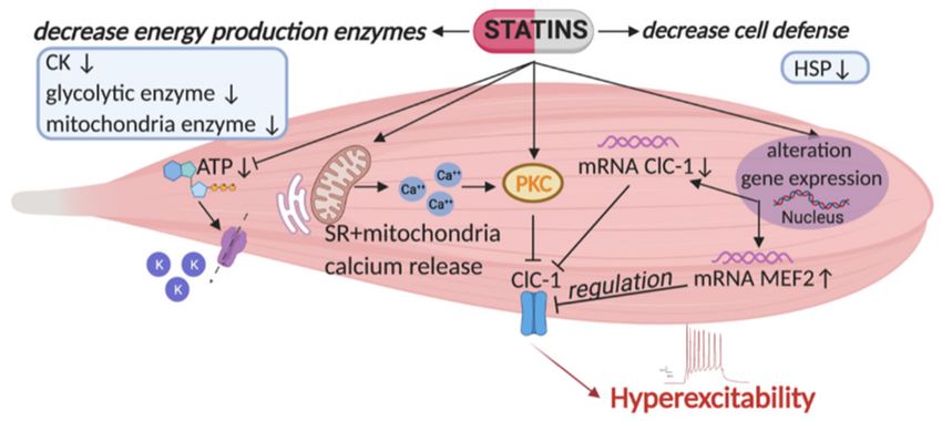
Int. J. Mol. Sci. 2021, 22, 2070                                                                                              2 of 19

                                   The term “statin intolerance” is referred to the incapacity of patients to tolerate a statin dose
                                   useful to contrast CVD. Adverse effects included headache, sleep disorders and peripheral
                                   neuropathy, gastro-intestinal distress, fatigue, arthritis, increase of liver enzymes, rash,
                                   alopecia, erectile dysfunction, gynecomastia [10]. Randomized clinical trials have also
                                   shown an increased risk of diabetes [11,12]. However, skeletal muscle symptoms are the
                                   most frequent and range from myalgia to life-threatening rhabdomyolysis. Skeletal muscle
                                   tissue is disseminated in all the body and its damage may lead to severe consequences [13].
                                   This is what happened with the use of cerivastatin, withdrawn from the market because of
                                   an increased risk of rhabdomyolysis, which caused muscle pain and weakness and 52 cases
                                   of renal failure and death [14]. Although new drugs are emerging (monoclonal antibodies,
                                   antisense oligonucleotides), statins remain the first line option for the treatment of hyperc-
                                   holesterolemia, making it essential to identify the causes of their toxicity. The European
                                   Atherosclerosis Society (EAS) Consensus Panel recently described the pathophysiology of
                                   statin-associated muscle symptoms (SAMS) and provided a guideline for diagnosis and
                                   management of the risk at the aim to ensure the right therapy [9]. In general, SAMS are
                                   not common to all statins since different factors may contribute to this condition (genetic,
                                   pharmacodynamic, pharmacokinetics, presence of comorbidity, polypharmacy). These
                                   recommendations can assist the clinicians in improving adherence to therapy and then
                                   the possibility that patients experiencing SAMS may receive the best cholesterol lowering
                                   therapy as to minimize CVD risk.

                                   2. The Causes of Statin-Induced Myopathy
                                         Although rare, SAMS can be severe. Observational studies describe the occurrence of
                                   myalgia in a low percent of patients, with rhabdomyolysis being rarer. Due to SAMS, it
                                   is often necessary to reduce the given dose, or to substitute statins, or even to completely
                                   stop therapy, thereby precluding the optimal lowering of plasma LDL level [15]. Many
                                   studies have reported that the stop of statin therapy leads to an increased occurrence of
                                   harmful cardiovascular events [16]. Sometimes, discontinuation of statin therapy may also
                                   be due to advice from friends or family members or on information from the lay media.
                                   Thus, at the aim to classify these symptoms and to identify the causes, many studies have
                                   been undertaken. The clinical description of SAMS covers a wide range of symptoms
                                   and the precise classification is still debated. Indeed, an accurate diagnosis of SAMS
                                   is hard to assess because symptoms are often subjective and there are no standardized
                                   diagnostic tests. The SAMS can arise after few weeks but also after several years of
                                   therapy, and the onset of symptoms can occur after an augmentation in statin dose or
                                   after the beginning of a concomitant therapy. Interestingly, the SAMS are more frequent
                                   in physically active individuals [17,18]. Thus, statin treatment needs to be personalized.
                                   Additionally, statins are generally considered as a class, but highlight that differences
                                   (pharmacokinetic and pharmacodynamic properties) exist among them and it should be
                                   taken into account. A list of adverse effects is available based on the results of clinical trials
                                   or on post-marketing surveillance [19]. The classification of adverse effects of statins in
                                   skeletal muscle is often different among authors and consensus groups. The definition
                                   of SAMS is an unexplained muscle pain, weakness or cramps (myalgia) accompanied
                                   or not by a creatine kinase (CK) increase in blood after statin therapy. The most feared
                                   statin-induced rhabdomyolysis is a grave form of myopathy with CK typically >40 times
                                   the normal value, which generally requires hospitalization, because of massive muscle
                                   fiber necrosis. This results in myoglobinuria that can cause a fatal acute renal failure [20].
                                         Different hypotheses have been proposed to understand the causes of myopathy
                                   induced by statin. First of all, it has been described that, due to statin effects, the depletion
                                   of cholesterol in muscle cell membranes may have an important role in causing myopathy,
                                   since its depletion may destabilize sarcolemma and alter ion balance [21,22]. Several lines
                                   of evidence have proposed that, due to mevalonate synthesis inhibition, also the reduction
                                   of intermediates of the cholesterol biosynthetic pathway, like farnesyl pyrophosphate
                                   and ubiquinone (coenzyme Q10) can be harmful for skeletal muscle and mitochondrial
Int. J. Mol. Sci. 2021, 22, 2070 3 of 19
function. Indeed, ubiquinone participates in the electron transport chain and in oxidative
phosphorylation in mitochondria. It is possible that the decrease of ubiquinone levels
causes a loss of ATP and energy production [23]. This is the reason why patients are
advised to take CoQ10 while receiving statin therapy. Additionally, the inhibition of
prenylation of small GTP-binding proteins like Rho, Ras, and Rac by geranylgeranyl
pyrophosphate has a role in the alteration of protein synthesis or organelle biogenesis,
signal transduction, and intracellular trafficking [24].
Additionally, the reduction of selenoprotein synthesis, due to the inhibition of the
mevalonate pathway, may be partially involved in the failing of myocyte regeneration
and in SAMS onset [25]. In addition, statin has been found to inhibit AKT/mTOR sig-
naling pathway, involved in muscle growth during development and regeneration. This
may be one of the causes of myotoxicity [26]. The adverse events of statins are dose
dependent and different predisposing factors are potentially involved in the generation
of myopathy, such as low body mass index (BMI), drug interaction, genetics, female sex,
alcohol abuse, hypothyroidism, fibromyalgia, extreme physical exercise, serum Vitamin
D deficiency [10]. The adverse events associated with statins occur essentially in patients
presenting comorbidities, due to drug interactions [27]. Previous studies found a higher
expression of atrogin-1, a key gene involved in skeletal muscle atrophy, in humans af-
fected by lovastatin-induced myopathy [28]. The same alteration was observed in vitro in
murine skeletal muscle cells exposed to lovastatin. In accord, simvastatin, by impairing
PI3K/Akt signaling and FOXO transcription factors, upregulates the genes involved in
protein catabolism, carbohydrate oxidation, oxidative stress, and inflammation (Murf1,
cathepsin, myostatin, IL6, etc.) in treated rats [29]. These animals also show an increase of
CK suggesting a direct involvement in the myopathic events.
Statins can also directly target subcellular components and interfere with skeletal
muscle function. Indeed, these drugs may alter cytoskeleton integrity with consequent
decrease of cell migration, adhesion and viability [30], may disturb endoplasmic reticulum
and Golgi vesicular trafficking inducing vacuolation and cell death [31]. Additionally,
peroxisome hyperplasia and increased catalase activity has been found due to statin effect,
although it is not well known how it can negatively impact on skeletal muscle function [26].
Moreover, in muscle tissue of statin-treated rats an increase of the slow isoforms of myosin
heavy chain (MHC) was described, which may suggest a fast-to-slow phenotype shift [32].
A rare form of statin-induced necrotizing autoimmune myopathy (SINAM) has been
observed in a low number of patients, in which autoantibodies directed against 3-hydroxy-
3-methyl-glutaryl-coenzyme-A reductase (HMGCR) were found [33,34]. In these patients,
statin treatment seems to be related to an increase of HMGCR expression, that together
with a particular genetic predisposition to autoimmune diseases can induce the production
of anti-HMGCR [35]. The histopathological picture showed myofiber necrosis [33,36]. In
this condition, the simple discontinuation of statin treatment does not improve clinical
symptoms and requires immunosuppressive medication [37,38]. SINAM can occur either
after months or years following statin exposure [39,40] and with a prevalence of 1 in
100,000 persons [41]. The observation that many patients with SINAM recover only after
immunosuppressive therapy, clearly suggests an immune-mediated pathogenesis [42]. As
in other autoimmune diseases, also in the SINAM, the individual genetic susceptibility
and the specific environmental triggers are implicated in the pathogenesis. In this case
the statins are the “environmental trigger” [43]. It is not excluded that also genetic factors
associated to the immune system, as human leukocyte antigen (HLA) are implicated in the
SINAM pathogenesis [44].
3. Ion Channels as Biomarkers of Statin-Induced Muscle Symptoms of Myopathy
Our previous preclinical studies in the course of many years have shown ion channels
as important targets of statin side effects [45–51]. We have demonstrated that lipophilic
statins affect ion channel activity and expression in skeletal muscle. Ion channels are impor-
tant for muscle function since they control excitability and contractility. A general schemeInt. J. Mol. Sci. 2021, 22, 2070 4 of 19
is reported in Figure 1. In particular, our previous studies show a significant reduction of
the resting chloride conductance (gCl) in adult rats treated with high doses of simvastatin,
fluvastatin and atorvastatin, likely through a mechanism independent of their capacity to
reduce endogenous cholesterol. The resting gCl, sustained by the muscle ClC-1 chloride
channel, plays a critical role in the maintenance of resting membrane potential and helps to
repolarize the sarcolemma after the action potential, allowing a correct contractile activity
and preservation of skeletal muscle integrity. It is known that loss-of-function mutation
of the ClC-1 channel leads to Myotonia Congenita, a rare disease with hyperexcitability
and impaired relaxation as known signs [52–54]. Additionally, skeletal muscle exposure to
ClC-1 blockers is known to reduce channel activity producing myotonia in vitro [55]. Thus,
it became clear that the reduction of gCl during statin treatment, caused by reduced ClC-1
channel activity, can be detrimental for muscle function. We have also shown that gCl
can be further reduced by an increased activity of Protein Kinase C (PKC), that is a potent
regulator of ClC-1 able to phosphorylate and close it [47,56,57]. Indeed, the PKC have an
important role in the onset of action potential by triggering a reduction of gCl and depolar-
ization [58]. This suggests that the PKC-dependent ClC-1 inhibition at this stage has the
physiological role to trigger muscle excitability for movement, but the sustained increased
activity of PKC can be dangerous for muscle function. In contrast, resting gCl was not
modified by pravastatin treatment, suggesting that the physico-chemical characteristics are
important in producing different effects. An increase in total potassium conductance (gK)
was also recorded in 30% of the rats administered a high dose of simvastatin (50 mg/kg)
and in only 15% of the rats administered a high dose of pravastatin (100 mg/kg). This
increase was re-established by in vitro application of glybenclamide, a specific compound
able to block the ATP-sensitive potassium channel (KATP), the main potassium channel
contributing to the total gK. This evidence suggests that the increase of gK can be related
to the reduction of the produced ATP, due to the cholesterol pathway inhibition. Indeed,
the KATP channels are involved in cell metabolism and open when ATP level is limited.
Indeed, their opening is protective for the muscle. However, recent findings suggest that
the opening of KATP, induced by statin may be dangerous in pancreatic beta cells because
it may lead to inhibition of insulin release [59].
Lipophilic statins are able to increase the release of calcium from mitochondria and
sarcoplasmic reticulum in rat and human skeletal muscle [60,61] which induces PKC-
dependent phosphorylation and closure of ClC-1 channel. In support, also other authors
have reported that the alterations in Ca2+ homeostasis can be responsible for the iatrogenic
effects of statins [60,62]. Indeed, acute applications of simvastatin on skeletal muscle fibers
from human biopsies triggered an increase of intracellular Ca2+ that mostly originates
from sarcoplasmic reticulum (SR) [63–67]. Genetic variants within the ryanodine (RyR)
and dihydropiridine (DHP) receptor genes are associated to the vulnerability to statin-
associated muscle symptoms [68].
We also found that regulators of gene expression, such as myocyte-enhancer factor-2
(MEF-2) and histone deacetylase (HDAC) were able to affect ClC-1 and PKC [50,57]. It
is hypothesized that PKC-theta, an isoform present in skeletal muscle, co-operates with
calcineurin (CN), particularly in slow-twitch muscles, to guarantee MEF-2 transcriptional
activation, sustaining the expression of slow muscle typical genes [69] and possibly to main-
tain a low ClC-1 amount [49]. Thus, the increase of MEF-2 found in statin-treated animals
can be the cause of ClC-1 reduction [50,70]. Moreover, it has been shown that PKC activa-
tion is also associated with reduced expression of the hepatic organic anion transporter
OATP1B1 in membrane and reduced function, increasing blood statin concentration [71].
A proteomic study identified a different pattern of expression in statin treated rats,
that suggests an alteration of skeletal muscle function. After statin treatment the oxidative
and glycolytic enzymes, creatine kinase and the energy production machinery were down-
regulated. Additionally, an alteration of proteins responsible for cellular defenses against
oxidative stress, such as the heat shock proteins, was demonstrated [48].Int. J. Mol. Sci. 2021, 22, 2070 5 of 19
Figure 1. Representation of the major risk factors causing skeletal muscle impairment as found
in statin-treated animal model [46,50]. Statins affect the activity and expression of ClC-1 chloride
channel, a protein typically present in skeletal muscle and important for its function. Indeed,
this channel controls sarcolemma excitability by stabilizing the resting potential, and preventing
repetitive runs of discharges after the action potential. As shown, statins reduce ClC-1 gene and
protein expression and increase that of the protein kinase C (PKC), which is known to induce
phosphorylation and closure of the channel. PKC activation is also sustained by an enhancement of
calcium release from the sarcoplasmic reticulum (SR) and mitochondria. This mechanism produces
a decrease of resting chloride conductance (gCl), maintained by the ClC-1 channel, leading to a
persistent hyperexcitability. It is likely that the modification of ClC-1 can be due to the observed
increase of MEF2 (related to PKC activation and found to be increased during myotonia). Moreover,
statin by reducing cholesterol, reduces coenzyme Q (CoQ) and ATP level. This reduction, together
with reduction of glycolytic and mitochondrial enzymes, can be detrimental for skeletal muscle
function. In addition, ATP reduction leads to the opening of ATP-sensitive potassium channels
(KATP) as a compensative mechanism. This is complicated by a decrease of proteins responsible
for cellular defenses against oxidative stress, for instance the heat shock proteins (HSP). All these
new pharmacological targets may be attractive in the control of statin induced side effects in skeletal
muscle. Arrows up and arrows down indicate an increase and a decrease, respectively.
Since with advanced age skeletal muscles undergo functional modifications, also
in terms of gCl reduction [72], muscle events due to statin therapy can be worsened in
this period of life. To validate this hypothesis, we performed a long-term treatment with
atorvastatin in 24-months-old rats. Our results show a significant reduction of resting gCl,
in parallel with a decrease of ClC-1 channel mRNA and protein expression in aged rats
treated with atorvastatin, as compared to treated adult animals. As anticipated above, the
increase in MEF-2 in treated animals can be responsible for ClC-1 expression modifica-
tion. The use of appropriate pharmacological tools (in vitro application of chelerythrine)
demonstrate that also the activity of PKC was increased, leading to further decrease of gCl
in aged rats treated with the statin. In parallel, a marked reduction of the expression of
glycolytic enzymes demonstrates an impairment of muscle metabolism and energy produc-
tion. These results indicate that a marked reduction of gCl together with an alteration of
muscle metabolism coupled to age-related sarcopenia can be responsible for the increased
risk of statin-induced myopathy in the elderly.
4. Translational Studies: Ion Channel Function and Statin-Induced Myopathy in
Patients
Translational studies focus on statin-induced side effects in skeletal muscle of patients
in therapy and it is important to understand the causes of the harm. Based on our previous
preclinical studies, we evaluated whether ClC-1 channel is modified in muscle biopsies
of statin treated patients. We examined patients who experienced myalgia and other
typical symptoms, such as hyper-CK-emia after starting statin therapy and we compared
the results to those obtained in non-myopathic subjects not using lipid-lowering drugs.
Importantly, the analysis of protein expression showed a 40% reduction of ClC-1 protein
and an increase of phosphorylated PKC-theta in muscle biopsies of statin-treated patientsInt. J. Mol. Sci. 2021, 22, 2070 6 of 19
with respect to untreated subjects, independently from their age and statin type. Real-time
PCR analysis showed that despite reduction of the protein, the ClC-1 mRNA was not
significantly modified, suggesting posttranscriptional modification (decrease of translation
or protein maturation, increased protein degradation). In this regard, Murf-1 is increased
suggesting protein degradation; MEF-2 and calcineurin (CN) mRNA were not increased
suggesting that in humans they are less involved in ClC-1 expression, indeed ClC-1 mRNA
was not modified [51]. GLUT-4 transporter was found to be reduced, suggesting alteration
of glucose entry and energy insufficiency. However, the phosphorylated form of AMPK
protein was increased, indicating the activation of compensative cytoprotective process [51].
In parallel, the expression of Notch-1, a gene implicated in the proliferation of muscle cells,
was highly expressed in muscle biopsies of statin-treated patients, indicating effective
regeneration. In this regard, it has been found that simvastatin was able to improve
arteriogenesis, in case of stroke, particularly by regulating Notch-mediated signaling
pathway [73]. Additionally, the expression of PGC-1-alpha and isocitrate dehydrogenase were
increased as well as the activity of citrate-synthase (CS) suggesting the mitochondrial
biogenesis [51,74]. Thus, in spite of the evident attempt of the muscle to counteract some
of the statin-induced damage, it is not able to counteract the reduction of ClC-1 protein
and consequent hyperexcitability of sarcolemma, as well as the energy production deficit
that appears to be one of the most important troubles associated with statin-related risk
of myopathy in humans. Accordingly, other studies showed reduced respiratory chain
activity in mitochondria of patients [64]. A disturbance of the structural integrity of the
muscle, with swelling, vacuolization and alteration of the transverse tubules (T-tubules)
of sarcolemma was found [75], together with increased expression of the SR ryanodine
receptor 3 (RYR3) and Ca2+ ATPase 3 (SERCA3) mRNAs [76]. Thus, the identification
of a biomarker able to evidence myopathy is crucial to avoid therapy interruption and
maybe to counteract statin-induced muscle damage. This study confirms the measure of
ClC-1 expression as a reliable clinical test in muscle biopsies (when possible) to detect
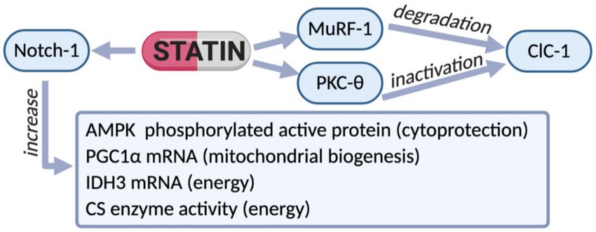
statin-dependent risk of myopathy (Figure 2).
Figure 2. Scheme of the effects of therapy with statins observed in biopsies of patients experiencing
myalgia and hyper-CK-emia. In these subjects a reduction of ClC-1 protein was observed, likely
due to the increased degradation via MuRF-1 increase. In parallel, an increase of phosphorylated
PKC-theta supports ClC-1 inactivation. However, an increased expression of Notch-1 (a protein found
to raise during muscle cell proliferation) was observed in statin-treated patients, indicating effective
regeneration. In accord, the phosphorylated form of AMPK was increased, suggesting the activation
of restorative cytoprotective process. In an attempt to counteract energy deficit, compensative
mechanisms were likely activated. Indeed, an increased expression of PGC-1-alpha and isocitrate-
dehydrogenase (IDH3), together with increased activity of the enzyme citrate-synthase (CS), suggest
mitochondrial biogenesis. Thus, the reduction of ClC-1 protein and consequent hyperexcitability in
sarcolemma together with energy deficit seems to be among the most important alterations associated
with statin-related risk of myopathy in humans.
5. Higher Risk with Statin Therapy: Role of Comorbidity, Genetic and Drug
Interactions
Numerous risk factors, in terms of age, gender, frailty, genetics, occurrence of other dis-
eases, and polypharmacy may accelerate and/or aggravate myopathy symptoms [9,26,77]Int. J. Mol. Sci. 2021, 22, 2070 7 of 19
(Table 1). Statin-related adverse effects can be more severe in the aged population which
uses statins for the prevention of cardiovascular diseases. It has been established that
aging process strongly affects skeletal muscle function, causing a loss of muscle mass
and functional disability [78,79]. In rodents, senescence of skeletal muscle is associated
to a reduction of resting gCl and ClC-1 mRNA expression, an increase of PKC activity
along with alteration of calcium homeostasis [72,80,81] and of the glycolytic and oxidative
pathways [82]. Thus, all of these alterations, also due to statin action [47], can additionally
worsen muscle damage. Moreover, the reduced activity of some metabolizing enzymes,
typical of aged subjects, may slow-down drug elimination in these subjects that will have a
higher risk to develop side effects.
Numerous studies suggest also a genetic predisposition to statins-intolerance [83–85].
Thus, modification of the genes codifying for proteins involved in statin uptake, metabolism,
or elimination may confer susceptibility to statin-induced myopathy. In patients with my-
opathy, high concentrations of statins and their metabolites have been found in plasma,
suggesting a pharmacokinetic modification [86]. Indeed, alterations in the metabolic pro-
cess able to activate the lactone forms of lipophilic statin or to biotransform the acid form
by the cytochrome P450 isoforms (CYP3A4, CYP2C9 and CYP2C8) to produce metabolites,
may have a role in statin-induced myopathy. In particular, statins are transferred into hepa-
tocytes by the organic anion transporting polypeptide (OATP1B1) encoded by the SLCO1B1
gene [87]. Some genetic variation of SLCO1B1 gene are associated to the increased risk of
myopathy. Two polymorphisms associated with OATP1B1 may cause statin pharmacoki-
netics alteration [88]. Regarding statin metabolism, the CYP3A4 systems are responsible
for simvastatin, lovastatin and atorvastatin modification [89], and the inhibition of this
system induces an increased risk of muscle complications [90]. The CYP3A4 show few
polymorphisms that can affect drug activity [91]. On the contrary, the polymorphisms of
CYP2D6 are linked to a higher incidence of simvastatin and atorvastatin intolerance [92,93].
Genetic risk factors correlated to the statin-induced myopathy also include modification of
genes encoding for plasma membrane calcium ATPase [94], or genes involved in creatine
phosphorylation [95] or in mitochondrial energy production [96–98].
Polypharmacy is another risk factor. Indeed, it is known that concomitant therapies
with fenofibrate or ciclosporin can worsen statin-related side effects since they can inhibit
the activity of metabolizing enzymes [99–101]. Thus, the assumption of the different
drugs, especially during aging, should be monitored. However, pravastatin represents an
exception because it is mainly biotransformed by a sulfotransferase [102,103].
Based on our previous studies, we deduce the importance to monitor the effects
of statin treatment in subjects affected by muscle chloride channel (ClC-1) malfunction.
Interestingly, a CLCN1 gene variant (a heterozygote truncating mutation) encoding for the
ClC-1 channel, with loss of function, has been observed to be more frequent in patients with
statin-induced myotoxicity than in a healthy population [104], strongly supporting our
data. Thus, there are other categories of patients with an increased risk of statin-induced
myopathy. For instance, subjects affected by Myotonia Congenita [53], as well as patients
with other pathologies in which the alteration of chloride channels induces skeletal muscle
malfunction, such as the amyotrophic lateral sclerosis (ALS) [105,106] and Huntington
disease [107].
It is known that statin, by reducing cholesterol biosynthesis, may reduce energy stores
in the muscle. Actually, patients with an impairment of muscle energy production due
to enzymatic malfunction at different levels of either glycogen or lipid metabolisms (i.e.,
PFK and beta-enolase) [108] may risk a worsening of muscle symptoms during statin
therapy. In addition, statin therapy may unmask a metabolic myopathy or may increase
susceptibility of asymptomatic carriers to develop symptoms when exposed to statins [109].
Among metabolic myopathies the most frequent types, such as myophosphorylase deficit
(McArdle’s disease) and carnitine palmityltransferase 2 (CPT2) deficiency, due to energy
failure because of a dysfunction of glycogen and lipid utilization in muscle, add risk for
myopathic outcomes [109]. Since the number of patients on statin therapy is expected toInt. J. Mol. Sci. 2021, 22, 2070 8 of 19
grow, a pharmacogenetic investigation may be helpful to evaluate and reduce side effects
and to improve the compliance.
Indeed, phosphofructokinase deficiency (Tarui’s disease), a hereditary disorder that
alters the ability of muscles to utilize carbohydrates (such as glucose) for energy production,
was reported in patients suffering from muscle cramps, exercise intolerance and recurrent
episodes of rhabdomyolysis during statin therapy [110]. Similar considerations have to
be extended to other metabolic myopathies as muscle β-enolase deficiency, an ultrarare
metabolic myopathy due to impairment of terminal glycolysis described in few adult
patients with muscle fatigability, myalgia and increased CK values after intense physical
exercise [111–113]. Elevated level of CK may be also observed in other diseases such as
Guillain–Barrè syndrome [114]. Our research pointed out that these metabolic pathways
may be affected during statin therapy and are severely altered in aged rats treated with
statin [48,50]. The downregulation of mRNA level of beta-enolase, a glycolytic enzyme
leading to the formation of phosphoenol-pyruvate (PEP), is a clear indication of the gly-
colytic suffering, which is particularly evident in the skeletal muscle of aged treated rats,
a tissue already compromised by aging process. The downregulation of the glycolytic
enzymes determines an energy production failure, and strongly contributes to muscle
damage. Interestingly, an increase of Pyruvate kinase M2 (PKM2) isoform was observed in
both adult and aged rats treated with atorvastatin. At the moment, it is difficult to under-
stand the reason for PKM2 increase since it has been found in rapidly proliferating tissues
and in tumor cells [115]. Its increase can be related to an accumulation of the upstream
glycolytic metabolites with a shift of glucose metabolism toward alternative pathways
responsible for the biosynthesis of important macromolecules required for increased prolif-
eration. It should be underlined that PKM2 was significantly decreased in untreated aged
muscles suggesting a surprising regenerative potential of statins. Accordingly, atorvastatin
was shown to improve the post-infarct microenvironment by inhibiting the RhoA/ROCK
pathway, and then promoting the survival and therapeutic efficacy of transplanted stem
cells [116].
Mitochondrial function was modified particularly in aged rodents treated with ator-
vastatin. Indeed, a significant decrease in mitochondrial NADH dehydrogenase subunit 5
(mt-ND5) (a part of complex I of mitochondrial respiratory chain) was observed in these
animals, again suggesting an impairment of energy production mediated by the oxidative
phosphorylation system. The perturbation in energy metabolism and ATP synthesis may
have a profound impact on the contractile function during aging. In accord, in treated rats,
we observed a slight increase of AMP-activated protein kinase (AMPK) mRNA level, a
crucial sensor of energy status, known to be activated during metabolic stress, hypoxia and
ATP depletion [50,117]. In support of this idea, the slow-twitch muscles seem to be less
affected by statin likely because the elevated number of mitochondria may compensate the
energy request.
It is still debated whether the use of statins enhances the risk of new-onset diabetes.
Recent large-scale meta-analyses support the concept of a diabetogenic effect of statins,
since there are clinical trials reporting changes in glycemia and insulin levels. However, a
definitive mechanism has not been elucidated yet [118,119]. Recent studies show a decrease
of the expression of GLUT4, able to transport glucose into the cells [51] and reduced insulin
secretion with consequent alteration of glucose homeostasis [120]. Previous studies show
the opening of KATP channels during administration of high statin doses due to low ATP
level, supporting reduced insulin secretion [121]. These effects can aggravate an already
compromised situation.
Some evidence suggests that patients undergoing intense physical activity suffer more
likely from muscle symptoms with respect to sedentary patients, suggesting that athletes
can be more intolerant to lipid-lowering therapy able to amplify the CK increases that
commonly occur after vigorous exercise [122–124].Int. J. Mol. Sci. 2021, 22, 2070 9 of 19
Table 1. Risk factors that may increase statin-induced muscle symptoms.
Baseline Characteristics
Risk Factors References
or Genetic Factors
Decreased metabolism (reduced activity
[89]
of cytochrome P450 isoenzymes);
Reduction of muscle chloride
Advanced age [50,81]
channel (ClC-1) activity and expression;
Increased intracellular Ca2+ ; [60,80]
Energy production defect; [96,97]
Lower glomerular filtration rate [16]
Female gender Different activity of metabolizing enzymes [26]
[16]
Polymorphism and genetic variation of metabolizing enzymes (cytochrome P450) and/or of membrane
[85]
transporters (i.e., SLCO1B1);
Ethnicity and [26]
predisposing genetic variants Variant in CACNA1H encoding
[104]
voltage-dependent calcium channel;
Variant of ClCN-1 gene encoding for ClC1 channel protein; [104]
Genetic variants within the ryanodine
[68]
and dihydropiridine receptor genes
Additional reduction of vitamin
Vitamin D deficiency [10]
D synthesis due to cholesterol reduction
Additional cholesterol decrease [123]
Low body mass index
Muscle protein degradation [98]
Fetal abnormalities due to
Pregnancy [16]
cholesterol reductionInt. J. Mol. Sci. 2021, 22, 2070 10 of 19
Table 1. Cont.
Baseline Characteristics
Risk Factors References
or Genetic Factors
Exogenous factors
Drug–drug interaction with inhibitors of cytochrome P450 metabolic [85]
isoenzymes (fibrates, immunosuppressant [26]
Polypharmacy: drugs or food interaction
drugs, cardiovascular antiplatelet or
anticoagulant drugs, antimicrobials, and antiviral drugs);
Inhibitors of OATP1B1 (gemfibrozil); [71,123]
High consumption of grapefruit juice
(containing furanocoumarins) that
inhibits CYP3A4 and increases the plasma concentration of statins
Strenuous exercise Amplification of the Creatine Kinase (CK) increase that commonly occurs after strenuous exercise [123,124]
Liver disease (additive transaminases
Alcohol abuse [120,123]
increase)
Energy production defect
Mitochondrial gene defect ATP synthesis reduction [96,97]
Comorbidity/Pre-existing diseases
Hypertrigliceridemia Fibrates administration [101]
Intracerebral hemorrhage Statin may potentiate the anticoagulant effect of coadministered drugs (i.e., warfarin) [16]
Skeletal muscle hypermetabolism;
Muscle mitochondria energy defect;
Decrease of nutrients supply to muscle; [106]
Amyotrophic lateral sclerosis
Cholesterol synthesis inhibition
and muscle membrane instability;
Reduction of muscle chloride [16]
channel (ClC-1) activity and [105]
expression with hyperexcitability
and alteration of contraction
Myotonia congenita ClC-1 malfunction and skeletal muscle involvement [53]
Huntington disease ClC-1 malfunction and skeletal muscle involvement [107]Int. J. Mol. Sci. 2021, 22, 2070 11 of 19
Table 1. Cont.
Baseline Characteristics
Risk Factors References
or Genetic Factors
Comorbidity/Pre-existing diseases
Guillain–Barrè syndrome Additional elevated CK [114]
GLUT4 mRNA reduction;
Diabetes Reduced secretion of insulin [50]
Inhibition of insulin release due to [120]
the opening of KATP channels in pancreatic beta cells [59]
Hypothyroidism can
Hypothyroidism [17,101]
cause hypercholesterolemia and raised serum CK
Chronic kidney disease Renal function impairment with increased drug systemic exposure [16]
Liver disease Elevated transaminases [10]
Mitochondrial myopathies Additive mitochondrial function deficit [123]
Impairment of glucose and
glycogen metabolism
Glycogen storage disease [109]
Potentiation of myalgia, cramps, fatigue
GSD V (McArdle disease) [110]
Rhabdomyolysis with
GSD VII (Tarui’s disease) [111]
myoglobinuria (dark urine)
GSD XIII [108,112]
Muscle cramps, exercise intolerance and rhabdomyolysis, symptoms that
can worsen during statin therapy
Lipid Storage myopathies Impairment of lipid metabolism
[109]
CPT2 deficiency Increasing of rhadomyolysis episodesInt. J. Mol. Sci. 2021, 22, 2070 12 of 19
However, positive results reported that statins are able to ameliorate muscle dys-
trophy [13,125]. Simvastatin reduced inflammation in dystrophic (mdx) mice together
with a shift toward an oxidative metabolism. Moreover, a reduction of fibrosis was ob-
served, a major cause of functional decline of skeletal muscle in mdx mice [126]. Moreover,
some authors claim that statin therapy is associated with a decreased risk of Alzheimer
disease, because of their ability to decrease the accumulation of cholesterol and amyloid
plaques [120,127], but this aspect needs to be elucidated.
6. Management of the Risk of Myopathy with Statin Therapy
Statin can be administered at low doses or once every two days in case of intolerance
(diagnosed in 15–30% of treated patients) [10,128]. Alternatively, other drugs can be pre-
scribed, such as ezetimibe, colesevelam, and also nutraceutical compounds to substitute
statin therapy [20,129–131]. More recently, the proprotein convertase subtilisin-kexin type
9 (PCSK9) inhibitors were introduced in therapy [132,133]. PCSK9 is a protease that de-
grades hepatic LDL-C receptors. Among these inhibitors, evolocumab and alirocumab
are fully human monoclonal antibodies in clinical use. These drugs are injected to in-
hibit PCSK9, and preserve receptor availability so to reduce circulating LDL-C. The most
common adverse effects observed in clinical studies were musculoskeletal manifestations,
airway infections, nasopharyngitis, hypersensitivity reactions (pruritus, rash, urticaria)
and injection-site reactions. These drugs are considered well tolerated but long-term safety
data are still lacking. Since these biotechnological drugs do not interact with the system of
the hepatic cytochrome, they may be associated with statins and other drugs. These drugs
are recommended in high risk patients, in which LDL-C reduction is not achieved also
with the highest tolerated dose of statin and in subjects intolerant to statins treatment [134].
These drugs are also effective in heterozygous FH patients. Another PCSK9 inhibitor is
bococizumab, a humanized monoclonal antibody, that was discontinued from use because
it induced antibodies production that led to attenuation of the cholesterol-lowering ef-
fect [128]. Mipomersen, an antisense oligonucleotide, approved by the FDA in 2013, binds
and silences the mRNA encoding for apolipoprotein B (apoB), consequently reducing the
apoB-containing lipoproteins. This therapy may induce adverse effects as injection-site
reactions, flu-like symptoms, nausea, hepatic steatosis, elevations in serum transaminases,
specifically alanine aminotransferase (ALT) and now is not clinically available [135]. Lomi-
tapide is a microsomal triglyceride transfer protein inhibitor, responsible for the assembly
and secretion of apo-B-containing lipoproteins in both liver (VLDL) and intestine (chy-
lomicron). Lomitapide treatment results in the impairment of triglycerides secretion from
the liver, that can lead to the accumulation of hepatic triglycerides and steatosis. Since
lomitapide is an inhibitor of cytochrome P450 3A4, it needs to be monitored because of
possible adverse effects when coadministered with statins [136]. Recent drugs include
bempedoic acid, inhibitor of the enzyme ATP citrate lyase, involved in the cholesterol
synthesis, upstream from the site of action of statins. Unlike statins, bempedoic acid is not
active in skeletal muscle, decreasing the possibility of muscle side effects. Volanesorsen a
second-generation antisense oligonucleotide administered s.c., is able to reduce apoC-III
(a protein present on VLDL, LDL, Lp(a), HDL lipoproteins) levels by specific binding of
the corresponding mRNA, and promoting its degradation [135]. Among the drugs still
in clinical development there is inclisiran, a small interfering RNA (siRNA) that blocks
DNA transcription and the synthesis of the PCSK9 protein in the liver. New monoclonal
antibodies include evinacumab, an inhibitor of ANGPTL3, a protein that inactivates a
specific lipase, reducing triglycerides and LDL cholesterol, independently from the LDL
receptor [137]. Antisense oligonucleotides against Lp(a), a cholesterol-rich lipoprotein, are
also under investigation.
Nutraceuticals are food components or derivatives that offer a safe and generally
well-tolerated alternative to statin intolerant patients. Red yeast rice (containing monacolin
K or lovastatin), berberine, plant sterols, omega-3 polyunsaturated fatty acids, policosanols,
polyphenols, flavonoids, can offer some benefit as lipid-lowering agents [137,138].Int. J. Mol. Sci. 2021, 22, 2070 13 of 19
7. Summary and Conclusions
This study provides a summary of the causes responsible for statin-induced myopathy.
Ion channels and in particular the ClC-1 chloride channel appears to be a susceptible target
for statin action. Since these channels are important for skeletal muscle excitability and
contraction their alteration can contribute to myopathy. Considering the wide statins use,
it is pivotal to better define the role of statins in exacerbating pre-existing myopathies.
An increased risk of myopathy due to statin therapy may occur during aging and/or
pathologies characterized by basic alteration of skeletal muscle ion channels. Additionally,
oxidative and glycolytic metabolism alteration and sarcopenia make the muscle fibers more
sensitive to stress conditions and to myopathy. A scheme of the proposed mechanism is
represented in Figure 1. All these findings suggest caution with statin therapy in aged
subjects and in predisposed adults (Table 1). The possibility to assess a clear role of
these biomarkers in determining skeletal muscle damage by genetic analysis or muscle
biopsies may pave the way for preventive therapies. For instance, drugs able to increase
chloride channel activity and/or PKC inhibitors, allowing gCl recovery, may reduce the
statin-induced muscle damage. In addition, the restoration of the energetic pathways, i.e.,
through Coenzyme Q10 supplementation or through AMPK-targeted pharmacological
strategies, may be useful.
Author Contributions: S.P., G.M.C., N.T., I.C., M.D.B. conceived, edited and performed a compre-
hensive review of the literature; S.P., G.M.C., O.M. wrote the manuscript. All authors have read and
agreed to the published version of the manuscript.
Funding: This research received no external funding.
Institutional Review Board Statement: Not applicable.
Informed Consent Statement: Not applicable.
Acknowledgments: The authors wish to thank Annamaria De Luca for critical reading of the
manuscript and the precious advice.
Conflicts of Interest: The authors declare no conflict of interest.
References
1. Endo, A.; Kuroda, M.; Tanzawa, K. Competitive inhibition of 3-hydroxy-3-methylglutaryl coenzyme a reductase by ML-236A and
ML-236B fungal metabolites, having hypocholesterolemic activity. FEBS Lett. 1976, 72, 323–326. [CrossRef]
2. Pang, J.; Chan, D.C.; Watts, G.F. The Knowns and Unknowns of Contemporary Statin Therapy for Familial Hypercholesterolemia.
Curr. Atheroscler. Rep. 2020, 22, 1–10. [CrossRef] [PubMed]
3. Cignarella, A.; Bellosta, S.; Corsini, A.; Bolego, C. Hypolipidemic therapy for the metabolic syndrome. Pharmacol. Res. 2006, 53,
492–500. [CrossRef]
4. Weitz-Schmidt, G. Statins as anti-inflammatory agents. Trends Pharmacol. Sci. 2002, 23, 482–487. [CrossRef]
5. Jeong, G.H.; Lee, K.H.; Kim, J.Y.; Eisenhut, M.; Kronbichler, A.; Van Der Vliet, H.J.; Shin, J.I.; Gamerith, G. Statin and Cancer
Mortality and Survival: An Umbrella Systematic Review and Meta-Analysis. J. Clin. Med. 2020, 9, 326. [CrossRef]
6. Schönbeck, U.; Libby, P. Inflammation, immunity, and HMG-CoA reductase inhibitors: Statins as antiinflammatory agents?
Circulation 2004, 109 (Suppl. 1), II18–II26. [CrossRef]
7. Oesterle, A.; Laufs, U.; Liao, J.K. Pleiotropic Effects of Statins on the Cardiovascular System. Circ. Res. 2017, 120, 229–243.
[CrossRef]
8. Urbach, D.; Awiszus, F.; Leiß, S.; Venton, T.; De Specht, A.V.; Apfelbacher, C. Associations of Medications With Lower Odds
of Typical COVID-19 Symptoms: Cross-Sectional Symptom Surveillance Study. JMIR Public Health Surveill. 2020, 6, e22521.
[CrossRef]
9. Stroes, E.S.; Thompson, P.D.; Corsini, A.; Vladutiu, G.D.; Raal, F.J.; Ray, K.K.; Roden, M.; Stein, E.; Tokgözoğlu, L.; Nordestgaard,
B.G.; et al. Statin-associated muscle symptoms: Impact on statin therapy—European Atherosclerosis Society Consensus Panel
Statement on Assessment, Aetiology and Management. Eur. Heart J. 2015, 36, 1012–1022. [CrossRef] [PubMed]
10. Banach, M.; Rizzo, M.; Toth, P.P.; Farnier, M.; Davidson, M.H.; Al-Rasadi, K.; Aronow, W.S.; Athyros, V.; Djuric, D.M.; Ezhov, M.V.;
et al. Statin intolerance-an attempt at a unified definition. Position paper from an international lipid expert panel. Expert Opin.
Drug Saf. 2015, 14, 935–955. [CrossRef] [PubMed]
11. Carter, A.A.; Gomes, T.; Camacho, X.; Juurlink, D.N.; Shah, B.R.; Mamdani, M.M. Risk of incident diabetes among patients treated
with statins: Population based study. BMJ 2013, 346, f2610. [CrossRef]Int. J. Mol. Sci. 2021, 22, 2070 14 of 19
12. Brault, M.; Ray, J.; Gomez, Y.-H.; Mantzoros, C.S.; Daskalopoulou, S.S. Statin treatment and new-onset diabetes: A review of
proposed mechanisms. Metabolism 2014, 63, 735–745. [CrossRef] [PubMed]
13. De Luca, A.; Pierno, S.; Camerino, D.C. Electrical properties of diaphragm and EDL muscles during the life of dystrophic mice.
Am. J. Physiol. Physiol. 1997, 272, C333–C340. [CrossRef]
14. Furberg, C.D.; Pitt, B. Withdrawal of cerivastatin from the world market. Curr. Control. Trials Cardiovasc. Med. 2001, 2, 205–207.
[CrossRef] [PubMed]
15. Zhang, H.; Plutzky, J.; Skentzos, S.; Morrison, F.; Mar, P.; Shubina, M.M.; Turchin, A. Discontinuation of statins in routine care
settings: A cohort study. Ann. Intern. Med. 2013, 158, 526–534. [CrossRef]
16. Newman, C.B.; Preiss, D.; Tobert, J.A.; Jacobson, T.A.; Page, R.L.; Goldstein, L.B.; Chin, C.; Tannock, L.R.; Miller, M.; Raghuveer,
G.; et al. Statin Safety and Associated Adverse Events: A Scientific Statement From the American Heart Association. Arter.
Thromb. Vasc. Biol. 2019, 39, e38–e81. [CrossRef]
17. Bruckert, E.; Hayem, G.; Dejager, S.; Yau, C.; Bégaud, B. Mild to Moderate Muscular Symptoms with High-Dosage Statin Therapy
in Hyperlipidemic Patients—The PRIMO Study. Cardiovasc. Drugs Ther. 2005, 19, 403–414. [CrossRef] [PubMed]
18. Noyes, A.M.; Thompson, P.D. The effects of statins on exercise and physical activity. J. Clin. Lipidol. 2017, 11, 1134–1144. [CrossRef]
[PubMed]
19. Collins, R.; Reith, C.; Emberson, J.; Armitage, J.; Baigent, C.; Blackwell, L.; Blumenthal, R.; Danesh, J.; Smith, G.D.; DeMets, D.;
et al. Interpretation of the evidence for the efficacy and safety of statin therapy. Lancet 2016, 388, 2532–2561. [CrossRef]
20. Nikolic, D.; Banach, M.; Chianetta, R.; Luzzu, L.M.; Stoian, A.P.; Diaconu, C.C.; Citarrella, R.; Montalto, G.; Rizzo, M. An overview
of statin-induced myopathy and perspectives for the future. Expert Opin. Drug Saf. 2020, 19, 601–615. [CrossRef]
21. Rosenson, R.S. Current overview of statin-induced myopathy. Am. J. Med. 2004, 116, 408–416. [CrossRef] [PubMed]
22. Vaughan, C.J.; Gotto, A.M., Jr. Update on statins. Circulation 2004, 110, 886–892. [CrossRef] [PubMed]
23. Reidenberg, M.M. Statins, lack of energy and ubiquinone. Br. J. Clin. Pharmacol. 2005, 59, 606–607. [CrossRef] [PubMed]
24. Bonetti, P.O.; Lerman, L.O.; Napoli, C.; Lerman, A. Statin effects beyond lipid lowering—Are they clinically relevant? Eur. Heart J.
2003, 24, 225–248. [CrossRef]
25. Moosmann, B.; Behl, C. Selenoprotein synthesis and side-effects of statins. Lancet 2004, 363, 892–894. [CrossRef]
26. Muntean, D.M.; Thompson, P.D.; Catapano, A.L.; Stasiolek, M.; Fabis, J.; Muntner, P.; Serban, M.-C.; Banach, M. Statin-associated
myopathy and the quest for biomarkers: Can we effectively predict statin-associated muscle symptoms? Drug Discov. Today 2017,
22, 85–96. [CrossRef] [PubMed]
27. Chatzizisis, Y.S.; Koskinas, K.C.; Misirli, G.; Vaklavas, C.; Hatzitolios, A.; Giannoglou, G.D. Risk factors and drug interactions
predisposing to statin-induced myopathy: Implications for risk assessment, prevention and treatment. Drug Saf. 2010, 33, 171–187.
[CrossRef]
28. Cao, P.; Hanai, J.; Tanksale, P.; Imamura, S.; Sukhatme, V.P.; Lecker, S.H. Statin-induced muscle damage and atrogin-1 in-duction
is the result of a geranylgeranylation defect. FASEB J. 2009, 23, 2844–2854. [CrossRef]
29. Mallinson, J.E.; Constantin-Teodosiu, D.; Sidaway, J.; Westwood, F.R.; Greenhaff, P.L. Blunted Akt/FOXO signalling and
ac-tivation of genes controlling atrophy and fuel use in statin myopathy. J Physiol. 2009, 587, 219–230. [CrossRef]
30. Copaja, M.; Venegas, D.; Aranguiz, P.; Canales, J.; Vivar, R.; Avalos, Y.; Garcia, L.; Chiong, M.; Olmedo, I.; Catalán, M.; et al.
Simvastatin disrupts cytoskeleton and decreases cardiac fibroblast adhesion, migration and viability. Toxicology 2012, 294, 42–49.
[CrossRef]
31. Sakamoto, K.; Wada, I.; Kimura, J. Inhibition of Rab1 GTPase and Endoplasmic Reticulum-to-Golgi Trafficking Underlies Statin’s
Toxicity in Rat Skeletal Myofibers. J. Pharmacol. Exp. Ther. 2011, 338, 62–69. [CrossRef] [PubMed]
32. Trapani, L.; Melli, L.; Segatto, M.; Trezza, V.; Campolongo, P.; Jozwiak, A.; Swiezewska, E.; Pucillo, L.P.; Moreno, S.; Fanelli, F.;
et al. Effects of myosin heavy chain (MHC) plasticity induced by HMGCoA-reductase inhibition on skeletal muscle functions.
FASEB J. 2011, 25, 4037–4047. [CrossRef]
33. Christopher-Stine, L.; Casciola-Rosen, L.A.; Hong, G.; Chung, T.; Corse, A.M.; Mammen, A.L. A novel autoantibody recog-nizing
200-kd and 100-kd proteins is associated with an immune-mediated necrotizing myopathy. Arthritis Rheum. 2010, 62, 2757–2766.
[CrossRef]
34. Selva-O’Callaghan, A.; Alvarado-Cardenas, M.; Pinal-Fernández, I.; Trallero-Araguás, E.; Milisenda, J.C.; Martínez, M.A.; Marín,
A.; Labrador-Horrillo, M.; Juárez, C.; Grau-Junyent, J.M. Statin-induced myalgia and myositis: An update on patho-genesis and
clinical recommendations. Expert Rev. Clin. Immunol. 2018, 14, 215–224. [CrossRef]
35. Mohassel, P.; Mammen, A.L. Statin-associated autoimmune myopathy and anti-HMGCR autoantibodies. Muscle Nerve 2013, 48,
477–483. [CrossRef]
36. Needham, M.; Fabian, V.; Knezevic, W.; Panegyres, P.; Zilko, P.; Mastaglia, F.L. Progressive myopathy with up-regulation of
MHC-I associated with statin therapy. Neuromuscul. Disord. 2007, 17, 194–200. [CrossRef] [PubMed]
37. Hamann, P.D.; Cooper, R.G.; McHugh, N.J.; Chinoy, H. Statin-induced necrotizing myositis-a discrete autoimmune entity within
the “statin-induced myopathy spectrum”. Autoimmun. Rev. 2013, 12, 1177–1181. [CrossRef]
38. Ramanathan, S.; Langguth, D.; Hardy, T.A.; Garg, N.; Bundell, C.; Rojana-Udomsart, A.; Dale, R.C.; Robertson, T.; Mammen, A.L.;
Reddel, S.W. Clinical course and treatment of anti-HMGCR antibody-associated necrotizing autoimmune myopathy. Neurol.
Neuroimmunol. Neuroinflammation 2015, 2, e96. [CrossRef]Int. J. Mol. Sci. 2021, 22, 2070 15 of 19
39. The SEARCH Collaborative Group; Link, E.M.; Parish, S.; Armitage, J.M.; Bowman, L.H.; Heath, S.; Matsuda, F.; Gut, I.; Lathrop,
M.; Collins, R.A. SLCO1B1Variants and Statin-Induced Myopathy—A Genomewide Study. N. Engl. J. Med. 2008, 359, 789–799.
[CrossRef]
40. Stenzel, W.; Goebel, H.-H.; Aronica, E. Review: Immune-mediated necrotizing myopathies-a heterogeneous group of diseases
with specific myopathological features. Neuropathol. Appl. Neurobiol. 2012, 38, 632–646. [CrossRef]
41. Bernatsky, S.; Joseph, L.; Pineau, C.A.; Bélisle, P.; Boivin, J.F.; Banerjee, D.; Clarke, A.E. Estimating the prevalence of poly-myositis
and dermatomyositis from administrative data: Age, sex and regional differences. Ann. Rheum. Dis. 2009, 68, 1192–1196.
[CrossRef] [PubMed]
42. Grable-Esposito, P.; Katzberg, H.D.; Greenberg, S.A.; Srinivasan, J.; Katz, J.; Amato, A.A. Immune-mediated necrotizing myopathy
associated with statins. Muscle Nerve 2009, 41, 185–190. [CrossRef]
43. Loganathan, P.; Oddis, C.V.; Aggarwal, R. Immune-mediated statin myopathy. Expert Rev. Clin. Immunol. 2015, 12, 33–38.
[CrossRef] [PubMed]
44. Tiniakou, E. Statin-Associated Autoimmune Myopathy: Current Perspectives. Ther. Clin. Risk Manag. 2020, 16, 483–492.
[CrossRef]
45. Pierno, S.; De Luca, A.; Tricarico, D.; Ferrannini, E.; Conte, T.; D’Alò, G.; Camerino, D.C. Experimental Evaluation of the Effects of
Pravastatin on Electrophysiological Parameters of Rat Skeletal Muscle. Pharmacol. Toxicol. 1992, 71, 325–329. [CrossRef] [PubMed]
46. Pierno, S.; Didonna, M.P.; Cippone, V.; De Luca, A.; Pisoni, M.; Frigeri, A.; Nicchia, G.P.; Svelto, M.; Chiesa, G.; Sirtori, C.; et al.
Effects of chronic treatment with statins and fenofibrate on rat skeletal muscle: A biochemical, histological and electrophysiological
study. Br. J. Pharmacol. 2006, 149, 909–919. [CrossRef]
47. Pierno, S.; Camerino, G.; Cippone, V.; Rolland, J.-F.; Desaphy, J.-F.; De Luca, A.; Liantonio, A.; Bianco, G.; Kunic, J.; George, A.L.;
et al. Statins and fenofibrate affect skeletal muscle chloride conductance in rats by differently impairing ClC-1 channel regulation
and expression. Br. J. Pharmacol. 2009, 156, 1206–1215. [CrossRef] [PubMed]
48. Camerino, G.M.; Pellegrino, M.A.; Brocca, L.; Digennaro, C.; Camerino, D.C.; Pierno, S.; Bottinelli, R. Statin or fibrate chronic
treatment modifies the proteomic profile of rat skeletal muscle. Biochem. Pharmacol. 2011, 81, 1054–1064. [CrossRef]
49. Camerino, G.M.; Bouchè, M.; De Bellis, M.; Cannone, M.; Liantonio, A.; Musaraj, K.; Romano, R.; Smeriglio, P.; Madaro, L.;
Giustino, A.; et al. Protein kinase C theta (PKCθ) modulates the ClC-1 chloride channel activity and skeletal muscle phenotype:
A biophysical and gene expression study in mouse models lacking the PKCθ. Pflügers Archiv-Eur. J. Physiol. 2014, 466, 2215–2228.
[CrossRef]
50. Camerino, G.M.; De Bellis, M.; Conte, E.; Liantonio, A.; Musaraj, K.; Cannone, M.; Fonzino, A.; Giustino, A.; De Luca, A.; Romano,
R.; et al. Statin-induced myotoxicity is exacerbated by aging: A biophysical and molecular biology study in rats treated with
atorvastatin. Toxicol. Appl. Pharmacol. 2016, 306, 36–46. [CrossRef]
51. Camerino, G.M.; Musumeci, O.; Conte, E.; Musaraj, K.; Fonzino, A.; Barca, E.; Marino, M.; Rodolico, C.; Tricarico, D.; Camerino,
C.; et al. Risk of Myopathy in Patients in Therapy with Statins: Identification of Biological Markers in a Pilot Study. Front.
Pharmacol. 2017, 8, 500. [CrossRef]
52. Desaphy, J.-F.; Pierno, S.; De Luca, A.; Didonna, P.; Camerino, D.C.; Conte, D. Different Ability of Clenbuterol and Salbutamol
to Block Sodium Channels Predicts Their Therapeutic Use in Muscle Excitability Disorders. Mol. Pharmacol. 2003, 63, 659–670.
[CrossRef] [PubMed]
53. Desaphy, J.F.; Gramegna, G.; Altamura, C.; Dinardo, M.M.; Imbrici, P.; George, A.L., Jr.; Modoni, A.; Lomonaco, M.; Conte
Camerino, D. Functional characterization of ClC-1 mutations from patients affected by recessive myotonia congenita pre-senting
with different clinical phenotypes. Exp. Neurol. 2013, 248, 530–540. [CrossRef] [PubMed]
54. Altamura, C.; Desaphy, J.-F.; Conte, D.; De Luca, A.; Imbrici, P. Skeletal muscle ClC-1 chloride channels in health and diseases.
Pflügers Archiv-Eur. J. Physiol. 2020, 472, 961–975. [CrossRef]
55. Altamura, C.; Mangiatordi, G.F.; Nicolotti, O.; Sahbani, D.; Farinato, A.; Leonetti, F.; Carratù, M.R.; Conte, D.; Desaphy, J.-F.;
Imbrici, P. Mapping ligand binding pockets in chloride ClC-1 channels through an integrated in silico and experimental ap-proach
using anthracene-9-carboxylic acid and niflumic acid. Br. J. Pharmacol. 2018, 175, 1770–1780. [CrossRef]
56. Cozzoli, A.; Liantonio, A.; Conte, E.; Cannone, M.; Massari, A.M.; Giustino, A.; Scaramuzzi, A.; Pierno, S.; Mantuano, P.;
Capogrosso, R.F.; et al. Angiotensin II modulates mouse skeletal muscle resting conductance to chloride and potassium ions and
calcium homeostasis via the AT1 receptor and NADPH oxidase. Am. J. Physiol. Physiol. 2014, 307, C634–C647. [CrossRef]
57. Pierno, S.; Camerino, G.M.; Cannone, M.; Liantonio, A.; De Bellis, M.; Digennaro, C.; Gramegna, G.; De Luca, A.; Germinario,
E.; Danieli-Betto, D.; et al. Paracrine Effects of IGF-1 Overexpression on the Functional Decline Due to Skeletal Muscle Disuse:
Molecular and Functional Evaluation in Hindlimb Unloaded MLC/mIgf-1 Transgenic Mice. PLoS ONE 2013, 8, e65167. [CrossRef]
58. Pedersen, T.H.; Macdonald, W.A.; de Paoli, F.V.; Gurung, I.S.; Nielsen, O.B. Comparison of regulated passive membrane
conductance in action potential-firing fast- and slow-twitch muscle. J. Gen. Physiol. 2009, 134, 323–337. [CrossRef]
59. Sehra, D.; Sehra, S.; Sehra, S.T. Cardiovascular pleiotropic effects of statins and new onset diabetes: Is there a common link: Do
we need to evaluate the role of KATP channels? Expert. Opin. Drug. Saf. 2017, 16, 823–831. [CrossRef] [PubMed]
60. Liantonio, A.; Giannuzzi, V.; Cippone, V.; Camerino, G.M.; Pierno, S.; Camerino, D.C. Fluvastatin and Atorvastatin Affect
Calcium Homeostasis of Rat Skeletal Muscle Fibers in Vivo and in Vitro by Impairing the Sarcoplasmic Reticulum/Mitochondria
Ca2+-Release System. J. Pharmacol. Exp. Ther. 2007, 321, 626–634. [CrossRef]You can also read