Neuroprotective Effect of Vascular Endothelial Growth Factor on Motoneurons of the Oculomotor System - MDPI
←
→
Page content transcription
If your browser does not render page correctly, please read the page content below
International Journal of
               Molecular Sciences

Review
Neuroprotective Effect of Vascular Endothelial Growth Factor
on Motoneurons of the Oculomotor System
Silvia Silva-Hucha, Angel M. Pastor                     and Sara Morcuende *

                                           Departamento de Fisiología, Facultad de Biología, Universidad de Sevilla, 41012 Sevilla, Spain;
                                           silvia_sh88@hotmail.com (S.S.-H.); ampastor@us.es (A.M.P.)
                                           * Correspondence: smorcuende@us.es; Tel.: +34-954-55-95-49

                                           Abstract: Vascular endothelial growth factor (VEGF) was initially characterized as a potent an-
                                           giogenic factor based on its activity on the vascular system. However, it is now well established
                                           that VEGF also plays a crucial role as a neuroprotective factor in the nervous system. A deficit
                                           of VEGF has been related to motoneuronal degeneration, such as that occurring in amyotrophic
                                           lateral sclerosis (ALS). Strikingly, motoneurons of the oculomotor system show lesser vulnerability to
                                           neurodegeneration in ALS compared to other motoneurons. These motoneurons presented higher
                                           amounts of VEGF and its receptor Flk-1 than other brainstem pools. That higher VEGF level could
                                           be due to an enhanced retrograde input from their target muscles, but it can also be produced by
                                           the motoneurons themselves and act in an autocrine way. By contrast, VEGF’s paracrine supply
                                           from the vicinity cells, such as glial cells, seems to represent a minor source of VEGF for brainstem
                                           motoneurons. In addition, ocular motoneurons experiment an increase in VEGF and Flk-1 level in
                                           response to axotomy, not observed in facial or hypoglossal motoneurons. Therefore, in this review,
                                           we summarize the differences in VEGF availability that could contribute to the higher resistance of
                                           extraocular motoneurons to injury and neurodegenerative diseases.

                                    Keywords: VEGF; oculomotor system; trophic factors; motoneurons; neurodegeneration; axotomy;
Citation: Silva-Hucha, S.; Pastor,
                                           amyotrophic lateral sclerosis
A.M.; Morcuende, S. Neuroprotective
Effect of Vascular Endothelial Growth
Factor on Motoneurons of the
Oculomotor System. Int. J. Mol. Sci.       1. Vascular Endothelial Growth Factor (VEGF)
2021, 22, 814. https://doi.org/            1.1. History
10.3390/ijms22020814                             VEGF was initially described as an angiogenic factor and, consequently, it was named
                                           vascular permeability factor (VPF) for its role in inducing vascular permeability in tumor
Received: 10 December 2020
                                           cells [1]. It was not until 1989 when the VEGF protein, whose molecular weight is approxi-
Accepted: 13 January 2021
                                           mately 45 kDa, was purified and sequenced, and it was definitively assigned the name of
Published: 15 January 2021
                                           vascular endothelial growth factor [2,3].
                                                 It is well known that this factor is a highly specific mitogen for vascular endothelial
Publisher’s Note: MDPI stays neu-
tral with regard to jurisdictional clai-
                                           cells, whose family consists of multiple cell signaling proteins involved in angiogenesis,
ms in published maps and institutio-
                                           lymphangiogenesis, vasodilation, and vascular leakage, among other functions [4]. In 2001,
nal affiliations.
                                           his essential role in motoneuronal protection was revealed for the first time [5], as will be
                                           discussed later in detail.

                                           1.2. VEGF Family
Copyright: © 2021 by the authors. Li-           Since the discovery of the first member of the VEGF family, known as VEGF-A, the
censee MDPI, Basel, Switzerland.           family has continued to grow and is currently constituted by VEGF-A, VEGF-B [6,7],
This article is an open access article     VEGF-C [8,9], VEGF-D [10,11], VEGF-E [12], VEGF-F [13] and placental growth factor
distributed under the terms and con-
                                           (PlGF [14]).
ditions of the Creative Commons At-
                                                VEGF-A stands out as the most studied VEGF family members because of its essential
tribution (CC BY) license (https://
                                           roles in neuroprotection. The gene encoding VEGF-A is located on chromosome 6p21.5,
creativecommons.org/licenses/by/
                                           giving rise to three different isoforms (VEGF-A 121, VEGF-A 145, VEGF-A 165), which
4.0/).

Int. J. Mol. Sci. 2021, 22, 814. https://doi.org/10.3390/ijms22020814                                              https://www.mdpi.com/journal/ijms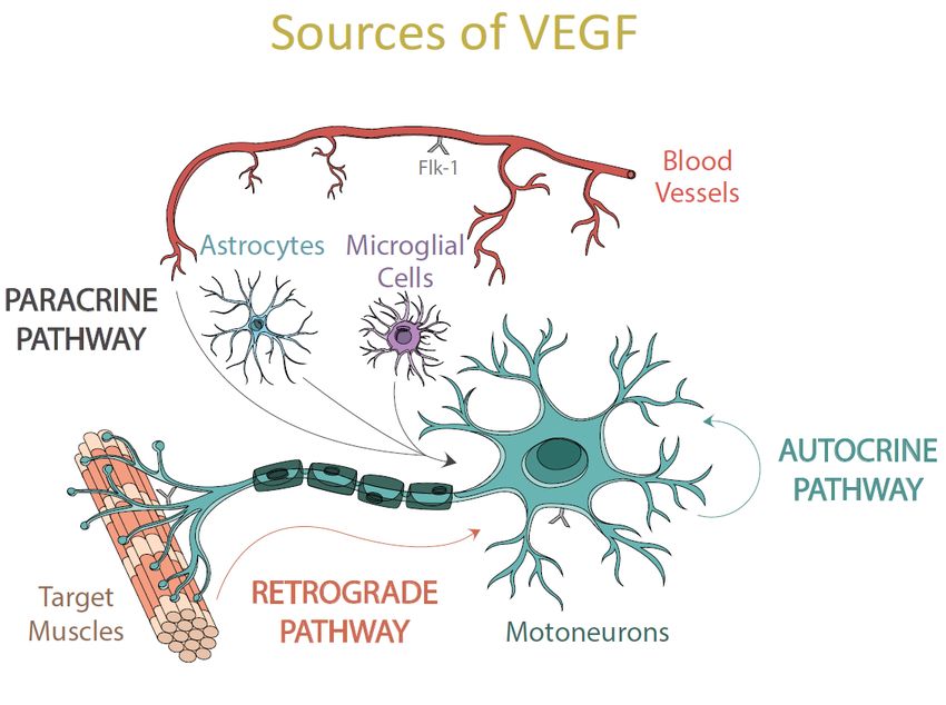
Int. J. Mol. Sci. 2021, 22, 814                                                                                            2 of 20

                                  are differentiated by their molecular weight, solubility, biological functions, binding affini-
                                  ties to the components of the extracellular matrix, and tyrosine kinase receptor subtypes
                                  (RTKs) [15]. Furthermore, VEGF 165 is the predominant isoform in the central nervous
                                  system (CNS), where it acts as a protection factor by promoting the survival of motoneu-
                                  rons [16,17]. This vital role is the one that interests us and the one that we will develop on
                                  throughout this review.
                                        On the other hand, VEGF-B is also expressed in the CNS and can regulate adult
                                  neurogenesis and even rescuing neurons from apoptosis, but with less vascular effects
                                  and worse neuroprotective function on motoneurons than VEGF-A [18]. It has recently
                                  been discovered that the presence of VEGF-B is neither necessary nor essential for the
                                  survival, maintenance, and development of motoneurons under normal physiological
                                  conditions [19,20]. Finally, mention should be made of the members VEGF-C and VEGF-D,
                                  which regulate lymphatic angiogenesis, and of VEGF-E, which is virally encoded and
                                  specifically expressed in the venom of the habu snake (Trimeresurus flavoviridis) [21].

                                  1.3. Functions of VEGF
                                       Throughout this review, we will refer to the VEGF-A 165 isoform, which is the one
                                  that exerts direct trophic and neuroprotective effects on many types of neural cells [22],
                                  including motoneurons [5,23], astroglia [24], microglia [25], hippocampal, dopaminergic,
                                  cortical, cerebellar, sympathetic neurons, and even muscle satellite cells [16,26–32].
                                       Furthermore, this trophic factor plays a fundamental role in stimulating neurogenesis
                                  in both developing and adult CNS [16], promoting Schwann cells’ proliferation [27]. It also
                                  supports synaptic plasticity [33] and favors the growth, survival, differentiation, and mi-
                                  gration of neuronal and glial cells [20,34]. Additionally, VEGF guarantees an optimal blood
                                  and glucose supply to the brain and spinal cord [5], protecting motoneurons from oxidative
                                  stress [35], hypoxia, hypoglycemia [23], and glutamate-mediated excitotoxicity [29,36–39].
                                       VEGF is also a potent inducer of the blood-brain barrier’s interruption by increasing
                                  its permeability and favoring the supply of oxygen and nutrients to neurons [34,40]. This
                                  trophic factor is involved in vasculogenesis during embryological development and pro-
                                  moting angiogenesis in many pathological conditions, such as tumor growth, rheumatoid
                                  arthritis, psoriasis, and diabetic retinopathy [4].

                                  1.4. VEGF Expression
                                       Several factors have been found to upregulate VEGF mRNA expression, including
                                  tumor necrosis factor (TNF-α), platelet-derived growth factor (PDGF), interleukins, an-
                                  giopoietins, and erythropoietins [41–45]. Another molecule that regulates the expression
                                  of VEGF is nitric oxide, which contributes to the processes of permeabilization of blood
                                  vessels and in vasodilation stimulated by VEGF [29,46,47].
                                       However, one of the main and more robust regulators of VEGF expression is hy-
                                  poxia [48]. VEGF mRNA has a half-life of 30–45 min under normoxic conditions, whereas
                                  the mRNA half-life is prolonged in hypoxia [49,50] and cells increase the production of the
                                  hypoxia-induced transcription factor 1 (HIF-1), a heterodimer consisting of three subunits
                                  (HIF-1α, HIF-1β, and HIF-3) [51,52]. HIF-1α and HIF-1β are produced continuously, but
                                  HIF-1α is highly labile in the presence of oxygen, so it degrades under aerobic condi-
                                  tions [53]. When the cell is in a hypoxic environment, HIF-1α persists and translocates
                                  to the nucleus, where it associates with HIF-1β and forms the HIF-1α/HIF-1β complex.
                                  This complex binds to the hypoxia response element (HRE) [54,55], whose transcriptional
                                  activation requires the recruitment of the CREB-binding protein, which is a transcrip-
                                  tional coactivator. Thus, as the 2019 Nobel Laureates in Medicine Kaelin, Ratcliff, and
                                  Semenza described, through this mechanism, cells perceive and adapt to changes in oxygen
                                  levels, modifying both their metabolism and physiological functions [52,56]. Therefore,
                                  transactivation of HRE by the HIF-1α/HIF-1β complex stimulates the gene expression
                                  of erythropoietin, glucose transporters, glycolytic enzymes, and VEGF [57,58], among
Int. J. Mol. Sci. 2021, 22, x FOR PEER REVIEW 3 of 20
modifying both their metabolism and physiological functions [52,56]. Therefore, transac-
Int. J. Mol. Sci. 2021, 22, 814 tivation of HRE by the HIF-1α/HIF-1β complex stimulates the gene expression of erythro-
3 of 20
poietin, glucose transporters, glycolytic enzymes, and VEGF [57,58], among others, the
latter being in charge of promoting angiogenesis after binding to its specific receptors [5].
Little is known about the expression and function of HIF-3 [59].
others, the latter being in charge of promoting angiogenesis after binding to its specific
receptors [5]. Little is known about the expression and function of HIF-3 [59].
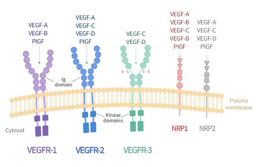
2. VEGF Receptors
2. VEGFThe biological
Receptorsactivity of the VEGF family is mediated through binding to two classes
of receptors: receptors
The biological withof
activity tyrosine
the VEGFkinase activity
family and receptors
is mediated throughwithout
bindingtyrosine kinase
to two classes
activity. The first group consists of three structurally related receptors characterized
of receptors: receptors with tyrosine kinase activity and receptors without tyrosine kinase by
the presence of seven immunoglobulin-like domains in the extracellular region,
activity. The first group consists of three structurally related receptors characterized a single
transmembrane
by the presence region,
of sevenand an intracellular consensus
immunoglobulin-like domains tyrosine kinase sequence
in the extracellular inter-
region, a
ruptedtransmembrane
single by a kinase insertion domain.
region, and These receptors
an intracellular are VEGR-1
consensus tyrosine (Flt-1), VEGFR-2
kinase sequence
(KDR/Flk-1),by
interrupted and VEGFR-3
a kinase (Flt-4).domain.
insertion On the other
Thesehand, the receptors
receptors without
are VEGR-1 kinase
(Flt-1), activ-
VEGFR-
2ity(KDR/Flk-1),
are neuropilin-1
and(NRP-1)
VEGFR-3 and neuropilin-2
(Flt-4). On the (NRP-2), which
other hand, theare also receptors
receptors withoutfor sem-
kinase
aphorinsare
activity [15,20] (Figure 1).
neuropilin-1 (NRP-1) and neuropilin-2 (NRP-2), which are also receptors for
semaphorins [15,20] (Figure 1).
Figure 1. VEGF receptors. The family of VEGF receptors includes three tyrosine kinase receptors (VEGFR-1, VEGFR-2,
VEGFR-3)
Figure 1. VEGFand two non-tyrosine
receptors. kinase neuropilin
The family receptorsincludes
of VEGF receptors (NP-1, NP-2).
three The different
tyrosine members
kinase of the
receptors VEGF family
(VEGFR-1, bind
VEGFR-2,
to the different types of VEGF receptors, as illustrated. The main effect of VEGF-A as a neurotrophic factor
VEGFR-3) and two non-tyrosine kinase neuropilin receptors (NP-1, NP-2). The different members of the VEGF family is mediated by
its
bind binding
to the to VEGFR-2
different (Flk-1).
types of VEGF receptors, as illustrated. The main effect of VEGF-A as a neurotrophic factor is medi-
ated by its binding to VEGFR-2 (Flk-1).
NRP-1 and 2 are expressed in different types of neurons [60,61] and play an essential
role in regulating
NRP-1 and 2andare developing
expressed in the cardiovascular
different types ofand nervous
neurons systems.
[60,61] Besides,
and play they act
an essential
as co-receptors for RTKs, presenting and improving VEGF binding to
role in regulating and developing the cardiovascular and nervous systems. Besides, theyFlk-1 and promoting
receptor phosphorylation
act as co-receptors andpresenting
for RTKs, neurotrophic andfactor-mediated
improving VEGF signal transduction
binding to Flk-1 [62,63].
and pro-
motingMany studiesphosphorylation
receptor indicate that both andFlt-1 and Flk-1 activation
neurotrophic could produce
factor-mediated neuroprotec-
signal transduction
tion, but there are differences between the functions of both receptors [5,26,29,30]. Flk-1
[62,63].
predominates in neuronal
Many studies indicateand thatSchwann
both Flt-1cells
andand is activation
Flk-1 necessary could
for endothelial prolifera-
produce neuropro-
tion and migration, while Flt-1 is expressed mainly in vessels, astrocytes,
tection, but there are differences between the functions of both receptors [5,26,29,30]. and reactive
Flk-
microglia [29,64]. Besides, it has also been described that Flt-1 acts as
1 predominates in neuronal and Schwann cells and is necessary for endothelial prolifera-a negative regulator
for
tionVEGF in endothelial
and migration, cells,
while Flt-1preventing
is expressed its mainly
bindingintovessels,
Flk-1 [4]astrocytes,
and that itsandfunctions
reactiveand
mi-
signaling properties may differ according to the stage of development of the
croglia [29,64]. Besides, it has also been described that Flt-1 acts as a negative regulator for animal, the
cell
VEGF type,
in and the binding
endothelial cells,ligand [65]. its binding to Flk-1 [4] and that its functions and
preventing
Unlike Flt-1, the Flk-1 receptor is an important survival promoter for endothelial
and CNS cells, being the primary mediator of VEGF functions [4,66]. The main ligand
of this Flk-1 receptor is the VEGF-A isoform, being the only one that triggers its auto-
phosphorylation and final glycosylated form [4]. Therefore, the VEGF receptor that we
will refer to throughout this review is Flk-1, which is expressed in motoneurons of theInt. J. Mol. Sci. 2021, 22, 814 4 of 20
human spinal cord [40], mouse [5], rat [38] and is reduced in some patients with amy-
otrophic lateral sclerosis (ALS) [40]. Flk-1 overexpression in spinal motoneurons of the ALS
SOD1 mouse model (with mutations in the gene encoding the antioxidant copper/zinc
superoxide dismutase) has been shown to delay both neurodegeneration and disease on-
set [67], being the primary mediator of the neuroprotective and anti-excitotoxic effects
of VEGF on motoneurons [37,38,68]. All these functions are performed by activating the
phosphatidylinositol-3 kinase-AKT (PI3-K/Akt) pathway, involved in the processes of cell
growth, proliferation, cell survival, and intracellular traffic, among others, in addition to
regulating the entry of glucose to the cell through an insulin signaling cascade [69]. Further-
more, VEGF binding to the Flk-1 receptor also exerts a protective effect by suppressing the
activation of the mitogen-activated protein kinase p38 (p38MAPK), a determining factor in
the cell death pathway [16,30,38,70].
3. Effects of Low Levels of VEGF
The experiments carried out by Oosthuyse et al. in 2001 [5] were the first to suggest
that VEGF acted as a neurotrophic factor at the CNS, as the reduction of VEGF function
induced a specific degeneration of motoneurons in the adult mice. In these experiments,
manipulation of the VEGF gene resulted in homozygous knock-in mice (VEGFδ/δ ), in
which the sequence of the hypoxia response element in the VEGF promoter region was
removed. Consequently, these mice lost the ability to increase VEGF expression in a hypoxic
situation. This alteration caused them severe muscle weakness due to the degeneration
of the lower motoneurons, and they became progressively less mobile, with symptoms
reminiscent of neuropathological signs of ALS. Although basal VEGF levels in muscles,
heart, and fibroblasts were unaffected by removing the hypoxia response element, an
overall 40% reduction of the neurotrophic factor was observed in neural tissue.
Those results obtained with the VEGFδ/δ mice allowed linking for the first time a
low level of VEGF with motoneuronal degeneration. Besides, mice resulting from the
crossing of the SOD1 mutant with VEGFδ/δ mice exhibited an even more drastic reduction
in VEGF levels, thence a more severe degeneration of motoneurons and an earlier onset
of symptoms of muscle weakness [17]. All these findings granted VEGF an unexpected
neuroprotective role in the degenerative processes that accompany the pathogenesis of
motoneurons, supporting the idea that motoneurons seem to be particularly sensitive to a
low VEGF support [5,34,71]. Thus, a link was established between low levels of VEGF and
neurodegeneration of motoneurons, such as occurs in ALS, and raised great expectation in
VEGF as a possible candidate for ALS specific treatment.
VEGF and ALS
ALS is characterized by being an adult neurodegenerative disease that causes pro-
gressive degeneration of motoneurons in the lower spinal cord, brainstem, and cortex.
Consequently, it triggers astrogliosis, progressive atrophy of the skeletal musculature, and
a reduction in voluntary movements, including those of the extremities and respiratory
movements [72,73].
The disease affects five out of every 100,000 people worldwide, is progressive, and is
generally fatal within 5 years after the onset of symptoms. 95% of cases are sporadic, and
only 5% of patients have a family history, with a fifth of these caused by mutations in the
SOD1 gene, located on chromosome 21 [15]. There are currently around 180 known muta-
tions in SOD1 which are related to the pathogenesis of the disease [74], with the SOD1G93A
mutant mouse model being the most widely used, studied, and well-characterized show-
ing symptoms similar to the disease [75], and to which we refer in this review as the
SOD1 model.
It has been shown that the expression of VEGF and its Flk-1 receptor undergo sig-
nificant downregulation in the motoneurons of the spinal cord of SOD1 mice [76]. These
findings correlate with other studies where SOD1 mice were crossed with transgenic mice
that overexpressed Flk-1, resulting in a delayed onset of motor impairment and degen-Int. J. Mol. Sci. 2021, 22, 814 5 of 20
eration of motoneurons, and prolonged survival [67]. Moreover, as indicated before, the
coincidence of both SOD1 mutation and VEGFδ/δ alteration produced an earlier and more
severe motoneuronal degeneration [17].
Although the predominant hypothesis is that ALS is a disease of neural origin, some
studies indicate that the disease involves a distal axonopathy and denervation of the
neuromuscular junctions (NMJs) in the muscles of the extremities in the presymptomatic
stage, that is, much before the loss of the motoneuron at the level of the spinal cord [77–79].
Several studies have shown that both the anterograde transport of VEGF and the in-
tracerebroventricular infusion of this factor help protect and preserve the NMJs in a rat
model SOD1 [67,80,81]. Likewise, experiments such as gene therapy of VEGF mediated by
lentiviral vectors, or transplantation of stem cells that overexpress VEGF, have managed to
significantly slow the progression of neurodegeneration, improving motor function and
significantly prolonging the survival of motoneurons in the brainstem and the cervical and
lumbar spinal cord [82,83]. All these findings give VEGF and its Flk-1 receptor an essential
role in the treatment of motoneuronal diseases.
4. Neuroprotective Effect of VEGF
Among the possible pathogenic mechanisms linked to the degenerative neuronal
process are oxidative stress, glutamate excitotoxicity, inflammation, mitochondrial and
neurofilament dysfunction, protein aggregation, axonal transport abnormalities, and, ul-
timately, the activation of pathways that trigger apoptosis [84]. Numerous studies show
the decisive neuroprotective role that VEGF plays in the CNS. These include experiments
where an intracerebroventricular administration of VEGF stimulates neurogenesis in the
adult hippocampus, promotes neurites growth, or provides greater protection to motoneu-
rons [85,86]. Furthermore, it has also been shown that the retrograde transport of VEGF,
after its intramuscular administration with lentiviral vectors, favors the survival of mo-
toneurons [17,82]. At the same time, the supply of VEGF at the site of a spinal cord injury
decreases lesion size, apoptosis levels, and retards neurodegeneration [36,87].
Two main hypotheses have been postulated to explain all of these VEGF effects. The
first affirms that this factor promotes the vascular niche necessary for motoneurons to
survive, and the second, that the binding of VEGF to Flk-1 promotes cell survival by
blocking the process of apoptosis.
Another protective effect of VEGF is also due to the induction of the expression of
the GluA2 subunit in AMPA receptors [64], which leads to a reduction in the entry of Ca2+
in neurons, which is a relevant mechanism involved in motoneuronal degeneration. All
this makes VEGF and its Flk-1 receptor attractive candidates for evaluating its therapeutic
potential in neurodegenerative disorders.
4.1. Anti-Apoptotic Effects of VEGF
One of the mechanisms by which the binding of the VEGF-A isoform to the Flk-1
receptor improves and promotes cell survival is by blocking the process of apoptosis
through the expression of anti-apoptotic proteins, such as the members of the Bcl-2 family
and the generation of neuronal progenitors in the nervous system [16,88,89].
It is well known that the binding of VEGF with Flk-1 directly activates the PI3-K/Akt
intracellular signaling pathway. This activation consequently causes an inhibition of the
phosphorylation of p38MAPK, which is an essential factor in the cell death pathway [38,64],
and an increase of the expression of the anti-apoptotic proteins Bcl-2 and A1, conceding
greater protection to motoneurons against excitotoxicity. That effect has been described in
diverse models of neurodegeneration, including ALS [38,70].
4.2. Role of Excitotoxicity in Neurodegeneration and VEGF Protection
Glutamate is the major excitatory neurotransmitter in the mammalian CNS and is
involved in many aspects of normal brain function. However, an excess in the synaptic
transmission of glutamate leads to an over-activation of the different types of receptorsInt. J. Mol. Sci. 2021, 22, 814 6 of 20
for this amino acid, which causes a massive entry of Ca2+ in the neurons and triggers the
uncontrolled activation of damaging processes that, eventually, produce the destruction of
the membrane, neurodegeneration and cell death [90]. Indeed, glutamate-mediated excito-
toxicity is considered the primary mechanism leading to a degeneration of motoneurons in
various neurodegenerative disorders, including Parkinson’s disease, Alzheimer’s disease,
and ALS [91–93].
Two broad categories of glutamate receptors are known: (i) ionotropic receptors,
which are ligand-activated ion channels, and comprise N-methyl-D-aspartate (NMDA), α-
amino-3-hydroxy-5-isoxazole propionate (AMPA), and kainate receptors; (ii) metabotropic
receptors, which are associated to G proteins and coupled to the production of intracellular
secondary messengers [94,95]. AMPA ionotropic receptors are heteromeric complexes
composed of four subunits, GluA1-GluA4 (formerly GluR1-GluR4), with different combi-
nations. The presence of the GluA2 subunit is known to decrease the permeability of these
receptors to Ca2+ [95]. Therefore, the hyperactivation of AMPA receptors that lack this
GluA2 subunit involves a massive entry of Ca2+ into the cell, which produces the activation
of phospholipases, proteases, and endonucleases, inducing apoptotic or necrotic cell death,
the production of reactive oxygen species (ROS) and a deficiency of mitochondrial function,
with the consequent interruption of energy metabolism [90,96]. All of this generates neu-
ronal degeneration, suggesting that the absence of the GluA2 subunit at AMPA receptors is
a critical factor for the selective vulnerability of neurons to excitotoxicity.
Motoneurons are particularly susceptible to excitotoxicity due to their low expression
of the GluA2 subunit [97,98]. Deficiency of this subunit exacerbates motoneuron degenera-
tion in SOD1 mouse models, whose mutation is implicated in the accumulation of oxidative
damage [99]. On top of that, many ALS patients have shown elevated glutamate levels
in the cerebrospinal fluid, supporting the excitotoxic hypothesis of degeneration of the
motoneurons [100]. Furthermore, glial cells that overexpress the SOD1 mutation are known
to adversely affect the viability of spinal motoneurons by producing elevated levels of
extracellular glutamate, leading to increased motoneuron degeneration and progressive
paralysis [101].
Interestingly, the administration of exogenous recombinant VEGF is capable of pre-
venting both excitotoxic neuronal death, induced by overactivation of AMPA receptors,
and the consequent motor disorders [36]. That reduction in glutamate toxicity is mediated
by the action of VEGF on PI3-K/Akt and MEK/ERK pathways [102]. Moreover, this
neurotrophic factor has been shown, both in vitro and in vivo, to induce an increase in
the expression of the GluA2 subunit in AMPA receptors [103], reducing the permeability
to Ca2+ and, therefore, granting protection to motoneurons against excitotoxicity. Those
experiments highlight the relevant role that VEGF plays in reducing excitotoxicity, making
this factor an essential piece for the survival of motoneurons.
5. Selective Vulnerability of Motoneurons to Neurodegeneration
As stated above, motoneurons are particularly sensitive to excitotoxic neurodegenera-
tion due, mainly, to their reduced capacity to blockade Ca2+ influx. However, a peculiarity
of neurodegenerative processes is that specific neuronal populations show superior resis-
tance to degeneration compared to other motor groups. In diseases such as ALS, motoneu-
rons of some motor nuclei offer selective resistance and persist until the last stages of the
disease, compared to other motoneurons that degenerate earlier [104]. Motoneurons of the
oculomotor system are among those resistant populations [104,105], while motoneurons
of the facial, hypoglossal, or trigeminal motor systems are vulnerable populations in the
brainstem [106].
Likewise, between the possible differences that mark the selective vulnerability of the
different motoneuronal groups is the differential expression of specific proteins. Several
studies have shown that ocular motoneurons have a distinct transcriptional profile from
other motoneurons in the expression of proteins related to synaptic transmission, includ-Int. J. Mol. Sci. 2021, 22, 814 7 of 20
ing several glutamate and GABA receptor subunits, Ca2+ -binding, ubiquitin-dependent
proteolysis, mitochondrial function, or immune system processes [107–109].
Moreover, a greater expression of laminins, synaptophysin, and p75 receptor has
been detected in muscle fibers of resistant motoneurons [110,111]. Thus, the differential
expression of specific proteins and neurotrophic factors on the target muscles seems to
influence the selective resistance of the different motor units against neurodegeneration and
the deterioration of the NMJs, granting a vital role to the retrograde trophic contribution in
motoneuronal survival.
6. Properties of Ocular Motoneurons
The ocular motoneurons present a series of morphological and functional charac-
teristics that differentiate them from the rest of the motoneuronal populations. Several
hypotheses have been proposed to explain the greater resistance of these cell populations
to neurodegeneration.
Motoneurons of the ocular system show an extensive buffering capability of intra-
cellular Ca2+ due to a greater expression of cytosolic Ca2+ binding proteins [112–114].
Overexpression of Ca2+ binding cytosolic proteins, such as calbindin D-28K (CaBP), calre-
tinin (CR), and parvalbumin (PV), seem to give high protection to motoneurons [115–118].
Likewise, it has been observed that 85–100% of the motoneurons of the primate ocular
motor nuclei contain PV, while only 20–30% of the neurons of the trigeminal, facial, and
hypoglossal nuclei present it [109]. This PV distribution pattern coincides with the se-
lective vulnerability between the brainstem motor nuclei [104]. Furthermore, additional
experiments in SOD1 mice revealed that PV levels were significantly higher in ocular
motoneurons compared to hypoglossal motoneurons [108].
Additionally, other studies in ALS models have related a low expression of the neu-
ropeptide calcitonin gene-related peptide (CGRP), with higher resistance of motoneurons.
Thus, CGRP could be a factor that promotes neuronal degeneration. Accordingly, mo-
toneurons of the rat oculomotor system show lower expression of CGRP compared to
other vulnerable motoneurons, such as facial or spinal ones, which would support these
results [119,120]. However, these results have only been demonstrated in rats since a
constitutive expression of CGRP has been observed in cats [121].
Notably, the extraocular motoneurons and the EOMs also express a higher proportion
of the insulin-like growth factor 2 (IGF-2), which acts as a survival factor for motoneurons,
and of its receptor IGF-1R, which mediates its survival effect [122,123]. Moreover, IGF-2
delivery to muscles preserved motoneurons and extended life-span in SOD mice [122]. It
has also been shown that receptor α1 of the inhibitory neurotransmitter GABA-A (Gabra1)
is preferably present in resistant motoneurons, such as ocular motoneurons, of symptomatic
SOD1 mice and patients with end-stage ALS [107,108]. In contrast, vulnerable motoneurons
show higher levels of GABA-A receptors α2 (Gabra2), dynein, and peripherin (intermediate
neurofilament), which are involved in excitability and retrograde transport, which put
these motoneurons at a higher risk [108]. Indeed, dysregulation in the dynein-dynactin
or peripherin complex is well known to cause degeneration of the spinal motor neuron in
mice due to faulty axonal transport [124,125], which is corroborated by the low levels of
dynactin shown by spinal motoneurons in ALS patients [126]. Thus, the lower expression
of dynein and peripherin is correlated with a lower vulnerability during neurodegenerative
processes since retrograde transport is not affected [127]. Therefore, ocular motoneurons
could continue receiving a correct trophic contribution from their target muscles, favoring
the maintenance of their NMJs.
Motoneurons present heterogenic neurotrophic dependence [128–130]. It is known
that these nerve cells receive NGF, BDNF, and NT-3 from the muscle [131] and that the
need for neurotrophic contribution, as well as the expression of the different RTKs, both
in a control situation and after inducing a lesion, vary among the diverse populations
of motoneurons [120,132–135]. Indeed, the adult rat spinal and cranial motoneurons are
known to express the TrkB and TrkC receptors but lack the TrkA receptor [132,136–139].Int. J. Mol. Sci. 2021, 22, 814 8 of 20
However, another peculiarity of ocular motoneurons is that they express TrkA both in
control and after axotomy in the adult [120,140]. This gives them greater efficiency in their
response to NGF [120,141], which acts as a potent survival factor for axotomized neonatal
motoneurons [142] and plays an essential synaptotrophic and functional role in axotomized
motoneurons of the abducens nuclei [140].
Remarkably, recently it has been shown that motoneurons of the oculomotor nuclei
present higher expression of VEGF and Flk-1 in the motoneuronal soma compared to other
more vulnerable groups of motoneurons, such as the facial and hypoglossal [143]. Previous
studies have also shown weak immunoreactivity for VEGF in hypoglossal and facial
motoneurons in control rats [144]. The neuroprotective role of VEGF has been extensively
exposed above and, therefore, could also contribute to the extended survival of ocular
motoneurons in neurodegenerative diseases.
6.1. VEGF and FLK-1 Expression in the Oculomotor System
A high level of VEGF expression has been broadly related to neuronal survival. The
expression of VEGF and Flk-1 is high during the embryonic stages but decreases during
the adult state, being restricted to some areas of the adult CNS [145]. A higher basal level
of VEGF and its receptor Flk-1 has been detected in ocular motoneurons compared to other
brainstem motoneurons that are more vulnerable to neurodegeneration [143]. Thus, these
oculomotor neurons could form one of these discrete CNS regions that retain the ability to
express VEGF and Flk-1 after development.
Likewise, VEGF decreases the levels of pro-apoptotic proteins caspase-3, caspase-9,
and Bax, and induces an increased expression of the anti-apoptotic protein Bcl-2 [146].
Therefore, the higher expression of VEGF observed in oculomotor neurons could yield a
greater expression of anti-apoptotic proteins, which may be one of the reasons why these
neurons show resistance against neurodegeneration. Furthermore, it has been observed that
an increased expression of VEGF leads to a greater expression of its Flk-1 receptor [147],
which correlates with the fact that a higher expression of Flk-1 was observed in those
motoneurons that in turn expressed more VEGF [143].
Motoneurons are known to be especially susceptible to changes in Flk-1 expression,
with a linear relationship between a lower expression of this RTK and a more significant
loss of motoneurons. This occurs as a consequence of the blockade of the neuroprotective
effect of VEGF, preventing activation of the PI3-K/Akt pathway, phospholipase C, and
the p38MAPK protein [38]. These claims are supported by other studies where it was
shown that the degeneration of spinal motoneurons could be delayed in transgenic SOD1
mice that overexpressed the Flk-1 receptor, thanks to the survival signals generated after
VEGF binding [67]. All these findings give the Flk-1 receptor a key role in selective
resistance that specific populations of motoneurons show against excitotoxic processes and
neurodegeneration.
6.2. VEGF Sources to Ocular Motoneurons
The high level of VEGF found in the soma of extraocular motoneurons compared to
the observed in other brainstem motoneurons [143] could be one more of the reasons for
the lower vulnerability of this population to neurodegeneration. But, which is the origin of
that higher VEGF content? Several possibilities could be considered: (i) VEGF could be
synthesized by the motoneurons themselves and act as an autocrine source for extraocular
motoneurons; (ii) it could reach motoneurons from surrounding cells, such as glial cells and
endothelial cells on the blood vessels; (iii) VEGF could also come from the target muscles
via retrograde to the innervating motoneurons (Figure 2).the observed in other brainstem motoneurons [143] could be one more of the reasons for
the lower vulnerability of this population to neurodegeneration. But, which is the origin
of that higher VEGF content? Several possibilities could be considered: (i) VEGF could be
synthesized by the motoneurons themselves and act as an autocrine source for extraocular
motoneurons; (ii) it could reach motoneurons from surrounding cells, such as glial cells
Int. J. Mol. Sci. 2021, 22, 814 9 of 20
and endothelial cells on the blood vessels; (iii) VEGF could also come from the target mus-
cles via retrograde to the innervating motoneurons (Figure 2).
Figure 2. In this scheme, the different pathways of VEGF supply for motoneurons are illustrated. (i) autocrine: self-
production
Figure 2. Inofthis
VEGF by thethe
scheme, motoneurons themselves;
different pathways (ii) paracrine:
of VEGF supply VEGF arriving at the
for motoneurons aremotoneurons from
illustrated. (i) the surrounding
autocrine: self-pro-
cells
duction of VEGF by the motoneurons themselves; (ii) paracrine: VEGF arriving at the motoneurons from themuscles.
and blood vessels; and (iii) retrograde: VEGF can also reach the soma of motoneurons from their target surrounding
cells and blood vessels; and (iii) retrograde: VEGF can also reach the soma of motoneurons from their target muscles.
6.2.1. Via
6.2.1. Via Autocrine
Autocrine
Motoneurons are known to synthesize trophic factors, including neurotrophins such
Motoneurons are known to synthesize trophic factors, including neurotrophins such
as BDNF, NGF, and NT-3 [148,149]. It is well-known that they also express their recep-
as BDNF, NGF, and NT-3 [148,149]. It is well-known that they also express their receptors,
tors, allowing these motoneurons to receive and use the trophic factors as an autocrine
allowing these motoneurons to receive and use the trophic factors as an autocrine source
source [141,149]. Their production has been shown to vary in response to diverse in-
[141,149]. Their production has been shown to vary in response to diverse insults
sults [120,150].
[120,150].
As aforementioned, ocular motoneurons can synthesize VEGF both in control situation
As aforementioned, ocular motoneurons can synthesize VEGF both in control situa-
and after injury [151], and they express VEGF receptors on their surface [143,151]. Therefore,
tion and after injury [151], and they express VEGF receptors on their surface [143,151].
it is possible that those motoneurons are also acting as an autocrine source of VEGF. Thus,
Therefore, it is possible that those motoneurons are also acting as an autocrine source of
the increased VEGF highlights the autocrine functions of the VEGF, as previously described
VEGF. Thus, the increased VEGF highlights the autocrine functions of the VEGF, as pre-
in the CNS [22,152], this pathway being one of the essential vias of VEGF supply to ocular
viously described in the CNS [22,152], this pathway being one of the essential vias of
motoneurons [151].
VEGF supply to ocular motoneurons [151].
6.2.2. Via Paracrine
The high level of VEGF located in extraocular motoneurons could also indicate that
VEGF is acting as a paracrine factor for the adjacent neurons. The fact that the presence of
the Flk-1 receptor is increased in this pool of motoneurons allows them to receive higher
amounts of the trophic factor from the neighboring cells. Thus, the upregulation of Flk-1 in
the ocular motoneurons highlights the paracrine functions of VEGF [151,152].
Astrocytes are involved in almost all physiological processes that ensure the well-
being of neurons [153,154]. Likewise, astrocytes also play a role in neurodegenerative
processes since the selective deterioration of the glutamate transporter EAAT2 causes
the extracellular accumulation of excitotoxic levels of this amino acid and an increase in
the entry of Ca2+ in neurons [155]. ROS are believed to induce this oxidative disruption
of glutamate transport and promote the spread of this damage, affecting motoneurons.
Consequently, glutamate levels increase further, inducing more ROS in motoneuronsInt. J. Mol. Sci. 2021, 22, 814 10 of 20
and triggering a progressive cascade of selective motoneuronal injury, with consequent
astrocytic and microglial activation [156]. On the other hand, the cells of the microglia are
the specialized macrophages of the CNS [157]. They are an essential component of the
inflammatory response to lesions and pathogens [158]. After an injury to the CNS they are
the first glial cells to respond, producing pro-inflammatory mediators [159] and promoting
the reaction of neurotoxic astrocytes [160].
However, the expression of VEGF driven by the glial cells surrounding brainstem mo-
toneurons is low under control circumstances [151]. A low expression of both mRNA and
VEGF protein in glial cells in a control situation was also previously described [161,162], rul-
ing out the possible role of these neural cells as a paracrine source of VEGF to motoneurons
at basal conditions. Therefore, astrocytes and microglia do not seem to be contributing to
the differential expression of VEGF detected between oculomotor, facial, and hypoglossal
motoneurons in a control situation. Nevertheless, glial cells have been reported to modify
their VEGF expression under adverse conditions [25,163].
It is well-known that VEGF also acts as a growth factor for vascular endothelial cells
forming the blood vessels [2], promoting vascular proliferation and permeability and
therefore providing oxygen and nutrients to neurons, which contribute to their wellness.
Administration of exogenous VEGF in the brainstem is not accompanied by either angiogen-
esis or a significantly increased vascular permeability around treated motoneurons [164].
Therefore, it could be assumed that the action of this factor on motoneuron survival was
likely due to a direct effect on the motoneurons instead of an indirect effect due to increased
blood perfusion. Besides, no differences were observed in the vascularization of these
motor nuclei, neither in control nor after an injury [151,164].
Therefore, paracrine actions of VEGF do not seem to be crucial for the differences
observed in resistance to degeneration between diverse pools of brainstem motoneurons.
6.2.3. Retrograde Via
Three pairs of extraocular muscles (EOMs) are inserted around the eye, functioning
as antagonistic to each other. These are: (i) the medial rectus and lateral rectus muscles,
producing eye movements in the horizontal plane; (ii) the superior and inferior rectus
muscles, in charge of vertical movements; and (iii) the superior and inferior oblique muscles,
which mediate oblique movements [165].
EOMs are anatomically and functionally quite different from other muscles (reviewed
in [166]). Most skeletal muscles exclusively have single innervation fibers (SIF), with
a single axon forming part of the NMJs and constituting the motor unit, in which a
motoneuron innervates 300–2000 muscle fibers (ratio 1: 300–2000). Furthermore, these
SIF fibers have a high content of mitochondria and oxidative enzymes, which results
in faster contractions. On the other hand, EOMs present a high percentage (20%) of
fibers with multiple innervations (MIF), characterized by forming smaller motor units
(1:5 ratio), with lower mitochondrial content and fewer oxidative enzymes, a relatively
slow, graduated contraction [166,167]. This constitution favors a fine and precise muscular
control, modulating the ocular movement and resulting in a more stable vision [168].
It is important to highlight that fast motor units degenerate before slow ones, due, at
least in part, to the fact that the motoneurons that supply the slow contraction muscles
can compensate the death of its neighboring motoneurons temporarily by generating
compensatory axonal branches and reinnervation of the denervated muscle [166]. In
contrast, motor-neuronal populations that exclusively present fibers with SIF innervation
lose contact with their target muscles much earlier and, consequently, are more vulnerable
to neurodegeneration. This resistance has been demonstrated in SOD1 mouse models,
where the EOMs remain fully innervated in stages in which the limb muscles show deep
denervation [108,169,170]. Therefore, the EOMs can maintain NMJs for a longer time,
which leads to a greater retrograde trophic support from the EOMs to the projecting
motoneurons.Int. J. Mol. Sci. 2021, 22, 814 11 of 20
EOMs have been shown to express VEGF and, therefore, are good candidates to
intervene in trophic supply towards motoneurons [151]. Previous studies described the
anterograde and retrograde transport of VEGF to neurons, the latter being crucial for
maintaining the integrity and functionality of NMJs [82,171]. The importance of trophic
supply to ocular motoneurons is also emphasized by the higher expression of BDNF, NGF,
and NT-3 found in the EOMs, compared to the buccinator and tongue muscles, target
muscles for facial and hypoglossal motoneurons, respectively [172], emphasizing the role
of the retrograde pathway as a source of trophic factors.
Although the level of VEGF expression is similar in buccinator, tongue, and EOMs,
there is a higher density of Flk-1 receptors in the pre-synaptic terminal of the EOMs
compared to the muscles innervated by facial and hypoglossal motoneurons [151]. Previous
studies have also shown the presence of the Flk-1 receptor at NMJs level of the abducens
motoneurons, projecting by the abducens nerve towards the lateral rectus muscle [173].
These data support the idea that, although extraocular, facial, and hypoglossal muscle
fibers were found to be positive for VEGF, not all target muscles appear to be acting to the
same extent as the retrograde source of this factor towards the motoneurons that innervate
them [151]. In this sense, the VEGF reaching the motoneurons of the ocular motor system
through the retrograde pathway may have a more significant influence than the VEGF that
comes to the facial or hypoglossal motoneurons.
All this evidence suggests that the retrograde function of VEGF is important and
determinant for the survival of brainstem motoneurons.
6.3. Characteristics of the Ocular Motor System after Axotomy
6.3.1. Regulation of Trophic Factors
Upregulation of VEGF expression seems to be a common phenomenon in response to
a lesion since any injury to the CNS is known to trigger a hypoxia process involving VEGF
expression [144,174]. These findings suggest that the upregulation of endogenous VEGF
may be related to its neuroprotective role, so there would be a selective and preferential
induction of this neurotrophic factor in some areas of the brain, which would allow greater
binding of the ligand to its receptors and would provide greater trophic support [175]. In
this sense, it is important to highlight that extraocular motoneurons suffer an increase in the
expression of VEGF and Flk-1 in response to axotomy, an increase that was not observed in
facial and hypoglossal motoneurons [151].
That increases in VEGF expression has been previously detected in response to is-
chemia or seizures in both neurons and glial cells of the hippocampus, thalamus, amygdala,
and neocortex [175,176]. These results have great functional relevance since VEGF seems
to maintain neuromuscular communication even during the denervation processes of the
target and are consistent with the neuroprotective role that VEGF exerts on motoneurons,
prolonging their survival and improving motor performance [17,82,177,178].
As discussed before, it is well known that the neuroprotective effects that VEGF plays
on motoneurons are mediated by the Flk-1 receptor, which is involved in the release of
growth factors [64] and mediates trophic functions [37,38,40]. Therefore, this evidence
emphasizes the importance of the increased expression of Flk-1 observed in the ocular
motoneurons in response to axotomy [151], this being one of the possible keys to the greater
resistance shown by this population against neurodegeneration.
Previous studies showed changes in the expression of other trophic factors, such as
neurotrophins, or their receptors in motoneurons after axotomy [120,132,179]. It has also
been described that axotomized motoneurons experience a decrease in the expression
of the protein acetylcholintransferase (ChAT) [120,132,164]. It is important to note that
both the immunoreactivity of ChAT and the activity and mRNA of this enzyme are also
markedly reduced in cases of ALS [180–182]. In fact, the motoneurons of the anterior
horn of the spinal cord suffer a decrease in ChAT activity from early stages, compared to
control neurons [183], which suggests that a reduction in ChAT expression is a specific
and initial change in disease pathogenesis [184]. It is worth mentioning that the exoge-Int. J. Mol. Sci. 2021, 22, 814 12 of 20
nous administration of VEGF at the site of the injury is capable of preventing the loss
of the cholinergic phenotype in the axotomized motoneurons, which allows the ocular
motoneurons to retain a neurotransmissive phenotype [164]. These studies emphasize the
importance of the neuroprotective effect that VEGF has on motoneurons by maintaining its
synaptic transmission capacity.
Therefore, all this evidence suggests that facing an injury to the CNS that involves
the loss of the muscular target, the ocular motoneurons are capable of expressing a greater
amount of VEGF and Flk-1 in their neuronal somas as a compensatory mechanism to keep
them protected and in an operational state [151,164]. In summary, the improvement in
endogenous VEGF levels highlights autocrine functions, and upregulation of the Flk-1
receptor emphasizes the paracrine functions of VEGF in its neuroprotective effect against
degeneration [152].
6.3.2. Administration of Trophic Factors
The axotomy of the oculomotor nerves in the adult, and therefore the loss of synaptic
connections with the EOMs target, does not trigger the death of ocular motoneurons [185].
However, alterations in their firing patterns and loss of synaptic inputs are observed, which
are reversed by the administration of different neurotrophins [140,186]. Thus, although the
survival of adult ocular motoneurons does not depend entirely on the neurotrophic supply,
this retrograde signaling is required for the maintenance and regulation of their activity
and synaptic properties [187–189].
It is known that the administration of trophic factors can rescue ocular motoneurons
from death after axotomy in early postnatal stages [142], as well as reversing the effects
of axotomy in adults by promoting the restoration of synaptic coverage and recovery
of tonic-phasic triggering [140,186]. Furthermore, neurotrophins have been shown to
promote recovery of the cholinergic phenotype after injury [142,190–192]. Therefore, it
can be concluded that the ocular motoneurons are characterized by exhibiting a great
neurotrophic dependence during the postnatal and adult stages.
In another series of experiments, the recovery of the electrophysiological charac-
teristics of the axotomized motoneurons was observed after the implantation of neural
progenitors at the site of the injury [193]. Those neural progenitors expressed NGF, NT-3,
and VEGF, revealing for the first time the possible neuroprotective role of VEGF in the
oculomotor system. However, it is known that the lack of neurotrophins does not cause
motoneuronal degeneration or muscular paralysis, as it does with VEGF deficiency, pro-
ducing motor alterations similar to that observed during ALS [5,84,194,195]. Therefore,
VEGF is probably the most potent of all the neurotrophic factors tested in experimental
ALS models [195].
Several experiments have shown that VEGF administration at the injury site can alle-
viate motoneuronal degeneration in animal models of ALS [36,38,67,82,196]. Recently, the
exogenous application of VEGF after axotomy of the abducens nerve prevented the changes
observed in axotomized motoneurons, restoring their electrophysiological, morphological
properties, and synaptic coverage [173]. These data are correlated with the recovery of
ChAT activity observed in axotomized oculomotor neurons due to the administration of
VEGF at the site of injury [164].
All these results, together with those that affirm that VEGF administration at the injury
supposes a reduction of retrograde axonal degeneration [80,81] and a decrease in injury
size and apoptosis levels [87], make this factor an exciting candidate to restore the effects
produced by brain damage.
7. Conclusions
In summary, all these data suggest that the higher level of VEGF and its receptor Flk-1
observed in extraocular motoneurons may contribute to their higher resistance shown in
adverse conditions, such as excitotoxicity, brain damage, or neurodegenerative diseases,
such as ALS. The extraocular motor system presents a series of characteristics that favorsInt. J. Mol. Sci. 2021, 22, 814 13 of 20
the correct contribution of VEGF to ocular motoneurons, even during degeneration and
denervation processes, compared to what is observed in other most vulnerable motoneu-
rons. The differential presence of VEGF detected in the soma of the oculomotor and
non-oculomotor brainstem motoneurons may be the result of a more generous retrograde
trophic contribution of VEGF from the EOMs. Furthermore, the fact that after induction
of various types of brain damage, there is an increase in the expression of VEGF and
Flk-1 in this specific population of motoneurons further highlights the importance of this
neurotrophic factor on motoneuronal survival.
Author Contributions: All authors contributed to the design and writing of this manuscript. All
authors have read and agreed to the published version of the manuscript.
Funding: This work was supported by the Ministerio de Economía y Competitividad (Grant ref-
erences: BFU2015-64515-P and PGC2018-094654-B-100), and Consejería de Economía, Innovación
Ciencia y Empleo, Junta de Andalucía, BIO-297, in Spain.
Conflicts of Interest: The authors declare no conflict of interest.
References
1. Senger, D.R.; Galli, S.J.; Dvorak, A.M.; Perruzzi, C.A.; Harvey, S.V.; Dvorak, H.F. Tumor cells secrete a vascular permeability factor
that promotes accumulation of ascites fluid. Science 1983, 219, 983–985. [CrossRef]
2. Ferrara, N.; Henzel, W.J. Pituitary follicular cells secrete a novel heparin-binding growth factor specific for vascular endothelial
cells. Biochem. Biophys. Res. Commun. 1989, 161, 851–858. [CrossRef]
3. Keck, P.J.; Hauser, S.D.; Krivi, G.; Sanzo, K.; Warren, T.; Feder, J.; Connolly, D.T. Vascular permeability factor, an endothelial cell
mitogen related to PDGF. Science 1989, 246, 1309–1312. [CrossRef]
4. Ferrara, N.; Gerber, H.-P.; LeCouter, J. The biology of VEGF and its receptors. Nat. Med. 2003, 9, 669–676. [CrossRef]
5. Oosthuyse, B.; Moons, L.; Storkebaum, E.; Beck, H.; Nuyens, D.; Brusselmans, K.; Van Dorpe, J.; Hellings, P.; Gorselink, M.;
Heymans, S.; et al. Deletion of the hypoxia-response element in the vascular endothelial growth factor promoter causes motor
neuron degeneration. Nat. Genet. 2001, 28, 131–138. [CrossRef]
6. Grimmond, S.; Lagercrantz, J.; Drinkwater, C.; Silins, G.; Townson, S.; Pollock, P.; Gotley, D.; Carson, E.; Rakar, S.; Nordenskjöld,
M.; et al. Cloning and characterization of a novel human gene related to vascular endothelial growth factor. Genome Res. 1996, 6,
124–131. [CrossRef]
7. Olofsson, B.; Pajusola, K.; Kaipainen, A.; Von Euler, G.; Joukov, V.; Saksela, O.; Orpana, A.; Pettersson, R.F.; Alitalo, K.; Eriksson,
U. Vascular endothelial growth factor B, a novel growth factor for endothelial cells. Proc. Natl. Acad. Sci. USA 1996, 93, 2576–2581.
[CrossRef]
8. Joukov, V.; Pajusola, K.; Kaipainen, A.; Chilov, D.; Lahtinen, I.; Kukk, E.; Saksela, O.; Kalkkinen, N.; Alitalo, K. A novel vascular
endothelial growth factor, VEGF-C, is a ligand for the Flt4 (VEGFR-3) and KDR (VEGFR-2) receptor tyrosine kinases. EMBO J.
1996, 15, 290–298. [CrossRef]
9. Lee, J.; Gray, A.; Yuan, J.; Luoh, S.M.; Avraham, H.; Wood, W.I. Vascular endothelial growth factor-related protein: A ligand and
specific activator of the tyrosine kinase receptor Flt4. Proc. Natl. Acad. Sci. USA 1996, 93, 1988–1992. [CrossRef]
10. Orlandini, M.; Marconcini, L.; Ferruzzi, R.; Oliviero, S. Identification of a c-fos-induced gene that is related to the platelet-derived
growth factor/vascular endothelial growth factor family. Proc. Natl. Acad. Sci. USA 1996, 93, 11675–11680. [CrossRef]
11. Yamada, Y.; Nezu, J.I.; Shimane, M.; Hirata, Y. Molecular cloning of a novel vascular endothelial growth factor, VEGF-D. Genomics
1997, 42, 483–488. [CrossRef]
12. Ogawa, S.; Oku, A.; Sawano, A.; Yamaguchi, S.; Yazaki, Y.; Shibuya, M. A novel type of vascular endothelial growth factor,
VEGF-E (NZ-7 VEGF), preferentially utilizes KDR/Flk-1 receptor and carries a potent mitotic activity without heparin-binding
domain. J. Biol. Chem. 1998, 273, 31273–31282. [CrossRef] [PubMed]
13. Yamazaki, Y.; Takani, K.; Atoda, H.; Morita, T. Snake venom vascular endothelial growth factors (VEGFs) exhibit potent activity
through their specific recognition of KDR (VEGF Receptor 2). J. Biol. Chem. 2003, 278, 51985–51988. [CrossRef] [PubMed]
14. Maglione, D.; Guerriero, V.; Viglietto, G.; Delli-Bovi, P.; Persico, M.G. Isolation of a human placenta cDNA coding for a protein
related to the vascular permeability factor. Proc. Natl. Acad. Sci. USA 1991, 88, 9267–9271. [CrossRef] [PubMed]
15. Sathasivam, S. VEGF and ALS. Neurosci. Res. 2008, 62, 71–77. [CrossRef] [PubMed]
16. Jin, K.; Zhu, Y.; Sun, Y.; Mao, X.O.; Xie, L.; Greenberg, D.A. Vascular endothelial growth factor (VEGF) stimulates neurogenesis
in vitro and in vivo. Proc. Natl. Acad. Sci. USA 2002, 99, 11946–11950. [CrossRef]
17. Lambrechts, D.; Storkebaum, E.; Morimoto, M.; Del-Favero, J.; Desmet, F.; Marklund, S.L.; Wyns, S.; Thijs, V.; Andersson, J.; Van
Marion, I.; et al. VEGF is a modifier of amyotrophic lateral sclerosis in mice and humans and protects motoneurons against
ischemic death. Nat. Genet. 2003, 34, 383–394. [CrossRef]Int. J. Mol. Sci. 2021, 22, 814 14 of 20
18. Li, X.; Tjwa, M.; Van Hove, I.; Enholm, B.; Neven, E.; Paavonen, K.; Juan, T.D.; Sievers, R.E.; Chorianopoulos, E.; Wada, H.; et al.
Reevaluation of the role of VEGF-B suggests a restricted role in the revascularization of the ischemic myocardium. Arter. Thromb.
Vasc. Biol. 2008, 28, 1614–1620. [CrossRef]
19. Poesen, K.; Lambrechts, D.; Van Damme, P.; Dhondt, J.; Bender, F.; Frank, N.; Bogaert, E.; Claes, B.; Heylen, L.; Verheyen, A.; et al.
Novel role for vascular endothelial growth factor (VEGF) receptor-1 and its ligand VEGF-B in motor neuron degeneration. J.
Neurosci. 2008, 28, 10451–10459. [CrossRef]
20. Carmeliet, P.; Ruiz de Almodovar, C. VEGF ligands and receptors: Implications in neurodevelopment and neurodegeneration.
Cell. Mol. Life Sci. 2013, 70, 1763–1778. [CrossRef]
21. Shibuya, M. Vascular endothelial growth factor (VEGF) and its receptor (VEGFR) signaling in angiogenesis: A crucial target for
anti- and pro-angiogenic therapies. Genes Cancer 2011, 2, 1097–1105. [CrossRef] [PubMed]
22. Ogunshola, O.O.; Antic, A.; Donoghue, M.J.; Fan, S.Y.; Kim, H.; Stewart, W.B.; Madri, J.A.; Ment, L.R. Paracrine and autocrine
functions of neuronal vascular endothelial growth factor (VEGF) in the central nervous system. J. Biol. Chem. 2002, 277,
11410–11415. [CrossRef] [PubMed]
23. Van Den Bosch, L.; Storkebaum, E.; Vleminckx, V.; Moons, L.; Vanopdenbosch, L.; Scheveneels, W.; Carmeliet, P.; Robberecht, W.
Effects of vascular endothelial growth factor (VEGF) on motor neuron degeneration. Neurobiol. Dis. 2004, 17, 21–28. [CrossRef]
24. Ijichi, A.; Sakuma, S.; Tofilon, P.J. Hypoxia-induced vascular endothelial growth factor expression in normal rat astrocyte cultures.
Glia 1995, 14, 87–93. [CrossRef] [PubMed]
25. Bartholdi, D.; Rubin, B.P.; Schwab, M.E. VEGF mRNA induction correlates with changes in the vascular architecture upon spinal
cord damage in the rat. Eur. J. Neurosci. 1997, 9, 2549–2560. [CrossRef] [PubMed]
26. Silverman, W.F.; Krum, J.M.; Mani, N.; Rosenstein, J.M. Vascular, glial and neuronal effects of vascular endothelial growth factor
in mesencephalic explant cultures. Neuroscience 1999, 90, 1529–1541. [CrossRef]
27. Sondell, M.; Lundborg, G.; Kanje, M. Vascular endothelial growth factor stimulates Schwann cell invasion and neovascularization
of acellular nerve grafts. Brain Res. 1999, 846, 219–228. [CrossRef]
28. Sondell, M.; Lundborg, G.; Kanje, M. Vascular endothelial growth factor has neurotrophic activity and stimulates axonal
outgrowth, enhancing cell survival and Schwann cell proliferation in the peripheral nervous system. J. Neurosci. 1999, 19,
5731–5740. [CrossRef]
29. Sondell, M.; Sundler, F.; Kanje, M. Vascular endothelial growth factor is a neurotrophic factor which stimulates axonal outgrowth
through the flk-1 receptor. Eur. J. Neurosci. 2000, 12, 4243–4254. [CrossRef]
30. Jin, K.L.; Mao, X.O.; Greenberg, D.A. Vascular endothelial growth factor: Direct neuroprotective effect in in vitro ischemia. Proc.
Natl. Acad. Sci. USA 2000, 97, 10242–10247. [CrossRef]
31. Svensson, B.; Peters, M.; König, H.G.; Poppe, M.; Levkau, B.; Rothermundt, M.; Arolt, V.; Kögel, D.; Prehn, J.H.M. Vascular
endothelial growth factor protects cultured rat hippocampal neurons against hypoxic injury via an antiexcitotoxic, caspase-
independent mechanism. J. Cereb. Blood Flow Metab. 2002, 22, 1170–1175. [CrossRef]
32. Wick, A.; Wick, W.; Waltenberger, J.; Weller, M.; Dichgans, J.; Schulz, J.B. Neuroprotection by hypoxic preconditioning requires
sequential activation of vascular endothelial growth factor receptor and Akt. J. Neurosci. 2002, 22, 6401–6407. [CrossRef]
33. Licht, T.; Goshen, I.; Avital, A.; Kreisel, T.; Zubedat, S.; Eavri, R.; Segal, M.; Yirmiya, R.; Keshet, E. Reversible modulations of
neuronal plasticity by VEGF. Proc. Natl. Acad. Sci. USA 2011, 108, 5081–5086. [CrossRef]
34. Ruiz de Almodovar, C.; Lambrechts, D.; Mazzone, M.; Carmeliet, P. Role and therapeutic potential of VEGF in the nervous
system. Physiol. Rev. 2009, 89, 607–648. [CrossRef]
35. Li, B.; Xu, W.; Luo, C.; Gozal, D.; Liu, R. VEGF-induced activation of the PI3-K/Akt pathway reduces mutant SOD1-mediated
motor neuron cell death. Mol. Brain Res. 2003, 111, 155–164. [CrossRef]
36. Tovar-y-Romo, L.B.; Zepeda, A.; Tapia, R. Vascular endothelial growth factor prevents paralysis and motoneuron death in a rat
model of excitotoxic spinal cord neurodegeneration. J. Neuropathol. Exp. Neurol. 2007, 66, 913–922. [CrossRef]
37. Tolosa, L.; Mir, M.; Asensio, V.J.; Olmos, G.; Lladó, J. Vascular endothelial growth factor protects spinal cord motoneurons against
glutamate-induced excitotoxicity via phosphatidylinositol 3-kinase. J. Neurochem. 2008, 105, 1080–1090. [CrossRef]
38. Tovar-y-Romo, L.B.; Tapia, R. VEGF protects spinal motor neurons against chronic excitotoxic degeneration in vivo by activation
of PI3-K pathway and inhibition of p38MAPK. J. Neurochem. 2010, 115, 1090–1101. [CrossRef]
39. Ruiz de Almodovar, C.; Fabre, P.J.; Knevels, E.; Coulon, C.; Segura, I.; Haddick, P.C.G.; Aerts, L.; Delattin, N.; Strasser, G.; Oh,
W.-J.; et al. VEGF mediates commissural axon chemoattraction through its receptor Flk1. Neuron 2011, 70, 966–978. [CrossRef]
40. Brockington, A.; Wharton, S.B.; Fernando, M.; Gelsthorpe, C.H.; Baxter, L.; Ince, P.G.; Lewis, C.E.; Shaw, P.J. Expression of vascular
endothelial growth factor and its receptors in the central nervous system in amyotrophic lateral sclerosis. J. Neuropathol. Exp.
Neurol. 2006, 65, 26–36. [CrossRef]
41. Folkman, J.; Shing, Y. Angiogenesis. J. Biol. Chem. 1992, 267, 10931–10934. [CrossRef]
42. Pertovaara, L.; Kaipainen, A.; Mustonen, T.; Orpana, A.; Ferrara, N.; Saksela, O.; Alitalo, K. Vascular endothelial growth factor
is induced in response to transforming growth factor-β in fibroblastic and epithelial cells. J. Biol. Chem. 1994, 269, 6271–6274.
[CrossRef]
43. Cohen, T.; Nahari, D.; Cerem, L.W.; Neufeld, G.; Levin, B.Z. Interleukin 6 induces the expression of vascular endothelial growth
factor. J. Biol. Chem. 1996, 271, 736–741. [CrossRef] [PubMed]You can also read