Culicoides Biting Midges-Underestimated Vectors for Arboviruses of Public Health and Veterinary Importance - MDPI
←
→
Page content transcription
If your browser does not render page correctly, please read the page content below
viruses
Review
Culicoides Biting Midges—Underestimated Vectors
for Arboviruses of Public Health and
Veterinary Importance
Franziska Sick 1 , Martin Beer 1 , Helge Kampen 2 and Kerstin Wernike 1, *
 1    Institute of Diagnostic Virology, Friedrich-Loeffler-Institut, Suedufer 10, 17493 Greifswald-Insel Riems,
      Germany; Franziska.Sick@fli.de (F.S.); Martin.Beer@fli.de (M.B.)
 2    Institute of Infectology, Friedrich-Loeffler-Institut, Suedufer 10, 17493 Greifswald-Insel Riems, Germany;
      Helge.Kampen@fli.de
 *    Correspondence: Kerstin.Wernike@fli.de; Tel.: +49-38351-71212

 Received: 26 March 2019; Accepted: 18 April 2019; Published: 24 April 2019

 Abstract: Culicoides biting midges, small hematophagous dipterans, are the demonstrated or putative
 vectors of multiple arboviruses of veterinary and public health importance. Despite its relevance
 in disease spread, the ceratopogonid genus Culicoides is still a largely neglected group of species,
 predominantly because the major human-affecting arboviruses are considered to be transmitted by
 mosquitoes. However, when a pathogen is detected in a certain vector species, a thorough search for
 further vectors often remains undone and, therefore, the relevant vector species may remain unknown.
 Furthermore, for many hematophagous arthropods, true vector competence is often merely suspected
 and not experimentally proven. Therefore, we aim to illuminate the general impact of Culicoides
 biting midges and to summarize the knowledge about biting midge-borne disease agents using
 the order Bunyavirales, the largest and most diverse group of RNA viruses, as an example. When
 considering only viruses evidentially transmitted by Culicoides midges, the Simbu serogroup (genus
 Orthobunyavirus) is presumably the most important group within the virus order. Its members are of
 great veterinary importance, as a variety of simbuviruses, e.g., the species Akabane orthobunyavirus or
 Schmallenberg orthobunyavirus, induces severe congenital infections in pregnant animals. The major
 zoonotic representative of this serogroup occurs in South and Central America and causes the so-called
 Oropouche fever, an acute febrile illness in humans.

 Keywords: Culicoides; biting midges; transmission; insect vector; Bunyavirales; orthobunyavirus;
 Simbu serogroup; Schmallenberg virus; Akabane virus

1. Introduction
     During the last decade, the incidence of emerging viral diseases has increased considerably in
various regions worldwide. Diseases such as Zika fever or West Nile fever have been among numerous
neglected viral diseases, until they have expanded to new regions and caused large outbreaks, thereby
attracting strong media attention [1,2]. Among the emerging or re-emerging pathogenic viruses are a
large number of so-called arthropod-borne (arbo) viruses, i.e., viruses that are transmitted between
their vertebrate hosts by insects and other arthropods [3].
     For the establishment and maintenance of “virus-vector-host” transmission cycles of arboviruses in
a given area, competent vectors and susceptible hosts need to encounter under favorable environmental
conditions [4]. Hence, as reasons for the increasing number of reported arbovirus-induced disease
outbreaks, factors such as climate change, increasing urbanization, and globalization with increasing
trade, livestock movement, and increasing traveling activities are discussed [5].

Viruses 2019, 11, 376; doi:10.3390/v11040376                                         www.mdpi.com/journal/viruses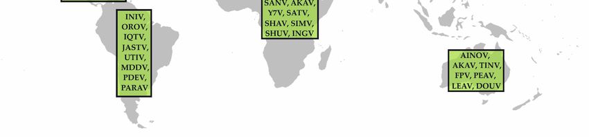
Viruses 2019, 11, 376                                                                                2 of 19

     Medically, one of the most highly important arthropod vectors is unquestionably mosquitoes.
Nonetheless, further, sometimes neglected arthropods such as ticks or biting midges can also act as
vectors of emerging disease agents [6]. However, when a pathogen has been detected in a certain
vector species, a thorough search for further putative vectors often remains undone. Furthermore,
the mere demonstration of a pathogen in a blood-feeding arthropod is often mistaken as evidence for
vector competence of that arthropod species without further experimental studies. Therefore, we want
to summarize here knowledge about ceratopogonid-transmitted disease agents and their impact on
human and animal health focusing on Culicoides biting midges and the new viral order Bunyavirales,
which contains the largest and most diverse families of RNA viruses [7]. In this review, we illuminate
the general impact of Culicoides biting midges, an often-neglected insect vector, but primarily, we
present evidence of their role in the transmission of selected orthobunyaviruses of public health or
veterinary importance.

2. How to Differentiate Mechanical from Biological Vectors?
     Vector competence refers to the physiological ability of arthropods to acquire and maintain microbial
agents from a host, and later transmit them to the next susceptible host [8]. The mere virus detection in a
midge does not necessarily indicate vector competence. A midge feeding on a viremic host can carry
virus from the blood meal in its gut without being infected itself. However, it can subsequently serve as
a so-called “mechanical vector” for virus transmission. To understand a disease and its epidemiology
entirely, it is essential to distinguish biological (competent) from pure mechanical vectors.
     One option to examine the vector competence is the separate analysis of the head and the salivary
glands and the rest of the insect body. Virus detection in the salivary glands is a sign for a biological
infection followed by virus dissemination, resulting in a potential virus transmission via saliva. Hence,
virus detection in the salivary glands of an insect hints at true vector competence. On the other hand,
detection of virus in the completely homogenized body of the insect may indicate just the ingestion of
a viremic blood meal, which not necessarily leads to an infection of the insect.
     To evaluate vector competence in more detail, infection studies under laboratory conditions either
with biting midges caught in the field or with laboratory-adapted colonies are a suitable method.
Insects can be artificially infected by intrathoracic inoculation of virus directly into the hemolymph
bypassing the intestinal (“gut”) barrier, or orally by feeding viremic blood. Subsequently, after an
extrinsic incubation period, samples are collected from surviving individuals. The insects themselves
could be examined as a whole or with head plus salivary glands and body separated as described
above. Furthermore, saliva could be collected from individual insects and tested for infectious virus or
viral genome. From the results, infection, dissemination and transmission rates are calculable [9].

3. Culicoides Biting Midges: Classification, Morphological Characteristics, and Distribution
      Culicoides is a genus of biting midges in the order Diptera, family Ceratopogonidae. Currently, the
genus contains 1368 species divided into numerous subgenera [10].
      Culicoides biting midges are among the most abundant hematophagous insects worldwide,
occurring from temperate areas to the tropics. Representing one of the smallest hematophagous
flies, they measure only 1–3 mm [11]. Their mouthparts form a proboscis well adapted for cutting
skin and sucking blood. Wings are well developed, and biting midges are commonly identified to
complex or species level based on the wing maculation [12]. However, this method of identification
is very time consuming and depends on the professional experience of the examiner. Alternative
methods use genome amplification by polymerase chain reaction (PCR) with subsequent sequencing
and phylogenetic comparison of DNA marker regions [13,14]. In addition, real-time PCR assays have
been established for a few Culicoides species [15,16], a DNA microarray was developed to identify
Culicoides species of the obsoletus group [17], and matrix-assisted laser desorption/ionization time-of
flight mass spectrometry (MALDI-TOF-MS) has been used to identify Culicoides species based on
peptide and protein signatures [18].
Viruses 2019, 11, 376 3 of 19
The life cycle of Culicoides, which passes through the egg stage, four larval stages, a pupa, and the
imago, requires a certain amount of free water or moisture, and some species occur in both fresh water
and estuarine environments. Breeding sites range from pools, streams, tree holes to saturated soil,
animal dung and rotting vegetation [19]. Females depend on blood for the maturation of the eggs; they
feed, depending on the species, on mammals and/or birds. Males do not feed on blood, and can survive
on nectar alone. The development of Culicoides species takes a few weeks—or even months, when
overwintering in a larval stage—and this process depends on the ambient temperatures, resulting in
a seasonal activity pattern in temperate regions. In mild climatic zones, the insect numbers start to
increase in late spring and early summer and usually peak in late summer or early autumn [11,20].
With the onset of low temperatures and the first frosts, the number of active insects drops dramatically.
In general, adult biting midges are short-lived and only a few individuals survive longer than 10
to 20 days. During this time, females may feed on hosts multiple times [11].
4. Public Health and Veterinary Impact of Culicoides Biting Midges
Biting midge bites can cause painful lesions; in some cases, the saliva even induces acute
allergic reactions such as the “common summer eczema (insect hypersensitivity)” in horses [21].
However, their veterinary or public health importance predominantly results from their role in the
transmission of pathogens, especially viruses, but also protozoans and filarial parasites [22] such
as avian hamosporidians [23,24] and Tetrapetalonema spp. [25–27]. More than 50 viruses have been
isolated from Culicoides spp. worldwide [28,29]. Most of the viruses belong to the families Reoviridae
(e.g., African horse sickness virus (AHSV), bluetongue virus (BTV), or epizootic hemorrhagic disease
virus (EHDV)), Rhabdoviridae (e.g., bovine ephemeral fever virus (BEFV)) and Peribunyaviridae (e.g.,
Akabane virus (AKAV), Schmallenberg virus (SBV), or Oropouche virus (OROV)). However, some
viruses isolated from Culicoides are only incidental findings, such as Rift Valley fever virus (RVFV),
which is mainly transmitted by mosquitoes [28,30]. Furthermore, vector competence studies are still
missing for a wide range of viruses suspected to be transmitted by Culicoides.
Surveillance on biting midges and biting midge-borne diseases is carried out in various countries
of several continents. As an example, the distribution and potential spread of BTV is monitored by
the collection of biting midges in virtually every affected European country, also to define e.g., a
so-called “seasonally vector free” period. To sample the adult Culicoides population, light-suction
traps placed at sentinel sites across outbreak areas are the most commonly used standardized trap
system [31]. Furthermore, detailed studies on local dispersal of Culicoides are available from Australia,
comparing various types of light traps with different outcomes in biting midge catches [32] and
analyzing distribution patterns related to weather and climate [33].
In search for potential vector species, an accurate estimate of species actually biting the vertebrate
host is vital. In the context of BTV vector search in Europe, it was demonstrated in a comparative
study of light-suction traps and drop catches that the biting midges caught in light-suction traps do
not provide an accurate reflection of the biting Culicoides population [31], which has to be considered
when assessing data collected in such surveillance studies.
Because of the needs of their agents’ vectors, Culicoides-borne diseases are strongly linked to
climate and weather. In temperate regions, the seasonal pattern of virus transmission coincides with
warm, moist summer and autumn months. In tropical and subtropical regions, high vertebrate infection
rates have been reported during wet summer and declining transmission rates in periods of lower
rainfall [34]. Although their flight range does not exceed a few hundred meters, biting midges can
be dispersed passively over great distances by wind [35–38], further contributing to their impact on
disease epidemiology.
The establishment of laboratory colonies poses great difficulties, especially because field-collected
specimens fail to mate under laboratory conditions. So far, very few Culicoides species have been successfully
reared under laboratory conditions [29,39,40] and, consequently, are available for studies on their vector
competence. Most laboratory colonies represent C. nubeculosus and C. sonorensis, rendering these two speciesViruses 2019, 11, 376 4 of 19
important models for studying arbovirus transmission by Culicoides biting midges [41]. Due to difficulties in
acquiring biting midges from the field for laboratory studies caused by the characteristics of the insects, the
major part of data available on experimental infections pertain to C. nubeculosus and C. sonorensis.
5. The Genus Orthobunyavirus
Only recently, the International Committee on Taxonomy of Viruses (ICTV) made some
fundamental changes in the taxonomy of the order Bunyavirales, which had been established in
2017 to accommodate viruses with linear, segmented, single-stranded RNA genome [42]. In early 2019,
the order was taxonomically revised by creating several new families, subfamilies, genera, and species.
As of February 2019, the order Bunyavirales consists of 46 genera assigned to 12 distinct families [7]. One
of the genera belonging to the newly established family Peribunyaviridae is the genus Orthobunyavirus,
which contains more than 170 unanimous insect-transmitted viruses in 18 serogroups, of which the
Simbu serogroup is not only one of the largest, but also the most important in terms of veterinary
public health. For human health, the California encephalitis serogroup is very likely the most relevant
one as it contains, e.g., La Crosse virus (LACV).
In general, orthobunyaviruses are spherical, about 100 nm in diameter and relatively simple in
their composition as the tri-partite RNA genome encodes only four structural and two non-structural
proteins. The small (S) genomic segment encodes for the nucleocapsid protein N and the non-structural
protein NSs, while the medium (M) segment encodes for the glycoproteins Gn and Gc, which form
spikes on the surface of the virus particle, and the non-structural protein NSm. The large (L) segment
encodes for the RNA dependent RNA polymerase [43].
The glycoproteins Gn and Gc are integral transmembrane proteins which are important for viral
attachment, membrane fusion and the induction of the host’s immune response [44–46]. The N-protein
is essential for viral RNA transcription and replication, and it forms ribonucleoprotein complexes
with the three viral RNA segments. These complexes are associated with the L-protein [47,48].
The NSs-protein is a major virulence factor in vertebrate hosts as it acts as an interferon antagonist and
is responsible for the shut-off of protein synthesis in mammalian hosts cells [48]. In vertebrate hosts,
the NSm-protein is associated with virus assembly and morphogenesis [49]. However, the function of
both non-structural proteins, NSs and NSm, in the insect vectors is largely unknown.
5.1. The Simbu Serogroup
When considering only viruses evidentially transmitted by Culicoides midges, the Simbu serogroup
is presumably the most important group within the genus Orthobunyavirus. Furthermore, its members
are of great veterinary importance, as a variety of simbuviruses induces severe congenital infections
in pregnant animals. The serogroup currently consists of 32 viruses grouped into 19 virus species
(Table 1). Simbuviruses are distributed worldwide (Table 1, Figure 1), and they persist in nature by
alternately infecting mammalian hosts and Culicoides vectors. In endemic regions, Simbu serogroup
viruses establish a pattern of cyclic circulation, with seasons of high virus appearance followed by
periods of only sporadic detection [50–55], which is presumably related to the overall immunity in the
host population and the abundance of Culicoides vectors.
Important representatives of the Simbu serogroup are AKAV and SBV, which predominantly
infect ruminants. Infections of adults are either asymptomatic or mild, associated with fever, diarrhea,
and decrease in milk yield for a few days. Infections of naïve dams during a critical phase of
gestation, however, may be followed by abortion, premature birth, mummification, stillbirth or
congenital deformations referred to as arthrogryposis-hydranencephaly syndrome [34,56]. Further
ruminant-infecting Simbu serogroup viruses that might induce similar clinical signs as AKAV or
SBV include Shuni virus (SHUV), Aino virus (AINOV) and the eponymous Simbu virus (SIMV) [57]
(Table 1). The only zoonotic virus of this serogroup is OROV (and reassortants such as Iquitos virus
and Madre de Dios virus), which is present in South America (Figure 1).Viruses 2019, 11, 376 5 of 19
Viruses 2019, 11, x FOR PEER REVIEW 5 of 21
Figure 1. Distribution of Simbu serogroup viruses. The meanings of the abbreviations (virus names) are listed in Table 1.
Figure 1. Distribution of Simbu serogroup viruses. The meanings of the abbreviations (virus names) are listed in Table 1.
Viruses 2019, 11, x; doi: FOR PEER REVIEW www.mdpi.com/journal/virusesViruses 2019, 11, 376 6 of 19
Table 1. Classification and distribution of Simbu serogroup viruses and vector species responsible for their transmission.
First Isolation Insect Vector Distribution
Virus Species Virus (Abbreviation)
Animal Hosts (Virus Reference (1st
Year Country Organism Putative Vectors Demonstrated Vectors Continent
Detection) Description)
Asia, Africa,
Akabane orthobunyavirus Akabane virus (AKAV) 1959 Japan mosquitoes biting midges, mosquitoes C. brevitarsis, C. variipennis 1 ruminants, swine [58]
Australia
Tinaroo virus (TINV) 1978 Australia Culicoides midges biting midges (C. brevitarsis) Australia ruminants [59]
Yaba-7 virus (Y7V) 1963 Nigeria mosquitoes mosquitoes Africa [60]
Aino orthobunyavirus Aino virus (AINOV) 1964 Japan mosquitoes mosquitoes, biting midges (C. brevitarsis) Asia, Australia ruminants [61]
Buttonwillow
Buttonwillow virus (BUTV) 1962 USA Cottontail rabbit midges (C. variipennis) North America leproids [62]
orthobunyavirus
mosquitoes (Culex spp., Anopheles spp.,
Cat Que orthobunyavirus Cát Quế virus (CQV) 2004 Vietnam mosquitoes Asia swine, birds [63]
Mansonia spp.)
Oya virus (OYAV) 1999 Malaysia swine mosquitoes Asia swine, humans [64]
Faceys paddock Facey’s paddock virus
1974 Australia mosquitoes mosquitoes (Culex annulirostris), midges Australia [65]
orthobunyavirus (FPV)
Ingwavuma mosquitoes (Culex spp., Monsonia spp.),
Ingwavuma virus (INGV) 1959 South Africa spectacled weaver Africa, Asia swine, dogs, birds [66]
orthobunyavirus biting midges
Jatobal orthobunyavirus Jatobal virus (JASTV) 1985 Brazil coati mosquitoes, biting midges South America coati [67]
Leanyer orthobunyavirus Leanyer virus (LEAV) 1974 Australia mosquitoes mosquitoes (Anopheles meraukensis) Australia cattle, wallabies, dogs [68]
Manzanilla mosquitoes (Culex tritaeniorhynchus), Central America,
Manzanilla virus (MANV) 1954 Trinidad Howler monkey [69]
orthobunyavirus biting midges Asia
Inini virus (INIV) 1973 French Guaiana Aracari bird South America birds [70]
Mermet orthobunyavirus Mermet virus (MERV) 1964 USA purple martin mosquitoes (Culex spp.) North America birds [71]
Oropouche orthobunyavirus Iquitos virus (IQTV) 1999 Peru human midges South America humans [72]
Madre de Dios virus
2007 Peru human mosquitoes, biting midges South America humans, monkeys [73]
(MDDV)
mosquitoes (Aedes spp., Coquillettidia sloths, non-human
Central/South
Oropouche virus (OROV) 1955 Trinidad human venezuelensis, Culex quinquefasciatus), C. paraensis primates, rodents, birds, [74]
America
biting midges humans
black-tufted
Perdões virus (PDEV) 2012 Brazil South America non-human primates [75]
marmoset
Pintupo virus (PINTV) Panama sloth biting midges Central America sloths [76]
biting midges (C. brevitarsis, C. imicola, C.
Peaton orthobunyavirus Peaton virus (PEAV) 1976 Australia Culicoides spp. Australia, Asia ruminants, horses [77]
jacobsoni)
Sabo orthobunyavirus Sabo virus (SABOV) 1966 Nigeria goat biting midges Africa goat [60]
Sango orthobunyavirus Sango virus (SANV) 1965 Nigeria cattle mosquitoes, biting midges Africa cattle [60]
Schmallenberg
Douglas virus (DOUV) 1978 Australia cattle biting midges (C. brevitarsis) Australia/Oceania ruminants [59]
orthobunyavirus
mosquitoes (Culex vishnui), biting midges
Sathuperi virus (SATV) 1957 India mosquitoes Asia, Africa ruminants [78]
(C. oxystoma)
biting midges (C. obsoletus,
Schmallenberg virus (SBV) 2011 Germany cattle biting midges C. scoticus, C. chiopterus, C. Europe ruminants [79]
imicola, C. sonorensis 1 )
Shamonda virus (SHAV) 1965 Nigeria cattle biting midges Africa, Asia ruminants [60]
Shuni orthobunyavirus Kaikalur virus (KAIV) 1971 India mosquitoes mosquitoes Asia [80]
biting midges (C.
ruminants, horse,
Shuni virus (SHUV) 1966 Nigeria cattle mosquitoes (Culex theileri), biting midges nubeculosus 1 , C. sonorensis Africa, Asia [60]
1) (humans?)
Simbu orthobunyavirus Para virus (PARAV) Argentina mosquitoes mosquitoes South America [81]
Simbu virus (SIMV) 1955 South Africa mosquitoes mosquitoes (Aedes spp.), biting midges Africa [82]
Thimiri orthobunyavirus Thimiri virus (THIV) 1963 India Indian Pond Heron biting midges (C. histrio) Asia birds [83]
brown-throated
Utinga orthobunyavirus Utinga virus (UTIV) 1965 Brazil ? South America sloths [84]
sloth
1 laboratory-adapted species, not naturally found in areas where the respective virus circulates.Viruses 2019, 11, 376 7 of 19
5.1.1. AKAV
AKAV was first described in the 1950s in Japan and is now endemic in large parts of Asia, the
Middle East, Australia and Africa [34,57,85,86]. The virus may induce abnormal courses of pregnancy
and fetal malformation in ruminants as described for both AKAV and SBV [34]. In addition, some
strains might occasionally cause encephalitis in newborn calves. The Iriki strain, which is present in
Japan and Korea, has been in rare cases associated with encephalitis in adult cattle [87–89].
Although AKAV was initially isolated from mosquitoes, they do not seem to play an important
epidemiological role in virus transmission [90]. In contrast, depending on geographical regions, various
Culicoides species are considered the main vectors and responsible for virus spread.
For Australia, the assumed main vector is C. brevitarsis [91,92], since it was demonstrated that
AKAV replicates in C. brevitarsis to high virus titers and reaches the salivary glands after 10 days of
incubation [93]. Furthermore, the virus has been isolated from C. wadai in Australia [94]. In Japan, C.
oxystoma is considered the major vector [51,95], although AKAV was also isolated from the mosquito
species Aedes vexans and Culex tritaeniorhynchus [58], which are most likely no competent vectors. In the
Middle East, C. imicola is probably the main vector responsible for virus circulation. In Israel, AKAV
was repeatedly detected in this vector species by RT-PCR, and in Oman, the isolation of AKAV was
possible from this culicoid species [96,97]. Also, throughout Africa, C. imicola seems to be an important
vector of AKAV. The virus was isolated multiple times from C. imicola in Zimbabwe [98], and in South
Africa AKAV was detected in a pool of mixed Culicoides spp. mainly consisting of C. imicola [99].
Another vector could be C. milnei, as AKAV was isolated from this species in Zimbabwe [98]. In Kenia,
AKAV was isolated from Anopheles funestus mosquitoes [100], but true vector competence has not been
demonstrated yet.
To investigate the vector competence of midges for AKAV, some experimental infection studies
were carried out with different Culicoides species. It could be demonstrated that AKAV replicates in
C. variipennis after oral infection for at least 9 days, while in C. nubeculosus, AKAV replicated only
after intrathoracic inoculation [101]. The extensive replication of AKAV within C. variipennis suggests
that Culicoides spp. can act as fully competent vectors. However, although C. variipennis serving as a
suitable surrogate model, this laboratory-adapted species is not the natural vector of the virus, as it is
native to North America while AKAV is endemic in Asia, Africa, and Australia.
5.1.2. SBV
SBV emerged in late 2011 in the German/Dutch border region [79] and is now endemic in most
European countries [50,102]. It predominantly infects ruminants, causing a mild, transient disease in
adult animals, but it may induce severe fetal malformation when naïve dams are infected during a
critical period of pregnancy [103]. Based on its close relationship to AKAV, it was assumed that Culicoides
act as vectors also for SBV and, indeed, SBV genome was subsequently detected in field-collected
Culicoides midges of various species repeatedly throughout Europe [104–108]. In contrast, mosquitoes
do not seem to play a major role, if any, in virus transmission [109–112].
In temperate European countries, biting midges of the C. obsoletus complex are considered the
main vectors, while in Mediterranean countries C. imicola and C. punctatus seem to significantly
contribute to virus transmission. Viral genome was also detected in C. dewulfi, C. pulicaris, C. newsteadi,
C. lupicaris, and C. nubeculosus by (real-time) RT-PCR [104–108,112–114]. To underpin the findings in
field-collected biting midges with experimental data, several vector competence studies were carried
out. The Nearctic species C. sonorensis was infected orally and intrathoracically. The detection of
viral genome after an extrinsic incubation period in the saliva and the isolation of infectious virus
from the head proved dissemination of SBV within the insect organism [115] and demonstrated that
C. sonorensis is a suitable laboratory model for SBV. Furthermore, from these infected individuals, a
range of Cq-values from decapitated heads (including salivary glands) was available to compare with
those produced from heads of field-collected Culicoides. The values provided for field-collected C.
obsoletus, C. scoticus and C. chiopterus from the Netherlands were very similar to those obtained fromViruses 2019, 11, 376 8 of 19
the laboratory infections, indicating that these species act as true vectors [115]. In contrast, multiple
infection studies with laboratory colonies of C. nubeculosus showed only low infection rates, although
viral genome was detected in field-collected C. nubeculosus [112,115].
Unfortunately, for the C. obsoletus complex, which consists of the species C. obsoletus, C. scoticus, C.
chiopterus and C. montanus and is considered to contain the major vectors of SBV in temperate European
countries, laboratory colonies are not available. However, experimental laboratory infection studies
were carried out with field-collected individuals of the C. obsoletus complex and with C. imicola, which,
according to field data, is considered the main vector in the Mediterranean. Both, i.e., midges of the C.
obsoletus complex and C. imicola, showed high infection rates and virus dissemination [116], confirming
the assumptions of their vector competence, and field-collected C. scoticus were able to replicate SBV to
a potentially transmissible level [112].
5.1.3. SHUV
SHUV was firstly isolated in Nigeria from healthy cattle [60] as well as from Culicoides biting
midges and mosquitoes in Africa [117]. In 2014, SHUV was for the first time detected outside of Africa,
and was isolated from malformed lambs in Israel [118]. In addition to typical Simbu virus-related
congenital defects in cattle and sheep, SHUV may occasionally induce severe neurological symptoms
in horses or cattle [119,120]. A general zoonotic potential cannot be ruled out, since SHUV was isolated
from a febrile child in Nigeria [121], and specific antibodies were found in large animal veterinarians
in South Africa [122].
In South Africa, SHUV was recovered twice from pools of Culex theileri [117]. However, the
mosquito taxa Culex pipiens biotype pipiens and Aedes aegypti showed only very low susceptibility
to SHUV following intrathoracic inoculation. Oral infection was not possible under experimental
conditions [9]. In contrast, C. nubeculosus and C. sonorensis could be orally infected, with the virus
disseminating well in both species [9], indicating that Culicoides biting midges are more likely the
natural vectors than mosquitoes.
5.1.4. OROV
First discovered in a febrile forest worker in 1955 in Trinidad [74], OROV is now endemic in many
South and Central American countries [123,124]. Oropouche fever is an acute febrile illness that affects
humans. Common symptoms include fever, headache, muscle pain and skin rash, and many infections
develop into meningitis or encephalitis [123]. With this zoonotic potential, OROV protrudes from
the other viruses in the Simbu serogroup. In South and Central America, OROV occurs in a sylvatic
cycle between its insect vectors, some wild bird species and mammals such as rodents, sloths and
non-human primates as amplifying hosts (Table 1). In the urban cycle, humans are most likely the only
vertebrate hosts, as domestic animals such as chickens, dogs or cats could be excluded as amplifying
hosts [124,125].
The main vector in the sylvatic cycle is still unclear; however, OROV was isolated from two sylvatic
mosquitoes in the forest: Aedes serratus, collected in the Brazilian Amazon region, and Coquillettidia
venezuelensis in Trinidad [125,126].
In the urban cycle, the biting midge species C. paraensis and the mosquito Culex quinquefasciatus are
believed to be the main vectors. Although the isolation rate from C. paraensis during epidemics is low,
its involvement as a vector is suggested based on transmission studies, where C. paraensis transmitted
OROV to hamsters five or more days after feeding on blood of viremic patients [125].
Likewise, Culex quinquefasciatus proved to be an ineffective vector of OROV in laboratory
transmission experiments [127], but virus is frequently detected in this species during outbreaks [128].
The link between the two transmission cycles, i.e., the sylvatic and the urban cycle, are probably
humans entering the forests, where they become infected, and returning to urbanized areas [125].Viruses 2019, 11, 376 9 of 19
5.1.5. Further Members of the Simbu Serogroup
There are several additional members of veterinary importance within the Simbu serogroup
(Table 1), which might induce the typical congenital malformation when naïve pregnant dams are
infected. Unfortunately, vector competence studies are missing for most of them. However, in general,
certain biting midge species are suspected to be able to transmit several members of the serogroup. As
an example, field-collected C. brevitarsis, the major vector for AKAV in Australia, were tested positive
for further Australian simbuviruses such as Douglas virus (DOUV) or Peaton virus (PEAV) [59,77].
In the Mediterranean and the Middle East, where C. imicola is the main vector of SBV or AKAV, also
PEAV was found in this species [129], confirming the concept of certain Culicoides species transmitting
several simbuviruses present in the respective region.
5.2. Further Orthobunyaviruses of Public Health Importance
In the California encephalitis serogroup of the genus Orthobunyavirus, several zoonotic agents
distributed in North America, Europe, Africa, or Asia can be found. However, for none of them Culicoides
spp. have been demonstrated to be vector-competent. Since they are transmitted by mosquitoes
between their vertebrate hosts, which range from small mammals to ungulates and humans, they are
among the viruses that are referred to as “mobovirus” (= mosquito-borne viruses) [130]. Some of the
most relevant representatives of zoonotic orthobunyaviruses include LACV, Jamestown Canyon virus
(JCV), Keystone virus (KEYV) and Ťahyňa virus (TAHV).
LACV, the most pathogenic agent within the California encephalitis serogroup, is the causative
agent of the so-called “rural encephalitis” in the Appalachian and midwestern regions of North America
and the leading cause of pediatric arboviral encephalitis. Clinical symptoms of an LACV-infection
include headache, fever, myalgia, and encephalitis, while in rare cases fatalities may occur (reviewed
in [131]). Within the natural infection cycle between small mammals and mosquito vectors, humans are
considered as dead-end hosts, meaning that the virus is not transmitted from humans to blood-feeding
insects and that, consequently, the transmission chain ends in human beings [132]. As LACV was
isolated multiple times from Aedes triseriatus [133–135] this mosquito species, which is native to North
America, is considered the main insect vector. Furthermore, LACV has been isolated from the invasive
Asian tiger mosquito Aedes albopictus [136,137] and the Asian bush mosquito Aedes japonicus [138,139].
In experimental infection studies, both proved to be competent vectors of LACV [140,141]. To date, this
virus has not been found in midges, suggesting that these do not play an important role in transmission.
Similar to LACV, JCV has a wide geographical distribution throughout North America and causes
a mild febrile illness or central nervous system infections inducing meningitis or meningoencephalitis
in humans [142]. However, in contrast to LACV, which is associated with clinical disease in children,
JCV induces symptoms also in adults. White-tailed deer and other ungulates are considered the
primary vertebrate hosts [143]. The first virus isolation succeeded from a pool of Aedes abserratus [144].
Since then, JCV has been found in varying but great numbers of different species of Culicidae (Aedes
spp., Anopheles spp., Culiseta spp., Psorophora spp.) and Tabanidae (Chrysops spp., Hybomitra spp.) [145].
In the US state of Connecticut, the predominant vector species seems to be Aedes canadensis, while in
northeastern New York and in Michigan, JCV is predominantly transmitted by Aedes provocans [146,147].
Aedes intrudens, Aedes abserratus and Aedes punctor seem to be important vectors in Massachusetts [148],
Aedes stimulans in northern Indiana [149], and Aedes vexans in North Dakota [150].
KEYV is distributed on the east coast of North America from Florida, where the virus was firstly
discovered in 1964 from Aedes atlanticus in Keystone [151], up to Maryland in the north and Texas
in the west. Vertebrate hosts seem to be mammals such as squirrels, raccoons, and white-tailed
deer [152–154], while the main insect vectors are mosquitoes such as Aedes atlanticus [155], various
Aedes spp. (e.g., vexans, tiseriatus, taeniorhynchus, infirmatus, canadensis), and some Culex spp. [156].
In humans, antibodies against KEYV have been repeatedly detected [157], and the virus itself was
isolated from individuals showing clinical symptoms such as fever and rash [158].Viruses 2019, 11, 376 10 of 19
TAHV is endemic in Asia and throughout Africa and represents the first mobovirus pathogenic
to humans found in Europe [130]. It may induce a febrile illness referred to as Valtice fever, which
occurs mainly in children and is characterized by influenza-like symptoms. In rare cases, it may cause
meningitis or atypical pneumonia [159]. Vertebrate hosts other than humans are small mammals
such as rabbits or hedgehogs [160,161]. Arthropod vectors are most likely mosquitoes of the genera
Aedes, Culex, and Anopheles; however, experimental vector competence studies are again missing.
Originally, TAHV was isolated from Aedes vexans and Aedes caspius mosquitoes in the villages Ťahyňa
and Križany, Czechoslovakia [162]. Following its initial discovery, TAHV has been isolated from several
mosquito species such as Aedes vexans, Aedes cantans, Aedes caspius, Aedes sticticus, Aedes diantaeus, Aedes
hexodontus, Culex pipiens and Anopheles hyrcanus throughout Europe [163–169].
6. Responses to Culicoides-borne Arbovirus Incursions
Since treatment options are not available for Culicoides-borne arbovirus infections, other
possibilities are discussed to prevent clinical disease in vertebrate hosts and virus spread into unaffected
regions. Trading and movement restrictions are put in place for livestock and their products (e.g.,
semen, embryos) to reduce the risk of virus introductions into new areas [22,103]. For Culicoides-borne
diseases with teratogenic effects such as AKAV, SBV, or SHUV, livestock-management measures could
be effective. The mating period of livestock could be adjusted to avoid that naïve females are in the
critical phase of pregnancy during the season of the highest biting midge activity. Another possibility is
to ensure that dams acquire immunity before they conceive for the first time, which could be achieved
by exposing the youngstock to potentially infected vectors. However, this concept requires a high
number of infected insect organisms every year and a very high transmission rate of the virus from the
vector to the vertebrate host to ensure that every young animal is bitten and infected. Hence, a much
more reliable method to acquire immunity is by vaccination [50]. For some of the livestock-affecting
simbuviruses including AKAV, AINOV, and SBV, vaccines are commercially available [170–173], and
European experiences during the bluetongue outbreak from 2006 to 2009 demonstrated that vaccination
campaigns against a Culicoides-borne disease can play a major role in reducing virus circulation or
even in eradicating the disease from a given region [174,175]. For the zoonotic OROV, however, there
is no vaccine available yet [176].
From the entomological point of view, the application of insecticides or repellents could be
taken into consideration to prevent vectors from biting susceptible animals. In addition, these could
be housed in insect-proof (e.g., screened) stable buildings. However, most of these measures seem
unhandy, expensive, impracticable and have either none or only limited effect [177,178]. For humans
with transient exposure, the use of repellents is a suitable measure to protect themselves from insect
bites; N,N-diethyl-m-toluamide (DEET) is considered the gold standard repellent [177,179].
In addition, measures controlling vector development should be considered. These may include
environmental interventions to remove larval breeding sites and the control of adult midges by residual
insecticide spraying of surfaces where adult biting midges rest within animal stables. However, due
to the broad range of habitats and breeding sites used by biting midges, insecticide treatment and
removal of breeding sites are of limited success in biting midge control [177].
In conclusion, Culicoides-borne diseases are difficult to control by vector management alone, while
vaccinating livestock may represents an effective tool for disease prevention or even for eradicating a
disease from a region.
7. Concluding Remarks
For veterinary medicine and veterinary public health, Culicoides biting midges are of great
importance as they transmit a multitude of viruses of great impact. In contrast, they only seem to
play a minor role for human public health. Due to their potential to contribute to unexpected disease
emergence they should be thoroughly surveyed. Multiple detections of novel viruses or introductionsViruses 2019, 11, 376 11 of 19
of known pathogens into previously unaffected regions during the past decade demonstrate in an
impressive fashion that arbovirus outbreaks are hard to predict in space and time [180].
From the genus Orthobunyavirus, the emergence of SBV in Europe or the incursion of SHUV into
the Middle East are prominent examples [79,118]. To be prepared for putative future outbreaks of
arboviral diseases, it is critical to carry out profound research and surveillance on biting midges and
their role as vectors.
Author Contributions: Conceptualization, M.B. and K.W.; methodology, F.S. and K.W.; writing—original draft
preparation, F.S. and K.W.; writing–review and editing, H.K. and M.B; visualization, F.S. and K.W.; all authors
read and approved the final version of the manuscript.
Funding: This research was funded by the German Federal Ministry of Food and Agriculture (BMEL) through the
Federal Office for Agriculture and Food (BLE), grant number 281B101816.
Conflicts of Interest: The authors declare no conflict of interest. The funders had no role in the design of the
study; in the collection, analyses, or interpretation of data; in the writing of the manuscript, or in the decision to
publish the results.
References
1. Baud, D.; Gubler, D.J.; Schaub, B.; Lanteri, M.C.; Musso, D. An update on Zika virus infection. Lancet 2017,
390, 2099–2109. [CrossRef]
2. Petersen, L.R.; Brault, A.C.; Nasci, R.S. West Nile virus: Review of the literature. JAMA 2013, 310, 308–315.
[CrossRef]
3. Young, P.R. Arboviruses: A family on the move. Adv. Exp. Med. Biol. 2018, 1062, 1–10. [PubMed]
4. Althouse, B.M.; Hanley, K.A. The tortoise or the hare? Impacts of within-host dynamics on transmission
success of arthropod-borne viruses. Philos. Trans. R. Soc. Lond. B Biol. Sci. 2015, 370, 20140299. [CrossRef]
[PubMed]
5. Medlock, J.M.; Leach, S.A. Effect of climate change on vector-borne disease risk in the UK. Lancet Infect. Dis.
2015, 15, 721–730. [CrossRef]
6. Hubálek, Z.; Rudolf, I.; Nowotny, N. Arboviruses pathogenic for domestic and wild animals. Adv. Virus Res.
2014, 89, 201–275.
7. ICTV. Virus Taxonomy: The Classification and Nomenclature of Viruses. Available online: https://talk.
ictvonline.org/ictv-reports/ictv_online_report/ (accessed on 25 March 2019).
8. Beerntsen, B.T.; James, A.A.; Christensen, B.M. Genetics of mosquito vector competence. Microbiol. Mol. Biol.
Rev. 2000, 64, 115–137. [CrossRef] [PubMed]
9. Möhlmann, T.W.R.; Oymans, J.; Wichgers Schreur, P.J.; Koenraadt, C.J.M.; Kortekaas, J.; Vogels, C.B.F. Vector
competence of biting midges and mosquitoes for Shuni virus. PLoS Negl. Trop. Dis. 2018, 12, e0006993.
[CrossRef]
10. Borkent, A. Numbers of Extant and Fossil Species of Ceratopogonidae. 6 July 2016. Available online:
https://www.inhs.illinois.edu/files/4014/6785/5847/WorldCatalogtaxa.pdf (accessed on 25 March 2019).
11. Mellor, P.S.; Boorman, J.; Baylis, M. Culicoides biting midges: Their role as arbovirus vectors. Annu. Rev.
Entomol. 2000, 45, 307–340. [CrossRef]
12. Mathieu, B.; Cêtre-Sossah, C.; Garros, C.; Chavernac, D.; Balenghien, T.; Carpenter, S.; Setier-Rio, M.L.;
Vignes-Lebbe, R.; Ung, V.; Candolfi, E.; et al. Development and validation of IIKC: An interactive identification
key for Culicoides (Diptera: Ceratopogonidae) females from the western Palaearctic region. Parasit. Vectors
2012, 5, 137. [CrossRef]
13. Pages, N.; Sarto, I.M.V. Differentiation of Culicoides obsoletus and Culicoides scoticus (Diptera: Ceratopogonidae)
based on mitochondrial cytochrome oxidase subunit I. J. Med. Entomol. 2005, 42, 1026–1034. [CrossRef]
14. Dallas, J.F.; Cruickshank, R.H.; Linton, Y.M.; Nolan, D.V.; Patakakis, M.; Braverman, Y.; Capela, R.; Capela, M.;
Pena, I.; Meiswinkel, R.; et al. Phylogenetic status and matrilineal structure of the biting midge, Culicoides
imicola, in Portugal, Rhodes and Israel. Med. Vet. Entomol. 2003, 17, 379–387. [CrossRef]
15. Cêtre-Sossah, C.; Mathieu, B.; Setier-Rio, M.L.; Grillet, C.; Baldet, T.; Delécolle, J.C.; Albina, E. Development
and evaluation of a real-time quantitative PCR assay for Culicoides imicola, one of the main vectors of
bluetongue (BT) and African horse sickness (AHS) in Africa and Europe. Res. Vet. Sci. 2008, 85, 372–382.
[CrossRef]Viruses 2019, 11, 376 12 of 19
16. Wenk, C.E.; Kaufmann, C.; Schaffner, F.; Mathis, A. Molecular characterization of Swiss Ceratopogonidae
(Diptera) and evaluation of real-time PCR assays for the identification of Culicoides biting midges. Vet.
Parasitol. 2012, 184, 258–266. [CrossRef]
17. Deblauwe, I.; de Witte, J.C.; de Deken, G.; de Deken, R.; Madder, M.; van Erk, S.; Hoza, F.A.; Lathouwers, D.;
Geysen, D. A new tool for the molecular identification of Culicoides species of the Obsoletus group: The glass
slide microarray approach. Med. Vet. Entomol. 2012, 26, 83–91. [CrossRef]
18. Uhlmann, K.R.; Gibb, S.; Kalkhof, S.; Arroyo-Abad, U.; Schulz, C.; Hoffmann, B.; Stubbins, F.; Carpenter, S.;
Beer, M.; von Bergen, M.; et al. Species determination of Culicoides biting midges via peptide profiling using
matrix-assisted laser desorption ionization mass spectrometry. Parasit. Vectors 2014, 7, 392. [CrossRef]
19. Wirth, W.W.; Hubert, A.A. The Culicoides of Southeast Asia (Diptera: Ceratopogonidae). Am. Entomol. Inst.
(USA) 1989, 44, 514.
20. Kline, D.L.; Axtell, R.C. Salt-marsh Culicoides (Diptera—Ceratopogonidae)—Species, seasonal abundance
and comparisons of trapping methods. Mosq. News 1976, 36, 1–10.
21. Van der Rijt, R.; van den Boom, R.; Jongema, Y.; van Oldruitenborgh-Oosterbaan, M.M. Culicoides species
attracted to horses with and without insect hypersensitivity. Vet. J. 2008, 178, 91–97. [CrossRef]
22. Carpenter, S.; Groschup, M.H.; Garros, C.; Felippe-Bauer, M.L.; Purse, B.V. Culicoides biting midges,
arboviruses and public health in Europe. Antiviral Res. 2013, 100, 102–113. [CrossRef]
23. Veiga, J.; Martinez-de la Puente, J.; Vaclav, R.; Figuerola, J.; Valera, F. Culicoides paolae and C. circumscriptus as
potential vectors of avian haemosporidians in an arid ecosystem. Parasit. Vectors 2018, 11, 524. [CrossRef]
24. Chagas, C.R.F.; Bukauskaite, D.; Ilgunas, M.; Iezhova, T.; Valkiunas, G. A new blood parasite of leaf warblers:
Molecular characterization, phylogenetic relationships, description and identification of vectors. Parasit.
Vectors 2018, 11, 538. [CrossRef]
25. Yates, J.A.; Lowrie, R.C., Jr.; Eberhard, M. Development of Tetrapetalonema llewellyni to the infective stage in
Culicoides hollensis. J. Parasitol. 1982, 68, 293–296. [CrossRef]
26. Lowrie, R.C., Jr.; Eberhard, M.L.; Orihel, T.C. Development of Tetrapetalonema marmosetae to the infective
stage in Culicoides hollensis and C. furens. J. Parasitol. 1978, 64, 1003–1007. [CrossRef]
27. Linley, J.R. Biting midges (Diptera: Ceratopogonidae) as vectors of nonviral animal pathogens. J. Med.
Entomol. 1985, 22, 589–599. [CrossRef]
28. Meiswinkel, R.; Nevill, E.M.; Venter, G.J. Vectors: Culicoides spp. In Infectious Diseases of Livestock with Special
Reference to Southern Africa; Coetzer, J.A.W., Thomson, G.R., Tustin, R.C., Eds.; Oxford University Press: Cape
Town, South Africa, 1994; Volume 1, pp. 68–89.
29. Carpenter, S.; Veronesi, E.; Mullens, B.; Venter, G. Vector competence of Culicoides for arboviruses: Three
major periods of research, their influence on current studies and future directions. Rev. Sci. Tech. 2015, 34,
97–112. [CrossRef]
30. Jennings, M.; Platt, G.S.; Bowen, E.T. The susceptibility of Culicoides variipennis Coq. (Diptera:
Ceratopogonidae) to laboratory infection with Rift Valley fever virus. Trans. R. Soc. Trop. Med. Hyg.
1982, 76, 587–589. [CrossRef]
31. Carpenter, S.; Szmaragd, C.; Barber, J.; Labuschagne, K.; Gubbins, S.; Mellor, P. An assessment of Culicoides
surveillance techniques in northern Europe: Have we underestimated a potential bluetongue virus vector? J.
Appl. Ecol. 2008, 45, 1237–1245.
32. Murray, M.D. Local dispersal of the biting midge Culicoides brevitarsis Kieffer (Diptera, Ceratopogonidae) in
Southeastern Australia. Austr. J. Zool. 1987, 35, 559–573. [CrossRef]
33. Murray, M.D.; Nix, H.A. Southern limits of distribution and abundance of the biting-midge Culicoides
brevitarsis Kieffer (Diptera, Ceratopogonidae) in Southeastern Australia—An application of the Growest Model.
Austr. J. Zool. 1987, 35, 575–585. [CrossRef]
34. Kirkland, P.D. Akabane virus infection. Rev. Sci. Tech. 2015, 34, 403–410. [CrossRef]
35. Sellers, R.F. Weather, host and vector—Their interplay in the spread of insect-borne animal virus diseases. J.
Hyg. (London) 1980, 85, 65–102. [CrossRef]
36. Sellers, R.F.; Maarouf, A.R. Possible introduction of epizootic hemorrhagic disease of deer virus (serotype 2)
and bluetongue virus (serotype 11) into British Columbia in 1987 and 1988 by infected Culicoides carried on
the wind. Can. J. Vet. Res. 1991, 55, 367–370.
37. Sellers, R.F.; Pedgley, D.E. Possible windborne spread to western Turkey of bluetongue virus in 1977 and of
Akabane virus in 1979. J. Hyg. (London) 1985, 95, 149–158. [CrossRef]Viruses 2019, 11, 376 13 of 19
38. Braverman, Y.; Chechik, F. Air streams and the introduction of animal diseases borne on Culicoides (Diptera,
Ceratopogonidae) into Israel. Rev. Sci. Tech. 1996, 15, 1037–1052. [CrossRef]
39. Hunt, G.J. A Procedural Manual for the Large-Scale Rearing of the Biting Midge, Culicoides variipennis (Diptera:
Ceratopogonidae); U.S. Department of Agriculture, National Agricultural Library (USA): Beltsville, MD, USA,
1994.
40. Carpenter, S.; Mordue, A.J.; Mordue, W. Oviposition in Culicoides impunctatus under laboratory conditions.
Entomol. Exp. Appl. 2001, 101, 123–129. [CrossRef]
41. Jones, R.H.; Foster, N.M. Relevance of laboratory colonies of the vector in arbovirus research—Culicoides
variipennis and bluetongue. Am. J. Trop. Med. Hyg. 1978, 27, 168–177. [CrossRef]
42. Maes, P.; Alkhovsky, S.V.; Bao, Y.; Beer, M.; Birkhead, M.; Briese, T.; Buchmeier, M.J.; Calisher, C.H.;
Charrel, R.N.; Choi, I.R.; et al. Taxonomy of the family Arenaviridae and the order Bunyavirales: update 2018.
Arch. Virol. 2018, 163, 2295–2310. [CrossRef]
43. Barr, J.N.; Walter, C.T. Recent advances in the molecular and cellular biology of bunyaviruses. J. Gen. Virol.
2011, 92, 2467–2484.
44. Shi, X.; Lappin, D.F.; Elliott, R.M. Mapping the Golgi targeting and retention signal of Bunyamwera virus
glycoproteins. J. Virol. 2004, 78, 10793–10802. [CrossRef]
45. Shi, X.; van Mierlo, J.T.; French, A.; Elliott, R.M. Visualizing the replication cycle of Bunyamwera
orthobunyavirus expressing fluorescent protein-tagged Gc glycoprotein. J. Virol. 2010, 84, 8460–8469.
[CrossRef] [PubMed]
46. Shi, X.; Goli, J.; Clark, G.; Brauburger, K.; Elliott, R.M. Functional analysis of the Bunyamwera orthobunyavirus
Gc glycoprotein. J. Gen. Virol. 2009, 90, 2483–2492. [CrossRef]
47. Elliott, R.M. Orthobunyaviruses: Recent genetic and structural insights. Nat. Rev. Microbiol. 2014, 12,
673–685. [CrossRef] [PubMed]
48. Elliott, R.M.; Blakqori, G. Molecular biology of orthobunyaviruses. In Bunyaviridae. Molecular and Cellular
Biology; Plyusnin, A., Elliott, R.M., Eds.; Caister Academic Press: Norfolk, UK, 2011; pp. 1–39.
49. Shi, X.; Kohl, A.; Léonard, V.H.J.; Li, P.; McLees, A.; Elliott, R.M. Requirement of the N-terminal region of
Orthobunyavirus nonstructural protein NSm for virus assembly and morphogenesis. J. Virol. 2006, 80, 8089.
[CrossRef]
50. Wernike, K.; Beer, M. Schmallenberg virus: A novel virus of veterinary importance. Adv. Virus Res. 2017, 99,
39–60. [PubMed]
51. Kato, T.; Shirafuji, H.; Tanaka, S.; Sato, M.; Yamakawa, M.; Tsuda, T.; Yanase, T. Bovine arboviruses in
Culicoides biting midges and sentinel cattle in southern Japan from 2003 to 2013. Transbound. Emerg. Dis.
2016, 63, e160–e172. [CrossRef]
52. Kato, T.; Yanase, T.; Suzuki, M.; Katagiri, Y.; Ikemiyagi, K.; Takayoshi, K.; Shirafuji, H.; Ohashi, S.; Yoshida, K.;
Yamakawa, M.; et al. Monitoring for bovine arboviruses in the most southwestern islands in Japan between
1994 and 2014. BMC Vet. Res. 2016, 12, 125. [CrossRef]
53. Hayama, Y.; Yanase, T.; Suzuki, M.; Unten, K.; Tomochi, H.; Kakehi, M.; Shono, Y.; Yamamoto, T.; Kobayashi, S.;
Murai, K.; et al. Meteorological factors affecting seroconversion of Akabane disease in sentinel calves in the
subtropical Okinawa Islands of Japan. Trop. Anim. Health Prod. 2018, 50, 209–215. [CrossRef]
54. Wernike, K.; Holsteg, M.; Szillat, K.P.; Beer, M. Development of within-herd immunity and long-term
persistence of antibodies against Schmallenberg virus in naturally infected cattle. BMC Vet. Res. 2018, 14,
368. [CrossRef]
55. Geoghegan, J.L.; Walker, P.J.; Duchemin, J.B.; Jeanne, I.; Holmes, E.C. Seasonal drivers of the epidemiology of
arthropod-borne viruses in Australia. PLoS Negl. Trop. Dis. 2014, 8, e3325. [CrossRef]
56. Wernike, K.; Elbers, A.; Beer, M. Schmallenberg virus infection. Rev. Sci. Tech. 2015, 34, 363–373. [CrossRef]
57. Saeed, M.F.; Li, L.; Wang, H.; Weaver, S.C.; Barrett, A.D. Phylogeny of the Simbu serogroup of the genus
Bunyavirus. J. Gen. Virol. 2001, 82, 2173–2181. [CrossRef]
58. Oya, A.; Okuno, T.; Ogata, T.; Kobayashii; Matsuyama, T. Akabane, a new arbovirus isolated in Japan. Jpn. J.
Med. Sci. Biol. 1961, 14, 101–108. [CrossRef]
59. Cybinski, D.H. Douglas and Tinaroo viruses: Two Simbu group arboviruses infecting Culicoides brevitarsis
and livestock in Australia. Aust. J. Biol. Sci. 1984, 37, 91–97. [CrossRef]Viruses 2019, 11, 376 14 of 19
60. Causey, O.R.; Kemp, G.E.; Causey, C.E.; Lee, V.H. Isolations of Simbu-group viruses in Ibadan, Nigeria
1964-69, including the new types Sango, Shamonda, Sabo and Shuni. Ann. Trop. Med. Parasitol. 1972, 66,
357–362. [CrossRef]
61. Takahashi, K.; Oya, A.; Okazda, T.; Matsuo, R.; Kuma, M. Aino virus, a new member of Simbu group of
arbovirus from mosquitoes in Japan. Jpn. J. Med. Sci. Biol. 1968, 21, 95–101. [CrossRef]
62. Reeves, W.C.; Scrivani, R.P.; Hardy, J.L.; Roberts, D.R.; Nelson, R.L. Buttonwillow virus, a new Arbovirus
isolated from mammals and Culicoides midges in Kern County, California. Am. J. Trop. Med. Hyg. 1970, 19,
544–551. [CrossRef]
63. Bryant, J.E.; Crabtree, M.B.; Nam, V.S.; Yen, N.T.; Duc, H.M.; Miller, B.R. Isolation of arboviruses from
mosquitoes collected in northern Vietnam. Am. J. Trop. Med. Hyg. 2005, 73, 470–473. [CrossRef]
64. Kono, Y.; Yusnita, Y.; Mohd Ali, A.R.; Maizan, M.; Sharifah, S.H.; Fauzia, O.; Kubo, M.; Aziz, A.J.
Characterization and identification of Oya virus, a Simbu serogroup virus of the genus Bunyavirus, isolated
from a pig suspected of Nipah virus infection. Arch. Virol. 2002, 147, 1623–1630. [CrossRef]
65. Doherty, R.L.; Carley, J.G.; Kay, B.H.; Filippich, C.; Marks, E.N.; Frazier, C.L. Isolation of virus strains from
mosquitoes collected in Queensland, 1972–1976. Aust. J. Exp. Biol. Med. Sci. 1979, 57, 509–520. [CrossRef]
66. McIntosh, B.M.; McGillivray, G.M.; Dickinson, D.B. Ingwavuma virus: An arbovirus isolated in South Africa.
S. Afr. J. Med. Sci. 1965, 30, 67–70.
67. Figueiredo, L.T.; Da Rosa, A.P. Jatobal virus antigenic characterization by ELISA and neutralization test
using EIA as indicator, on tissue culture. Mem. Inst. Oswaldo Cruz 1988, 83, 161–164. [CrossRef]
68. Doherty, R.L.; Carley, J.G.; Filippich, C.; Kay, B.H.; Gorman, B.M.; Rajapaksa, N. Isolation of Sindbis
(alphavirus) and Leanyer viruses from mosquitoes collected in the Northern Territory of Australia, 1974.
Aust. J. Exp. Biol. Med. Sci. 1977, 55, 485–489. [CrossRef]
69. Anderson, C.R.; Spence, L.P.; Downs, W.G.; Aitken, T.H. Manzanilla virus: A new virus isolated from the
blood of a howler monkey in Trinidad, W.I. Am. J. Trop. Med. Hyg. 1960, 9, 78–80. [CrossRef] [PubMed]
70. Pajot, F.X. Diseases transmitted by insects in French Guaiana. Bol. Oficina Sanit. Panam. 1980, 88, 218–227.
(In Spanish) [PubMed]
71. Calisher, C.H.; Kokernot, R.H.; De Moore, J.F.; Boyd, K.R.; Hayes, J.; Chappell, W.A. Arbovirus studies in the
Ohio-Mississippi Basin, 1964–1967. VI. Mermet: A Simbu-group arbovirus. Am. J. Trop. Med. Hyg. 1969, 18,
779–788. [CrossRef] [PubMed]
72. Aguilar, P.V.; Barrett, A.D.; Saeed, M.F.; Watts, D.M.; Russell, K.; Guevara, C.; Ampuero, J.S.; Suarez, L.;
Cespedes, M.; Montgomery, J.M.; et al. Iquitos virus: A novel reassortant Orthobunyavirus associated with
human illness in Peru. PLoS Negl. Trop. Dis. 2011, 5, e1315. [CrossRef] [PubMed]
73. Ladner, J.T.; Savji, N.; Lofts, L.; Travassos da Rosa, A.; Wiley, M.R.; Gestole, M.C.; Rosen, G.E.; Guzman, H.;
Vasconcelos, P.F.; Nunes, M.R.; et al. Genomic and phylogenetic characterization of viruses included in the
Manzanilla and Oropouche species complexes of the genus Orthobunyavirus, family Bunyaviridae. J. Gen.
Virol. 2014, 95, 1055–1066. [CrossRef] [PubMed]
74. Anderson, C.R.; Spence, L.; Downs, W.G.; Aitken, T.H. Oropouche virus: A new human disease agent from
Trinidad, West Indies. Am. J. Trop. Med. Hyg. 1961, 10, 574–578. [CrossRef]
75. Tilston-Lunel, N.L.; Hughes, J.; Acrani, G.O.; da Silva, D.E.; Azevedo, R.S.; Rodrigues, S.G.; Vasconcelos, P.F.;
Nunes, M.R.; Elliott, R.M. Genetic analysis of members of the species Oropouche virus and identification of
a novel M segment sequence. J. Gen. Virol. 2015, 96, 1636–1650. [CrossRef]
76. Seymour, C.; Peralta, P.H.; Montgomery, G.G. Viruses isolated from Panamanian sloths. Am. J. Trop. Med.
Hyg. 1983, 32, 1435–1444. [CrossRef] [PubMed]
77. St George, T.D.; Standfast, H.A.; Cybinski, D.H.; Filippich, C.; Carley, J.G. Peaton virus: A new Simbu group
arbovirus isolated from cattle and Culicoides brevitarsis in Australia. Aust. J. Biol. Sci. 1980, 33, 235–243.
[CrossRef] [PubMed]
78. Dandawate, C.N.; Rajagopalan, P.K.; Pavri, K.M.; Work, T.H. Virus isolations from mosquitoes collected in
North Arcot district, Madras state, and Chittoor district, Andhra Pradesh between November 1955 and
October 1957. Indian J. Exp. Biol. 1969, 57, 1420–1426.
79. Hoffmann, B.; Scheuch, M.; Höper, D.; Jungblut, R.; Holsteg, M.; Schirrmeier, H.; Eschbaumer, M.; Goller, K.V.;
Wernike, K.; Fischer, M.; et al. Novel orthobunyavirus in cattle, Europe, 2011. Emerg. Infect. Dis. 2012, 18,
469–472. [CrossRef] [PubMed]You can also read