Mangosteen Pericarp and Its Bioactive Xanthones: Potential Therapeutic Value in Alzheimer's Disease, Parkinson's Disease, and Depression with ...
←
→
Page content transcription
If your browser does not render page correctly, please read the page content below
International Journal of
            Molecular Sciences

Review
Mangosteen Pericarp and Its Bioactive Xanthones:
Potential Therapeutic Value in Alzheimer’s Disease,
Parkinson’s Disease, and Depression with
Pharmacokinetic and Safety Profiles
Ha Thi Thu Do and Jungsook Cho *
 College of Pharmacy, Dongguk University-Seoul, Dongguk-ro 32, Ilsandong-gu, Goyang, Gyeonggi 10326,
 Korea; doha201191@gmail.com
 * Correspondence: neuroph@dongguk.edu

 Received: 27 July 2020; Accepted: 25 August 2020; Published: 27 August 2020

 Abstract: Alzheimer’s disease (AD), Parkinson’s disease (PD), and depression are growing burdens
 for society globally, partly due to a lack of effective treatments. Mangosteen (Garcinia mangostana
 L.,) pericarp (MP) and its xanthones may provide therapeutic advantages for these disorders. In this
 review, we discuss potential therapeutic value of MP-derived agents in AD, PD, and depression with
 their pharmacokinetic and safety profiles. MP-derived agents have shown multifunctional effects
 including neuroprotective, antioxidant, and anti-neuroinflammatory actions. In addition, they target
 specific disease pathologies, such as amyloid beta production and deposition as well as cholinergic
 dysfunction in AD; α-synuclein aggregation in PD; and modulation of monoamine disturbance
 in depression. Particularly, the xanthone derivatives, including α-mangostin and γ-mangostin,
 exhibit potent pharmacological actions. However, low oral bioavailability and poor brain penetration
 may limit their therapeutic applications. These challenges can be overcome in part by administering
 as a form of MP extract (MPE) or using specific carrier systems. MPE and α-mangostin are generally
 safe and well-tolerated in animals. Furthermore, mangosteen-based products are safe for humans.
 Therefore, MPE and its bioactive xanthones are promising candidates for the treatment of AD, PD,
 and depression. Further studies including clinical trials are essential to decipher their efficacy,
 and pharmacokinetic and safety profiles in these disorders.

 Keywords: mangosteen (Garcinia mangostana L.) pericarp;                 neurodegenerative diseases;
 Alzheimer’s disease; Parkinson’s disease; depression; α-mangostin; γ-mangostin; pharmacokinetics; safety

1. Introduction
     Neurodegeneration is described as the progressive loss of neurons in the central nervous system
(CNS) with consequent clinical deficiencies. Major neurodegenerative diseases, such as Alzheimer’s
disease (AD) and Parkinson’s disease (PD) are medical and economical burdens for patients, caregivers,
and society worldwide [1]. AD is the most common cause of dementia, which is characterized
by progressive memory loss, language problems, and defective cognitive abilities [2]. There are
consistent age-dependent increases in the incidence of AD, from approximately 0.5% per year among
individuals aged 65–70 to approximately 6–8% among individuals over 85 years old [3]. Notably, AD is
reported to be the fifth leading cause of death for individuals over 65 years of age in the United
States (U.S.) [2]. In addition, PD is the most common movement disorder and second most prevalent
neurodegenerative disease after AD. PD is typically identified by motor symptoms, such as resting
tremors, muscle rigidity, bradykinesia, and postural instability; and non-motor symptoms, including
depression, sleep disturbances, and cognitive decline [4]. By 2016, PD prevalence had increased to

Int. J. Mol. Sci. 2020, 21, 6211; doi:10.3390/ijms21176211                      www.mdpi.com/journal/ijms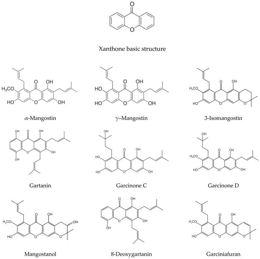
Int. J. Mol. Sci. 2020, 21, 6211                                                                   2 of 24

6.1 million people worldwide from 2.5 million in 1990 [5]. It affects approximately 1–2% of individuals
over 65 years of age [4].
      Depression, a mood disorder that can interfere with daily functioning, is another significant cause
of global burden [6]. It is described as persistent feelings of sadness, a loss of pleasure or interest,
tiredness, disturbed appetite and sleep, poor concentration, feelings of guilt and low self-worth,
and suicidal ideation [7]. In 2017, depression was the leading cause of disability worldwide, affecting
more than 264 million people [8]. In addition to the high prevalence, the lack of effective treatments
for AD, PD and depression significantly augment the disease burden. Therefore, new remedies or
conjunctive treatments for these disorders are unmet needs for future research. Growing evidence has
indicated that natural products may provide therapeutic advantages to these brain diseases [9–11].
In the present review, we will focus on the potential therapeutic value of various agents derived
from mangosteen.
      Mangosteen, Garcinia mangostana L., is a tree cultivated mostly in Southeast Asian countries.
Mangosteen fruit, known as “the queen of fruits”, is edible, with a sweet and tangy taste.
Some mangosteen-based products, such as juice and tablets, are among the best-selling food
supplements in the U.S. [12]. Additionally, mangosteen is well-recognized for its medicinal benefits.
Mangosteen pericarp (MP) has long been used as a traditional medicine to treat infection, wounds,
inflammation, and diarrhea [13]. Phytochemical studies have explored medicinal properties using
xanthone derivatives—the major bioactive secondary metabolites derived from MP. Xanthones are
polyphenolic compounds, which are characterized with a distinctive skeleton of a xanthene-9-one that
are distinguished by the tricyclic aromatic ring system (Figure 1) [14]. At least 68 distinct xanthones,
which exist as oxygenated or prenylated forms, have been identified in different portions of the plant,
with more than 50 xanthones being present in MP at higher concentrations than in other parts of the
fruit [14]. The most investigated xanthones in MP are α-mangostin (α-MG) and γ-mangostin (γ-MG).
Other xanthones found in MP include β-mangostin, gartanin, 3-isomangostin, garcinones A, B, C, D,
and E, 8-deoxygartanin, mangostanol, isomangostin, and garciniafuran [14,15]. The chemical structures
of representative bioactive xanthones are shown in Figure 1. MP and its bioactive xanthones have
been reported to exhibit numerous biological activities, including antiparasitic, antitumor, antioxidant,
anti-inflammatory, and neuroprotective effects in various experimental studies [16].
      MP and xanthones derived from MP have recently attracted researchers’ attention for their
pharmacological actions in neurons. Firstly, MP-derived xanthones such as garcinone D or gartanin
exerted neuroprotective activities and mitigated oxidative injury, possibly via Nrf-2/HO-1 and
STAT3/Cyclin D1 [17], or AMPK/SIRT1/PGC-1α signaling pathways [18], respectively. In addition,
the xanthone derivatives exhibited anti-inflammatory effects in the brain [19,20]. Besides, the xanthones
alleviated lead-induced neurotoxicity in mice via suppression of oxidative damage and reversion
of acetylcholinesterase (AChE) activity, resulting in the restoration of learning deficits and memory
loss [21]. Several studies have reported the neuroprotective and antioxidant properties of α-MG
via the amelioration of mitochondrial toxin 3-nitropropionic acid or iodoacetate-induced reactive
oxygen species (ROS) production in primary culture of cerebellar granule cells [22,23]. Intriguingly,
the polar fraction of ethanol MP extract (MPE) augmented antioxidant capability and attenuated
oxidative damage to proteins in the blood cells of healthy adults, demonstrating its antioxidant effects
in humans [24].
      With the neuroprotective, antioxidant, anti-neuroinflammatory activities, MP-derived agents
such as MPE and its bioactive xanthones may exert beneficial actions in many brain diseases.
Wang et al. provided a comprehensive review on the pharmacological properties of mangostins
and their derivatives, which promised their potential utilities in the treatment of certain diseases
including AD [25]. In addition, Ashton et al. extensively reviewed the potential of MP as an adjunctive
therapy for serious mental illnesses such as bipolar disorder and schizophrenia [26]. In the present
study, we will discuss the potential therapeutic value of MP and its bioactive xanthones, particularly
focusing on the treatment of AD, PD and depression. We first provide a concise overview of the major
Int. J. Mol. Sci. 2020, 21, 6211 3 of 24
pathologies and therapeutic strategies of AD, PD, and depression, and then discuss pharmacological
effects of MP-derived agents in these diseases along with their pharmacokinetic and safety profiles.
Figure 1. Chemical structures of basic xanthone and representative bioactive xanthones in
mangosteen pericarp.
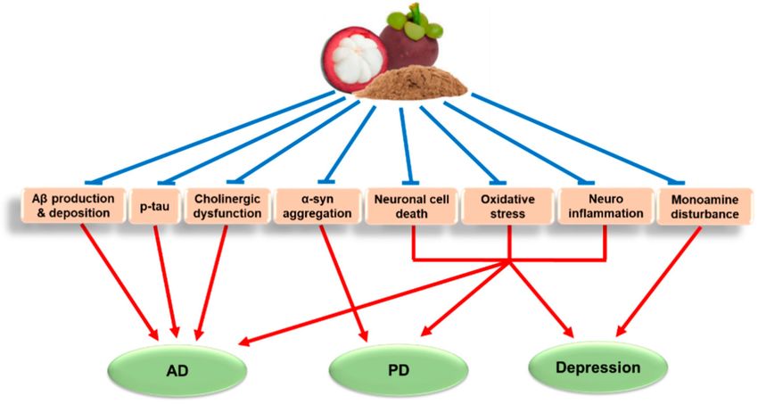
2. Major Pathologies and Therapeutic Strategies of AD, PD, and Depression
From a neuropathological point of view, AD, PD, and depression involve specific pathologies
associated with each disorder, but also share several common ones. In this section, we briefly describe
specific and common pathologies associated with AD, PD, and depression. In addition, we discuss the
current treatments of these diseases and propose potential therapeutic strategies for future research.
AD is a slowly progressive neurodegenerative disease that begins many years before symptoms
emerge. The most widely accepted hallmarks of the clinical features of AD are: (1) the formation
and deposition of extracellular amyloid beta (Aβ) plaques and (2) the accumulation of intracellular
hyperphosphorylated tau proteins, known as neurofibrillary tangles, in the brain [2]. Aβ is a
short peptide, with its most common alloforms being Aβ1-40 and Aβ1-42 , derived from the cleavage
of an amyloid precursor protein by β- and γ-secretases [27–29]. As a consequence, Aβ peptides
spontaneously form soluble oligomers, coalesce into insoluble beta-sheet conformation, and are
extracellularly deposited in diffuse senile plaques [30]. This process interferes with neuron-to-neuron
communication at synapses, and leads to neuronal cell death [2]. Tau protein is involved in the assembly
and stabilization of microtubules in neurons [31]; however, the hyperphosphorylation of tau causesInt. J. Mol. Sci. 2020, 21, 6211 4 of 24
the loss of microtubule stabilization, which leads to the abnormal shape and functionality of neurons,
and eventually, neuronal cell death [32,33]. In addition, Aβ peptides and hyperphosphorylated tau
destabilize calcium homeostasis and increase the vulnerability of neurons to excitotoxicity, mainly via
interactions with N-methyl-D-aspartate (NMDA) receptors [34,35]. These processes eventually induce
the impairment in cognitive and memory function seen in AD patients [32,36]. Moreover, the intellectual
deficiency in AD patients has been linked to a loss of cholinergic function in the cortex and hippocampus,
which also contributes to AD pathology [37]. To date, the approved drugs for AD treatment are
cholinesterase inhibitors and a NMDA receptor antagonist. Numerous clinical trials were performed
so far to improve the cognitive and behavioral symptoms of AD [38]. In particular, disease-modifying
therapies targeting Aβ or tau protein, neuroprotective strategies, and immunotherapies have been
intensely attempted. Unfortunately, however, due to extremely low success rates, there has been no
new approved drug for the treatment of AD since 2003 [38].
PD is characterized by a preferential loss of dopaminergic (DAergic) neurons within the substantia
nigra pars compacta (SNpc) and the deposition of intracellular inclusions, known as Lewy bodies [39,40].
Lewy bodies are predominantly formed by misfolding and aggregation of the presynaptic protein,
α-synuclein (α-syn), in surviving neurons [39,40]. Presynaptic compensation in the nigrostriatal
dopamine (DA) pathway may account for PD progression. Typically, parkinsonian motor symptoms
become apparent when more than 30% of nigral DA neurons or 50% of striatal DA contents are lost [41,42].
Current PD treatment primarily consists of DA replacement therapies, such as levodopa, DA agonists,
and inhibitors of catechol-O-methyl transferase and monoamine oxidase (MAO) B. Additionally,
anticholinergics and amantadine are used to ameliorate motor symptoms, while antipsychotics and
antidepressants are prescribed to treat non-motor symptoms of PD [43,44].
Monoamine dysregulation is recognized as a primary pathology of depression. DA and serotonin
(also known as 5-HT) are dysregulated in depression. DAergic and serotonergic functional loss in the
striatum and hippocampus, respectively, is associated with disturbances in mood regulation, which is
linked to depressive disorder [45–47]. Moreover, downregulation of norepinephrine (NE) [48] and
disturbances in γ-aminobutyric acid (GABA) and glutamate are also recognized in this disease [49].
Since monoamine imbalance is the primary pathology of depression, it has been the target of
pharmacological treatments. Modulation of DA, serotonin, and NE imbalance is the major mechanisms
of action by which antidepressant drugs exert their actions. The major antidepressants include
selective serotonin reuptake inhibitors, serotonin-NE reuptake inhibitors, NE-DA reuptake inhibitors,
5-HT2 receptor antagonists, MAO inhibitors, and tricyclic antidepressants (TCAs) [50].
In addition to the specific pathologies associated with AD, PD, or depression as discussed above,
these disorders share some pathologies in common. The common pathologies associated with AD, PD,
and depression include neuroinflammation, oxidative stress, mitochondrial dysfunction, and reduction
of neurotrophic factors such as brain-derived neurotrophic factor (BDNF). Neuroinflammation is the
process of innate or adaptive immune activation occurring in the CNS in response to the perturbed
homeostasis [51]. Overactivation of the immune system during neuroinflammatory processes may
lead to neuronal damage in AD and PD [52–54]. Likewise, there is growing evidence supporting the
involvement of inflammation in depression [55–57]. One of the potential underlying mechanisms is the
suppression of serotonin synthesis from tryptophan by pro-inflammatory cytokines through activation
of indoleamine 2,3-dioxygenase, a rate-limiting enzyme involved in tryptophan metabolism [58].
Oxidative stress is also a common feature of AD, PD and depression. Free radicals produced by
oxidative stress affect the structure and function of neuronal cells, contributing to the development
of AD and PD [59,60]. Elevated levels of ROS and nitrogen species are associated with increased
damage of intracellular biomolecules such as DNA in the depressed brain [61]. Another study suggests
that depression correlates with oxidative alterations of nucleotides and polymorphisms of genes
associated with metabolism of ROS [62]. These findings strongly support critical roles of oxidative
stress in the pathologies of AD, PD, and depression. Due to its direct relationship with oxidative
stress, mitochondrial dysfunction also plays a key role in the pathogenesis of AD [63], PD [64],Int. J. Mol. Sci. 2020, 21, 6211 5 of 24
and depression [61,65]. Meanwhile, neurotrophic factors such as glial cell-derived neurotrophic factor,
nerve growth factor, and especially, BDNF are crucial for survival, maintenance, and regeneration
of specific neurons in the adult brain [66]. Reductions of these neurotrophic factors are commonly
recognized in AD and PD [66–68] as well as in depression [69]. Therefore, agents targeting these
common features may possess promising therapeutic values for these diseases.
Unfortunately, however, the current therapies for AD, PD and depression are not fully adequate for
growing treatment needs. The drugs clinically used for the treatment of AD or PD show limited efficacy
with only slowing the disease progress or just alleviating symptoms [38,70,71]. Similarly, current
therapies for depression exhibit a number of limitations such as delayed onset of action, insufficient
efficacy, and unwanted side effects [72]. Therefore, novel effective agents are urgently required to
enter the therapeutic pipeline. New agents with neuroprotective, anti-inflammatory, and antioxidant
properties or targeting AD-, PD-, or depression-related specific pathologies would be promising for
curative outcomes in these diseases. In the following sections, we assess natural MP-derived agents as
promising candidates to intervene in AD, PD and depression.
3. Pharmacological Effects of MP-Derived Agents in AD Models
Agents derived from MP have been shown to exhibit neuroprotective, anti-inflammatory,
and antioxidant effects, supporting their efficacy as potential treatments or co-adjuvant therapies for
brain disorders including AD.
3.1. Pharmacological Effects of MPE and MP Diet in AD Models
Various preparations of MPEs and MP diet were found to inhibit neurotoxicity and oxidative
stress induced by Aβ, NMDA, H2 O2 or other stimuli in in vitro and in vivo studies. An in vitro
study revealed that pretreatment with the water-soluble partition of methanol MPE successfully
prevented Aβ1-42 -induced cytotoxicity, and reduced ROS levels and caspase-3 activity in SK-N-SH
neuroblastoma cells. Interestingly, the MPE diminished Aβ1-42 -induced changes of several proteins
including FK506-binding protein 4 and HLA-B associated transcript 1, suggesting that these proteins
might be responsible for the neuroprotective effects of MPE [73]. Moreover, the water extract of MP and
the butanol fraction of methanol MPE significantly attenuated NMDA- and Aβ25-35 -induced neuronal
cell damage in primary cultured rat cortical neurons, possibly via inhibition of ROS generation and
apoptotic events. In parallel, the antioxidant properties of these extracts and their inhibitory effects on
β-secretase activity were also reported based on the in vitro studies [74,75]. In another study, the water
and 50% ethanol MPE exhibited potent antioxidant effects with high free-radical scavenging and
neuroprotective actions in H2 O2 -treated NG108-15 neuroblastoma cells [76]. Similarly, the water-soluble
partition of ethanol MPE partially antagonized the effects of H2 O2 and polychlorinated biphenyls on
cell viability, ROS production, and caspase-3 activity in SK-N-SH cells [77]. The MPE was demonstrated
to improve scopolamine-induced memory dysfunction in mice, as assessed by Morris water maze
(MWM) and passive avoidance tests. Moreover, the increased ROS level and caspase-3 activity in the
brain of scopolamine-treated mice were inhibited by the MPE treatment [77]. Besides, MP diet exerted
neuroprotective and antioxidant effects in triple transgenic AD (3×Tg-AD) mice, and reduced Aβ
deposition in the hippocampus, which might further attenuate the deficit in spatial memory retrieval in
the MWM test [78]. Furthermore, 50% ethanol MPE significantly enhanced the antioxidant parameters,
such as superoxide dismutase, glutathione peroxidase, glutathione, and catalase in the brain tissues of
streptozotocin-treated male Swiss albino (SA) mice [79].
Several studies have demonstrated inhibition of AChE activities by MPE. Notably, the water-soluble
partition of ethanol MPE reduced AChE activity of SK-N-SH cells to ~ 60% of the control activity [77].
The suppression of AChE by the water MPE was confirmed in another study [75]. Furthermore,
pretreatment of male SA mice with 50% ethanol MPE prior to streptozotocin injection induced a
dose-dependent reduction in AChE activities in the brain, and increased habituation memory and
cognitive function as measured by open field test (OFT) and Y-maze test [79].Int. J. Mol. Sci. 2020, 21, 6211 6 of 24
The effects of MP on the regulation of BDNF expression, tau phosphorylation, and neuroinflammation
have been reported [78]. MP significantly decreased the cell death and increased BDNF expression
in an organotypic hippocampal slice culture. In addition, 8-month dietary supplementation with MP
significantly attenuated the cognitive impairment in aged C57BL/6J (B6) mice, primarily associated
with anti-inflammation, increased BDNF level and decreased phosphorylation of tau. The MP diet
in 3×Tg-AD mice also exerted anti-inflammatory effects and reduction of phosphorylated tau in the
hippocampus, which might further be reflected in the improvement of spatial memory retrieval [78].
These data highlight that different preparations of MPE and MP diet have multiple pharmacological
effects targeting specific and common AD pathologies including: prevention of Aβ production,
deposition, and toxicity; inhibition of tau hyperphosphorylation, NMDA excitotoxicity, and AChE
activity; and antioxidant and anti-inflammatory activities. Furthermore, improved cognitive and
memory functions by MPEs and MP diet have been established in different AD animal models.
Therefore, these agents may be beneficial for the prevention or treatment of AD. The pharmacological
effects of MP-derived agents in various in vitro and in vivo AD models are summarized in Table 1.
Table 1. Pharmacological effects of MP-derived agents in Alzheimer’s disease models.
Experimental Experimental
No. Agents Results References
Models Conditions
Water-soluble ↓ Neurotoxicity
1 partition of methanol SK-N-SH cells Aβ1-42 ↓ Caspase-3 [73]
MPE ↓ ROS
↓ Neurotoxicity &
Primary cultured rat NMDA
apoptotic events
cortical neurons Aβ25-35
↓ ROS
Butanol fraction of
2 [74]
methanol MPE Rat brain
Fe2+ /ascorbic acid ↓ Lipid peroxidation
homogenates
Cell-free bioassay ↓ β-Secretase activity
↓ Neurotoxicity &
Primary cultured rat NMDA
apoptotic events
cortical neurons Aβ25-35
↓ ROS
Rat brain
3 Water MPE Fe2+ /ascorbic acid ↓ Lipid peroxidation [75]
homogenates
↓ β-Secretase activity
Cell-free bioassay
↓ AChE activity
ICR mice Scopolamine ↓ Memory impairment
↓Oxidative neurotoxicity
Water/50% ethanol
4 NG108-15 cells H2 O2 Free radical scavenging [76]
MPE
activity
↓Oxidative neurotoxicity
H2 O2 ↓ Caspase-3
SK-N-SH cells
PCBs ↓ ROS
Water-soluble
↓ AChE activity
5 partition of ethanol [77]
↓ Memory impairment
MPE
ICR mice Scopolamine ↓ Brain ROS
↓ Caspase-3Int. J. Mol. Sci. 2020, 21, 6211 7 of 24
Table 1. Cont.
Experimental Experimental
No. Agents Results References
Models Conditions
↓ Aβ deposition
3×Tg-AD mice ↓ Phosphorylated tau
NA ↓ Memory impairment
↓ Systemic IL-6
6 MP diet ↑ BDNF level [78]
B6 mice
↓ Phosphorylated tau
↓ Cognitive impairment
↓ Neurotoxicity
MP OHSC
↑ BDNF level
↑ Antioxidant
parameters: superoxide
dismutase, glutathione
peroxidase, glutathione,
7 50% Ethanol MPE Male SA mice Streptozotocin [79]
and catalase
↓ AChE levels
↓ Cognitive & memory
impairment
↓ Neurotoxicity
HT22 cells Glutamate ↑ HO-1 level
↓ DPPH radicals
α-MG, γ-MG,
8 gartanin, garcinone E. coli/Cell-free ↓ Self-induced Aβ [80]
C bioassay aggregation
Cell-free bioassay ↓ β-Secretase activity
↓ β- & γ-Secretase
Primary cultured rat activity
9 α-MG NA [81]
cortical neurons ↓ Aβ1-40 & Aβ1-42
production
↓ Neurotoxicity
Primary cultured rat Aβ1-40 or Aβ1-42
10 α-MG ↓ Aβ fibril formation & [82]
cortical neurons oligomers
pre-formed fibrils
↓ Oxidative
H2 O2 or neurotoxicity
Primary cultured rat
xanthine/xanthine ↓ ROS
cortical neurons
oxidase ↓ DNA fragmentation
↓ Caspases-3 & 9
11 γ-MG [83]
Rat brain ↓ Lipid peroxidation &
Fe2+ /ascorbic acid
homogenates DPPH radicals
Cell-free bioassay ↓ β-Secretase activity
ICR mice Scopolamine ↓ Memory impairment
α-MG, γ-MG,
mangostanol, ↓ AChE activity
12 Cell-free bioassay NA [84]
3-isomangostin, &
garcinone C
13 α-MG Female B6 mice LPS ↓ IL-6, COX-2 & TSPO [20]
Aβ, amyloid beta; AChE, acetylcholinesterase; α-MG, α-mangostin; B6, C57BL/6J; BDNF, brain-derived
neurotrophic factor; COX-2, cyclooxygenase-2; DPPH, 2,2-diphenyl-1-picrylhydrazyl; E. coli, Escherichia coli;
γ-MG, γ-mangostin; GSH, glutathione; HO-1, heme oxygenase-1; ICR, Institute for Cancer Research; IL, interleukin;
LPS, lipopolysaccharide; MP, mangosteen pericarp; MPE, mangosteen pericarp extract; NA, not applicable; NMDA,
N-methyl-D-aspartate; OHSC, organotypic hippocampal slice culture; PCBs, polychlorinated biphenyls; ROS,
reactive oxygen species; SA, Swiss albino; TSPO, 18 kDa translocator protein.
3.2. Pharmacological Effects of Xanthones Isolated from MP in AD Models
Bioactive compounds derived from MP were shown to mitigate the neurotoxicity induced by
glutamate or Aβ, and diminish Aβ production and aggregation. Xanthones such as α-MG, γ-MG,Int. J. Mol. Sci. 2020, 21, 6211 8 of 24
gartanin, and garcinone C exhibited neuroprotective effects against glutamate-caused HT22 hippocampal
neuronal cell death, partly by up-regulating HO-1 protein level. Additionally, these xanthones
suppressed self-induced Aβ aggregation and β-secretase activity [80]. In particular, α-MG was
reported to diminish Aβ1-40 and Aβ1-42 production in primary cultured rat cortical neurons through
inhibition of β- and γ-secretase activities in the amyloidogenic pathway [81]. Consistently, α-MG
concentration-dependently attenuated the neurotoxicity induced by Aβ1-40 or Aβ1-42 oligomers in the
cultured neurons. It was speculated that α-MG may disturb the pre-formed Aβ fibrils by stabilizing its
α-helical conformation, and thereby, block the fibril formation [82].
Additionally, the xanthones were found to contribute to the antioxidant effects of MP in the
brain. α-MG, γ-MG, gartanin, and garcinone C showed promising antioxidant properties with
scavenging activities of 2,2-diphenyl-1-picrylhydrazyl (DPPH) free radicals. Intriguingly, however,
only γ-MG, not α-MG, predominantly protected cultured cortical neurons against either H2 O2 - or
xanthine/xanthine oxidase-induced oxidative neuronal death, and mitigated ROS generation [83].
γ-MG significantly abolished H2 O2 -induced DNA fragmentation and caspases-3 and -9 activation,
indicating its anti-apoptotic action. In agreement with these findings, only γ-MG exhibited effective
suppression of lipid peroxidation and DPPH radical formation in cell-free bioassays. In contrast,
β-secretase activity was inhibited by both α-MG and γ-MG [83]. Remarkably, oral administration of
γ-MG significantly reversed scopolamine-induced memory deficits in mice. These in vitro and in vivo
findings suggest that γ-MG may be the preferable candidate over α-MG in combatting oxidative
stress-associated neurodegenerative diseases including AD [83].
Prenylated xanthones derived from MP, including α-MG, γ-MG, mangostanol, 3-isomangostin,
and garcinone C, showed inhibition of AChE activities in vitro [84]. Interestingly, molecular docking
studies revealed that α-MG, γ-MG and garcinone C may interact differently with the key regulatory
regions of AChE, mainly through hydrophobic interactions and hydrogen bonding. These promising
results should be further investigated in animal studies [84].
As previously discussed, neuroinflammation is considered as one of the common pathologies
associated with AD, PD [52–54] and depression [55–58]. It was demonstrated that oral gavage of α-MG
significantly attenuated the lipopolysaccharide (LPS)-induced brain inflammation in B6 mice [20].
α-MG suppressed the brain levels of LPS-induced pro-inflammatory cytokine interleukin-6 (IL-6),
cyclooxygenase-2, and 18 kDa translocator protein, a sensitive biomarker of brain inflammation [85].
The anti-inflammatory effects of α-MG may contribute to beneficial impacts on not only AD, but also
PD and depression.
Collectively, the above findings demonstrated the multiple pharmacological effects of the
MP-derived xanthones, especially α-MG and γ-MG, which could contribute to the treatment of
AD (Table 1). Specifically, the compounds attenuated Aβ production, dissociated the Aβ aggregation,
and thereby, inhibited Aβ-induced neurotoxicity. Furthermore, the xanthones were shown to suppress
oxidative stress, as well as alleviate AChE activities. These findings illustrated the utility of natural
xanthones as novel neuroprotective candidates through the intervention of multiple pathological
processes of AD.
4. Pharmacological Effects of MP-Derived Agents in PD Models
Since AD and PD share common pathological features, the MP-derived products can also be
potential candidates for PD treatment. Unfortunately, no studies have reported the effects of MPE on
PD. However, there are several studies illustrating the pharmacological effects of α-MG and γ-MG in
PD models. In this section, we discuss the potential value of these xanthones in PD treatment.
α-MG has been shown to exert neuroprotective and antioxidant functions in in vitro models
of PD. Evidence showed that α-MG alleviated 1-methyl-4-phenylpyridinium-induced apoptosis
in SH-SY5Y neuroblastoma cells, which may be associated with inhibition of ROS formation,
modulation of the balance between pro- and anti-apoptotic proteins, and suppression of caspase-3
activation [86]. Another study showed that α-MG significantly ameliorated rotenone-inducedInt. J. Mol. Sci. 2020, 21, 6211 9 of 24
cytotoxicity in SH-SY5Y cells in a concentration-dependent manner [87]. α-MG treatment also
reversed rotenone-induced overproduction of ROS, activation of caspases-3 and -8, and mitochondrial
dysfunction [87]. Notably, α-MG protected DAergic neurons from α-syn-induced neurotoxicity in
primary rat mesencephalic neuron-glia co-cultures. Inhibition of ROS production by α-MG was also
observed in α-syn-stimulated primary rat microglia [88].
The effects of α-MG on PD were further illustrated in vivo. Intraperitoneal (i.p.) injection of α-MG
to adult male Sprague Dawley (SD) rats significantly restored rotenone-reduced locomotor activity and
neuromuscular weakness, as assessed by rotarod and grip strength tests, respectively. Furthermore,
treatment with α-MG demonstrated antioxidant effects in the striatum of the rats by diminishing
rotenone-induced oxidative damage, such as increased malondialdehydes, nitrite concentrations,
and decreased glutathione levels [89]. Importantly, α-MG inhibited α-syn aggregation and rescued
tyrosine hydroxylase (TH), a rate-limiting enzyme involved in the synthesis of DA. Specifically,
α-MG alleviated rotenone-induced aggregation of α-syn and loss of TH in SH-SY5Y cells [87].
Furthermore, α-MG also significantly reduced phosphorylation of α-syn, and thereby protecting from
TH+ -DAergic neuronal loss in the SNpc of the rat model of PD [89].
Considering the hydroxyl groups in the structure (Figure 1), γ-MG is also expected to possess free
radical scavenging effects. Unfortunately, only a few studies have reported neuroprotective effects of
γ-MG in PD models. γ-MG pretreatment was found to attenuate 6-hydroxyDA-induced neuronal cell
death. The protective effect of γ-MG was associated with its antioxidant potential and modulation
of apoptotic signaling [90]. These results illustrate the immense potential of γ-MG for preventing
oxidative stress-induced neurodegeneration in PD.
It is well-known that microglial activation can initiate a cascade of events associated with
neuroinflammation, which leads to progressive neurodegeneration of nigral DAergic neurons.
Therefore, inhibitions of microglial activation and neuroinflammation are also attractive strategies for
PD treatment [91]. As described earlier, orally administered α-MG suppressed the LPS-induced
inflammation in the brain of B6 mice [20]. In addition, α-MG abated the increased levels of
pro-inflammatory cytokines, including tumor necrosis factor-α, IL-1β, IL-6, and nitric oxide,
in α-syn-stimulated primary rat microglia, possibly through the inhibition of nuclear factor kappa B
and NADPH oxidase [88]. Although detailed mechanisms remain to be further defined, these results
suggest that α-MG can be a promising therapy for PD.
Altogether, the above findings indicate that α-MG and γ-MG possess neuroprotective, antioxidant,
and anti-inflammatory activities. Additionally, these xanthones were shown to arrest α-syn aggregation,
TH loss, and mitochondrial dysfunction, illustrating their therapeutic roles in preventing PD
progression. The pharmacological effects of these xanthones in PD models are summarized in
Table 2. Further mechanistic studies of α-MG, γ-MG, and other bioactive compounds or extracts from
MP are required on both preclinical and clinical levels to probe their potential value in the management
of PD.
Table 2. Pharmacological effects of α-mangostin and γ-mangostin in Parkinson’s disease models.
Experimental Experimental
No. Agents Results References
Models Conditions
↓ Apoptosis
1 α-MG SH-SY5Y cells MPP+ [86]
↓ ROS
↓ Cell death
↓ Caspases-3 & 8
↓ ROS
2 α-MG SH-SY5Y cells Rotenone ↓ Mitochondrial [87]
dysfunction
↓ Aggregation of α-syn
and TH lossInt. J. Mol. Sci. 2020, 21, 6211 10 of 24
Table 2. Cont.
Experimental Experimental
No. Agents Results References
Models Conditions
↓ ROS
Primary rat ↓ TNF-α, IL-1β, IL-6,
α-Syn
microglia cells NO, NF-κB & NADPH
oxidase
3 α-MG [88]
Primary rat
↓ DAergic neuronal cell
mesencephalic
α-Syn death
neuron-glia
co-culture
4 α-MG Female B6 mice LPS ↓ IL-6, COX-2 & TSPO [20]
↓ MDA, nitrite
↑ GSH
↓ Phosphorylated α-syn
↓ TH+ -DAergic neuronal
5 α-MG Adult male SD rats Rotenone [89]
loss in SNpc
↑ Locomotor activity
↑ Neuromuscular
function
↓ Neurotoxicity
6 γ-MG SH-SY5Y cells 6-OHDA ↓ Apoptosis [90]
↑ Antioxidant potential
6-OHDA, 6-hydroxydopamine; α-MG, α-mangostin; α-syn, α-synuclein; DAergic, dopaminergic; γ-MG,
γ-mangostin; GSH, glutathione; IL, interleukin; MDA, malondialdehyde; MPP+ , 1-methyl-4-phenylpyridinium;
NF-κB, nuclear factor kappa B; NO, nitric oxide; ROS, reactive oxygen species; SD, Sprague Dawley; SNpc, substantia
nigra pars compacta; TH, tyrosine hydroxylase; TNF-α, tumor necrosis factor-α.
5. Pharmacological Effects of MP-Derived Agents in Depression Models
The tricyclic structure of xanthones in MP (Figure 1) may link to antidepressive functions as
seen in TCAs. As described above, MP-derived agents exhibit redox-modulatory actions [18,22,23],
anti-inflammatory effects [19,20], and probable reversion of mitochondrial dysfunction [87].
These effects may contribute to beneficial actions to surmount common pathologies of AD and
PD as well as depression. However, limited evidence is available to support antidepressant effects of
MP-derived agents.
Recent preclinical evidence has illustrated the antidepressant-like and pro-cognitive effects of
MPE in Flinders Sensitive Line (FSL) rats, a genetic model of depression [92]. Acute administration
of ethyl acetate MPE demonstrated antidepressant-like effects in OFT and forced swim test (FST),
with the comparable effects to a reference antidepressant, imipramine (IMI, a TCA). Similarly, chronic
treatment with this MPE displayed significant antidepressant- and pro-cognitive effects in FST and
novel object recognition test, respectively. Again, the antidepressant effect of the MPE was comparable
to that of IMI. Interestingly, however, the neurotransmitters associated with antidepressant effects
of chronically treated MPE and IMI appeared to be different. A more prominent serotonergic action
for MPE was observed as opposed to a noradrenergic action for IMI treatment. IMI significantly
altered cortico-hippocampal NE levels, possibly by chronic blocking the NE transporter, which was
consistent with a previous study [93]. In contrast, chronic MPE-treated FSL rats showed elevated
levels of 5-hydroxyindoleacetic acid, a main metabolite of 5-HT, in both the frontal cortex and
hippocampus, indicating serotonergic action. Numerous animal studies have shown the associations
of oxidative stress with cognitive impairment [94,95] and depressive-like behaviors [96]. Indeed,
reversion of hippocampal lipid peroxidation was observed with IMI or MPE treatment. These findings
indicate that modulation of dysregulated brain monoamines and suppression of oxidative stress are
possibly involved in the antidepressant activity of MPE [92]. Another study revealed that α-MG
markedly exhibited antidepressant-like activity, which was reversed by pretreatment with haloperidol
(a D2 receptor antagonist), p-chlorophenylalanine (an inhibitor of 5-HT synthesis), and bicucullineInt. J. Mol. Sci. 2020, 21, 6211 11 of 24
(a competitive GABA antagonist). Furthermore, α-MG effectively accentuated brain DA, 5-HT and
GABA levels in mice, indicating that these neurotransmitter systems may mediate antidepressant-like
effect of α-MG [97]. As mentioned previously, orally administered α-MG exhibited anti-inflammatory
effects in B6 mice, which may also contribute to beneficial impacts on depression [20]. In addition,
both ethyl acetate MPE and α-MG revealed significant antidepressant-like properties in a rat model
of schizophrenia, suggesting that these agents may specifically address the depressive symptoms of
schizophrenia [98].
Although there are no clinical data directly focusing on the effects of MP agents in the depressive
patients, several studies have evaluated their impacts on mental health outcomes in humans. In a small
randomized controlled trial (RCT), mangosteen-based supplementation containing 305 mg of α-MG
and 278 mg of hydroxycitric acid was orally administered to healthy adults prior to cycle ergometer
exercise and the parameters directly reflecting the condition of physical fatigue were measured [99].
All parameters measured showed no differences between the supplementation group and placebo
group, except the Profile of Mood States (POMS) scores. The single dose of mangosteen supplementation
showed a positive impact on the scores of POMS, a psychological rating scale used to assess transient,
distinct mood states [99].
Intriguingly, α-MG was found to reduce body weight (b.w.) gain in high-fat diet-induced obese
mice [100]. Moreover, there are several clinical results exhibiting the efficacy of mangosteen-based
products in obese individuals. Among them, a RCT assessed the efficacy and tolerability of Meratrim,
an herbal formulation consisting of extracts from Sphaeranthus indicus flower heads and MP mixed
together in a ratio of 3:1, in controlling b.w. in healthy overweight subjects [101]. In this study,
while the reduction of b.w. from the baseline to the end of 16 weeks-treatment period was evaluated
as a primary endpoint, total mood disturbance (TMD) scores were also measured as a secondary
endpoint using POMS-short form. Notably, a statistically significant decrease in the mean TMD
score was reported in subjects receiving Meratrim over placebo [101]. In addition, a pilot study of
double-blind placebo-controlled trial was conducted using encapsulated MP powder in 80 patients
with schizophrenia or schizoaffective disorder [102]. As the primary outcomes, significant reduction
of symptom domains associated with schizophrenia was observed in the MP group. Furthermore,
secondary outcomes, assessed by Montgomery Åsberg Depression Rating Scale to measure the severity
of depressive episodes at 180-day endpoint, were also significantly different between the two groups.
The findings from these randomized trials demonstrate that MP has beneficial impact on depression.
However, this study possesses several limitations; small sample size and participants with mild
depression at baseline [26]. Nevertheless, this is the only clinical study which directly evaluates the
potential therapeutic value of MP in the treatment of a serious mental illness accompanied by depression.
These positive results may initiate further research of MP for the treatments of depression and other
psychiatric disorders with depression. In fact, one RCT (Trial registry ID: ACTRN12616000028404) is
currently in progress investigating the efficacy of adjunctive MP for bipolar depression [103].
Taken together, both MPE and α-MG showed the antidepressant activities via modulation of
dysregulated monoamines and oxidative stress in the brain, suggesting novel potential therapies
for depression. The pharmacological effects of the MP-derived agents in depression models are
summarized in Table 3. However, since our knowledge regarding the underlying mechanisms
associated with the antidepressant effects of MPE, α-MG, or other xanthones in MP remains poor,
further investigation is required.Int. J. Mol. Sci. 2020, 21, 6211 12 of 24
Table 3. Pharmacological effects of MP-derived agents in depression models.
Experimental Experimental
Agents Results References
Models Conditions
Antidepressant-like effect
Antidepressant &
Acute treatment pro-cognitive effects
Ethyl acetate MPE FSL rats [92]
Chronic treatment Prominent serotonergic action
↓ Hippocampal lipid
peroxidation
Ethyl acetate Pregnant female Antidepressant-like effect in
MIA [98]
MPE/α-MG SD rats schizophrenia
Mangosteen-based Positive impact on POMS
Healthy adults RCT [99]
products scores
Mangosteen-based
Overweight ↓ Body weight
products RCT [101]
subjects ↓ Total mood disturbance
(Meratrim)
Patients with
Encapsulated MP schizophrenia or ↓ PANSS
RCT [102]
powder schizoaffective ↓ MADRS
disorder
Patients with
Water MPE RCT No published results [103]
bipolar depression
Antidepressant-like activity
(reversed by pretreatment
with HAL, bicuculline &
α-MG Mice TST [97]
p-CPA)
↑ Brain DA, 5-HT & GABA
levels
α-MG Female B6 mice LPS ↓ IL-6, COX-2 & TSPO [20]
α-MG, α-mangostin; DA, dopamine; FSL, Flinders Sensitive Line; GABA, gamma-aminobutyric acid; HAL,
haloperidol; MIA, maternal immune-activation; MADRS, Montgomery Åsberg Depression Rating Scale; MP,
mangosteen pericarp; MPE, mangosteen pericarp extract; PANSS, Positive and Negative Syndrome Scale; POMS,
Profile of Mood States; p-CPA, p-chlorophenylalanine; RCT, randomized controlled trial; SD, Sprague Dawley; TST,
tail suspension test.
6. Pharmacokinetic (PK) Profiles of MP-Derived Agents
We discussed in vitro and in vivo pharmacological effects of MP-derived agents in the experimental
models of AD, PD, or depression in the previous sections. The MPE and several xanthones including
α-MG are described to be promising candidates for the treatment of AD, PD, and depression.
In the following section, we discuss the current evidence of the PK profiles of MP-derived agents,
including commercially available MP-based products, in the preclinical and clinical conditions and
propose solutions to improve their PK profiles.
6.1. In Vitro and In Vivo PK Profiles of MP-Derived Agents
PK studies for herbal agents containing multicomponent mixtures with numerous active
phytochemicals are generally extraordinarily complex [104], which may explain the lack of PK
data for MP-derived agents. Intravenous (i.v.) injection of α-MG exhibited a biphasic deposition with
a fast distribution phase and a slow terminal elimination phase, suggesting high tissue binding in
rats [104]. The bioavailability after oral administration of α-MG was very low, probably due to a
poor gastrointestinal absorption or an extensive metabolism in the small intestine and liver [104,105].
The distribution of α-MG was relatively high in the intestine, liver, kidney, and lung, but not in the brain.
Interestingly, however, it was reported that some xanthones such as γ-MG, gartanin, and garcinone
C were able to penetrate the blood-brain barrier (BBB) in vitro [80]. It was proposed that α-MGInt. J. Mol. Sci. 2020, 21, 6211 13 of 24
underwent phase II, and possibly, phase I metabolism [105,106]. Although α-MG was metabolized
primarily by CYP1A2 in vitro [107], an in vivo study indicated that phase I metabolism played a
minimal role in the overall metabolism of α-MG. Phase II metabolism appeared to be the primary
route, which was consistent with previous reports [105]. To date, only one study has measured the
absolute bioavailability of γ-MG; this study reported similar PK profiles to that of α-MG. After i.v.
administration, γ-MG exhibited a two-compartmental body model, indicating its distribution into the
peripheral compartments. Both α-MG and γ-MG exhibited an intensive first-pass metabolism with
rapid conjugation, fast distribution, and slow elimination after oral administration [108].
Intriguingly, the low bioavailability of α-MG and γ-MG was partly improved when they were
administered as a form of an extract, despite still being low [108,109]. Therefore, we hypothesize that
the PK parameters of α-MG and γ-MG may be influenced by other polyphenols present in the MPE.
When administered as an extract, the total absorption of α-MG and γ-MG was not changed [108],
or increased [109]; however, the conjugation was slower, resulting in increased exposure to free
(unconjugated) compounds. Since beneficial biological activities of xanthones from MP were based on
the free, unconjugated compounds, food supplements containing MPE would be preferred products
over that containing pure xanthones [108].
Taken together, xanthones from MP, such as α-MG and γ-MG, have low oral absorption and are
highly distributed into the peripheral compartments, but not in the brain. Additionally, these xanthones
undergo phase II metabolism exhibiting a slow terminal elimination phase. The low bioavailability of
the xanthones limits their pharmacological actions, especially in the brain. Interestingly, the usage of
these xanthones as a form of an extract partially improves the PK parameters and bioavailability of
the active components; however, these studies should be repeated in humans to fully elucidate the
PK properties.
6.2. PK Profiles of MP-Derived Agents in Humans
Several reports have measured the PK profiles of extracts or xanthones from MP in humans.
The bioavailability of xanthones from 100% mangosteen juice was determined in healthy adult
participants [110]. A single ingestion of mangosteen juice with a high-fat breakfast led to absorption
and partial conjugation of xanthones. Notably, both free and glucuronidated/sulfated xanthones
of α-MG, γ-MG, 8-deoxygatanin, garcinones D and E, and gartanin were detected in the serum
and urine following ingestion. It was also found that approximately 15.4% of total xanthones in
the juice partitioned into mixed micelles during in vitro digestion. This observation was supported
by a study using a human epithelial colorectal adenocarcinoma Caco-2 cell line. Transepithelial
transport of α-MG and γ-MG was dependent on taurocholate micellarization in Caco-2 cells [111].
Collectively, these findings demonstrate that xanthones in mangosteen juice are absorbed when
ingested with a high-fat meal, even though the release of xanthones from the pericarp particles may be
limited during digestion [111].
Two randomized double-blind, placebo-controlled clinical trials with a similar design were
performed to investigate the absorption of a mangosteen fruit-based functional beverage in healthy
adults [112,113]. α-MG gradually increased to reach the maximum concentration after 1 h and sustained
up to 6 h of the trial period [112,113]. These studies considerably contributed to our understanding
of PK properties of mangosteen-based functional beverages in healthy adults; however, there were
limitations in this study. The plasma samples were collected only up to 6 h after the ingestion of the
product, and the xanthone metabolites were not investigated. In addition, the beverage included other
components, such as aloe vera, green tea, and multivitamins, which might affect the effectiveness and
bioavailability of α-MG [112].
In summary, these studies showed that xanthones in mangosteen juice are absorbed, especially
when administered with a high-fat meal. Unfortunately, all the PK studies in humans have been
conducted using mangosteen fruit-based products. Therefore, further studies using specific MPE or
bioactive xanthones are necessary. Moreover, in order to assure therapeutic efficacy of MP-derivedInt. J. Mol. Sci. 2020, 21, 6211 14 of 24
agents in AD, PD, and depression, solutions to improve their PK profiles with more distribution into
the brain should be elucidated.
6.3. Proposed Solutions to Improve the PK Profiles of MP-Derived Agents
Among the bioactive xanthones, α-MG is the most studied compounds in the brain diseases;
however, its efficacy may be limited due to the poor penetration through BBB [114]. In recent years,
numerous studies have sought to overcome the poor bioavailability of α-MG in the brain and augment
its potential clinical efficacy in the treatment of neurological diseases.
One study tested PK properties of the α-MG-loaded microemulsion in i.p. injected rodents.
α-MG was universally distributed into almost all organs, including the liver, kidney, and brain.
This study demonstrated that the microemulsion was an efficient delivery system for α-MG to cross
BBB [115]. Another proposed delivery system was transferrin-modified liposomes (Tf(α-MG) liposome)
administered through i.v. injection [114]. This liposome improved the bioavailability of α-MG in
the plasma and augmented its delivery and accumulation in the brain. These results proved that
the Tf(α-MG) liposome targeting the brain could be a potential carrier of α-MG for the treatment
of AD [114]. However, i.p. and i.v. administrations may not be appropriate to apply to the actual
treatment regimen of AD in humans [115]. Oral administrations of α-MG may possess limitations
due to its hydrophobic nature, poor aqueous solubility and stability, low bioavailability, and lack of
accumulation in the target organs including brain [116,117]. Therefore, more appropriate delivery
system is absolutely required to improve the efficacy of orally administered α-MG. Noticeably, a soft
capsule containing vegetable oil as a dispersion matrix of α-MG exhibited its quick absorbance after
oral administration with a wide distribution in many tissues including brain [118].
Recently, advances in nanotechnology have promised an opportunity to overcome the obstacles
in the treatment of various brain disorders including AD [119]. First, polyvinylpyrrolidone
(PVP) was explored as a hydrophilic matrix to carry α-MG in the ethanol MPE (MPE:PVP
nanofibers). These nanofibers were demonstrated to markedly increase the release rate
and antioxidant activity of α-MG, improving the therapeutic effects offered by the ethanol
MPE [16]. Furthermore, a nanoencapsulation of α-MG with a core of poly(ethylene
glycol)-poly(l-lactide) nanoparticles [NP(α-MG)] improved the distribution of α-MG in the brain
and liver, and specifically, enhanced the brain clearance of 125 I-radiolabeled Aβ1-42 in a low-density
lipoprotein receptor (LDLR)-dependent manner. LDLR mediates Aβ uptake and clearance by
astrocytes; therefore, increased glial LDLR expression may promote Aβ degradation within the
brain [120]. Consequently, the NP(α-MG) reduced Aβ deposition, attenuated neuroinflammatory
responses, ameliorated neurologic changes, and reversed behavioral deficits in AD models [121].
Another biomimetic delivery system of α-MG is apolipoprotein E-reconstituted high-density lipoprotein
nanocarrier (ANC-α-MG). It possessed improved brain delivery efficiency and high Aβ-binding affinity
to intervene AD progression. As a result, ANC-α-MG decreased amyloid deposition, attenuated
microgliosis, and rescued memory defects in mice, providing a promising platform for brain drug
delivery for the treatment of AD [122].
Taken together, these studies highlight several solutions to improve the PK profiles of MP-derived
agents for AD therapy, including nanotechnology to increase bioavailability and penetration through
BBB. These novel techniques improving the delivery of MP-derived agents to the brain may also be
applicable to drugs for a wide variety of other CNS disorders. However, it should be considered that
enhanced bioavailability and wider distribution in tissues including liver may induce more toxicity.
Safety profiles of MP-derived agents are discussed in the following section.
7. Safety Profiles of MP-Derived Agents
MP has been used as a traditional medicine for a long time; therefore, it is considered as generally
safe, even in humans [13]. However, a comprehensive awareness of safety profiles is very important toInt. J. Mol. Sci. 2020, 21, 6211 15 of 24
further evaluate the therapeutic potential of MP-derived agents for disease treatments. In this section,
we summarize the safety profiles of the agents in both animals and humans.
7.1. Safety Profiles of MP-Derived Agents in Animals
Cumulative studies have demonstrated that MP-derived agents show no acute or chronic
toxicity. Single oral administration of MPEs fractionated with different percentages of ethanol up
to 5000 mg/kg b.w. or α-MG up to 2000 mg/kg did not exhibit any signs or symptoms of toxicity,
mortality, and behavioral changes in rodents during the study periods [25,123–128]. Similarly, daily oral
doses up to 2000 mg/kg of the ethanol MPEs or Meratrim appeared to be safe and well-tolerated in
rodents [124–126,129].
Nevertheless, the MPEs and α-MG may induce mortality in animals with increased
dosages [109,123,129,130]. Several side effects have been reported, such as intestinal dysbiosis in several
strains of mice [131,132], cardiac dysfunction, and disrupted erythropoiesis in zebrafish embryos [130].
The increased bioavailability of α-MG using techniques described above may enhance toxicity risks.
In fact, side effects including significant b.w. loss, liver ascites, and high mortality were observed in
mice after repeated i.p. administration of the α-MG-loaded microemulsion at 10 mg/kg for 10 days [115].
This may be due to the lipophilic nature and specific targeting property of α-MG, leading to its high
accumulation in the liver and inducing hepatic injury [115]. Similarly, the enhanced distribution of α-MG
in the liver was noticed in the other delivery systems of Tf-liposome and nanoencapsulation [114,121].
This may also increase the risk of α-MG-induced hepatotoxicity. Further evaluations of the safety
profiles of these carrier systems are required before their clinical uses.
In light of the diverse findings, the ethanol MPEs and α-MG exhibited broad spectrum of
safety profiles in animals. However, potential induction of adverse effects such as dysbiosis,
cardiac dysfunction, and hepatic toxicity should be taken into account with increased dosages
or repeated administrations.
7.2. Safety Profiles of MP-Derived Agents in Humans
Although toxicity profiles of MP-derived agents are generally safe and tolerable in most animal
studies, it is difficult to extrapolate their safety profiles to humans. Unfortunately, there are currently
no clinical studies for these agents in patients with AD, PD, or depression; therefore, their safety
profiles in humans lack evidence. Nevertheless, these products have been studied in individuals with
obesity because of their effects on weight management [101]. In an 8-week randomized, double-blind,
placebo-controlled study of a juice blend containing mangosteen whole fruit puree, no side effects
or significant laboratory or electrocardiogram changes were reported in 44 obese individuals [133].
Similarly, two randomized, double-blind, placebo-controlled clinical trials evaluated safety profile of a
product similar to Meratrim in 100 overweight participants, and reported no alterations across organ
function panels and multiple vital signs, or major adverse events [134,135]. Based on these findings,
mangosteen-based products may be considered tolerable in obese individuals.
In addition, several studies have indicated the safety of mangosteen-based products in healthy
individuals. A randomized, double-blind, placebo-controlled clinical trial enrolling 30 men and
30 women determined no side effects on either immune, hepatic, or renal functions following long-term
consumption of a mangosteen fruit-based beverage [136]. In another study, the safety profile of
orally administered polar fraction from ethanol MPE was investigated in 11 healthy Thai volunteers.
Only minor and tolerable adverse effects were documented during the 24-week period without
serious side effects, liver damage, or kidney dysfunction in the participants. However, this study had
limitations because it did not include a placebo control group, had a small sample size, and administered
a relatively small dose of the MPE; therefore, it was difficult to validate the safety profile of MPE
administration [24].
In a RCT conducted using encapsulated MP powder in schizophrenia patients, MP was well
tolerated in the participants with high adherence during the study period. Nonetheless, the relativelyYou can also read