POSTER PRESENTATIONS - FULL TEXT - Acta Medica
←
→
Page content transcription
If your browser does not render page correctly, please read the page content below
POSTER PRESENTATIONS - FULL TEXT
          https://doi.org/10.32552/2021.ActaMedica.611
Hacettepe AAV Workshop - 2020 Acta Medica 2021; 53 (Supplement 2)
PP-1: A Case Report of Eosinophilic Granulomatosis Polyangiitis
(EGPA) Presenting with Foot Drop
E vasculitis (AAV) accompanied by asthma, eosinophilia, and eosinophilic inflammation of various
Ali Karakaş, MD, osinophilic granulomatosis with polyangiitis (EGPA) is a rare systemic necrotizing small-vessel
ORCID: 0000-0002-4667-0603
tissues including the peripheral nerves. Herein, we reported a case of EGPA presenting with drop foot.
Tuba Yüce İnel, MD,
ORCID: 0000-0001-9026-9641
CASE REPORT
Yeşim Erez, MD,
A 37-year-old male patient, diagnosed with asthma for 4 years, applied to the emergency department with
ORCID: 0000-0002-2669-2880
complaints of shortness of breath, numbness in the right foot, and weakness. There were bilateral multiple
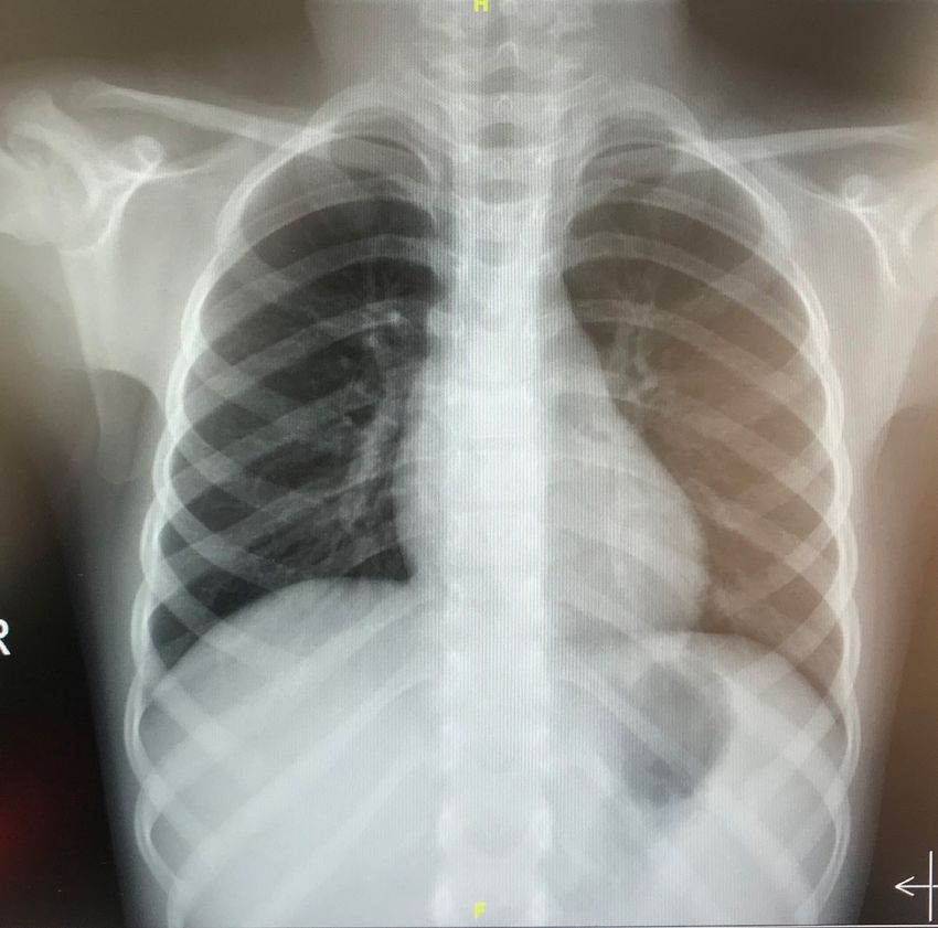
Semih Gülle, MD, infiltration areas in the thorax computed tomography (Figure 1). COVID-19 PCR test was negative. The
ORCID: 0000-0002-8814-9648 patient was hospitalized for further examination. Bronchoalveolar lavage showed alveolar macrophages,
Tuba Demirci eosinophils, and neutrophils, some of which were degenerated. Laboratory tests revealed hemoglobin:
14.9 g/dL, WBC: 17500/μl, neutrophil: 7200, eosinophil: 8100, platelet: 220000 /μl, creatinine: 0.74 mg/
Yıldırım, MD,
dL, AST: 40 U/L, ALT: 46 U/L and CRP, 142.6 mg/L. There was no proteinuria and the urine sediment
ORCID: 0000-0002-8814-9648
was inactive. ANA, ENA, and ANCA tests were negative. Electromyographic examination revealed
Assoc. Prof. Gerçek electrophysiological findings consistent with axonal degeneration in the common peroneal branch and
Can, MD, tibialis posterior branch of the right sciatic nerve. A diagnosis of EGPA was made with a history of
ORCID: 0000-0001-8347-0873 asthma, increased acute phase reactants, marked eosinophilia, multiple lung infiltrations and peripheral
neuropathy. 1mg/kg/day methylprednisolone and intravenous cyclophosphamide(CYC) treatment were
Professor İsmail
started. Clinical and biochemical response was obtained during follow-up. The patient’s respiratory
Sarı, MD, symptoms regressed and the dorsiflexor muscle strength of the foot increased with the simultaneous
ORCID: 0000-0001-7737-4180 physical therapy program.
Professor Merih
Birlik, MD, DISCUSSION
ORCID: 0000-0001-5118-9307 Peripheral neuropathy (PN) is a prevalent and important manifestation of EGPA[1]. Diagnosis of EGPA
Professor Fatoş is difficult especially when PN is the initial symptom. Pathological findings of vasculitic neuropathy are
Önen, MD characterized by axonal degeneration of nerve fibers caused by vasculitis-induced ischemia [2]. In the
largest published series of patients with EGPA, 51.4–60% had peripheral neuropathy at presentation.
ORCID: 0000-0002-6341-2622
Mononeuritis multiplex was slightly more common than symmetric polyneuropathy, and the lower limbs
were predominantly affected [3]. Our patient had right drop foot. The ANCA test of our patient was
negative, and less than 40% of patients with EGPA had positive ANCA test. Steroids are the mainstay of
treatment in EGPA [1]. Adding CYC to the treatment may reduce recurrence, morbidity, and mortality
in the presence of multiple organ involvement. A good clinical and biochemical response was obtained
in our patient with steroid and CYC treatment.
Dokuz Eylül University
Faculty of Medicine,
Department of Internal
Medicine, Division of
Rheumatology, İzmir Figure 1: Multiple infiltration areas in the HRCT of the patient
© 2021 Acta Medica. All rights reserved. 71
Hacettepe AAV Workshop - 2020                                                                           Acta Medica 2021; 52 (Supplement 2)

                  KEY MESSAGE
                  •      Vasculitic processes should also be kept in mind in patients presenting with drop foot.

                  References
                      1. A. Greco et al., “Churg-Strauss syndrome,” Autoimmunity Reviews, vol. 14, no. 4. 2015, doi: 10.1016/j.autrev.2014.12.004.
                      2. C. R. Camara-Lemarroy, A. Infante-Valenzuela, H. J. Villareal-Montemayor et al., “Eosinophilic Granulomatosis with
                         Polyangiitis Presenting as Acute Polyneuropathy Mimicking Guillain-Barre Syndrome,” Case Rep. Neurol. Med., vol. 2015,
                         2015, doi: 10.1155/2015/981439.
                      3. J. Wolf, V. Schmitt, F. Palm et al.,“Peripheral neuropathy as initial manifestation of primary systemic vasculitides,” J. Neurol.,
                         vol. 260, no. 4, 2013, doi: 10.1007/s00415-012-6760-7.

72                                                                                                         © 2021 Acta Medica. All rights reserved.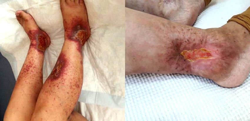
Hacettepe AAV Workshop - 2020                                                                 Acta Medica 2021; 53 (Supplement 2)

                                      PP-2: Eosinophilic Granulomatosis with Polyangitis
                                                             Presenting with Myocarditis

                             E characterized by eosinophilic infiltration of organs with necrotizing vasculitis and interstitial and
Bekir Torun, MD,               osinophilic Granulomatosis with Polyangiitis(EGPA) is a small and medium vessel vasculitis
ORCID: 0000-0002-9117-9514
                             perivascular granulomas. Three phases have been described in the natural history of the disease
Burak Okyar, MD,             (prodromal, eosinophilic, and vasculitic phases) although they do not always occur successively [1].
ORCID: 0000-0002-9028-9930
Fatih Albayrak, MD, Usually the patients’ age range is between 20 and 40 years, and both men and women are equally affected.
                             The etiology of EGPA still unknown, but it has been attributed to hypersensitivity to an inhaled agent.
ORCID: 0000-0002-6052-3896
                             Rarely a parasitic infection or antigenic drug for desensitization represents a triggering event. Asthma
Assoc. Prof. Fatih           is the main characteristic of this syndrome. Lungs are the organs most frequently involved, followed by
Yıldız,                      kidneys. Pulmonary hemorrhage and glomerulonephritis are much less common than in other small
ORCID: 0000-0003-3628-8870   vessels vasculitis [2].
Assoc. Prof. Gözde
Yıldırım Çetin               CASE REPORT
ORCID: 0000-0001-9680-7535   In 2018, when she was 18 year-old presented in a few-weeks history of pain and petechial rash on both
                             legs. For the past one week, she had low back pain radiating to left lower limb and weakness of the
                             left foot. She was diagnosed with astma 8 months ago and receving inhaler treatment. A diagnosis of
                             EGPA was made because of astma, eosinophilia, displaced pulmoner infiltrations (Figure 1), tissue
                             biopsy compatible with vasculitis and polyneuropaty. In 2020, when she was 20 year-old, she admitted
                             to our rheumatology department with acute respiratory distress and severe chest pain. Initial laboratory
                             findings revealed Troponin I elevation: 0.89 μg/l (reference value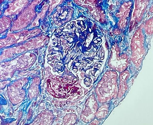
Hacettepe AAV Workshop - 2020                                                                 Acta Medica 2021; 52 (Supplement 2)

                  Serum troponin I level was increased to 1.30 ng/mL, CK-MB 32 IU/L, aspartate transaminase 55 IU/L,
                  lactate dehydrogenase 606 IU/L, CK-MB 32 IU/L. The patient was transferred to the intensive care unit
                  for further diagnosis and treatment. Beta blocker treatment was discontinued. Coronary CT angiography
                  was normal. She was diagnosed with myopericarditis. We gave 500 mg methylprednisolon for 3 days,
                  then we continued as 40 mg daily and 1000 mg rituximab treatment. Intravenous immunoglobulin
                  (IVIG) was given at a dose of 400 mg/weight/day for 5 days. After the treatment troponin I and ck-
                  mb decreased. Chest pain and shortness of breath improved. Pericardial effusion regresssed in control
                  ECHO, EF: %50. Lung consolidations regressed after the treatment. She was followed up in the servise
                  for four weeks. We gave 40 mg methylprednisolone for 1 month and then tapered, on day 15 received
                  second dose of rituximab treatment.

                  DISCUSSION
                  In the long term this systemic vasculitis has a 90% 1 year survival and a 62% 5 years survival, without
                  treatment the survival is significantly diminished with 5 year survival being only 25% [3]. Relapse is
                  common, and long-term steroid therapy is often required. Relapse can be myocarditis in EGPA due to
                  the high morbidity an mortality, close follow up and early intervention is required.

                  KEY MESSAGE
                  •      Our case presents cardiac involvement in EGPA. We recommend that pay attention to diagnosis and
                         early treatment.

                  References
                      1. Subhasish Ghosh, MaitreyeeBhattacharya, Sandipan Dhar Indian J Dermatol. Churg-Strauss syndrome, 2011; 56:718-721.
                      2. GabrielLacerdaFernandes, ArivaldoAraújoTeixeira, Ana Graziela Santana Antón, Alan TimóteoRodrigues Reis, Ana
                         Carolina Rezende de Freitas, DunyaBachourBasílioRadiolBras. 2014; 47(4): 259–261.
                      3. J. R. E. Rees, P. Burgess. Case RepMed. 2010; 2010: 290654.

74                                                                                               © 2021 Acta Medica. All rights reserved.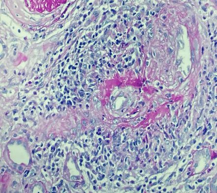
Hacettepe AAV Workshop - 2020                                                                      Acta Medica 2021; 53 (Supplement 2)

                                 PP-3: A Case of Granulomatosis with Poliangiitis (GPA)
                                                            Presenting with Joint Pain

                             A (GPA), microscopic polyangiitis (MPA), eosinophilic granulomatous polyangiitis (EGPA)) are
Mehmet Ali Aşık,                 nti-neutrophil cytoplasmic antibodies (ANCA) -related vasculitis (Granulomatosis polyangiitis
MD,
ORCID: 0000-0001-7336-2342
                             immune-mediated, progressive, necrotizing vasculitis with small vessel involvement. Lung and kidney
                             involvements are associated with mortality. The most common presentation in GPA can be multisystemic
Emrah Koç, MD,               involvement such as upper respiratory tract, eye, lung, renal and joint. Symptoms and signs of
ORCID: 0000-0002-7889-3051
                             granulomatosis polyangiitis might include; crusts in nose, stuffiness, sinus infections and nosebleeds,
Professor Süleyman           coughing sometimes with bloody phlegm. It may sometimes present with joint symptoms similar to
Özbek                        rheumatoid arthritis. Corticosteroid and immunosuppressive treatments are used in GPA treatment [1].
ORCID: 0000-0002-8548-8126
                             CASE REPORT
                             26-years-old female patient who is a doctor, presented with weakness, fatigue and pain in the great
                             joints of the lower extremities six months ago. No pathology was detected in the laboratory and physical
                             examination of the patient who applied to the internal medicine outpatient clinic. Meanwhile, the patient
                             had no lung symptoms. There were only arthralgia symptoms. The patient was followed up with the
                             present findings. The patient was referred to rheumatology because her complaints didn’t regress. The
                             patient was evaluated by rheumatology. Physical examination was unremarkable except for arthralgia.
                             Laboratory examination was planned. Leukocyte (7.8x10³ / µL), hemoglobin (13gr/dL), platelet (322x103
                             / µL), BUN (4.7 mg/dL), creatinine (0.7 mg/dL), AST (26 U/L), ALT (20 U/L), creatine kinase (120 U/L),
                             anti-nuclear antibody (ANA) negative, dsDNA negative, rhematoid factor (RF)
Hacettepe AAV Workshop - 2020                                                                  Acta Medica 2021; 52 (Supplement 2)

                  Kaynaklar
                   1. ANCA-Associated Vasculitis: Core Curriculum 2020. Am J Kidney Dis. 2020 Jan;75(1):124-137.
                   2. Stone J.H., Merkel P.A. et al, Rituximab versus cyclophosphamide for ANCA-associated vasculitis. N Engl J Med. 2010; 363:
                      221-232.
                   3. Jayne D.R., Gaskin G et al. Randomized trial of plasma exchange or high-dosage methylprednisolone as adjunctive therapy
                      for severe renal vasculitis. J Am Soc Nephrol. 2007; 18: 2180-2188.
                   4. Pagnoux C., Mahr A. Et al. Azathioprine or methotrexate maintenance for ANCA-associated vasculitis. N Engl J Med. 2008;
                      359: 2790-2803.

76                                                                                                 © 2021 Acta Medica. All rights reserved.
Hacettepe AAV Workshop - 2020                                                                                 Acta Medica 2021; 53 (Supplement 2)

                                     PP-4: Little Known Aspect of Microscopic Polyangiitis;
                                                                   Interstitial Lung Disease

                             I affected small calibre vessels and is associated with presence of antineutrophilic cytoplasmic
Ozan Cemal İçaçan¹,              ntroduction; Microscopic Polyangiitis (MPA) is type of systemic nectotizing vasculitis, predominantly
MD,
                             autoantibodies (ANCA). The organs most common affected in this type of vasculitis are the lungs and
ORCID: 0000-0002-1054-5034
                             kidneys. The common presentation of pulmonary involvement in MPA is usually pulmonary capillaritis
Selda Çelik¹, MD,            and alveolar hemorhhage. However, it has been reported that MPA may rarely occur as interstitial lung
ORCID: 0000-0003-4328-3189   disease ( ILD) and even without other systemic findings [1]. Herein, we present a case of MPA presenting
Melek Yalçın                 as ILD.
Mutlu¹, MD,
ORCID: 0000-0003-0598-5737   CASE REPORT
Fatih Yıldırım¹, MD, A 63 years old female patient applied to the Chest Diseases Policlinic in an external center with the
                             complaint of dry cough that started 5 months ago. Computed tomography of the thorax showed bilateral
ORCID: 0000-0003-3909-7500
                             ground glass opacities and traction bronchiectasis in the lower zones of the lungs, and a diagnosis of
Professor Cemal              ILD was made. She was referred to our clinic when detected an increase in kidney function tests in
Bes²                         her laboratory tests. She had constitutional symptoms such as fatigue and weight loss, hemoptysis and
ORCID: 0000-0002-1730-2991   other systemic findings were absent. Physical examination revealed fine crackles bilateral mid and
                             lower zones of lungs, vital signs and other systems were normal. In laboratory tests; urea; 82 mg/ dl,
                             creatinin 1.94 mg/ dl, normocytic normochromic anemia in complete blood count, CRP 113 mg/L,
                             ESR 90 mm/h. In urine we observed proteinuria (1.4 gr/ day in 24 hours urine test) and erythrocytes
                             (5 cells/ hpf). Proteinase 3 ANCA was negative, Myeloperoxidase (MPO) ANCA was positive (161,
                             normal value 0-12). Renal biopsy revealed cellular crescent in 4 of 34 glomeruli, fibrocellular crescent in
                             12 and fibrinoid necrosis in 2 glomeruli. The patient was diagnosed with MPA, pulse steroid and pulse
                             cyclophosphamide treatments were initiated.

                             DISCUSSION
                             It is known that ILD can be seen in ANCA associated vasculitis (AAV). In AAV, ILD is more common
                             in patients with advanced age and MPO-ANCA positive MPA [2]. Although rare, ILD may also be first
                             sign of MPA.

                             KEY MESSAGE
1
 University of Health        •      MPA is among the possible causes while intestigating the etiology of ILD.
Science, Bakırköy Dr.
Sadi Konuk Research
and Training Hospital,       References
Istanbul                         1. Nishkantha Arulkumaran, Naomi Periselneris, Gill Gaskin, Nicola Strickland, Philip W. Ind, Charles D. Pusey, Alan D.
2
 University of Health               Salama, Interstitial lung disease and ANCA-associated vasculitis: a retrospective observational cohort study, Rheumatology,
Science, Başakşehir                 2011; 50(11):2035–2043
Çam ve Sakura City               2. Katsuyama T, Sada KE, Makino H. Current concept and epidemiology of systemic vasculitides. Allergol Int. 2014;63(4):505-
Hospital, Istanbul                  13.

© 2021 Acta Medica. All rights reserved.                                                                                                                    77
Hacettepe AAV Workshop - 2020                                                                              Acta Medica 2021; 53 (Supplement 2)

                     PP-5: Granulomatosis with Poliangiitis (GPA) Case
      Presenting with Recurrent Conjunctival and Postnasal Discharge

                             G is characterized by granulomatous inflammation, tissue necrosis and varying degrees of vasculitis in
Damla Karataş1,                    ranulomatous with Polyangiitis (GPA) is a rare systemic autoimmune disease of unknown etiology. It
MD,
                             small and medium vessels. It can manifest itself with varying severity and wide clinical symptoms. It has
ORCID: 0000-0002-4755-0443
                             two forms. The limited form is characterized by upper and lower respiratory tract involvement without
Zeynep Öztürk1, MD,          renal involvement. In the systemic form, it can involve other organs, along with a more severe respiratory
ORCID: 0000-0001-6439-000X   and kidney disease. Ocular and orbital signs can be found in approximately half of the patients with
Sümeyye Merve                GPA and as the first presentation sign of the disease. It may be the result of disease in the neighboring
Türk1, MD,                   paranasal sinuses or may occur focal with orbital granuloma formation, vasculitis.
ORCID: 0000-0003-0662-4837
Rumeysa Kurt2, MD, CASE REPORT
                             A 55-year-old female patient was followed up by an ear-nose-throat physician with complaints of
ORCID: 0000-0001-5425-4418
                             postnasal drip for 6 months. Chronic dactriocystitis and episcleritis were found in the patient who
Professor Emel               was evaluated by the ophthalmology department with the addition of conjunctival discharge to his
Gönüllü1                     complaints. Orbital MRI revealed a heterogeneous mass of 25x12 mm at the left lacrimal gland. C-reactive
ORCID: 0000-0002-6990-4206   protein: 97 mg/dl, erythrocytesedimentation rate: 97 mm/h in the examinations, when we evaluated her.
                             No other abnormality was found in the systemic evaluation and examination. Biopsy taken from the
                             mass in the eye excluded malignancy and infiltrative infections, but was insufficient for GPA. ANCA
                             (indirect immunofluorescence) was negative. Working with ELISA, PR3 ANCA was positive (+++). The
                             BVAS-WG score of the patient who was accepted as GPA was 4. We started to the patient 1 mg /kg/
                             day methylprednisolone and 15 mg/week methotrexate for treatment. Methotrexate was stopped and
                             azathioprine was switched to the patient whose liver tests increased. Azathioprine was discontinued
                             in the patient whose liver tests did not improve. BVAS-WG activity of the patient who was found to
                             have proptosis, ptosis, limitation of gaze in all directions, conjunctival hyperemia and episcleritis was
                             calculated as 10. 1 mg/kg/day methylprednisolone + rituximab (RTX) 1000 mg every 6 months with
                             2-week intervals was planned. The patient is still being followed up with RTX therapy in remission.

                             DISCUSSION
                             In the presence of clinical findings, it would be beneficial to request PR3-ANCA and MPO-ANCA
                             tests performed by ELISA in cases with high suspicion of ANCA-associated vasculitis, especially if the
                             pathology is not guiding, although the ANCA screening test is negative.

                             KEY MESSAGE
                             •      In the presence of clinical suspicion, PR3 and MPO-ANCA ELISA tests may be helpful even in the case
                                    of negative ANCA screening test.
1
 Sakarya University
Faculty of Medicine,
Department of Internal       References
Medicine, Division of            1. Tarabishy AB, Schulte M, Papaliodis GN, Hoffman GS. Wegener’s granulomatosis: clinical manifestations, differential
Rheumatology, Sakarya               diagnosis, and management of ocular and systemic disease. Surv Ophthalmol. 2010;55(5):429–444.
2
 Sakarya University              2. Greco A, Marinelli C, Fusconi M, et al. Clinic manifestations in granulomatosis with polyangiitis. Int J Immunopathol
Faculty of Medicine,                Pharmacol. 2016;29(2):151–159.
Department of Internal           3. Comarmond C, Cacoub P. Granulomatosis with polyangiitis (Wegener): clinical aspects and treatment. Autoimmun Rev.
Medicine, Sakarya                   2014;13(11):1121–1125.

78                                                                                                            © 2021 Acta Medica. All rights reserved.
Hacettepe AAV Workshop - 2020 Acta Medica 2021; 53 (Supplement 2)
PP-6: Prostate Abscess: A Rare Clinical Prezentation for
Granulomatosis Polyangiitis
G findings, often with respiratory and renal involvement. However, although it is rare, urogenital
Emrah Koç, MD, ranulomatous with polyangiitis (GPA) is a necrotizing granulomatous vasculitis with constitutional
ORCID: 0000-0002-7889-3051
involvement can also be seen. Here, we present a case of GPA with lung, prostate, eye and soft palate
Mehmet Ali Aşık, involvement, whose initial presentation was a prostate abscess.
MD,
ORCID: 0000-0001-7336-2342
CASE REPORT
Professor Süleyman A 44-year-old male patient who applied to the urology clinic in an external center with the complaint
Özbek of urinary burning and frequent urination revealed high ESR and CRP, hematuria and pyuria. The
ORCID: 0000-0002-8548-8126 prostate gland of the patient, who underwent lower abdominal computed tomography, was observed
as hypodense and was found to be compatible with a prostate abscess. When the sample taken in the
transureteral resection performed after antibiotherapy was evaluated in the pathology, the result was
“Necrotic fibrinoid exudate”. During this period, the patient was transferred to internal medicine intensive
care because of respiratory distress and hypoxia. In the thorax computed tomography performed for
respiratory distress, cavitation areas up to 6 cm in size, nodular lesions, soft tissue lesions adjacent to
cavity areas were observed. The cavities were thought to be metastatic lung abscess in the foreground and
aspiration was made for diagnosis, and the pathology result sent from the sample came in accordance
with the suppurative abscess. Due to the development of a perforated lesion on the palate during the
follow-up, the patient was referred to Çukurova University Faculty of Medicine Internal Medicine
Intensive Care with a pre-diagnosis of vasculitis and the arrangement of the examination and treatment.
On admission to our hospital, the patient had a cavitary lesion on the palate, multiple cavities in the
lung, and soft tissue lesions. He had a fever. Hb value was 9.1 g/dl, CRP 160 mg/L, Procalcitonin 0.7 ng/
mL. ANCA was studied from the patient and the result was positive (pr3 positive).In the follow-up, the
patient developed episcleritis in the eye. The patient was diagnoised with GPA accepted. Plasmapheresis
and pulse steroid were administered every other day, followed by intravenous cyclophosphamide. After
the treatment, a significant improvement was observed in the lesions in the lung and prostate and in the
perforated lesion of the palate in the episcleritis clinic.
DISCUSSION
Granulomatosis with Poliangiitis (GPA) is an associated systemic vasculitis, but it is characterized
by mainly respiratory tract involvement. Urogenital system involvement is rarely observed in the
literature (2.3-7.4%). In the presence of urogenital system involvement, the most frequently affected
area is the prostate, but also epididymis, bladder, seminal vesicles, testicles, ureters, urethra, cervix,
vagina, perineum and penile involvement has been reported [1]. In prostate involvement, patients may
present with prostate abscess or prostatitis. The initial symptom may only be dysuria, as in our case.
Infections, simple granulomatous prostatitis, sarcoidosis should be kept in mind in the differential
diagnosis [2]. As in other involvement of Granulomatosis with Poliangiitis (GPA), patients benefit
from cyclophosphamide treatment [3]. Urogenital system involvement and especially prosthesis
involvement should be kept in mind in patients with a diagnosis of GPA, and these symptoms should
not be overlooked while managing patients.
KEY MESSAGE
• Urogenital system involvement and especially prostate involvement should be kept in mind in patients
with a diagnosis of GPA, and these symptoms should not be overlooked while managing patients.
References
1. Bacon PA. (2005) The spectrum of Wegener’s granulomatosis and disease relapse. New England Journal of Medicine 352:
Cukurova University 330–332.
Department of Internal 2. Alba MA, Moreno-Palacios J, Beça S, et al. . Urologic and male genital manifestations of granulomatosis with polyangiitis.
Diseases, Division of Autoimmun Rev 2015;14:897–902.
Rheumatology, Adana 3. Smith R, Jones R, Jayne D. Progress in treatment of ANCAassociated vasculitis. Arthritis Res Ther. 2012;14(2):210.
© 2021 Acta Medica. All rights reserved. 79Hacettepe AAV Workshop - 2020 Acta Medica 2021; 53 (Supplement 2)
PP-7: Presenting with Episcleritis in ANCA Associated
Vasculitis: A Case Report
A ımmune character that concern many systems. It is heterogeneous group of diseases that presents
Bengisu Aslan, MD, NCA associated vasculitis (AAV), is a group of diseases that present different clinical signs of auto-
ORCID: 0000-0001-6892-1090
many differrent clinical findings. Upper airway tract lesions and pulmoner symptoms are often found.
Professor Veli Pulmoner infiltrations in imaging and kidney abnormalities in laboratory examinations are the first
Yazısız findings that bring to mind te possibility of AAV. AAV affects classically pulmonary-renal systems but
ORCID: 0000-0002-3176-4850 these types of vasculitis can present with unusual symptoms. The eye is important target in AAV and
various eye lesions can develop. In this case report, an AAV case with episcleritis as the first finding is
presented.
CASE REPORT
A 45 year old male patient had a rash in the right eye three months ago. Local treament recommended
by the ophtalmalogist. Later, similar redness in the same eye repeated four times. For the last month,
there was pain in the left knee with red eye. Fever and cough have been added for a week. He was
hospitalized with pnemonia. Patient’s symptoms did not improve despite antibiotic treatment. It was
observed “Cavitary lesions containing calcification in the upper lobe apical of right lung, cavitary lesions
of about 1,5x1,5 in the lower lobe posterior – noduler density of about 1,5 cm in yhw lower lobe, two
fissure- based noduler lesions 4 mm diameter in the left lung lower lob” in high resolution computer
tomography (HRCT). Neutrophilia (7.7x10³ /µL) and thrombocytosis (391x103 / µL), increase in ESR
(63 mm/h) and CRP (9.5 mg/dL) and p-ANCA positivity (IFA 1/10) were present in the laboratory
examination. Liver function tests, creatinine and glomerular filtration values were in the normal range.
He had no abnormality in complete urine test. Episcleritis was diagnosed in eye examination. The patient
was evaluated as AAV particularly granulomatosis poliangiitis with clinical and laboratory findings. It
was decided to biopsy from lung lesions for different diagnose. It was started metil prednisolone for
induction therapy. Necrotizing granulamotosis inflamation was observed in pathology specimen.
Tuberculosis examination was negative.
The patient was diagnosed granulomatosis poliangitis with clinical and histopathological findings and
ıt was started 500-700 mg/m2 pulse cyclophospomide with high metil prednisolone. Metil prednisolone
was gradually reduced and maintenance azathioprine 2.5 mg/kg was started after cyclophospomide
cycles. The reason for respiratory complains in the tenth month of treatment was re-evaluated. Cavitary
lesions were seen in HRCT (Figure 1). It was accepted as a relapse disease and rituximab therapy was
started. Low dose metil prednizolone and azathioprine were continued, a second rituximab infusion was
administerd in the sixth month. He has been followed up as AAV in remission for five years.
Akdeniz University
Faculty of Medicine,
Department of Internal Figure 1: Caviter lesion in
Medicine, Division of lung with granulomatosis
Rheumatology, Antalya poliangitis
80 © 2021 Acta Medica. All rights reserved.Hacettepe AAV Workshop - 2020 Acta Medica 2021; 52 (Supplement 2)
DISCUSSION
The eye is an organ frequently affected by AAVs. Episcleritis, scleritis, corneal melting, uveitis, orbital
inflamation or retroorbital masses are seen in eye involvement. In a Patompong’s study, they examined
183 patient with eye involvement retrospectively, ıt was found that approximately half of the patients had
ocular involvement at the time of the diagnosis. Eye lesions that started an average of one year before
AAV diagnosis have been described [1].
Granulomatosis poliangitis; it affects small-medium size vessel; ıt has many different clinical findings.
It causes damage most often lung, kidney, upper airway tract. Pulmoner and renal involvement have
worse prognosis and increased mortality and morbidity [2]. Eye lesions can develop any period of
disease, and they respond to high dose corticosteroid and cyclosphospomide remission of the disease.
Eye involvement that is resistant to conventional treatments and with relapses has also been reported [3].
The case presented here is a case of granulomatosis poliangitis that starts with recurrent episcleritis
attacks, musculoskeletal symptoms are added eventually pulmoner lesions develop. It shows AAV can
present different clinical symptoms. It reveals that serious inflammatory diseases may be the basis of
recurrent episcleritis attacks and systemic evaluation and clinical follow up are important.
KEY MESSAGE
• This case is good example for showing that AAVs may present with symptoms other than classical
pulmonary and renal syndrome clinic and that AAVs may be the cause of persistent inflammatory eye
lesions.
References
1. Patompong U, Cynthia S.Crowson, Rodrigo Cartin-Ceba et al. Rheumatology Oxford 2017;10 1763-1770
2. Lee T, Gasim A, Derebail VK. et al. Predictors of treatment outcomes in ANCA-associated vasculitis with severe kidney
failure. Clin J Am Soc Nephrol 2014;9:905–13
3. Babu K, Dharmanand BG. Worsening of posterior scleritis and orbital pseudotumor in a patient with granulomatosis
polyangiitis with rituximab-A case report. Indian J Ophthalmol. 2020 Sep;68(9):1986-1988.
© 2021 Acta Medica. All rights reserved. 81Hacettepe AAV Workshop - 2020 Acta Medica 2021; 53 (Supplement 2)
PP-8: A Granulomatosis Polyangiitis Patient Presenting with
Recurrent Upper Airway Infection
G vessels of unknown etiology, which can be seen all ages especially in the middle ages. It may progress
Özlem Doğan ranulomatosis poliangiitis is a disease that develops due to inflammation of small, middle-sized
Ağbuga, MD,
with signs of inflammation in the upper respiratory tract unresponsive to antibiotics, as well as vital
ORCID: 0000-0002-4998-9177
organs such as lungs and kidneys.
Didem Arslan, MD
ORCID: 0000-0002-9654-2183
Case Report
28 years-old male patient. On his medical history it was learned that he did not have any illness. He admitted
to otorhinolaryngology department of hospital with ear pain. Otitis was diagnosed and antibiotic treatment
was started. After a month, the patient had complaints of ear pain again and cough with mucus was also added.
Sinus CT was taken in the hospital which he admitted and reported as: septum minimally deviated,
middle ear vertical areas were bilaterally pneumatized. Lung CT: It has been reported as a subpleural
approximately 2.5 cm central cavitated, slightly irregularly demarcated, nodular lesion in the upper lobe
of the left lung, and several thick-walled cavities of which the largest was 44x29 mm in the lower lobe
of both lungs. The patient was referred to our hospital for advanced examination and treatment. The
patient’s laboratory results were Hb:9.7g/dL, wbc:10.6x10³/µL, plt:331x103/µL, crp:155 mg/dL, ESR
51/sa ,procalcitonin 0.08 ng/l, ALT:19 U/L,AST:30 U/L, ALP:134 U/L, GGT:159 U/L, kr:0.6 mg/dL,
BUN:23 mg/dL and viral hepatitis were negative. Spot urine protein/creatinine ratio: resulted as 138.
Urine sediment was evaluated as inactive. The patient was hospitalized with a pre-diagnosis of vasculitis
and infection. Blood and urine cultures were taken because the fever of patient was 38 C and above.
Direct ARB test and culture were sent for tuberculosis. Sampling was performed for galactomannan
with suspected opportunistic infection. ANA and dsDNA were sent and the results were both negative.
ANCA was sent in terms of vasculitis. Abdominal USG was used to illuminate the increase in Liver
enzymes; minimal dilatation in intrahepatic bile ducts was detected. MRCP was applied for dilatation in
intrahepatic bile ducts and evaluated as normal. AMA was sent; the result was negative. c-ANCA result
was positive. The patient’s diagnosis was considered to be Granulomatosis with Poliangiitis (GPA). The
increase in liver functonal tests was attributed to disease involvement. Anemia was evaluated as chronic
disease anemia. Since secondary bacterial infection could not be ruled out, empirical antibiotherapy and
immunosuppressive treatment with 100 mg methylprednisolone (iv 5 days) were started simultaneously.
Clinical response was obtained on the third day of treatment. The CRP level was decreased to 26 mg/dL.
The patient whose general condition improved during the follow-up was discharged from the service
and followed up in the outpatient clinic. No significant reproduction was detected in his blood,urine
and mucus cultures. Rituximab treatment was planned for the patient. Trimethoprim-sulfomethaxazole
prophylaxis was initiated due to upper respiratory tract involvement.
DISCUSSION
Granulomatosis polyangiitis is a systemic vasculitis of unknown etiology, mostly affecting the upper
and lower airways and kidney. The onset of the disease can be with symptoms of upper respiratory tract
involvement (mostly in the nasal cavity and paranasal sinuses) and can be seen in 70–100% of cases [1].
There may be multiple antibiotic use and persistent sinusitis and rhinitis that do not respond to these
treatments [2]. As it is not included in differential diagnoses, as in our case, patients consult a doctor
with similar complaints multiple times, and other organ involvement may occur until the disease is
diagnosed. The purpose of the presentation of this case is to draw attention to the necessity of further
examination in terms of Granulomatosis with Poliangiitis (GPA) in case of recurrent persistent head and
neck inflammation.
Çukurova University
Faculty of Medicine, KEY MESSAGE
Department of Internal
Medicine, Division of • Inflammation is a common symptom of infections and autoimmune diseases, and it should be
Rheumatology, Adana considered especially in the case of antibiotic unresponsiveness; In cases of inflammation involving
82 © 2021 Acta Medica. All rights reserved.Hacettepe AAV Workshop - 2020 Acta Medica 2021; 52 (Supplement 2)
the head and neck region such as recurrent otitis and sinusitis, granulomatosis polyangiitis should be
considered in differential diagnosis.
References
1. Greco A, Marinelli C, Fusconi M, Macri GF, Gallo A, De Virgilio A, Zambetti G, de Vincentiis M. Clinic manifestations in
granulomatosis with polyangiitis. Int J Immunopathol Pharmacol. 2016;29(2):151-9.
2. Qaisar H, Shenouda M, Shariff M, Cheema A, Tang X, Kaplan A. Granulomatosis with Polyangiitis Manifesting as Refractory
Otitis Media and Mastoiditis. Arch Iran Med. 2019;22(7):410-413.
© 2021 Acta Medica. All rights reserved. 83Hacettepe AAV Workshop - 2020 Acta Medica 2021; 53 (Supplement 2)
PP-9: Coexistence of Necrotizing Episcleritis, Sacroileitis and
Familial Mediterranean Fever due to Granulomatosis Polyangiitis
G system autoimmune disorder characterized by necrotizing granulomatous inflammation and pauci-
Burak Karakaş, MD ranulomatosis polyangiitis (GPA), previously known as Wegener granulomatosis, is a rare multi-
ORCID: 0000-0003-3624-4958
immune vasculitis. Respiratory symptoms such as recurrent bloody rhinorrhea, rhinosinusitis and
nodular lesions in the lungs are seen in 45% of cases and in 87% during the course of the disease [1,2].
Ocular involvement is common and can range from mild conjunctivitis to scleritis, episcleritis, uveitis,
ciliary vasculitis and retro-orbital mass lesion [3]. In this case, we wanted to present a GPA case with
lung, kidney and eye involvement.
CASE REPORT
A 64-year-old male patient was admitted to the emergency department in 2010, after complaints of
fatigue, nausea-vomiting, shortness of breath and weight loss for the last 2 months. WBC: 13000 /µL,
Hb: 7.7 g/dL, Htc: 25.5%, urea: 141.6 mg/dl, creatinine: 8.2 mg/dl, albumin: 2.1 g/dl, CRP: 118 mg/L,
ESR was> 140 mm/hour. There were protein 2+ and 5-6 leukocytes in each field in urine analysis. The
patient, whose parenchyma echo patterns of bilateral kidneys were evaluated as grade 2 on renal USG,
is admitted to the nephrology service for examination and treatment. Micro total protein (MTP) in 24-
hour urine was 2.7 g/day. Hemodialysis is applied intermittently to the patient because of oliguric urine
output, dyspnea and metabolic acidosis in arterial blood gas.
Minimal mucosal thickening is detected in the right sphenoid sinus on CT of the patient who has
frequent sinusitis history. In the thoracic CT taken upon the presence of dyspnea, fibrotic changes in the
apex of both lungs and the infiltration area leading to consolidation in the right lower lobe posterobasal
and infiltration areas in both lungs are observed. No pathology was found in abdominal CT. When the
C-ANCA was> 100 in the etiology-based examinations of the patient, whose infection was excluded,
renal biopsy was performed on the patient. The pathology report resulted as “kidney tissue characterized
by crescent development in 1/3 of glomeruli and minimal mesangial matrix increase and cell proliferation
in other glomeruli”. The patient was diagnosed with Granulomatosis with Poliangiitis (GPA) because of
kidney and lung involvement. The patient’s BVAS-WG score was calculated as 7.
The patient was started on cyclophosphamide with pulse steroid. Azathioprine 100 mg / day was
started as maintenance therapy for the patient, who received 6 cycles of cyclophosphamide treatment.
The patient applied to us again after 4 years with complaints of inflammatory hip pain, morning
stiffness lasting half an hour, intermittent abdominal pain attacks, redness in the right eye, headache
and nosebleeds. MEFV gene analysis M694V, which was studied due to proteinuria, was found to be
homozygous in the patient with abdominal pain attacks and family history of FMF in the anamnesis.
CRP: 98 mg/L, ESR: 51 mm / hour, creatinine: 1.2 mg/dl and bilateral sacroielitis was detected on
sacroiliac graphy. Due to pain and loss of vision in the right eye, a diagnosis of necrotizing episcleritis
and corneal melting was made on eye examination. 1 gram of pulse methylprednisolone for 3 days,
followed by 1 mg/kg/day methylprednisolone, rituximab 1000 mg 0-15. days / 6 months given. In
addition, prophylactic trimethoprim-sulfamethoxazole was started. In the maintenance treatment, he
was discharged by starting methotrexate 15 mg/week, folbiol and colchicine treatment and reducing the
methylprednisolone treatment dose. In the follow-up of the patient, BVAS-WG score decreased. The
patient is in remission at outpatient clinic controls.
DISCUSSION
GPA is necrotizing vasculitis of small to medium vessels, which is a component of a wide range of
Sivas Cumhuriyet diseases called anti neutrophil-cytoplasmic-antibody (ANCA) -related vasculitis. Its diagnosis is made
University, Department clinically and histologically. The clinic can range from mild organ involvement to severe multisystem
of Internal Medicine,
Division of organ involvement [4]. In our case, upper respiratory tract, lung, kidney and eye involvement was
Rheumatology, Sivas observed.
84 © 2021 Acta Medica. All rights reserved.Hacettepe AAV Workshop - 2020 Acta Medica 2021; 52 (Supplement 2)
It has been reported that c-ANCA (cytoplasmic-ANCA) formed against proteinase 3 target antigen in
GPA is highly specific for active GPA and c-ANCA titers are directly related to GPA disease activity. In
patients with GPA with c-ANCA positivity, the possibility of kidney and respiratory tract involvement is
high and the tendency to relapse has increased. It has been reported that there is a correlation between
the c-ANCA level and the risk of exacerbation in patients in remission [5]. Treatment is individually
tailored to the severity of the clinic. RAVE and EUVAS studies have shown that rituximab is equally
effective as cyclophosphamide in induction of remission. In addition, Rituximab has demonstrated
superiority in the induction treatment of relapsed cases [6]. When remission occurs, it is recommended
to reduce the corticosteroid dose gradually, discontinue cyclophosphamide, and maintain remission
with methotrexate or azathioprine [7].
KEY MESSAGE
• GPA is a rare, difficult to diagnose disease that can involve many organs and has high mortality. Early
diagnosis and treatment are of great importance in terms of prognosis.
References
1. Cordier JF, Valeyre D, Guillevin L, Loire R, Brechot JM. Pulmonary Wegener’s granulomatosis. A clinical and imaging study
of 77 cases. Chest. 1990;97(4):906-912.
2. Rodrigues CE, Callado MR, Nobre CA, et al. Wegener’s granulomatosis: prevalence of the initial clinical manifestations—
report of six cases and review of the literature. Rev Bras Reumatol. 2010;50(2):150-164.
3. Harman LE, Margo CE. Wegener’s granulomatosis. Surv Ophtalmol. 1998;42(5):458-480.
4. Catanoso M, Macchioni P, Boiardi L, et al. Epidemiology of granulomatosis with polyangiitis (Wegener’s granulomatosis) in
Northern Italy: a 15-year population-based study. Semin Arthritis Rheum. 2014;44(2):202–7.
5. Hewins P, Tervaert JW, Savage CO, et al. (2000) Is Wegener’s granulomatosis an autoimmune disease? Current Opinion in
Rheumatology 12: 3–10
6. Van der Woude FJ, Rasmussen N, Lobatto S, et al. (1985) Autoantibodies against neutrophils and monocytes: Tool for
diagnosis and marker of disease activity in Wegener’s granulomatosis. Lancet 1: 425–429.
7. Holle JU, Laudien M, Gross WL. Clinical manifestations and treatment of Wegener’s granulomatosis. Rheum Dis Clin North
Am 2010;2002:507–26.
© 2021 Acta Medica. All rights reserved. 85Hacettepe AAV Workshop - 2020 Acta Medica 2021; 53 (Supplement 2)
PP-10: Granulomatosis with Poliangiitis (GPA); Systemic Vasculitis
of Small Vessels Associated with the Presence of ANCA
I patient who first applied to chest diseases with the complaint of dyspnea. Pulmonary malignancy
Neşe Çabuk Çelik, n 2015, high sedimentation (ESR) and low hemoglobin (Hb) were observed in a 52-year-old male
MD,
was considered as a pre-diagnosis. A mass consistent with malignancy and accompanying lymph nodes
ORCID: 0000-0003-2717-4804
and pleural effusion were detected in the thorax computed tomography (CT). Malignancy was ruled
Burak Karakaş, MD, out as a result of the bronchoscopic biopsy. The patient was referred to our outpatient clinic in terms
ORCID: 0000-0003-3624-4958 of arthritis and high ESR etiology. In physical examination; Bilateral PIP (proximal interphalangial
İlker Yalçın, MD, palanx) and MCF (metacarpophalangial) joints and wrists were present in the hands. Apart from this,
ORCID: 0000-0003-4924-5642 no pathological finding for the upper respiratory tract was found. Considering rheumatoid arthritis
(RA) in the foreground, examinations were made. The patient who had high ESR and CRP (C reactive
Beliz Karataş, MD,
protein) accompanying anemia had RF: 198 IU/L too. In advanced examinations; C-ANCA (cytoplasmic
ORCID: 0000-0003-1894-8062
ANCA) was significantly positive in ELISA/IFA. Anti-CCP was negative. Otorhinolaryngology and
Yılmaz Savaş, MD, eye department was consulted. Echocardiogram was done. In addition, protein was measured in 24-
ORCID: 0000-0002-0181-5450 hour urine. The patient was diagnosed with GPA with all these results. Pulmonary hemorrhage was
Professor Ali Şahin detected in the imaging performed on the patient whose shortness of breath and hemoptysis recurred.
Pulse steroid therapy was given as the monitored creatinine value (6.5 g / dl) increased. In addition,
ORCID: 0000-0002-6953-4276
plasmapheresis was performed. The patient’s clinic was stabilized. The BVAS-WG score of the patient who
was diagnosed with Wegener was calculated as 13. Looking at the total number of components; While
arthritis, pleurisy, nodule / cavity and secondary infiltration to Wegener met 4 minor / new worsening
criteria, alveolar hemorrhage, respiratory failure and creatinine increase by more than 30% met 3 major
/ new worsening criteria. Five factor score; It was calculated as 2 due to the development of kidney
failure and the absence of upper respiratory tract symptoms. Vasculitis damage index was calculated
as 1 due to fibrosis developing in the lung parenchyma. Remission was achieved with rituximab. In the
imaging performed during the outpatient clinic controls, it was observed that the lesions in the lungs
were completely resolved (Figure 1). After treatment, BVAS-WG score decreased to 1. Maintenance
treatment was given with weekly treatments such as methotrexate, methylprednisolone, trimethoprim /
sulfomethaxazole. The patient, whose steroid treatment was discontinued, has been followed for 5 years
with rituximab (every 6 months) and methotrexate.
Sivas Cumhuriyet
University Department Figure 1: Comparative
of Internal Diseases, views of regressing
Division of lesions taken before and
Rheumatology, Sivas after treatment.
86 © 2021 Acta Medica. All rights reserved.Hacettepe AAV Workshop - 2020 Acta Medica 2021; 52 (Supplement 2)
DISCUSSION
GPA is a type of autoimmune vasculitis involving small vessels, with necrotizing glomerulonephritis. The
diagnosis of GPA is made clinically and histologically. The disease may progress with mild involvement in
the upper respiratory tract such as the ear and nose during the active period, as well as severe multi-organ
failure [2]. In our case, the main involvement was predominantly pulmonary and renal involvement. The
frequency of renal involvement in GPA is over 70% [3]. It must meet international criteria for diagnosis.
CT and biopsy are used for these criteria. In 2011, rituximab, the B cell inhibitor anti CD-20, was among
the treatment options for remission in active patients [4]. Steroid therapy should be discontinued in the
patient in remission, as in our case [5].
KEY MESSAGE
• GPA may present with arthralgia/arthritis. Rituximab is an effective option. Steroid should be tapered
slowly in patient during remission.
References
1. Millet A, Pederzoli-Ribeil M, Guillevin Let al. Antineutrophil cytoplasmic antibody-associated vasculitides: is it time to split
up the group? Ann Rheum Dis 2013; 72:1273-1279.
2. Watts RA, Mahr A, Mohammad AJ, et al. Classification, epidemiology and clinical subgrouping of antineutrophil cytoplasmic
antibody (ANCA)-associated vasculitis. Nephrol Dial Transplant 2015;30 Suppl 1:i14–i22.
3. Sinico RA, Di Toma L, Radice A. Renal involvement in anti-neutrophil cytoplasmic autoantibody associated vasculitis.
Autoimmun Rev 2013;12:477–82.
4. Holle JU, Laudien M, Gross WL. Clinical manifestations and treatment of Wegener’s granulomatosis. Rheum Dis Clin North
Am 2010;2002:507–26.
5. Lutalo PM, D’Cruz DP. Diagnosis and classification of granulomatosis with polyangiitis (aka Wegener’s granulomatosis). J
Autoimmun 2014;48-49:94–8.
© 2021 Acta Medica. All rights reserved. 87Hacettepe AAV Workshop - 2020 Acta Medica 2021; 53 (Supplement 2)
PP-11: Granulomatosis with Polyangiitis: Case Report
G antineutrophil cytoplasmic antibody (ANCA) positivity and an inflammatory reaction pattern
Sümeyye Merve ranulomatous with polyangiitis (GPA) is an autoimmune small vessel vasculitis with frequent
Türk1, MD,
such as necrosis, granulomatous inflammation, and vasculitis. GPA may show either classic or limited
ORCID: 0000-0003-0662-4837
involvement. In classic involvement, GPA may involve the upper respiratory tract, lungs, and kidneys.
Damla Karataş1, In limited involvement, however, GPA does not show renal involvement and is often seen in women.
MD,
ORCID: 0000-0002-4755-0443
CASE REPORT
A 61-year-old male patient presented with a history of weakness, weight loss, fever, burning with
Zeynep Öztürk1, MD, urination, red eyes, and nasal obstruction. In March 2019, the patient presented to another healthcare
ORCID: 0000-0001-6439-000X center with the complaints of weakness and burning with urination and was hospitalized and then
İlhan Yıldırım1, MD, initiated on an ertapenem therapy, and then was referred to our Infectious Diseases department. The
ORCID: 0000-0002-4553-1054 patient was also examined by an ophthalmologist for the redness in the right eye and then was diagnosed
with scleritis. Paranasal computed tomography (CT) that was performed due to nasal obstruction
Professor Emel persisting for one month showed an appearance of high-density soft tissue with hyperdense components
Gönüllü2, MD in the right maxillary sinus causing aeration loss in the right maxillary sinus and also showed obliteration
ORCID: 0000-0002-6990-4206 of the right ostiomeatal complex and a lesion in the right maxillary sinus extending to the right ethmoid
sinuses and causing bone erosion in the lateral wall of the ethmoid sinus. Additionally, CT also revealed
mild mucosal thickening in the left maxillary sinus. Although chest X-ray was normal, CT revealed an
appearance of mild paraseptal emphysema in the right upper lobe apicoposterior segment of the right
lung. Purpuric rash was present in the medial aspect of both legs.
Laboratory parameters were as follows: hemoglobin (Hg) 11.8 g/dl, white blood cell count (WBC) 18.5
K/Ul, neutrophil (Neu) 16.1 K/Ul, platelet (Plt) 507 K/Ul, alanine transaminase (ALT) 80 U/L, C-reactive
protein (CRP) 228, procalcitonin (PCT) 0.05 ng/Ml, erythrocyte sedimentation rate (ESR) 135 mm/h,
blood culture: no growth, creatinine 0.56 mg/dl, complete urinalysis: 9 leukocyte, 76 erythrocyte, protein
(+), ANA (-), Anti ds DNA (-), CCP (-), RF (-), ANCA 1/32 (+), MPO ANCA (-), and PR3 ANCA (+++).
Throughout the follow-up period, the patient was evaluated by our clinic and had no fever, no growth
in blood and urine cultures, was negative for PCT and positive for PR3 ANCA, and had no decrease in
CRP level. The patient was initiated on methylprednisolone 1 mg/kg/day. On day 15 of the treatment,
creatinine level was 1.69 mg/dl and complete urinalysis indicated +2 protein. Subsequently, renal biopsy
that was performed by the Nephrology department indicated focal glomerular crescent formation (8/33
glomeruli) and signs of vasculitis, and thus cyclophosphamide 50 mg 2x1 was added to the treatment.
The Birmingham Vasculitis Activity Score (BVAS) of the patient was 19 and the BVAS for Wegener’s
Granulomatosis (BVAS-WG) score was 21. Remission was achieved with steroids and cyclophosphamide.
The maintenance therapy was continued with the addition of azathioprine 2 mg/kg/day.
The patient is currently being followed up in remission and receiving azathioprine 50 mg alone. His ESR
is 8 mm/h, creatinine level is 1 mg/dl, he has no protein in urine, and his chest X-ray is normal.
DISCUSSION
Granulomatous with polyangiitis (GPA) is an important clinical picture that needs to be recognized
early and treated appropriately. Throughout the course of GPA, vasculitis accompanied by necrotizing
granulomas can be seen in different organs and organ-specific symptoms may occur. Both morbidity and
mortality have been significantly reduced with corticosteroids and novel immunosuppressive treatments.
KEY MESSAGES
1
Sakarya University • GPA can manifest with a wide variety of symptoms and can be included in the differential diagnosis
Faculty of Medicine,
of numerous diseases. Of particular importance, its differential diagnosis with infection should be
Department of Internal
Medicine, Division of
evaluated in detail.
Rheumatology, Sakarya • GPA has a high mortality unless treated. With treatment, one-year survival is 90%, two-year survival
2
Görele Op. Dr. Ergün is 87.5%, and five-year survival is 76%.
Özdemir State Hospital
Internal Diseases References
1. Greca A, Marinelli C, Fusconi M, Macri GF, Gallo A, De Virgilio A, Zambetti G, Vincentiis M. Clinic manifestationsin
Department, Giresun
granulomatosis with poliangiitis. Int J Immunopathol Pharmacol 2016 Jun; 29(2): 151– 159.
88 © 2021 Acta Medica. All rights reserved.Hacettepe AAV Workshop - 2020 Acta Medica 2021; 53 (Supplement 2)
PP-12: Are ANCA Associated Vasculitides Really PAUCI-IMMUN?:
Vasculitis in Kidney Biopsy
K Kidney biopsy plays a pivotal role for diagnosis and differential diagnosis. Even though they are
Zarifa Rustamova1, idney involvement in ANCA-associated vasculitis is a significant cause of morbidity and mortality.
MD,
named as pauci-immune glomerulonephritis, vasculitis can also sometimes be found. In this case report,
ORCID: 0000-0002-4553-1054
we aimed to draw attention to the findings of fibrinoid necrosis and vasculitis in the kidney biopsy of a
Zehra Ozsoy2, MD, newly diagnosed MPO - ANCA vasculitis patient.
ORCID: 0000-0003-3274-3258
Gizem Ayan2, MD,
CASE REPORT
ORCID: 0000-0003-1889-9619
Emine Duran2, MD, A 46-year-old male patient applied to Chest diseases department due to cough and shortness of breath
lasting for 5 months. . It was found out that the patient had lost 7 kilograms involuntarily during a period
ORCID: 0000-0003-0257-1061
of 3 months and he had developed a reddish skin rash around his chest and neck before his complaints
Professor Şule Apraş started. The patient did not have hemoptysis Mild sensory axonal polyneuropathy was detected in the
Bilgen2, lower extremities in the electromyography (EMG), which was taken when he had presented with the
ORCID: 0000-0001-8208-1585 complaints of pain in the legs, loss of strength and fatigue after standing for a while that was lasting for
Professor Umut 5 months.
Kalyoncu2, Patient was directed to Rheumatology Department. Physical examination showed a respiratory rate of
ORCID: 0000-0001-7129-2109 20 / min , heart rate of 90 / min , blood pressure of 115 /80 mm/Hg. Velcro rales were heard during the
Professor Ömer auscultation of the lungs. Extremity strength examination was 5/5 in all extremities. In the laboratory
Karadag2, analysis of the patient, first stage of proteinuria (758 mg/day) was found. His kidney functions were
found to be within normal range (creatinine 0.91 mg/dL , urea nitrogen (BUN) - 16.8 mg/dl, uric acid
ORCID: 0000-0002-3443-3117
- 4.6 mg/dL ) and his acute phase reactants were found to be elevated (sedimentation 119 mm/hour
Professor Meral c-reactive protein - 14, 0 mg/dL ). Rheumatological marker evaluation of the patient was as follows:
Uner3 ANA (+) ( titer 1/100) , RF (+), antidsDNA (-), CCP (-) ENA (-), p-ANCA (+) (MPO, IF 1 /320).
ORCID: 0000-0001-9927-1246 In thorax basal and peripheral irregular reticular densities were observed in both lungs and traction
bronchiectasis and honeycomb appearances, which were more prominent at the posterior costophrenic
sinus level, were found to be consistent with usual interstitial pneumonia (Figure 1).
Before renal biopsy, renal USG of the patient was evaluated as within normal limits. Renal biopsy result
was: Focal necrotizing extracapillary proliferative glomerulonephritis, pauci-immune , ANCA-related.
Cellular/ fibrocellular crescent ratio of the was 7/15. Vasculitis that contains fibrinois necrosis. Interstitial
inflammation predominantly around the glomeruli, patchy tubular atrophy and interstitial fibrosis. In
our case, a diagnosis of MPO-ANCA-associated vasculitis was made with the presence of proteinuria in
urine, increased acute phase reactants, p-ANCA positivity and kidney biopsy findings. The treatment
was started with pulse steroids + cyclophosphamide.
DISCUSSION
In the kidneys, the characteristic lesion in AAV is segmental necrosis of glomerular capillary loops, with
little or no deposition of immunoglobulin or complement, termed ‘pauci-immune’ focal necrotizing (and
1
Hacettepe University
crescentic) glomerulonephritis. Different lesions in different glomeruli within the same biopsy specimen
Faculty of Medicine, reveal the asynchronous nature of the vasculitic injury. Acute glomerular injury is characterized by
Department of Internal segmental necrosis with extravasation of fibrin and erythrocytes into the urinary space, followed by
Medicine, Ankara
proliferation of parietal glomerular epithelial cells forming a cellular crescent.
2
Hacettepe University
Faculty of Medicine,
Department of Internel Kidney biopsy is important in the diagnosis of these patients. Glomerular lesions are used for staging the
Medicine, Division of renal disease. In histopathological classification, the dominant lesion is linked to the prognosis. There
Rheumatology, Ankara are four patterns of injury, namely sclerotic (≥50% globally sclerosed glomeruli, worst prognosis), focal
3
Hacettepe University
(≥50% normal glomeruli, best prognosis), crescentic (≥50% cellular crescents, intermediate prognosis)
Faculty of Medicine,
Department of and mixed (no single dominant type of lesion, prognosis is better than the sclerotic pattern but worse
Pathology, Ankara than the crescentic pattern) [1].
© 2021 Acta Medica. All rights reserved. 89Hacettepe AAV Workshop - 2020 Acta Medica 2021; 52 (Supplement 2)
a
b
Figure 1: A. “Honeycomb” appearancesl, B. Vasculitis with
fibrinoid necrosis (PAS staining), C. Fibrinoid necrosis
MTC staining
c
The combination of glucocorticoids with either cyclophosphamide or rituximab is the current standart
of care for induction of remission in severe disease. Cylophosphamide treatment is given by intermittent
intravenous pulse or by daily oral dosage. Doses are reduced for increased age and renal impairment.
Close monitoring is essential to minimize the risk of myelotoxicity [2,3].
KEY MESSAGE
• Though ANCA associated vasculitides are called as pauci immune, there can be signs of vasculitis in
the kidney biopsy.
References
1. Rahmattulla, C. , Bruijn, JA & Bajema, IM Histopathological classification of antineutrophil cytoplasmic antibody-associated
glomerulonephritis: an update. Curr. Opin. Nephrol. Hyperten. 2014;23, 224--231.
2. Harper, L. et al. Pulse versus daily oral cyclophosphamide for induction of remission in ANCA-associated vasculitis: long-
term follow-up. Ann. Rheum Tooth. 2012;71, 955--960.
3. Smith, R., Jayne, D. & Merkel, P. A randomized, controlled trial of rituximab versus azathioprine after induction of remission
with rituximab for patients with ANCA-associated vasculitis and relapsing disease (abstract). Arthritis Rheumatol. 2019;71
(Suppl. 10).
90 © 2021 Acta Medica. All rights reserved.You can also read