Huanglian Jiedu decoction remodels the periphery microenvironment to inhibit Alzheimer's disease progression based on the "brain-gut" axis through ...
←
→
Page content transcription
If your browser does not render page correctly, please read the page content below
Gu et al. Alzheimer's Research & Therapy   (2021) 13:44
https://doi.org/10.1186/s13195-021-00779-7

 RESEARCH                                                                                                                                          Open Access

Huanglian Jiedu decoction remodels the
periphery microenvironment to inhibit
Alzheimer’s disease progression based on
the “brain-gut” axis through multiple
integrated omics
Xinru Gu1, Junyi Zhou1, Yanyan Zhou1, Hongjie Wang1, Nan Si1, Wei Ren2, Wei Zhao3, Xiaorui Fan1, Wenya Gao1,
Xiaolu Wei1, Jian Yang1, Baolin Bian1* and Haiyu Zhao1*

  Abstract
  Background: In recent years, excellent results have suggested an association between the “brain-gut” axis and
  Alzheimer’s disease (AD) progression, yet the role of the “brain-gut” axis in AD pathogenesis still remains obscure.
  Herein, we provided a potential link between the central and peripheral neuroinflammatory disorders in AD
  progression.
  Methods: The Morris water maze (MWM) test, immunohistochemistry, ELISA, ProcartaPlex Multiplex immunoassay,
  multiple LC-MS/MS methods, and the V3-V4 regions of 16S rRNA genes were applied to explore potential biomarkers.
  Results: In Tg-APP/PS1 mice, gut dysbiosis and lipid metabolism were highly associated with AD-like
  neuroinflammation. The combination of inflammatory factors (IL-6 and INF-γ), phosphatidylcholines (PCs) and SCFA-
  producing bacteria were expected to be early diagnostic biomarkers for AD. Huanglian Jiedu decoction (HLJDD)
  suppressed gut dysbiosis and the associated Aβ accumulation, harnessed neuroinflammation and reversed cognitive
  impairment.
  Conclusion: Together, our findings highlighted the roles of neuroinflammation induced by gut dysbiosis and lipid
  metabolism disorder in AD progression. This integrated metabolomics approach showed its potential to understand
  the complex mechanisms of HLJDD in the treatment of AD.
  Keywords: Huanglian Jiedu decoction (HLJDD), Alzheimer’s disease (AD), Gut microflora, Neuroinflammation

* Correspondence: blbian@icmm.ac.cn; hyzhao@icmm.ac.cn
1
 Institute of Chinese Materia Medica, China Academy of Chinese Medical
Sciences, Beijing 100700, China
Full list of author information is available at the end of the article

                                       © The Author(s). 2021 Open Access This article is licensed under a Creative Commons Attribution 4.0 International License,
                                       which permits use, sharing, adaptation, distribution and reproduction in any medium or format, as long as you give
                                       appropriate credit to the original author(s) and the source, provide a link to the Creative Commons licence, and indicate if
                                       changes were made. The images or other third party material in this article are included in the article's Creative Commons
                                       licence, unless indicated otherwise in a credit line to the material. If material is not included in the article's Creative Commons
                                       licence and your intended use is not permitted by statutory regulation or exceeds the permitted use, you will need to obtain
                                       permission directly from the copyright holder. To view a copy of this licence, visit http://creativecommons.org/licenses/by/4.0/.
                                       The Creative Commons Public Domain Dedication waiver (http://creativecommons.org/publicdomain/zero/1.0/) applies to the
                                       data made available in this article, unless otherwise stated in a credit line to the data.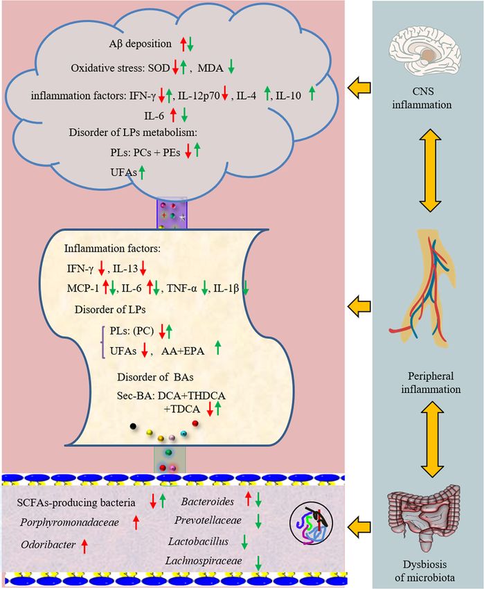
Gu et al. Alzheimer's Research & Therapy   (2021) 13:44                                                         Page 2 of 18

Introduction                                                   expression of amyloid-beta [16, 17]. So the accumulation
Alzheimer’s disease (AD), a chronic neurodegenerative          of Aβ throughout the brain is the failure of microglia
disease, is a major cause of disability and mortality if not   and peripherical monocytes to remove extracellular
effectively treated. Over 40 million people worldwide are      amyloid [18, 19], which reversely activates microglia and
suffering from AD, especially in elderly people [1]. There     releases inflammatory cytokines, such as interleukin-1β
are currently no preventative or disease-modifying treat-      (IL-1β), IL-6 and tumour necrosis factor (TNF-α) [20].
ments available, despite the countless investments that        In addition, they secrete oxidative stress-related mole-
have been made in the war against AD. Beyond all               cules, such as nitric oxide (NO), reactive oxygen species
doubt, the complexity of the aetiology of AD is the big-       (ROS) and superoxide anions [21]. Glucose is normally
gest challenge to overcome this problem. Now, as many          the major energy source for the brain, but in AD, glu-
as 1141 anti-AD drugs worldwide were in development.           cose metabolism dramatically decreases [15, 22–24].
However, only 6 of them stood out and were approved            However, docosahexaenoic acid (DHA) facilitates the
by the Food and Drug Administration (FDA). Regret-             transport of glucose into the brain by regulation of glu-
tably, these drugs are relatively against a single target      cose transporter protein-1 (GLUT1) transporters and re-
and are mainly acetylcholinesterase inhibitors, which is       ducing Aβ plaque aggregation in individuals with
not an optimal choice for AD patients with multi-              moderate dementia and AD [25]. Furthermore, phospho-
pathogenesis. GV-971, a mixture of acidic linear oligo-        lipids (PLs), which act as storage depots from a complex
saccharides ranging from dimers to decamers (molecular         meshwork of lipid mediators in cell membranes, are es-
weight up to ~ 1 kDa) that were approved by the FDA to         pecially important in controlling neuroinflammation
carry out a phase 3 clinical for AD in 2020, suppresses        [26]. PLs influence the formation of Aβ peptides by af-
gut dysbiosis and the associated phenylalanine/isoleucine      fecting membrane proteins, such as APP, and β- and γ-
accumulation, harnesses neuroinflammation and re-              secretase. The decrease of PCs in both the plasma and
verses the cognition impairment [2]. The success of GV-        the brain are associated with impaired cognitive per-
971 in anti-AD reveals that multi-target intervention          formance in elderly people and AD patients [27, 28].
breaks new ground for the drug development of AD.                 It should be noted that neuroinflammation is not
Traditional Chinese medicine (TCM), characterized by           solely restricted to contributions from resident biochem-
multiple components and multiple targets, closely coin-        ical factors in the brain, as perturbations in microbial di-
cides with this strategy.                                      versity are related to propagating neuroinflammation in
   HLJDD, a classical TCM formula used for fever relief        preclinical models of AD. Gut microbiota could affect
and detoxification, consists of Rhizoma coptidis (Rc),         the immune system directly via activation of the vagus
Radix scutellariae (Rs), Cortex phellodendri (Cp) and          nerve [29, 30], which in turn triggers bidirectional com-
Fructus Gardeniae (Fg) in a weight ratio of 3:2:2:3. It has    munication with the CNS and links them to the cogni-
been widely applied to treat cerebrovascular diseases, is-     tive and emotional centres of the brain [31–33]. The
chaemic stroke, and AD in many Asian countries for             results of a recent clinical trial performed on elderly sub-
centuries [3–6]. In our previous studies, the chemical         jects with dementia support the evidence of the role of
profile and haemodynamics of HLJDD were described in           amyloid and related bacterial accumulation in the patho-
detail [7, 8]. Sixty-nine compounds in HLJDD were iden-        genesis of cognitive damage [34]. Additionally, the dys-
tified, including mainly iridoids, alkaloids and flavonoids.   regulation of gut microbiota is responsible for the
Berberine (Ber), baicalin and geniposide were also repre-      increased permeability of intestinal barriers and the
sentative components [7]. Recently, excellent results on       blood-brain barrier (BBB) [35]. Short-chain fatty acids
the pharmacological effects of HLJDD or its components         (SCFAs) produced by bacterial fermentation of dietary
have been achieved in the treatment of AD, involving           carbohydrates can cross the BBB, modulate brain de-
ameliorating cognitive dysfunction, lessening of the           velopment and behaviour and regulate microglia hom-
plaque burden and oxidative stress and altering lipid me-      oeostasis [36–38]. In addition to SCFAs, gut bacteria
tabolism [9, 10]. However, the underlying mechanisms           also produce a range of substances with neuroactivity
of HLJDD on the amelioration of AD are still a mystery.        and immunomodulatory effects, including dopamine,
   Neuroinflammation is a key factor in the neurodegen-        γ-aminobutyric acid (GABA) and histamine [39–41].
erative process of AD [11]. This process involves an ini-      Tryptophan (Trp) is metabolized in the periphery
tial inflammatory stimulus (Aβ, pro-inflammatory               within the gut neurons and enterochromaffin cells,
cytokines, chemokines and the generation of reactive           and centrally in the neurons of the raphe of the brain
oxygen species) that triggers microglia, the resident          stem [42]. Dysregulation of Trp metabolism causes
macrophage within the central nervous system (CNS)             shifts in the balance between the Kyn and 5-HT path-
[12–15]. In addition, monocytes recruited from the per-        ways and is associated with psychiatric and neuro-
iphery can interact with microglia to influence the            logical disorders [43].
Gu et al. Alzheimer's Research & Therapy      (2021) 13:44                                                                               Page 3 of 18

  Bile acids (BAs) are critically important for the                           Plenty of evidence shown that peripheral and central
maintenance of a healthy gut microbiota, a balance                          inflammations play an important role in the pathogen-
between lipid and carbohydrate metabolism, energy                           esis of AD. The hypothesis of “brain-gut” axis will be an
homeostasis and innate immunity [44, 45]. Disturb-                          useful and promising exploration. In this study, we
ance of BAs has been demonstrated in AD progres-                            aimed to reveal the phenotypic features of APP/PS1
sion [46, 47]. Circulating BAs were thought to                              mice as comprehensively as possible, and provide a po-
provide a communication bridge between the gut and                          tential effective, safe and economical intervention strat-
the brain, and their alteration reflects gut dysbiosis                      egy from TCM.
[48–50]. Bile salt hydrolase-rich (BSH) bacteria readily
modify the BA profile. In turn, intestinal BAs control                      Results
the growth and maintenance of commensal bacteria                            Cognitive deficiency in Tg-APP/PS1 mice
[51–53]. Metagenomic analyses have demonstrated                             The cognitive functions of 8-month-old Tg-APP/PS1
that functional BSH is present in all major bacterial                       (Tg) mice were significantly impaired compared to age-
divisions and archaeal species in the human gut, in-                        matched wild-type (WT) mice (Fig. 1). In the navigation
cluding members of Lactobacilli, Bifidobacteria, Clos-                      test, Tg mice normally took longer time to reach the
tridium and Bacteroides [51, 54–56].                                        platform as compared to the WT mice over a 3-day

 Fig. 1 HLJDD ameliorates cognitive deficiency in APP/PS1 mice. a Acquisition of spatial memory was evaluated by the MWM test (n = 11). In the
 navigation test, mice were evaluated for the total time of four trainings spent searching for the platform location during the day. The Tg-APP/PS1
 mice required a longer time than the control group to locate the platform on the training days (P < 0.0001). This significant difference constantly
 appeared from the first day. b In the space probe trial, the distance percentage, time percentage, and times across platform in the target
 quadrant were used for evaluation. All of them were reduced in Tg-APP/PS1 mice. In contrast, the chronic administration of HLJDD for 4 months,
 including H-L and H-H, significantly ameliorated memory and spatial learning deficits of Tg-APP/PS1 mice. c Real-time monitoring of the mouse
 motion track MWM test experiment. WT, wide mice; Tg, Tg-APP/PS1 mice; H-L, HLJDD with low dosage; H-H, HLJDD with high dosage; Ber,
 berberine; Don, donepezil. All data were analysed by one-way ANOVA with Dunnett-test. All the results are expressed as the mean ± SEM; *P <
 0.05, **P < 0.01, ***P < 0.001, ****P < 0.0001
Gu et al. Alzheimer's Research & Therapy (2021) 13:44 Page 4 of 18
training period (P < 0.0001) (Fig. 1a). In the space probe glutamate (Glu) and GABA, and arginine, tyrosine and
test, the times across the platform was reduced to more asparaginate. Simultaneously, L-cysteine decreased after
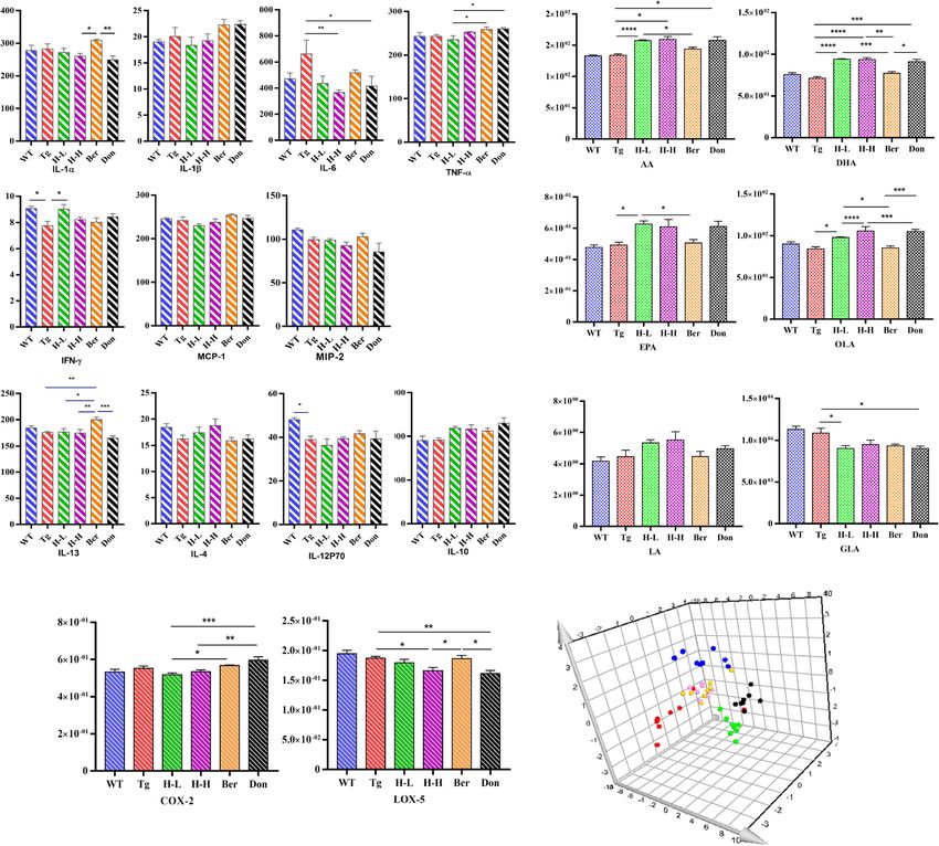
than half of that in WT mice (Fig. 1b). Meanwhile, the intervention with HLJDD. As far as PUFAs (polyunsatur-
trajectory map of Tg mice was disorganized and pur- ated fats) were concerned (Fig. 3b), the overall pattern of
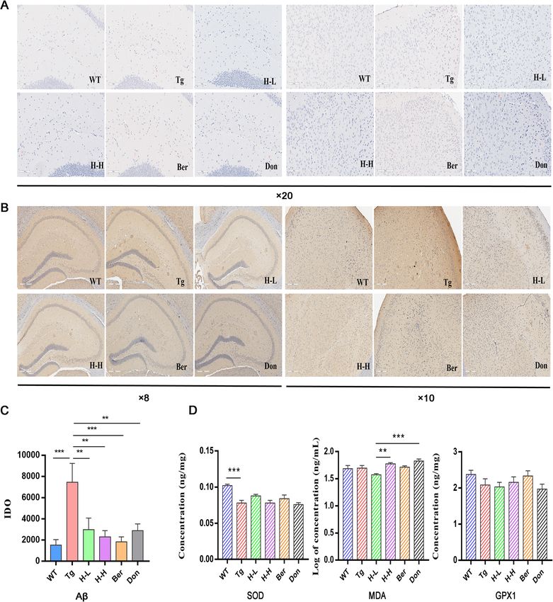
poseless (Fig. 1c). Aβ plaques markedly accumulated in results showed no significant differences in brain PUFAs
the brain cortex and hippocampus of Tg mice compared of Tg mice relative to WT mice. Nevertheless, treatment
with WT mice. Congo red staining has shown that Aβ with HLJDD significantly increased the brain levels of ara-
deposition with higher density and larger area existed in chidonic acid (AA), DHA, eicosapentaenoic acid (EPA),
the hippocampus and cortex of Tg mice than that of linoleic acid (LA) and oleic acid (OLA) and decreased the
WT mice (Fig. 2a). Meanwhile, the results of immuno- content of γ-linolenic acid (GLA). Moreover, HLJDD
histochemistry in the hippocampus and cortex of Tg treatment inhibited cyclooxygenase 2 (COX-2) and 5-
mice were also positive (Fig. 2b, c). Furthermore, the lipoxygenase (5-LOX) expression in the brain of Tg mice
brain contents of SOD dramatically decreased in Tg (Fig. 3c).
mice (P < 0.001) (Fig. 2d). Chronic oral administration of In addition, dysregulation of lipid metabolism in the
H-L (HLJDD with low dosage: 172 mg/kg/day) and H-H brains of Tg mice was confirmed by supervised OPLS-
(HLJDD with high dosage: 344 mg/kg/day) for 4 months DA with the values of R2Y and Q2 (93% and 79%, re-
significantly ameliorated the memory and spatial learn- spectively). In the PLS-DA plots (Fig. 3d), the HLJDD
ing deficits of Tg mice by suppressing the accumulation administration groups were relatively independent and
of Aβ plaques in the cortex and hippocampus. In had no intersection with Tg mice, but were closer to
addition, H-L could increase the level of SOD in the WT mice. In detail, 40 pathological lipid biomarkers in
brain of Tg mice to 1.23-fold and has a tendency to brain tissue were identified (Table 2). The levels of 17/
downregulate MDA. Therefore, we speculated that 21 PCs, 5/5 PEs, 3/3 glucosylceramides (GlcCers), 4/4
HLJDD has the potential to attenuate oxidative stress. ceramides (Cers) and 5/7 sphingomyelins (SMs) were
lower in Tg mice than in WT mice. HLJDD attenuated
The CNS neuroinflammation in Tg-APP/PS1 mice this lesion by increasing the contents of PCs and PEs in
Changes in inflammatory cytokines the brains of mice.
Cytokines of total brain homogenates were measured by
ProcartaPlex Multiplex Immunoassay to further assess The peripheral inflammation in Tg-APP/PS1 mice
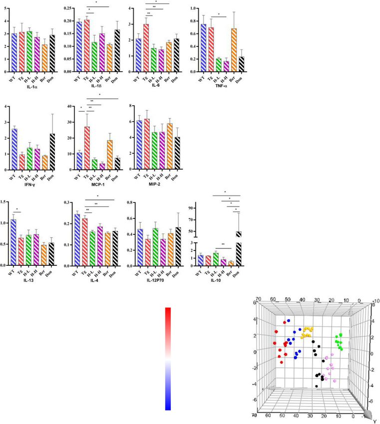
the neuroinflammatory profile (Fig. 3a). Compared with Changes in cytokines
WT mice, IFN-γ and IL-12p70 were significantly de- As shown in Fig. 4a, the levels of IFN-γ, IL-13 and IL-
creased in Tg mice with the content of 7.75 and 38.98 12P70 in the serum of Tg mice (0.96, 0.65 and 0.34 pg/
pg/g. The content of IL-6 in Tg mice was about 1.4 mL, respectively) were lower than these in WT mice
times greater than that in WT mice. After the adminis- (2.58, 1.08 and 0.47 pg/mL, respectively), while the con-
tration of HLJDD, the levels of IFN-γ tended to be a tents of monocyte chemotactic protein 1 (MCP-1, 2.53-
normal value (H-L: 9.02 pg/g; H-H: 8.22 pg/g), and the fold) and IL-6 (1.43-fold) were higher. Meanwhile, we
level of IL-6 observably decreased (P < 0.05). Further- found that the change tendency of IFN-γ, IL-6 and IL-
more, anti-inflammatory cytokine IL-4 and IL-10 both 12p70 in the periphery of Tg mice were consistent with
considerably increased in HLJDD mice compared with those in the brain. Oral administration of HLJDD sup-
Tg mice. pressed pro-inflammatory cytokines IL-1β, IL-6, MCP-1
and TNF-α expression in the serum of Tg mice.
Disorder of endogenous metabolites
Increasing evidence suggests that metabolic perturbations Alteration of endogenous metabolites
in various pathways mediate the occurrence of AD path- An overall level of peripheral omega-6 acid and omega-9
ology as well as the onset of cognitive impairment in pa- acid decreased in Tg mice compared to WT mice
tients. UPLC-QQQ MS/MS was employed to evaluate the (Fig. 4b), including LA (P < 0.05), AA (P < 0.001), EPA
alteration of endogenous metabolites in Tg mice. Overall, (P < 0.05), DHA (reduced by 25%), GLA (reduced by
there was no significant fluctuation in the neurotransmit- 28%) and OLA (P < 0.05). Nevertheless, the levels of
ters (NTs) levels in the brain of Tg mice compared with serum of PUFAs showed no significant changes after
WT mice (Table 1). However, the levels of citrulline and HLJDD. PUFAs are required for maintaining the struc-
methionine in Tg mice were higher than that in WT mice ture, function and vascular integrity of the brain. Non-
(fold: 1.66 and 1.28, respectively). HLJDD significantly in- essential fatty acids are synthesized in the brain, but essen-
creased the levels of NTs in the brain of Tg mice, includ- tial PUFAs (e.g., AA, DHA and EPA) are largely acquired
ing essential amino acids (phenylalanine: Phe, Trp, from the peripheral circulation [3]. Therefore, to investi-
leucine, isoleucine and threonine), proline, choline, gate whether HLJDD affected central PUFAs in Tg mice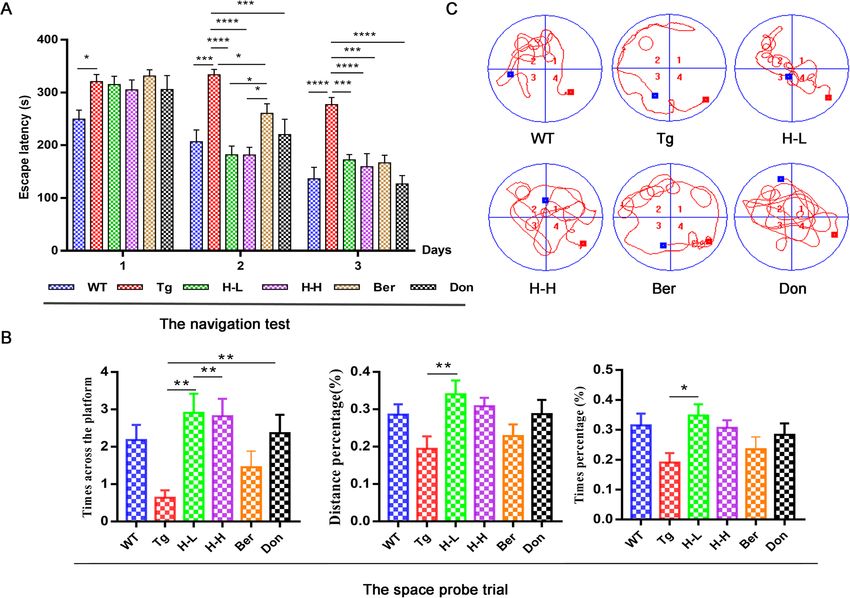
Gu et al. Alzheimer's Research & Therapy       (2021) 13:44                                                                                Page 5 of 18

 Fig. 2 HLJDD reduced Aβ plaque pathology in Tg-APP/PS1 mice. a Congo red staining reveals that Tg-APP/PS1 mice harbour more Aβ plaque with
 higher density and larger area in the hippocampus (left) and cortex (right) than WT mice, × 20 magnification, (n = 3). b Meanwhile, the results of
 immunohistochemistry in the hippocampus (left, × 8 magnification) and cortex (right, × 10 magnification) of Tg-APP/PS1 mice were also positive,
 (n = 3). c The level of Aβ in the CNS of mice was quantified by detecting the value of Integrated option density (IOD). HLJDD significantly suppressed
 the accumulation of A plaques in the cortex and hippocampus. d The levels of brain SOD (n = 5), MDA (n = 6), and GPX1 (n = 6) were detected by ELIS
 A. All data were analysed by one-way ANOVA with Dunnett-test. All the results are expressed as the mean ± SEM; *P < 0.05, **P < 0.01,
 ***P < 0.001, ****P < 0.0001

by regulating peripheral PUFAs, correlation network ana-                      PUFAs (DHA, EPA, LA and GLA) were negatively correl-
lysis was employed based on the measured contents                             ating with those of central PUFAs, and the level of serum
(Fig. 4c). Results showed that the contents of serum                          AA was a positive correlation with that of central AA.
Gu et al. Alzheimer's Research & Therapy     (2021) 13:44                                                                           Page 6 of 18

  A                                                                               B

                                                                                   ng/mg
    Pg/g

                                                                                   ng/mg
     Pg/g
    Pg/g

                                                                                    ng/mg

            C                                                                     D
                                                                                                                                       Don
                                                                                                                                       Ber
                                                                                                                                       H-H
            ng/mg

                                                                                                                                       H-L
                                                                                                                                       Tg
                                                                                                                                       WT

 Fig. 3 HLJDD improved the CNS inflammatory microenvironment in Tg-APP/PS1 mice. a Changes in pro-inflammatory and anti-inflammatory
 factors (n = 5). b Changes in UFAs (n = 6). c Changes in COX-2 and 5-Lox (n = 5). d PLS-DA plot. All data were analysed by one-way ANOVA with
 Dunnett-test. All the results are expressed as the mean ± SEM; *P < 0.05, **P < 0.01. ***P < 0.001, ****P < 0.0001

Dysregulation of lipid metabolism in the serum of Tg mice                 WT mice (Table S1). CA was hardly detected in the per-
was also confirmed by OPLS-DA, with values of R2x and                     iphery of WT mice, but they remained high in the serum
Q2 of 72% and 40%, respectively. The PLS-DA plot                          of AD mice. An increase of tauro-conjugated BAs (taur-
showed that HLJDD groups (including H-L and H-H)                          odeoxycholic acid (TDCA), tauroursodeoxycholic acid
were relatively independent and had no intersection with                  (TUDCA), taurohyodeoxycholic acid (THDCA), tauro-α-
the WT and Tg groups (Fig. 4d), which was similar to the                  muricholic acid (T-α-MCA)) and deoxycholic acid
pattern in the brain. In detail, 13 LP biomarkers in serum                (DCA, almost a 30-fold increase) were observed in Tg
were screened, including 11 PCs, 1 PE and 1 GlcCers                       mice compared with WT. Interestingly, HLJDD signifi-
(Table 3). Almost all serum lipid biomarkers decreased in                 cantly decreased the levels of DCA, T-MCA and THDC
Tg mice compared with WT mice. Interestingly, all of                      A. It has been reported that the ratio between BAs
these biomarkers were reversed by HLJDD.                                  reflected enzymatic activities in the liver and the gut
  Additionally, we found that some significant changes                    microbiome. The cholic acid (CA) to chenodesoxycholic
occurred in peripheral BAs in Tg mice compared with                       acid (CDCA) ratio reflected if a possible shift in BA
Gu et al. Alzheimer's Research & Therapy (2021) 13:44 Page 7 of 18
Table 1 The effect of HLJDD on neurotransmitters in the CNS of Tg-APP/PS1 mice
Changes in NTs (n = 6–8), absolute quantification (μg/g); the remaining NTs were measured in relative quantities (AS/AI); colour coded according to the contents.
The colour from light to dark represents the content from low to high. All data were analysed by one-way ANOVA with Dunnett-test. All the results are expressed
as the mean ± SEM; *P < 0.05, **P < 0.01. ***P < 0.001, ****P < 0.0001 (comparing with WT mice); #P < 0.05, ##P < 0.01. ###P < 0.001, ####P < 0.0001 (comparing with
Tg mice)
synthesis from the primary to the alternative BA path- mice. However, HLJDD could shift the gut microbiota
way has occurred in the liver. Ratios of secondary to pri- composition. At the phylum level (Fig. S1f), Tg mice ex-
mary BAs was used to assess the activity of intestinal hibited Firmicutes populations more abundantly than
microbiome enzymes which influenced the production WT (41% and 35%, respectively), as previously reported
of secondary BAs [47]. In this study, the ratio of CA: [57, 58], while the Bacteroidetes population was lower
CDCA and the secondary to primary BAs were higher in (54%, 60% respectively) [59]. Treatment with HLJDD in
Tg mice than those in WT mice, which were signifi- Tg mice decreased the population of Firmicutes and in-
cantly reversed by HLJDD. creased the population of Proteobacteria. At the family
level (Table 4), higher relative abundances of Lachnos-
Alteration of the gut microbiota in Tg-APP/PS1 mice piraceae (1.24-fold), Rikenellacae (1.3-fold) and Porphyr-
Pyrosequencing was employed to monitor the faecal omonadaceae (2.24-fold) were observed in Tg mice than
microbiota composition in Tg mice. Bacterial richness those in WT, while the relative abundances of Bacteroi-
and α-diversity in Tg mice showed no significant differ- dales_S24-7_group, Coriobacteriaceae and Alcaligen-
ences relative to WT, as demonstrated by the rarefaction aceae were lower (Tg: 4.13%, 0.13%, 0.16%; WT: 5.37%,
curve, Chao index, and Shannon index (Fig. S1a-c). 0.29%, 0.58%). Treatment with HLJDD increased the
Moreover, HLJDD treatment did not affect the diversity relative abundances of Prevotellaceae, Lactobacillaceae,
of microflora in Tg mice, but Ber treatment remarkably Peptococcaceae, Alcaligenaceae and Helicobacteraceae
decreased the levels of the Shannon index. According to and reduced the relative abundances of Bacteroidales_
ANOSIM analysis, the intra-group difference was less S24-7_group, Lachnospiraceae and Porphyromonadaceae.
than the inter-group difference. (Fig. S1d). For PLS-DA At the genus level (Table 5), the relative abundances of
analysis (Fig. S1e), the faecal microbiota composition of unidentified, Parasutterella, Blautia, Lachnospiraceae-
Tg mice was significantly different from that of WT UCG-001 and Ruminococcaceae-UCG-014 were lower inGu et al. Alzheimer's Research & Therapy (2021) 13:44 Page 8 of 18
Table 2 The metabolic changes of 40 potential lipids markers in the CNS of Tg mice
Colour coded according to the fold change, n = 8; Colour bar: ; tR: time retention; Actua M: actual mass; All the results are expressed as mean ± SEM;
All data were analysed by one-way ANOVA with Dunnett-test; *P < 0.05, **P < 0.01, ***P < 0.001, ****P < 0.0001 (comparing with the WT group), #P < 0.05, ##
P < 0.01,
### ####
P < 0.001, P < 0.0001 (comparing with the Tg group)Gu et al. Alzheimer's Research & Therapy (2021) 13:44 Page 9 of 18
A B 3 × 10 03 6 ×10 03
ng/mL
2 × 10 03 4 ×10 03
Pg/mL
1 × 10 03 2 ×10 03
0 0
WT Tg H-L H-H B e r Don WT Tg H-L H-H B e r Don
AA DHA
8 × 10 02 3 ×10 04
6 × 10 02
ng/mL
2 ×10 04
Pg/mL
4 × 10 02
1 ×10 04
2 × 10 02
0 0
WT Tg H-L H-H B e r Don C on Mod H-L H-H B e r Don
EPA OLA
5 × 10 04 6 ×10 00
4 × 10 04
4 ×10 00
3 × 10 04
ng/mL
Pg/mL
2 × 10 04
2 ×10 00
1 × 10 04
0 0
WT Tg H-L H-H B e r Don WT Tg H-L H-H B e r Don
LA GLA
C S-AA/B-AA
D
*
0.5
WT
S-DHA/B-DHA *
Tg
S-EPA/B-EPA
0
H-L
S-LA/B-LA * H-H
S-OLA/B-OLA Ber
-0.5
S-GLA/B-GLA
Don
H-L H-H Be r Don
Fig. 4 HLJDD improved the peripheral inflammatory microenvironment in Tg-APP/PS1 mice. a Changes in pro-inflammatory and anti-inflammatory
factors (n = 4–5); b Changes in UFAs (n = 6). c Correlation analysis of the CNS UFAs and the peripheral UFAs in each drug treatment group. The colour
scale illustrates the magnitude of correlation between the examined indexes on the plot. d PLS-DA plot. All data were analysed by one-way ANOVA
with Dunnett-test. All the results are expressed as the mean ± SEM; *P < 0.05, **P < 0.01
Tg mice (0.13%, 1.28%, 0.36% respectively) than those in specific altered bacterial phenotype. A total of 44 bac-
WT mice (0.5%, 2.35%, 0.69% respectively), while the teria changed significantly among the six groups, with
abundances of Lachnospiraceae_NK4A136_ group (1.5- a linear discriminant analysis (LDA) score log 10 > 3
fold), Bacteroides (5.4-fold) and Odoribacte (1.3-fold) (Fig. S2).
were higher. However, treatment of Tg mice with
HLJDD increased Prevotellaceae_UCG_001, Lactobacil- Phenotypic features of Tg-APP/PS1 mice after
lus, Helicobacter, Lachnospiraceae-UCG-001, Tyzzerella- administration of Ber
3, Ruminococcaceae-UCG-014 and Parasutterella, and In general, Ber was not as effective as HLJDD in
reduced the relative abundances of g-unidentified, Lach- ameliorating cognitive deficiency in Tg mice, which
nospiraceae_NK4A136_group, Bacteroides, Roseburia, was reflected in its poor performance in the MWM
Anaerotruncu, Lachnospiraceae_FCS020_group and task and more Aβ plaques in the CNS. In terms of
Odoribacte. In addition, LEfSe was used to identify the the therapeutic mechanism, the differences betweenGu et al. Alzheimer's Research & Therapy (2021) 13:44 Page 10 of 18
Table 3 The metabolic changes of 13 potential lipids markers in the periphery of Tg mice
Colour coded according to the fold change, n = 10; Colour bar: ; tR: time retention; Actua M: actual mass; All the results are expressed as mean ± SEM;
All data were analysed by one-way ANOVA with Dunnett-test; *P < 0.05, **P < 0.01, ***P < 0.001, ****P < 0.0001 (comparing with the WT group), #P < 0.05, ##
P < 0.01,
### ####
P < 0.001, P < 0.0001 (comparing with the Tg group)
Table 4 Relative abundance of the top 20 bacteria family among six groups
Colour coded according to the fold change (F). Colour bar: ; n = 11; Since the relative abundance of f_Rickettsiales_Incertae_Sedis in Tg group
was zero, its fold change between groups were not calculated; ND: not detected; : Less than 10 times; :more than 10 times; All the results are
expressed as mean ± SEMGu et al. Alzheimer's Research & Therapy (2021) 13:44 Page 11 of 18 Table 5 Relative abundance of the top 20 bacteria genus among six groups Colour coded according to the fold change (F). Colour bar: ; n = 11; Since the relative abundance of f_Rickettsiales_Incertae_Sedis in the Tg group was zero, its fold change between groups were not calculated; : Less than 10 times; :more than 10 times; All the results are expressed as mean ± SEM Ber and HLJDD were mainly manifested in the regu- regulating gut microflora was different from that of lation of neuroinflammation, lipid metabolism, and HLJDD (Tables 4, 5). After Ber administration, the the gut microbiota. In detail, HLJDD could upregulate proportion of special enterobacterium (Prevotellaceae, a variety of neurotransmitters, which was similar to Odoribacter, Tyzzerella_3, Alloprevotella, Lachnospira- Don. Ber mainly regulated citrulline, urea, and me- ceae_UCG-001 and Prevotellaceae_UCG-001) in Tg- thionine. The effects of Ber on central and peripheral APP/PS1 mice decreased sharply. Moreover, it was inflammatory cytokines were not as good as HLJDD. found that Ber had the opposite regulatory effect But Ber significantly increased the levels of IL-13 in compared with HLJDD on Prevotellaceae, Clostridia- the brain. Combining the performance of the lipid ceae, Bacteroidaceae, Lactobacillaceae, Bacteroidales_ metabolic profiling of the brain with that of serum, S24-7_group, Lachnoclostridium, Tyzzerella_3, Allopre- these plot for PLS-DA presented clear separation be- votella, Lachnospiraceae_UCG-001, Bacteroides, Lacto- tween HLJDD (including H-L and H-H group) and bacillus and Prevotellaceae_UCG-001. Ber groups (Figs. 3d, 4d). As shown in the Venn plots (Fig. S3a), PC (O-18:0/16:0) and Cer (d18:1/24:1) were the characteristic markers of the HLJDD groups in Discussion the CNS. PC (18:1/17:0), PC (16:0/18:1), PE (22:6/0:0), In the present study, the central neuroinflammation was PE (18:0/18:1) and PE (0:0/18:1) were the characteris- closely related to the onset of AD. The disorders in LPs tic markers of the HLJDD groups in the periphery. metabolism and intestinal flora were the potential PC (18:0/18:2) was the characteristic marker of the drivers (Fig. 5). 16S rRNA gene sequencing of the gut Ber group (Fig. S3b). Moreover, we found that Ber microbiome and integrated metabolomics were adopted was similar to HLJDD in regulating the CNS patho- to monitor the phenotypic features of APP/PS1 mice. logical lipids, but its effect on peripheral pathological The disorder of both biochemical factors in the brain lipids was not as good as HLJDD. Compared with and the microbial diversity in intestine contributed to HLJDD, Ber had a weaker regulatory effect on DHA, the neuroinflammation. HLJDD could reverse them and EPA and OLA. In addition, the mechanism of Ber in then improve the cognitive impairment in Tg mice.
Gu et al. Alzheimer's Research & Therapy   (2021) 13:44                                                      Page 12 of 18

  The elevation of Aβ level induced the overexpression          PUFAs act as precursors for biosynthesis of the lipid
of pro-inflammatory cytokines and chemokines to robust       mediators, which are actively involved in the inflamma-
inflammatory response [20]. The more severe inflamma-        tory response. We found that the levels of brain PUFAs
tory degree was found in the brain of Tg mice than that      had no significant difference in Tg mice compared to
in WT mice, followed with Aβ plaques accumulation            WT mice. Others have shown that the brain PUFAs dif-
and abnormal expression of inflammatory cytokines,           fer very little in AD compared to healthy people [78–
such as IFN-γ, IL-12p70 and IL-6. Meanwhile, the ele-        80]. However, the contents of PUFAs and OLA in the
vated level of Aβ42 was closely associated with the in-      periphery of Tg mice decreased significantly compared
creased level of oxidative stress [60], which was mainly     with those of WT mice. A higher level of AA is strongly
reflected in the decrease of SOD and the increase of me-     associated with AD by yielding some inflammatory me-
thionine in Tg-APP/PS1 mice. Methionine could in-            diators [81, 82]. Conversely, some study still believes that
crease the levels of Aβ and nitro-tyrosinated protein,       a lower level of AA is associated with cognitive decline
which further induced neuroinflammation [61, 62]. In         [83, 84]. In this study, the level of AA in the periphery
addition, the evidence showed that inflammation in Tg        of Tg mice decreased compared with WT mice, which
mice was not only confined to the CNS but also spread        was attributed to the decline of upstream molecules, in-
to the periphery. Compared with WT mice, both IFN-γ          cluding OLA, LA and GLA. LA, GLA and OLA partici-
and IL-6 in the CNS and periphery of Tg mice showed          pate in numerous cellular functions by affecting
significant changes. Upregulation of IL-6 was predictive     membrane fluidity, membrane enzymatic activities and
of progression to AD, which was verified in various AD       eicosanoid synthesis [84].
models and patients with varying degrees of AD [9, 63].         The present study in Tg mice indicated that alterations
Study reported that INF-γ has dual roles in Alzheimer’s      in the gut microbiome contributed to neuroinflamma-
disease [64]. Browne et al. reported that the release of     tion. Study reported that the circulating omega-3 fatty
INF-γ from infiltrating Th1 cells plays a diabolical role    acid (DHA, EPA) can influence the composition of the
in AD pathogenesis. There was still reports that INF-γ at    host gut flora, especially SCFA-producing bacteria [85].
the brain’s choroid plexus were reduced in brain ageing      SCFA-producing bacteria decreased in Tg mice com-
[65, 66] and under neurodegenerative condition [67].         pared with the WT mice, including Parasutterella and
The availability of INF-γ at the brain of AD mice might      Blautia. SCFAs can interact with nerve cells by stimulat-
be affected by systemic immune suppression. Mean-            ing the sympathetic and autonomic nervous system [86,
while, the higher level of MCP-1 in the serum was found      87]. SCFAs even cross the BBB and regulate microglial
in Tg mice compared with WT mice, which appeared to          homeostasis. A decrease of SCFA-producing bacteria ag-
be associated with greater severity and a faster cognitive   gravated cognitive decline. Porphyromonadaceae is
decline [68, 69].                                            highly associated with inflammatory diseases [88, 89],
  Many Aβ-produced proteins have been found in lipid         and even induces cognitive decline and anxiety-like be-
rafts such as Aβ protein precursor (βAPP), β-secretase,      haviour [90]. Increases in bacterial taxa from this family
γ- secretase and neprilysin [70]. In addition, Aβ directly   have also been observed in faecal samples from individ-
disrupt bilayer integrity by interacting with PLs [71].      uals with major depressive disorders, especially Odori-
Data from targeted lipidomics showed that Tg mice were       bacter [91]. An increase in intestinal Odoribacter was
characterized by a decrease in PCs, PEs and SMs of the       certified in a variety of AD models [58, 90, 92]. Likewise,
CNS or periphery. Previous evidence has shown that a         we observed that the proportion of Porphyromonadaceae
reduced concentration of PCs in the CNS and peripheral       and Odoribacter increased in Tg mice compared with
system was associated with impaired cognitive perform-       WT mice, which expected to be a potential biomarker to
ance in older individuals and AD patients [27, 28, 72].      predict the occurrence of AD.
Among them, PC (16:0/16:0) was predictive of progres-           Circulating BAs provide an important mechanism for
sion to AD dementia in individuals with mild cognitive       communication between the gut and the brain [48, 93,
impairment (MCI) [73, 74]. Oxidative and lipid peroxi-       94]. Alterations in the gut microbiome significantly
dation were early events in AD [75]. In this study, the      affect BAs transformation through various microbial en-
results of PEs, PCs and SOD were in accordance with          zymes such as bile salt hydrolase (BSHs) and hydroxyste-
the previous paper, which suggested the implication of       roid dehydrogenases [95]. Compared with WT mice, an
oxidative stress in the progressive degradation of brain     increase of tauro-conjugated BAs was observed in Tg-
PLs in AD. PCs and PEs were rich in readily oxidizable       APP/PS1 mice, probably a result of lower BSHs activity
AAs and DHAs [76, 77]. We speculated that the meta-          from Lactobacillus. Indeed, Lactobacillus have BSHs ac-
bolic disorder of PC and PE induced the abnormal ex-         tivity for catalyzing deconjugation of tauro-conjugated
pression of Aβ by breaking down the lipid rafts              BAs [96]. Increased amounts of secondary BAs in the
homeostasis, which deteriorated neuroinflammation.           blood may enter the brain through the permeability of
Gu et al. Alzheimer's Research & Therapy     (2021) 13:44                                                                            Page 13 of 18

 Fig. 5 Schematic diagram of gut-brain axis in AD progression and the intervention strategy. During AD progression, peripheral inflammation was
 highly correlated with neuroinflammation suggests that altered systemic inflammatory markers reflect neurodegenerative disease. HLJDD attenuated
 brain damage and memory deficits in Tg-APP/PS1 mice by coordinating CNS inflammation and peripheral inflammation. Red arrow: represent a major
 change in Tg mice. Green arrow: represent a major change that occurs after the HLJDD group
Gu et al. Alzheimer's Research & Therapy   (2021) 13:44                                                     Page 14 of 18

the BBB and affect brain physiology and metabolism           integrity of the host, BA, glucose and lipid metabolism
[97]. DCA represent aetiological agents and mediators of     [107]. In addition, HLJDD could remodel serum BAs
the pathogenic process [98, 99]. A higher level of DCA       homeostasis in Tg mice by decreasing the populations of
was significantly associated with worse cognitive per-       Lachnospiraceae and its followed genera and increasing
formance [100]. The decline of CA level is thought to be     the populations of Bacteroides and Lactobacillus. Bacter-
one of the causes of cognitive impairment in AD pa-          oides and Lactobacillus were reported to be actively in-
tients [47]. The decline of CA in Tg-APP/PS1 mice may        volved in BAs metabolism, including deconjugation, the
be caused by species difference [47, 100]. In addition,      hydroxyl groups at C3, C7 and C12, and desulfatation
the ratios of secondary to primary BAs increased in Tg       [108]. The ratios of secondary to primary BAs changed
mice relative to WT mice. This indicated that the en-        in Tg mice, which proved that HLJDD affected BAs me-
zymatic steps in the conversion of primary to secondary      tabolism by alternating microbiota structure. The ratio
BAs in the gut might contribute to disease.                  of secondary BAs to primary BAs is used to assess differ-
   The present study demonstrated that HLJDD with            ences in the activity of intestinal microbiome enzymes
strict quality control could reverse cognitive impairment    that can induce BAs changes [47].
in Tg mice. It is worth mentioning that HLJDD de-
creased the expression of IL-6 in both of brain and
serum of Tg mice. Upregulation of IL-6 forced microglia      Limitations
polarization to pro-inflammatory and neurodegeneration       This study had some limitations. Only MWM test was
[101]. In addition, the regulation of HLJDD on central       performed to assess short term memory which is im-
NTs was similar to that of Don. The previous literature      paired in AD. Actually, various behavioural assessments
reported that HLJDD ameliorates the cognitive dysfunc-       were indeed the best strategy for the comprehensive
tion by regulating LP metabolism [9]. The present study      evaluation of memory. In addition, although we have re-
indicated that HLJDD could relieve the neuroinflamma-        vealed existing communication disorders of the “brain-
tion by increasing the levels of PCs, PEs, DHA and EPA.      gut” axis in the pathogenesis of AD, further studies to
In the correlation network analysis, GLA, OLA, LA, EPA       identify the key bacterial strains and functional enzymes
and DHA were negatively correlated at the central and        that may account for metabolites and neuroinflamma-
peripheral levels after administration of HLJDD. How-        tion changes will be critical. Further research on these
ever, the contents of AA in periphery showed a positive      key issues may help gain a better understanding of the
association with that in the CNS. These results sug-         pathogenesis of AD and perfectly elucidate the potential
gested that HLJDD regulated the performance of central       mechanism of HLJDD in the treatment of AD.
PUFAs by alleviating peripheral PUFAs, since these es-
sential PUFAs in the brain are primarily derived from
the peripheral circulation [102]. In addition, we observed   Conclusion
that HLJDD effectively reshaped the gut microbiota           In the present study, neuroinflammation was closely re-
structure in Tg mice. HLJDD enriched the gut micro-          lated to the onset of AD, and disorders in LP metabol-
biota SCFA-producing bacteria population. The circulat-      ism and intestinal flora, which were the potential
ing DHA was positively correlated with SCFA-producing        drivers. Peripheral inflammation displayed the crosstalk
bacteria. Therefore, we speculated that HLJDD may alter      with CNS inflammation and cognitive impairment. The
the microbiota composition by affecting the levels of        combination of inflammatory factors (IL-6 and INF-γ),
DHA in Tg mice. Meanwhile, HLJDD could increase the          PCs and SCFA-producing bacteria was a promising early
populations of Prevotellaceae and its genus, including       diagnostic biomarker for AD, which worths further val-
Prevotellaceae_UCG-001, Prevotellaceae_Ga6A1_group           idation in numerous experiments. HLJDD suppressed
and Parasutterella. These bacteria were reported to be       gut dysbiosis and the associated Aβ accumulation,
reduced in PD patients and Tg mice [103, 104]. Prevo-        harnessed neuroinflammation and reversed cognitive
tella is known to breakdown complex carbohydrates,           impairment. More importantly, the identified anti-
providing SCFAs as well as thiamine and folate as by         inflammatory actions of HLJDD will open a new thera-
products that promote a healthy intestinal environment.      peutic avenue for AD treatment through an intervention
The decrease of Prevotella may reduce the levels of these    pattern in the “brain-gut” axis and guide the future devel-
important micronutrients.                                    opment of effective therapies by the central-peripheral
   The previous study demonstrated that HLJDD re-            contrast study. Further research should focus on the
stored the dysregulated microbiota structure and func-       causal relationship between the peripheral and central in
tion by increasing SCFA-producing bacteria in T2DM           AD models and patients, which may help gain a better un-
rats [105]. Lachnospiraceae can induce cognitive defect      derstanding of the pathogenesis of AD and the potential
in AD patients [106] by intervening in the mucosal           mechanism of HLJDD in the treatment of AD.
Gu et al. Alzheimer's Research & Therapy            (2021) 13:44                                                                                   Page 15 of 18

Supplementary Information                                                         Chinese Materia Medica, China Academy of Chinese Medical Sciences for
The online version contains supplementary material available at https://doi.      providing infrastructure.
org/10.1186/s13195-021-00779-7.
                                                                                  Authors’ contributions
 Additional file 1. Materials and Methods.                                        Conceived and designed the experiments: Haiyu Zhao, Baolin B. Wrote the
                                                                                  paper: Xinru Gu, Haiyu Zhao. Performed the experiments: Xinru Gu, Junyi
 Additional file 2: Table S1. Levels and ratios of Bile acids measured in         Zhou, Yanyan Zhou, Xiaorui Fan and Wenya Gao. Analysed the data: Xinru
 the Tg mice (n = 5). Table S2. Peak areas of three main compounds in             Gu, Hongjie Wang, Nan Si, Wei Ren and Wei Zhao. Contributed to reagents/
 HLJDD. Table S3. The selected detecting ions, Declustering potential             materials/analysis tools: Jian Yang, Hongjie Wang. All authors read and
 (DP), and collision energy (CE) of neurotransmitters. Table S4. The              approved the final manuscripts.
 regression equations and linear range of NTs. Table S5. The selected
 detecting ions, declustering potential (DP), and collision energy (CE) of        Funding
 UFAs. Table S6. The regression equations and linear range of UFAs.               This work was financially supported by the National Natural Science Fund
 Table S7. The selected detecting ions, declustering potential (DP), and          Project under grants [NO.81974523] and [NO. 81573967], and National
 collision energy (CE) of BAs. Table S8. The regression equations and             Science and Technology Major Projects “Major New Drugs Innovation and
 linear range of BAs. Fig. S1. HLJDD altered the overall gut microbiota           Development” (No. 2019ZX09201005-006-002 and No. 2019ZX09201005-002-
 structure in Tg mice (n = 11). (a) Rarefaction analyses; (b) Chao1 index         005).
 (data expressed as mean ± SEM); (c) Shannon index (data expressed as
 mean ± SEM); (d) ANOSIM analysis, R-value > 0 means the intra group dif-
                                                                                  Availability of data and materials
 ference was less than the inter group difference. (e) PLS-DA analyses; (f)
                                                                                  The datasets used and/or analysed during the current study are available
 Bacterial taxonomic profiling in the phylum level of gut microbiota. #P <
                                                                                  from the corresponding author on reasonable request.
 0.05 (comparing with the Mod group). Fig. S2. LEfSe rank plots of differ-
 entially abundant microbial clades in gut microbiome (n = 11). LDA scores
                                                                                  Ethics approval and consent to participate
 (log 10) for differentially abundant microbial clades in stool among six
                                                                                  All experiments and animal care in this study were conducted in accordance
 groups and the threshold on the logarithmic LDA score for discriminative
                                                                                  with the National Institute of Health guide for the care and use of laboratory
 feature is > 3.0. Fig. S3. Venn diagram of the brain (a) and serum (b)
                                                                                  animals (NIH Publications No. 8023, revised 1978), and the Provision and
 showed the shared and unique correlated lipid compounds, in which
                                                                                  General Recommendation of Chinese Experimental Animals Administration
 each ellipse represented the potential lipid markers based on compari-
                                                                                  Legislation.
 sons of the Tg group vs. drug treatment groups. Fig. S4. Profile of HPLC-
 UV chromatograms of mixed standard (a), HLJDD-1st (b) and HLJDD-7th
 (c) . peaks: 1. Geniposide; 2. Beiberine; 3. Baicalin. The chemical pattern of   Consent for publication
 HLJDD aqueous did not change significantly after 7 days of preparation.          Not applicable.

                                                                                  Competing interests
Abbreviations                                                                     The authors declare that they have no competing interests.
5-HT: Serotonin; 5-LOX: 5-Lipoxygenase; AA: Arachidonic acid;
AD: Alzheimer’s disease; ApoE: Apolipoprotein E; APP: Amyloid precursor           Author details
protein; Aβ: Amyloid β-protein; BA: Bile acid; BBB: Blood-brain barrier;          1
                                                                                   Institute of Chinese Materia Medica, China Academy of Chinese Medical
BSH: Bile salt hydrolase-rich; Ber: Berberine; CA: Cholic acid;                   Sciences, Beijing 100700, China. 2Drug Research Center of Integrated
CDCA: Chenodesoxycholic acid; Cer: Ceramide; CNS: Central nervous system;         Traditional Chinese and Western Medicine, Affiliated Traditional Chinese
COX-2: Cyclooxygenase 2; Cp: Cortex phellodendri; DCA: Deoxycholic acid;          Medicine Hospital, Southwest Medical University, Luzhou 646000, China.
                                                                                  3
DHA: Docosahexaenoic acid; Don: Donepezil; ELISA: Enzyme-linked                    Center for Drug Evaluation, National Medical Products Administration,
immunosorbent assay; EPA: Eicosapentaenoic acid; FDA: Food and Drug               Beijing 100022, China.
Administration; Fg: Fructus Gardeniae; GABA: γ-Aminobutyric acid; GLA: γ-
Linolenic acid; GlcCers: Glucosylceramides; Glu: Glutamate; GLUT1: Glucose        Received: 11 May 2020 Accepted: 25 January 2021
transporter protein-1; GPX-1: Glutathione peroxidase 1;
HETEs: Hydroxyeicosatetraenoic acids; HLJDD: Huanglian Jiedu decoction; H-
L: HLJDD with low dosage; H-H: HLJDD with high dosage; Rs: Radix                  References
scutellariae; Rc: Rhizoma coptidis; IBD: Inflammatory bowel disease; IFN-         1. Alzheimer’s Association. 2016 Alzheimer’s disease facts and figures.
γ: Interferon-γ; IL-1β: Interleukin-1β; IS: Internal standard; Kyn: Kynurenine;       Alzheimers Dement. 2016;12:459–509.
LA: Linoleic acid; LCA: Lithocholic acid; LDA: Linear discriminant analysis;      2. Wang X, Sun G, Feng T, Zhang J, Huang X, Wang T, et al. Sodium
LTs: Leukotrienes; MCI: Mild cognitive impairment; MCP-1: Monocyte                    oligomannate therapeutically remodels gut microbiota and suppresses gut
chemotactic protein 1; MDA: Malonic acid; MIP-2: Murine microphage                    bacterial amino acids-shaped neuroinflammation to inhibit Alzheimer's
inflammatory protein-2; MWM: Morris water maze; NO: Nitric oxide; OPLS-               disease progression. Cell Res. 2019;29:787–803.
DA: Orthogonal partial least squares discriminant analysis;                       3. Kondo Y, Kondo F, Asanuma M, Tanaka K, Ogawa N. Protective effect of
PC: Phosphatidylcholine; PE: Phosphatidyl ethanolamine;                               oren-gedoku-to against induction of neuronal death by transient cerebral
PEMT: Phosphatidylethanolamine N-methyltransferase; PG: Prostaglandin;                ischemia in the C57BL/6 mouse. Neurochem Res. 2000;25:205–9.
Phe: Phenylalanine; PLA2: Phospholipase A2; PLs: Phospholipids; PLS-              4. Xu J, Murakami Y, Matsumoto K, Tohda M, Watanabe H, Zhang S, et al.
DA: Partial least squares discriminant analysis; ROS: Reactive oxygen species;        Protective effect of Oren-gedoku-to (Huang-Lian-Jie-Du-Tang) against
SCFAs: Short-chain fatty acids; SMs: Sphingomyelins; SOD: Superoxide                  impairment of learning and memory induced by transient cerebral ischemia
dismutase; T2DM: Type 2 diabetes mellitus; TCA: Taurocholate;                         in mice. J Ethnopharmacol. 2000;73:405–13.
TCDCA: Taurochenodeoxycholate acid; TCM: Traditional Chinese medicine;            5. Fang H, Wang Y, Yang T, Ga Y, Zhang Y, Liu R, et al. Bioinformatics analysis
TDCA: Taurodeoxycholic acid; Tg mice: APP/PS1 transgenic mice;                        for the antirheumatic effects of huang-lian-jie-du-tang from a network
THDCA: Taurohyodeoxycholic acid; TNF-α: Tumour necrosis factor α;                     perspective. Evid Based Complement Alternat Med. 2013;2013:245357.
Trp: Tryptophan; TUDCA: Tauroursodeoxycholic acid; T-α-MCA: Tauro-α-              6. Okamoto H, Chino A, Hirasaki Y, Ueda K, Iyo M, Namiki T. Orengedoku-to
muricholic acid; UDCA: Ursodeoxycholic acid; PUFAs: Polyunsaturated fatty             augmentation in cases showing partial response to yokukan-san treatment:
acids; WT: Wild mice                                                                  a case report and literature review of the evidence for use of these Kampo
                                                                                      herbal formulae. Neuropsychiatr Dis Treat. 2013;9:151–5.
Acknowledgements                                                                  7. Yang Y, Wang HJ, Yang J, Brantner AH, Lower-Nedza AD, Si N, et al.
Thanks to Beijing Animals-Science Biotechnology Co., Ltd. for helping raise           Chemical profiling and quantification of Chinese medicinal formula Huang-
animals, Thanks for the technical guidance of Basic Medical Experimental              Lian-Jie-Du decoction, a systematic quality control strategy using ultra high
Teaching Center of Capital Medical University. Thanks to the Institute of             performance liquid chromatography combined with hybrid quadrupole-
Gu et al. Alzheimer's Research & Therapy (2021) 13:44 Page 16 of 18
orbitrap and triple quadrupole mass spectrometers. J Chromatogr A. 2013; 32. Mayer EA, Tillisch K. The brain-gut axis in abdominal pain syndromes. Annu
1321:88–99. Rev Med. 2011;62:381–96.
8. Ren W, Zuo R, Wang YN, Wang HJ, Yang J, Xin SK, et al. Pharmacokinetic- 33. Grenham S, Clarke G, Cryan JF, Dinan TG. Brain-gut-microbe communication
pharmacodynamic analysis on inflammation rat model after oral in health and disease. Front Physiol. 2011;2:94–109.
administration of Huang Lian Jie Du decoction. PLoS One. 2016;11:e0156256. 34. Cattaneo A, Cattane N, Galluzzi S, Provasi S, Lopizzo N, Festari C, et al.
9. Sun LM, Zhu BJ, Cao HT, Zhang XY, Zhang QC, Xin GZ, et al. Explore the effects Association of brain amyloidosis with pro-inflammatory gut bacterial taxa
of Huang-Lian-Jie-Du-Tang on Alzheimer’s disease by UPLC-QTOF/MS-based and peripheral inflammation markers in cognitively impaired elderly.
plasma metabolomics study. J Pharm Biomed Anal. 2018;151:75–83. Neurobiol of Aging. 2017;49:60–8.
10. Durairajan SSK, Iyaswamy A, Shetty SG, Kammella AK, Malampati S, et al. A 35. Quigley EMM. Microbiota-brain-gut axis and neurodegenerative diseases.
modified formulation of Huanglian-Jie-Du-Tang reduces memory Curr Neurol Neurosci Rep. 2017;17:94–103.
impairments and beta-amyloid plaques in a triple transgenic mouse model 36. Macfabe DF, Cain NE, Boon F, Ossenkopp KP, Cain DP. Effects of the enteric
of Alzheimer’s disease. Sci Rep. 2017;7:6238–51. bacterial metabolic product propionic acid on object-directed behavior,
11. Tejera D, Mercan D, Sanchez-Caro JM, Hanan M, Greenberg D, Soreq H, social behavior, cognition, and neuroinflammation in adolescent rats:
et al. Systemic inflammation impairs microglial Abeta clearance through relevance to autism spectrum disorder. Behav Brain Res. 2011;217:47–54.
NLRP3 inflammasome. EMBO J. 2019;38:e101064. 37. Macfabe DF. Short-chain fatty acid fermentation products of the gut
12. Calsolaro V, Edison P. Neuroinflammation in Alzheimer’s disease: current microbiome: implications in autism spectrum disorders. Microb Ecol Health
evidence and future directions. Alzheimers Dement. 2016;12:719–32. Dis. 2012;23:19260–84.
13. Da Mesquita S, Louveau A, Vaccari A, Smirnov I, Cornelison RC, Kingsmore 38. Erny D, Hrabě de Angelis AL, Jaitin D, Wieghofer P, Staszewski O, David E,
KM, et al. Publisher correction: functional aspects of meningeal lymphatics et al. Host microbiota constantly control maturation and function of
in ageing and Alzheimer’s disease. Nature. 2018;560:185–91. microglia in the CNS. Nat Neurosci. 2015;18:965–77.
14. Subhramanyam CS, Wang C, Hu Q, Dheen ST. Microglia-mediated 39. Tsavkelova EA, Botvinko IV, Kudrin VS, Oleskin AV. Detection of
neuroinflammation in neurodegenerative diseases. Semin Cell Dev Biol. neurotransmitter amines in microorganisms with the use of high-
2019;94:112–20. performance liquid chromatography. Dokl Biochem. 2000;372:115–7.
15. Butterfield DA, Halliwell B. Oxidative stress, dysfunctional glucose 40. Barrett E, Ross RP, O'Toole PW, Fitzgerald GF, Stanton C. γ-Aminobutyric acid
metabolism and Alzheimer disease. Nat Rev Neurosci. 2019;20:148–60. production by culturable bacteria from the human intestine. J Appl
16. Saresella M, Marventano I, Calabrese E, Piancone F, Rainone V, Gatti Andrea, Microbiol. 2012;113:411–7.
et al. A complex proinflammatory role for peripheral monocytes in 41. Thomas CM, Hong T, van Pijkeren JP, Hemarajata P, Trinh DV, Hu W, et al.
Alzheimer’s disease. J Alzhermers Dis 2014; 38: 403–413. Histamine derived from probiotic Lactobacillus reuteri suppresses TNF via
17. Cabinio M, Saresella M, Piancone F, LaRosa F, Marventano I, Guerini FS, et al. modulation of PKA and ERK signaling. PLoS One. 2012;7:e31951.
Association between hippocampal shape, neuroinflammation, and cognitive 42. Richard DM, Dawes MA, Mathias CW, Acheson A, Hill-Kapturczak N,
decline in Alzheimer’s disease. J Alzhermers Dis. 2018;66:1131–44. Dougherty DM. L-Tryptophan: basic metabolic functions, behavioral
18. Thériault P, ElAli A, Rivest S. The dynamics of monocytes and microglia in research and therapeutic indications. Int J Tryptophan Res. 2009;2:45–60.
Alzheimer’s disease. Alzheimers Res Ther. 2015;7:41–51. 43. Erhardt S, Schwieler L, Imbeault S, Engberg G, Erhardt S. The kynurenine
19. Hickman SE, Allison EK, Khoury JE. Microglial dysfunction and defective pathway in schizophrenia and bipolar disorder. Neuropharmacology. 2017;
beta-amyloid clearance pathways in aging Alzheimer’s disease mice. J 112:297–306.
Neurosci. 2008;28:8354–60. 44. Wahlström A, Sayin SI, Marschall HU, Bäckhed F. Intestinal crosstalk between
20. Minter MR, Taylor JM, Crack PJ. The contribution of neuroinflammation to bile acids and microbiota and its impact on host metabolism. Cell Metab.
amyloid toxicity in Alzheimer’s disease. J Neurochem. 2016;136:457–74. 2016;24:41–50.
21. Lee SH, Suk K. Identification of glia phenotype modulators based on select 45. Melin T, Qi C, Nilsson A. Bile but not chyle lipoprotein is an important
glial function regulatory signaling pathways. Expert Opin Drug Discov. 2018; source of arachidonic acid for the rat small intestine. Prostag Leukotr Ess.
13:627–41. 1996;55:337–43.
22. Connolly NMC, Theurey P, Pizzo P. Glucose dysregulation in pre-clinical 46. Nho K, Kueider-Paisley A, MahmoudianDehkordi S, Arnold M, Risacher SL,
Alzheimer’s disease. Aging. 2019;11:5296–7. Louie G. Altered bile acid profile in mild cognitive impairment and
23. Martins RN, Villemagne V, Sohrabi HR, Chatterjee P, Shah TM, Verdile G, et al. Alzheimer’s disease: relationship to neuroimaging and CSF biomarkers.
Alzheimer's disease: a journey from amyloid peptides and oxidative stress, Alzheimers Dement. 2019;15:232–44.
to biomarker technologies and disease prevention strategies-gains from 47. MahmoudianDehkordi S, Arnold M, Nho K, Ahmad S, Jia W, Xie G, et al.
AIBL and DIAN cohort studies. J Alzheimers Dis. 2018;62:965–92. Altered bile acid profile associates with cognitive impairment in Alzheimer’s
24. Di Domenico F, Barone E, Perluigi M, Butterfield DA, Domenico D. The disease-an emerging role for gut microbiome. Alzheimers Dement. 2019;15:
triangle of death in Alzheimer’s disease brain: the aberrant cross-talk among 76–92.
energy metabolism, mammalian target of rapamycin signaling, and protein 48. Cryan JF, Dinan TG. Mind-altering microorganisms: the impact of the gut
homeostasis revealed by redox proteomics. Antioxid Redox Signal. 2017;26: microbiota on brain and behaviour. Nat Rev Neurosci. 2012;13:701–12.
364–87. 49. Dinan TG, Cryan JF. Gut instincts: microbiota as a key regulator of brain
25. Patrick RP. Role of phosphatidylcholine-DHA in preventing APOE4- development, ageing and neurodegeneration. J Physiol. 2017;595:489–503.
associated Alzheimer’s disease. FASEB J. 2019;33:1554–64. 50. Dinan TG, Cryan JF. Gut-brain axis in 2016: brain-gut-microbiota axis-mood,
26. Bazan NG. Lipid signaling in neural plasticity, brain repair, and metabolism and behaviour. Nat Rev Gastroenterol Hepatol. 2017;14:69–70.
neuroprotection. Mol Neurobiol. 2005;32:89–103. 51. Archer RH, Chong R, Maddox IS. Hydrolysis of bile acid conjugates by
27. Whiley L, Sen A, Heaton J, Proitsi P, García-Gómez D, Leung R, et al. Clostridium bifermentans. Appl Microbiol Biot. 1982;14:41–5.
Evidence of altered phosphatidylcholine metabolism in Alzheimer’s disease. 52. Sánchez B. Bile acid–microbiota crosstalk in gastrointestinal inflammation
Neurobiol Aging. 2014;35:271–8. and carcinogenesis: a role for bifidobacteria and lactobacilli? Nat Rev
28. Varma VR, Oommen AM, Varma S, Casanova R, An Y, Andrews RM, et al. Brain Gastroenterology Hepatol. 2018;15:205.
and blood metabolite signatures of pathology and progression in Alzheimer 53. Jia W, Xie G, Jia W. Bile acid-microbiota crosstalk in gastrointestinal
disease: a targeted metabolomics study. Plos Med. 2018;15:e1002482. inflammation and carcinogenesis. Nat Rev Gastroenterol Hepatol. 2018;15:
29. Bravo JA, Julio-Pieper M, Forsythe P, Kunze W, Dinan TG, Bienenstock J, et al. 111–28.
Communication between gastrointestinal bacteria and the nervous system. 54. Ridlon JM, Dae-Joong K, Hylemon PB. Bile salt biotransformations by human
Curr Opin Pharmacol. 2012;12:667–72. intestinal bacteria. J Lipid Res. 2006;47:241–59.
30. Borovikova LV, Ivanova S, Zhang M, Yang H, Botchkina GI, Watkins LR, et al. 55. Jones BV, Begley M, Hill C, Gahan CG, Marchesi JR. Functional and
Vagus nerve stimulation attenuates the systemic inflammatory response to comparative metagenomic analysis of bile salt hydrolase activity in the
endotoxin. Nature. 2000;405:458–62. human gut microbiome. Proc Natl Acad Sci U S A. 2008;105:13580–5.
31. Rogers GB, Keating DJ, Young RL, Wong ML, Licinio J, Wesselingh S. From 56. Gilliland SE, Speck ML. Deconjugation of bile acids by intestinal lactobacilli.
gut dysbiosis to altered brain function and mental illness: mechanisms and Appl Environ Microbiol. 1977;33:15–8.
pathways. Mol Psychiatry. 2016;21:738–48.You can also read