Infant and Child MRI: A Review of Scanning Procedures
←
→
Page content transcription
If your browser does not render page correctly, please read the page content below
REVIEW
                                                                                                                                                  published: 12 July 2021
                                                                                                                                         doi: 10.3389/fnins.2021.666020

                                            Infant and Child MRI: A Review of
                                            Scanning Procedures
                                            Anni Copeland 1,2* , Eero Silver 1,2 , Riikka Korja 1,3 , Satu J. Lehtola 1 , Harri Merisaari 1,4 ,
                                            Ekaterina Saukko 4 , Susanne Sinisalo 1 , Jani Saunavaara 5 , Tuire Lähdesmäki 1,6 ,
                                            Riitta Parkkola 1,4 , Saara Nolvi 1,7 , Linnea Karlsson 1,2,8 , Hasse Karlsson 1,2 and
                                            Jetro J. Tuulari 1,2,9,10
                                            1
                                             FinnBrain Birth Cohort Study, Turku Brain and Mind Center, Department of Clinical Medicine, University of Turku, Turku,
                                            Finland, 2 Department of Psychiatry, Turku University Hospital, University of Turku, Turku, Finland, 3 Department
                                            of Psychology, University of Turku, Turku, Finland, 4 Department of Radiology, Turku University Hospital, University of Turku,
                                            Turku, Finland, 5 Department of Medical Physics, Turku University Hospital, Turku, Finland, 6 Department of Pediatric
                                            Neurology, Turku University Hospital, University of Turku, Turku, Finland, 7 Department of Psychology and Speech-Language
                                            Pathology, Turku Institute for Advanced Studies, University of Turku, Turku, Finland, 8 Centre for Population Health Research,
                                            Turku University Hospital, University of Turku, Turku, Finland, 9 Turku Collegium for Science, Medicine and Technology,
                                            University of Turku, Turku, Finland, 10 Department of Psychiatry, University of Oxford, Oxford, United Kingdom

                                            Magnetic resonance imaging (MRI) is a safe method to examine human brain. However,
                                            a typical MR scan is very sensitive to motion, and it requires the subject to lie still
                                            during the acquisition, which is a major challenge for pediatric scans. Consequently,
                           Edited by:       in a clinical setting, sedation or general anesthesia is often used. In the research setting
                        Roberto Viviani,    including healthy subjects anesthetics are not recommended for ethical reasons and
       University of Innsbruck, Austria
                                            potential longer-term harm. Here we review the methods used to prepare a child for an
                       Reviewed by:
                                            MRI scan, but also on the techniques and tools used during the scanning to enable
                     Houchun H. Hu,
     Clinical Science, Hyperfine, Inc.,     a successful scan. Additionally, we critically evaluate how studies have reported the
                        United States       scanning procedure and success of scanning. We searched articles based on special
                       Douglas Dean,
    University of Wisconsin-Madison,        subject headings from PubMed and identified 86 studies using brain MRI in healthy
                        United States       subjects between 0 and 6 years of age. Scan preparations expectedly depended
                  *Correspondence:          on subject’s age; infants and young children were scanned asleep after feeding and
                      Anni Copeland
                                            swaddling and older children were scanned awake. Comparing the efficiency of different
                      anmaco@utu.fi
                                            procedures was difficult because of the heterogeneous reporting of the used methods
                   Specialty section:       and the success rates. Based on this review, we recommend more detailed reporting
         This article was submitted to
              Brain Imaging Methods,
                                            of scanning procedure to help find out which are the factors affecting the success of
               a section of the journal     scanning. In the long term, this could help the research field to get high quality data, but
            Frontiers in Neuroscience       also the clinical field to reduce the use of anesthetics. Finally, we introduce the protocol
        Received: 09 February 2021          used in scanning 2 to 5-week-old infants in the FinnBrain Birth Cohort Study, and tips
           Accepted: 04 May 2021
           Published: 12 July 2021          for calming neonates during the scans.
                              Citation:     Keywords: magnetic resonance imaging, infant, child, neuroimaging, brain
         Copeland A, Silver E, Korja R,
   Lehtola SJ, Merisaari H, Saukko E,
              Sinisalo S, Saunavaara J,
   Lähdesmäki T, Parkkola R, Nolvi S,
                                            INTRODUCTION
 Karlsson L, Karlsson H and Tuulari JJ
(2021) Infant and Child MRI: A Review
                                            Magnetic resonance imaging (MRI) is a non-invasive and safe method to examine the human
              of Scanning Procedures.       brain across the entire lifespan. Compared to the computer tomography (CT) and X-ray, MRI
           Front. Neurosci. 15:666020.      does not use ionizing radiation and has excellent soft-tissue contrast (Lee et al., 2017). Thus,
      doi: 10.3389/fnins.2021.666020        it is well-suited also for clinical investigations carried out with pediatric population as well as

Frontiers in Neuroscience | www.frontiersin.org                                     1                                              July 2021 | Volume 15 | Article 666020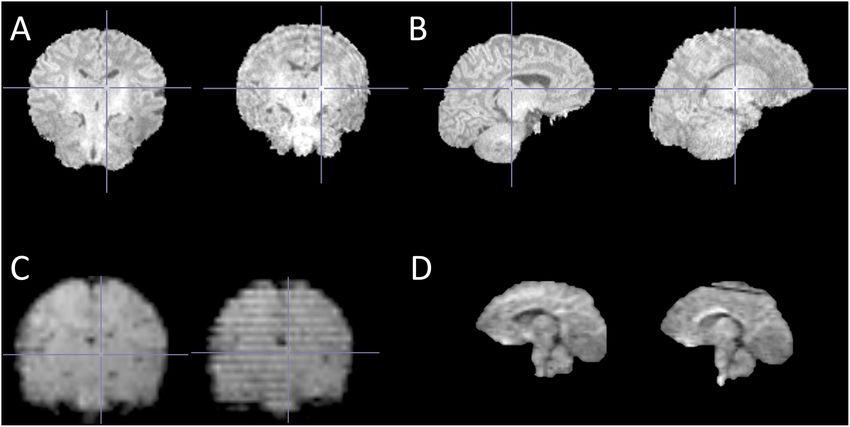
Copeland et al.                                                                                                            Infant and Child MRI Scanning

for research settings including healthy subjects. However, MRI is              2017). A specific vacuum fixation immobilizer is commonly used
very sensitive to motion, and therefore, the examination requires              to swaddle the infant in the feed and wrap technique. Using a
the subject to lie still during the scan. The acoustic noise of                vacuum immobilizer is safe, low cost, and obviates the need of
the scanner can rise up to 132 dB(A) (Foster et al., 2000), and                anesthesia (Golan et al., 2011). Questionnaire survey by Heller
acquisition time varies normally between 15 to 60 min, depending               et al. (2017) proved retrospectively that the primary technique for
on the set up and amount of sequences acquired. When scanning                  conducting neonatal MRI in NICU in the United States was the
pediatric populations for clinical purposes, moderate sedation                 feed and swaddle technique (64%), while the rest of the NICUs
or general anesthesia is often used to reduce anxiety and                      used primarily sedation or general anesthesia to aid the scans.
motion (Royal and Road, 2000). For ethical reasons, such as the                The same study expectedly showed a lower success rate of quality
risks related to anesthesia, sedation is not a generally accepted              data in the feed and swaddle group comparing to the sedation and
option for neuroimaging research examining healthy subjects                    general anesthesia groups. Further, after the first few months after
(Edwards and Arthurs, 2011). However, even the participants                    birth, the feed and swaddle technique becomes more ineffective
that are not able to co-operate during the scan, can be scanned                and scanning without sedation becomes more demanding. Still,
without motion during natural sleep, but this method in turn                   Dean et al. (2014a) outlined a protocol for scanning healthy
creates substantial challenges for the scan preparations. Despite              children under the age of four during natural, non-sedated sleep.
these challenges, MRI plays an important role in pediatric                     In that longitudinal study, 384 MRI datasets were successfully
neuroimaging research field (Zhang et al., 2019). A recent review              acquired from 220 healthy subjects with an overall 97% success
focuses on challenges in pediatric MRI and it also covers many of              rate. The scans were scheduled for the evening hours, and in some
the technological advances that may improve the success rate in                cases the participants were sleep deprived. There are even more
the future (Barkovich et al., 2019).                                           studies that have reported techniques to scan children mostly
    Neuroimaging studies in healthy infants using MRI as an                    older than 4 years while awake (Raschle et al., 2009; De Bie
imaging method have increased recently, and simultaneously, the                et al., 2010). For example, Raschle et al. (2009) provided general
field of research has expanded to various branches of science                  guidelines highlighting comfort, appropriateness, and motivation
such as psychology, logopedics, and social sciences. The human                 (CAM). A step-by-step protocol with a video report designed
brain develops and grows in size extremely fast during the first               for pediatric neuroimaging sessions in young children were also
2 years after birth (Knickmeyer et al., 2008) and is consequently              presented. In this age group, MRI compatible weighted blankets
sensitive to many environmental influences (Pulli et al., 2018). To            might be helpful to limit movement during acquisition as well
investigate what is normal development, a number of studies have               (Horien et al., 2020).
been conducted in healthy individuals (Bompard et al., 2014; Li                    Although motion prevention is carried out in the best
et al., 2014c; Deoni et al., 2015), at-risk populations (Hazlett et al.,       possible way, there is always a possibility of subtle, involuntary
2012; Dean et al., 2014b; Grewen et al., 2014; Donald et al., 2015;            movements during the acquisition. Even heart beats, breathing,
Langer et al., 2015; Monk et al., 2015; Ou et al., 2015; Qiu et al.,           or blinking can cause motion artifacts and reduce MRI data
2015a, 2013b; Chang et al., 2016; Jha et al., 2016; Salzwedel et al.,          quality. Any kind of motion is a challenge and concerns
2016), and clinical populations (Karmacharya et al., 2018; Moran               clinical and research imaging equally. To improve the data
et al., 2019), among others. To minimize postnatal environmental               quality, numerous methods have been developed to mitigate
influences, the most common imaging time point of interest                     or correct motion (Zaitsev et al., 2015). Methods can be
has been during infancy as close to birth as possible, generally               classified into prospective and retrospective approaches, which
during the first few postnatal weeks. This is surprisingly one of              both contain various techniques. Prospective techniques use a
the most convenient time points to perform imaging as infants                  real-time correction (Brown et al., 2010), while retrospective
sleep a lot during the period right after birth (Galland et al.,               techniques modify data during the reconstruction (Loktyushin
2012). Nevertheless, there are various challenges starting from                et al., 2013). Both methods have been applied in brain imaging
recruitment and timing of imaging without intruding parents’                   (Godenschweger et al., 2016). However, all methods have
day-to-day schedules. Further, pediatric scanning requires special             limitations and to date, no single method can completely
expertise as these scans are seldom a routine in the clinic or in              eliminate motion artifacts. Thus, minimization of the motion
research settings.                                                             remains crucial (Reuter et al., 2015).
    Previously, a few publications have especially focused on                      This review had specific goals to focus on the reporting of
scanning methods of infants and young children without                         scanning protocols and success rates of extant studies, which
sedation. The main factor affecting the selection of preparation               has not been covered in prior reviews on the field. We dedicate
techniques is age of the participants and developmental needs.                 a section to our own procedures that we hope will help future
Mathur et al. (2008) have published guidelines to perform brain                data collection. The first aim of this systematic review is to
MRI without sedation with neonatal intensive care unit (NICU)                  summarize the methods used to scan 0–6-year-old subjects in
patients. Later on, Arthurs et al. (2012) reviewed key techniques              the MRI scanner focusing on the studies published during the
to avoid sedation in neonatal imaging and focused on challenges                last 9 years, with a special emphasis on procedure to prepare
like physiological changes, equipment compatibility, and acoustic              a child for an MRI scan, but also on the techniques and
noise. The key technique with neonates is feed and wrap (also                  tools used during the scanning to enable a successful scan. We
termed feed and sleep, feed and swaddle, feed and bundle) and it               focused on the studies conducted on healthy, full-term subjects
is mainly used in infants less than 3 months old (Antonov et al.,              because the preterm born subjects are often scanned with clinical

Frontiers in Neuroscience | www.frontiersin.org                            2                                       July 2021 | Volume 15 | Article 666020
Copeland et al. Infant and Child MRI Scanning
implications. Descriptions of the scanning procedures and a as in the first phase. Given that we were particularly interested in
summary of the most commonly used techniques are reported. studies using healthy infants and young children who underwent
We also examined how the scans have succeeded, and on the MRI without sedation (noting that preterm-born children are
other hand, considered the reasons behind the failed scans. The frequently scanned under anesthesia), only articles that met the
second aim was to provide strategies for scanning infants and following criteria were included:
young children without sedation. To increase sample sizes and
(1) All subjects were scanned between 0 and 6 years of age.
the quality of data and decrease the number of drop-outs in the
(2) All scans were made without sedation and MRI was not
follow-up scans, it is important to know these methods well. In
clinically indicated. To make sure no sedatives were not
the future, these methods could even be introduced to the clinical
used, the study had to state it or mention scans were
setting as well to reduce the need of sedation during MRI. Finally,
made during natural sleep or awake. If this was not told,
we introduce the neonatal MR protocols that were used in the
publication was excluded due to insufficient information of
FinnBrain Birth Cohort Study Neuroimaging Lab (finnbrain.fi).
the scanning procedure.
(3) In accordance with the study’s inclusion or exclusion
METHODS criteria, only subjects born at gestational age (GA) 35 weeks
or later were included. If a study set a lower limit than
Literature Search 35 weeks for GA, it was excluded regardless of the subjects’
The primary targets of interest were study populations consisting GAs. If a study did not set a limit for GA and the
of term born infants and young children with focus on examining range was not reported the mean GA was ≥ 37 weeks
brain growth and development using MRI. A literature search with standard deviation ≤ 2 weeks (and mean GA minus
using PubMed database was originally conducted on the 30th of SD was ≥ 35 weeks). Finally, regarding studies with no
June in 2016. The search comprised of the following keywords mention on GA, only longitudinal studies were included.
(‘Magnetic Resonance Imaging’[Mesh] OR ‘MR imaging∗ ’ OR All information was obtained from the article full-texts
‘MRI’ OR ‘NMRI’ OR ‘fMRI’ OR ‘DTI’ OR ‘diffusion tensor and their supplementary data when applicable. While we were
imaging’) AND (‘Brain/growth and development’[Mesh] OR interested in the methods to calm subjects in the scanner, we
‘brain growth∗ ’ OR ‘brain developm∗ ’) AND (‘Infant’[Mesh] OR also investigated how studies have reported the used procedures
‘infant∗ ’ OR ‘toddler∗ ’). No languages were excluded at this point. before and at the scanner. Thus, reviewed studies may contain
To capture the most recent and relevant work in the field, a overlapping participant populations. 291 publications did not
starting date limit was enforced to include only papers published meet the criteria and were excluded.
after the 1st of January in 2012. The search was updated on the Finally, a total of 86 original articles published in English were
9th of March in 2021 and the final search included literature identified and included in this review. The included studies are
published between 1st of January in 2012 to 1st of January in 2021. listed in Supplementary Table 1. While we acknowledge that the
After duplicates were removed, the search resulted in a total of inclusion of a longer time frame and studies with prematurely
1098 publications. born participants would provide somewhat more information on
Titles and abstracts were used to screen articles in the first the topic, we opted to include the most recent studies performed
phase. Exclusion criteria were the following, in a descending without clinical grounds.
order of priority:
(1) The publication was written in a language other
RESULTS
than English.
(2) The study was not a human study.
(3) The study focused on the prematurely born or low birth
Study Characteristics
For all the included study populations (n = 86) MRI scans were
weight subjects of any age.
performed on the subjects between 0 and 6 years of age. The
(4) The study was focusing on a disease or treatment.
number of participants per one study ranged from 9 to 288.
A potential risk of carrying a disease was not a reason to
Majority of the subjects were term born, but in some cases
be excluded as long as the disease was not detected.
gestational age at birth was not provided. Sample sizes and ages
(5) 0–2-year-old living subjects were not MR imaged in the
at scan are shown in Supplementary Table 1. Fifty-two studies
study.
were completely cross-sectional and performed only one scan
If a publication met more than one criterion, only the highest per each subject (Hazlett et al., 2012; Deniz Can et al., 2013;
criterion was marked as a reason for exclusion. If the exclusion Deoni et al., 2013, 2015; Gao et al., 2013; O’Muircheartaigh et al.,
criterion was found from the title, the abstract was not used 2013, 2014; Qiu et al., 2013a, 2015b; Broekman et al., 2014; Dean
to find a higher priority criterion for exclusion. 721 articles et al., 2014b,c, 2017, 2018a,b; Grewen et al., 2014; Travis et al.,
were screened out. 377 publications were identified as potentially 2014; Zhang et al., 2014; Donald et al., 2015; Langer et al., 2015;
relevant (Figure 1). Ou et al., 2015; Poh et al., 2015; Spann et al., 2015a,b, 2020a,b;
These 377 publications were reviewed based on the abstracts Ferradal et al., 2016, 2019; Li et al., 2016; Sethna et al., 2016;
and full texts. At this second phase, we first excluded all review Adibpour et al., 2018, 2020; Lugo-Candelas et al., 2018; Monnelly
articles and after that, the exclusion criteria (1–5) were applied et al., 2018; Chen et al., 2019; Hernandez-Castillo et al., 2019;
Frontiers in Neuroscience | www.frontiersin.org 3 July 2021 | Volume 15 | Article 666020Copeland et al. Infant and Child MRI Scanning FIGURE 1 | Flow diagram outlining the literature search. Lebenberg et al., 2019; Lehtola et al., 2019; Ong et al., 2019; et al., 2020a,b, 2021; Alexander et al., 2020; Bruchhage et al., 2020; Tuulari et al., 2019; Acosta et al., 2020a,b, 2021; Alexander et al., Camacho et al., 2020; Dowe et al., 2020; Fenchel et al., 2020; Gale- 2020; Bruchhage et al., 2020; Camacho et al., 2020; Dowe et al., Grant et al., 2020; Merhar et al., 2020; Merz et al., 2020; Remer 2020; Fenchel et al., 2020; Gale-Grant et al., 2020; Graham et al., et al., 2020; Schmied et al., 2020), while 1.5 Tesla scanners were 2020; Merhar et al., 2020; Merz et al., 2020). The remaining less commonly used (n = 12) (Choe et al., 2013; Deniz Can et al., studies (n = 34) were longitudinal and conducted serial scans on 2013; Qiu et al., 2013a,b, 2015b; Broekman et al., 2014; Travis the same individuals (Geng et al., 2012, 2016; Choe et al., 2013; et al., 2014; Ou et al., 2015; Poh et al., 2015; Li et al., 2016; Sethna Li et al., 2013, 2014a,b,c, 2015a,b; Qiu et al., 2013b; Sadeghi et al., et al., 2016; Ong et al., 2019). Four studies did not report the field 2013; Alcauter et al., 2014, 2015; Bompard et al., 2014; Chen et al., strength of the used MR scanner (O’Muircheartaigh et al., 2013, 2014; Dean et al., 2014a, 2015a,b; Gao et al., 2014a,b; Swanson 2014; Deoni et al., 2016; Graham et al., 2020). et al., 2015; Chang et al., 2016; Croteau-Chonka et al., 2016; Deoni et al., 2016; Kim et al., 2016; Meng et al., 2017; Ahn et al., 2019; Participant’s State During the Scan Dai et al., 2019a,b; Hu et al., 2019; Wang et al., 2019, 2012; Remer All subjects underwent the MRI scanning non-sedated. Infants et al., 2020; Schmied et al., 2020). Serial scans included two to and children under the age of 4 slept during acquisition in seven scans per subject, most typically three scans per subject. majority of studies (n = 73/86) (Acosta et al., 2020a,b, 2021; The majority of the studies (n = 70) used 3 Tesla MRI scanners Adibpour et al., 2018, 2020; Ahn et al., 2019; Alcauter et al., 2014, (Geng et al., 2012, 2016; Hazlett et al., 2012; Deoni et al., 2013, 2015; Alexander et al., 2020; Bompard et al., 2014; Broekman 2015; Gao et al., 2013, 2014a,b; Li et al., 2013, 2014a,b,c, 2015a,b; et al., 2014; Bruchhage et al., 2020; Camacho et al., 2020; Chang Sadeghi et al., 2013; Alcauter et al., 2014, 2015; Bompard et al., et al., 2016; Chen et al., 2019; Choe et al., 2013; Croteau- 2014; Chen et al., 2014, 2019; Dean et al., 2014a,b,c, 2015a,b, 2017, Chonka et al., 2016; Dai et al., 2019a,b; Dean et al., 2014a,b,c, 2018a,b; Grewen et al., 2014; Zhang et al., 2014; Donald et al., 2015a,b, 2017, 2018a,b; Deniz Can et al., 2013; Deoni et al., 2015; Langer et al., 2015; Spann et al., 2015a,b, 2020a,b; Swanson 2013, 2015, 2016; Donald et al., 2015; Dowe et al., 2020; Ferradal et al., 2015; Chang et al., 2016; Croteau-Chonka et al., 2016; et al., 2016, 2019; Gale-Grant et al., 2020; Gao et al., 2013, Ferradal et al., 2016, 2019; Kim et al., 2016; Meng et al., 2017; 2014a,b; Geng et al., 2016; Graham et al., 2020; Grewen et al., Adibpour et al., 2018, 2020; Lugo-Candelas et al., 2018; Monnelly 2014; Hazlett et al., 2012; Hernandez-Castillo et al., 2019; Hu et al., 2018; Ahn et al., 2019; Dai et al., 2019a,b; Hernandez- et al., 2019; Kim et al., 2016; Langer et al., 2015; Lebenberg Castillo et al., 2019; Hu et al., 2019; Lebenberg et al., 2019; Lehtola et al., 2019; Lehtola et al., 2019; Li et al., 2014b, 2015a,b, 2016; et al., 2019; Tuulari et al., 2019; Wang et al., 2019, 2012; Acosta Lugo-Candelas et al., 2018; Meng et al., 2017; Merhar et al., 2020; Frontiers in Neuroscience | www.frontiersin.org 4 July 2021 | Volume 15 | Article 666020
Copeland et al.                                                                                                                          Infant and Child MRI Scanning

Merz et al., 2020; Monnelly et al., 2018; O’Muircheartaigh et al.,                   TABLE 2 | Challenges at motor coordination, emotional and attention
                                                                                     development in a different age groups.
2013, 2014; Ong et al., 2019; Poh et al., 2015; Qiu et al., 2013a,b,
2015b; Remer et al., 2020; Sadeghi et al., 2013; Schmied et al.,                                           Challenges at motor coordination, emotional and
2020; Sethna et al., 2016; Swanson et al., 2015; Tuulari et al., 2019;                                     attention development
Wang et al., 2019; Zhang et al., 2014). Seven studies reported
                                                                                     0–3 months            • Irregular daily rhythm, fragmented sleep
that infants were given time to fall asleep before scanning, but
                                                                                                           • Spontaneous movement of head, body and limbs
the subject’s state during the scan was not specifically reported
                                                                                                           • Startle response to hard/sudden noise
(Geng et al., 2012; Chen et al., 2014; Travis et al., 2014; Spann
                                                                                                           • Entirely dependent on caregiver in emotional and physical
et al., 2015a,b, 2020a,b). One study reported that infants were                                            regulation
scanned during natural sleep or while resting quietly (Ferradal                                            • Limited communicative abilities and underdeveloped
et al., 2016). Furthermore, a few studies (n = 6) reported scans                                           capabilities to reflect on the surroundings
without sedation, but subject’s state during the scan was not                        4 months – 1 year     • Sleep cycle maturates, longest continuous sleep during
specifically reported (Wang et al., 2012; Li et al., 2013, 2014a,c;                                        nighttime
Ou et al., 2015; Fenchel et al., 2020). See Table 1 for a summary.                                         • Depended on caregivers in emotional and physical
                                                                                                           regulation
                                                                                                           • Separation anxiety
Special Notes on Scanning 4–6-Year Old
                                                                                                           • Close relationship with primary caregivers
Children                                                                                                   • Limited communicative abilities, receptive vocabulary
Fifteen studies of all the total 86 studies in this review scanned                                         starts to develop
also children between the ages 4 to 6 years (O’Muircheartaigh                        2–3 years             • Sleep cycle maturates, longest continuous sleep during
et al., 2013, 2014; Chen et al., 2014, 2019; Dean et al., 2014a,c,                                         nighttime, no need for daytime sleep for some children
                                                                                                           • Rapid language development, inability to follow long
                                                                                                           instructions
TABLE 1 | Reporting of scanning procedures in included studies.                                            • Self-regulation capacity starts to develop (ability to
                                                                                                           regulate internal and external signals without adult’s help)
                              N   Method                                                                   • Testing boundaries, temper tantrums
                                                                                     4–6 years             • Characteristics and personality comes more visible
Timing of the visit           14 Scheduled for the naptime or bedtime
                                                                                                           • Better attention and self-regulation capacity (better ability
                              72 Not reported
                                                                                                           to regulate internal and external signals without adults help)
Preparations at home          3   Sleep deprivation
                                                                                                           • Ability to follow long verbal instructions
                              3   MRI sounds
                              81 Not reported
Preparations at MRI           32 Feeding before the scan                             2015a,b; Deoni et al., 2015, 2016, 2013; Croteau-Chonka et al.,
facilities                     5 Replicating typical naptime routines
                                                                                     2016; Dai et al., 2019a,b; Remer et al., 2020). At this age, the
                              5   MRI sounds
                                                                                     child has typically more ability to cooperate, but in contrast to
                              1   Stimulating tasks served to fatigue
                                                                                     infants, it might be more difficult to get the child to fall asleep
                              48 Not reported
                                                                                     in a strange environment (see Table 2). Eleven studies reported
Subject’s state during        80 Reported (sleep or awake)
the scan
                                                                                     that if tolerated by the child (in most cases ≥ 4 years old), the
                               6 Not reported
                                                                                     scan was made while the child was awake, e.g., when watching a
Motion prevention             51 Immobilization (various methods used)
                                                                                     movie or a TV show. The remaining four studies scanned during
                              35 Not reported
Noise attenuation             58 Ear protection (various methods used)
                                                                                     natural sleep. Otherwise preparations, motion prevention, sound
                              20 Acquisition parameter optimization
                                                                                     attenuation, and monitoring during the scan did not differ from
                              17 Sound insulating bore liner or foam insert
                                                                                     that of the younger children.
                                 placed inside of the scanner bore
                              6   “Precautions”                                      Timing of Visit
                              21 Not reported                                        Timing of the MRI sessions were frequently scheduled according
Monitoring during the         28 Pulse oximeter                                      to participant’s normal sleeping/diurnal rhythm. Scanning
scan                           1 Pulse socks                                         schedules were reported in 14 studies (Choe et al., 2013; Deniz
                              26 Visually monitored                                  Can et al., 2013; Dean et al., 2014a,b, 2017, 2018b; Gao et al.,
                              8   Camera                                             2014b; Travis et al., 2014; Langer et al., 2015; Lehtola et al.,
                              4   Electrocardiography                                2019; Tuulari et al., 2019; Bruchhage et al., 2020; Camacho
                              47 Not reported                                        et al., 2020; Dowe et al., 2020), and the remainder 72 studies
Duration of the scan          38 Reported†                                           did not report the timing of the visit. Imaging was frequently
                              48 Not reported                                        performed on naptimes or bedtime. In seven studies, MRI visits
Total duration of the visit   1   Reported                                           were scheduled for the subject’s naptime or bedtime, but the time
                              85 Not reported                                        was not specified (Gao et al., 2014b; Langer et al., 2015; Dean
† Somethingmentioned about the duration of the scan. The total duration of the       et al., 2017, 2018b; Bruchhage et al., 2020; Camacho et al., 2020;
scan was not required to be informed.                                                Dowe et al., 2020). One study scheduled visits for naptime in the

Frontiers in Neuroscience | www.frontiersin.org                                  5                                              July 2021 | Volume 15 | Article 666020
Copeland et al. Infant and Child MRI Scanning
late morning (Deniz Can et al., 2013) and three others in the late studies explained this so called feed and sleep or feed and wrap
afternoon or early evening (Choe et al., 2013; Lehtola et al., 2019; method in more detail: children were fed 15–30 min prior to the
Tuulari et al., 2019). In a few studies (n = 3), the majority or all scan and swaddled in warm sheets (Ou et al., 2015; Li et al., 2016).
the scans were performed in the evening around bedtime or at
night (Dean et al., 2014a,b; Travis et al., 2014). Naptime imaging Motion Prevention
was typically used with the youngest participants, while evening Swaddling or wrapping was used not only to make falling asleep
hours and nighttime were typical scan times not only for infants, easier, but also to reduce potential body movement during the
but also for the older participants. scan. A number of different approaches to wrapping the infants
were provided, varying from only wrapping them in sheets to
Preparations at Home placing them into an immobilizer. Several studies (n = 23) used
Getting ready for the upcoming MRI scan often started already specific vacuum immobilization mats, bags, or pillows to stabilize
at home. To habituate the infant to the scanner noise, families the child and reduce natural movement from breathing (Deoni
were provided a CD of the scanner sound, and parents were et al., 2013; Zhang et al., 2014; Ou et al., 2015; Croteau-Chonka
instructed to play the CD, while subjects were sleeping at home et al., 2016; Li et al., 2016; Dean et al., 2014a,b,c, 2015b, 2017,
(Hazlett et al., 2012; Langer et al., 2015). In one study, mothers 2018a,b; Dai et al., 2019a,b; Hernandez-Castillo et al., 2019;
were given an MRI prep kit including earplugs and a portable Lehtola et al., 2019; Ong et al., 2019; Tuulari et al., 2019; Camacho
speaker pre-loaded with MRI sounds. In this study, mothers were et al., 2020; Dowe et al., 2020; Graham et al., 2020; Merhar et al.,
also encouraged to start swaddling their infant to sleep if they had 2020; Remer et al., 2020). Dean et al. (2014a) reported placing a
not already started to do so (Camacho et al., 2020). In two studies, mat under the child before the child fell asleep and once asleep the
the child was deprived of sleep prior to scans by asking parents immobilizer was wrapped around the child. Subject’s head was
to wake the child up earlier in the morning or skip a nap on a separately secured in a vacuum fixation device in eight studies
research day (Geng et al., 2012; Dean et al., 2014a). To promote (Geng et al., 2012, 2016; Grewen et al., 2014; Li et al., 2015a,b;
sleep at the imaging site, Dean et al. (2014a) reported that parents Ahn et al., 2019; Wang et al., 2019, 2012), while only half of these
were asked to keep the child busy throughout the day before the also reported swaddling the child. Foam cushions, foam pads, and
scan. In total, 81 studies did not report preparations at home. visco-elastic matters were also commonly used to keep the head
in place and occupy the space between the subjects and the head
Preparations at MRI Facilities coil. All in all, the majority of studies (n = 51/86) mentioned some
Replicating the typical bed or naptime routines in the MRI method to stabilize the infant prior to the scan.
facilities was reported in five studies (Choe et al., 2013; Deniz
Can et al., 2013; Dean et al., 2014a; Langer et al., 2015; Camacho Noise Attenuation
et al., 2020). Parents were asked to bring along comfort items to Acoustic noise levels of the MRI scanner were reduced, and the
create a homely environment at the imaging site (Langer et al., hearing of the subjects was protected with different methods.
2015). Private rooms with diaper changing and bathing facilities, The major part of the studies (n = 65/86) reported the use of
rocking chairs, portacribs, blankets, soft lullaby music, and other passive or active measures during acquisition, while 21 studies
objects were attempted to make the environment cozier (Choe made no mention of sound attenuation. Six studies out of these
et al., 2013; Deniz Can et al., 2013; Dean et al., 2014a; Langer et al., 65 reporting noise attenuation mentioned taking precautions
2015). Dimmed lights at the MRI site and also around the facility, to reduce the noise, but did not delineate the methods (Qiu
when carrying the sleeping child, were also provided (Dean et al., et al., 2013a,b, 2015b; Broekman et al., 2014; Poh et al., 2015;
2014a; Dowe et al., 2020). While creating a comfortable and Ong et al., 2019). Most commonly used passive measure was
homely environment, the children got to familiarize with the MRI ear protection: 25 studies used double or triple ear protection
sound simultaneously before the scanning and when they fell (Deniz Can et al., 2013; Deoni et al., 2013, 2015, 2016; Dean
asleep in five studies (Langer et al., 2015; Spann et al., 2015a,b, et al., 2014a,c, 2017, 2018a,b; Donald et al., 2015; Spann et al.,
2020a,b). Langer et al. (2015) reported the use of interesting tasks 2015a,b, 2020a,b; Li et al., 2016; Monnelly et al., 2018; Dai et al.,
prior to the scan to fatigue the child. 2019a,b; Hernandez-Castillo et al., 2019; Lehtola et al., 2019;
Tuulari et al., 2019; Alexander et al., 2020; Camacho et al., 2020;
Feeding Dowe et al., 2020; Graham et al., 2020) and the remainder (n = 33)
Feeding the child before scanning was the most commonly used single ear protection. For example, MiniMuffs, earplugs,
reported preparation (n = 32/86) (Geng et al., 2012; Wang et al., headphones, sound attenuating ear protectors, electrodynamic
2012; Gao et al., 2013, 2014a; Li et al., 2013, 2014a,c, 2016; Sadeghi headphones and a custom-made acoustic hood were used. In four
et al., 2013; Alcauter et al., 2014, 2015; Bompard et al., 2014; studies, electrodynamic headphones played white noise (Dean
Chen et al., 2014; Grewen et al., 2014; Zhang et al., 2014; Donald et al., 2017, 2018a; Camacho et al., 2020; Dowe et al., 2020)
et al., 2015; Ou et al., 2015; Spann et al., 2015a,b, 2020a,b; Kim and in one study soothing rain sounds (Bruchhage et al., 2020)
et al., 2016; Dean et al., 2017, 2018b; Ahn et al., 2019; Hernandez- during image acquisition. In addition to ear protection, noise
Castillo et al., 2019; Lehtola et al., 2019; Tuulari et al., 2019; levels were lessened by a noise insulating bore liner or foam
Alexander et al., 2020; Camacho et al., 2020; Dowe et al., 2020; insert fitted inside of the scanner bore in 17 studies (Deoni et al.,
Merhar et al., 2020). Sleep was promoted by adjusting a feeding 2013, 2015, 2016; Dean et al., 2014a,b,c, 2015b, 2017, 2018a,b;
schedule prior to the scan. After children were fed, they were Croteau-Chonka et al., 2016; Dai et al., 2019a,b; Lebenberg et al.,
swaddled or wrapped, or otherwise helped to fall asleep. A few 2019; Bruchhage et al., 2020; Dowe et al., 2020; Remer et al.,
Frontiers in Neuroscience | www.frontiersin.org 6 July 2021 | Volume 15 | Article 666020Copeland et al. Infant and Child MRI Scanning
2020). Furthermore, some studies reported reducing scanner MRI session) (Ferradal et al., 2016), trying to keep them short to
noise actively by selecting specific imaging parameters, slowing prevent the child from waking up during the acquisition (Dean
the gradient switching rate and reducing the maximum gradient et al., 2014a). The child’s cooperation and ability to remain asleep
amplitudes (Deoni et al., 2013, 2015, 2016; O’Muircheartaigh enabled additional imaging sequences and longer imaging times
et al., 2013, 2014; Dean et al., 2014a,b,c, 2015b, 2017, 2018a,b; (Dean et al., 2015b). In some studies, the imaging times varied
Croteau-Chonka et al., 2016; Dai et al., 2019a,b; Lehtola et al., depending on the subject’s age due to different protocols used in
2019; Tuulari et al., 2019; Adibpour et al., 2020; Dowe et al., 2020; various age groups (Deoni et al., 2013, 2016; Dean et al., 2014c;
Remer et al., 2020). The changes in the imaging parameters were O’Muircheartaigh et al., 2014; Dai et al., 2019a,b). Finally, only
shown to provide approximately a 35 dB noise reduction during one study reported the duration of the whole scanning visit (Dean
the acquisition (Dean et al., 2014a). et al., 2014a). In this study, the duration of the visit was highly
variable from less than 1 h to more than 5 h.
Monitoring
Numerous studies (n = 39/86) mentioned monitoring subjects Success Rate and Missing Scans
throughout the scan. To confirm that the child remained asleep, All 86 studies had successful MRI scans and the number of
a physician, a nurse, a research assistant or a member of the included scans varied between 9 (Zhang et al., 2014) to 445
research team was presented, who visually monitored the subject (Dai et al., 2019b). In addition to the successful scans, reporting
in 26 studies (Geng et al., 2012; Wang et al., 2012; Qiu et al., about data losses it varied considerably between studies. 41
2013a,b, 2015b; Broekman et al., 2014; Dean et al., 2014a, 2017, studies (n = 41/86) reported the number of excluded scans or
2018b; Grewen et al., 2014; Li et al., 2014b, 2015b; Donald alternatively the success rate for included data (Hazlett et al.,
et al., 2015; Poh et al., 2015; Chang et al., 2016; Croteau- 2012; Deniz Can et al., 2013; Qiu et al., 2013a,b, 2015b; Sadeghi
Chonka et al., 2016; Meng et al., 2017; Monnelly et al., 2018; et al., 2013; Bompard et al., 2014; Broekman et al., 2014; Dean
Hernandez-Castillo et al., 2019; Lehtola et al., 2019; Ong et al., et al., 2014a,b, 2017, 2018a,b; Li et al., 2014c, 2016; Travis
2019; Tuulari et al., 2019; Bruchhage et al., 2020; Camacho et al., et al., 2014; Alcauter et al., 2015; Donald et al., 2015; Langer
2020; Dowe et al., 2020; Gale-Grant et al., 2020). Three studies et al., 2015; Poh et al., 2015; Spann et al., 2015a, 2020b; Chang
reported using an MRI compatible camera (Ou et al., 2015; et al., 2016; Ferradal et al., 2016, 2019; Sethna et al., 2016;
Bruchhage et al., 2020; Graham et al., 2020), and in additional Adibpour et al., 2018, 2020; Lugo-Candelas et al., 2018; Dai
five studies an infrared camera to monitor subjects during the et al., 2019a; Ong et al., 2019; Tuulari et al., 2019; Acosta
scan (Deoni et al., 2013; Dean et al., 2014c; Croteau-Chonka et al., 2020a,b, 2021; Bruchhage et al., 2020; Camacho et al.,
et al., 2016; Dai et al., 2019a,b). Besides visual monitoring, 28 2020; Dowe et al., 2020; Fenchel et al., 2020; Graham et al.,
studies used a pulse oximeter to follow the heart rate and oxygen 2020; Merz et al., 2020). Additionally, some studies mentioned
saturation (Geng et al., 2012; Wang et al., 2012; Deoni et al., reasons for exclusion during the data preprocessing steps but
2013; Qiu et al., 2013a,b, 2015b; Broekman et al., 2014; Dean did not report the number of excluded scans. Remainder of the
et al., 2014a,c; Grewen et al., 2014; Li et al., 2014b, 2015b, 2016; studies used only subjects with successful scans or for some
Donald et al., 2015; Ou et al., 2015; Poh et al., 2015; Spann et al., other reason did not report the number of missing scans. The
2015a,b, 2020a,b; Croteau-Chonka et al., 2016; Ferradal et al., number of included and excluded scans, and if available, reasons
2016; Meng et al., 2017; Monnelly et al., 2018; Dai et al., 2019a,b; for exclusion are shown in Supplementary Table 1. The most
Hu et al., 2019; Ong et al., 2019). Bruchhage et al. (2020) reported common specified reason for losing data was movement during
using pulse socks to monitor pulse and behavior when asleep. the scan causing motion artifacts to the data. Other mentioned
Electrocardiography (ECG) was used in four studies (Spann et al., reasons related to the scanning procedure were that the subject
2015a,b, 2020a,b). One study reported monitoring heart rate, did not fall asleep prior to scanning, woke up during transition
oxygen saturation, and temperature, but did not specify the to the scanning bed or during the MRI acquisition. Exclusions
used equipment (Gale-Grant et al., 2020). In addition to these were made also due to demographic reasons, problems with
monitoring methods made by a professional or a member of a the analysis, age under or over the study-specific range, missed
research team, only six studies reported parents being invited to measurements in other parts of the study, and due to a brain
remain in the imaging site during the acquisition (Dean et al., anatomical anomaly. In other words, not all exclusion criteria
2014a, 2017, 2018b; Lehtola et al., 2019; Tuulari et al., 2019; were a consequence of failure at imaging. Due to the different
Dowe et al., 2020). reasons of exclusion, the success rates between studies are
not comparable and do not represent exclusively the success
Scan Duration of scanning. Instead of the overall success rate, some studies
The majority of studies did not directly report the total duration reported separate rates for different parts of the sample, for
of the imaging protocol. The acquisition time could often be example, for age groups or for cases and controls separately.
calculated using given sequences and the number of planes, this For example, Li et al. (2014c) used data acquired at three time
however, does not tell the total/maximum time in the scanner. points with following success rates: 90% for neonates, 66% for 1-
Most of the studies reported using different imaging sequences year-olds, and 60% for 2-year-olds. Dean et al. (2014a) observed
(e.g., T1, T2, DTI, fMRI), but did not report the individual that scanning during the second or third visit succeeded more
acquisition times for them. When reported, the acquisition times often (success rate 100%) than scanning during the first visit
ranged from 2 min (Chen et al., 2019) up to 2 h (for a typical (success rate 90%).
Frontiers in Neuroscience | www.frontiersin.org 7 July 2021 | Volume 15 | Article 666020Copeland et al. Infant and Child MRI Scanning
The Infant Scanning Procedures of the the available good data from 92% estimated at the scanner to
69% in the final data. Figure 2 shows representative examples
FinnBrain Study and How to Calm Infants
of successful and unsuccessful neonate MRI scans (randomly
in the Scanner selected from our data).
We refer to FinnBrain infant MRI measurements in the current To share practical advice, we also report some procedures
article. They were carried out in accordance with the Declaration that we used to calm the infants if they woke up during the
of Helsinki, and the protocol was approved by the joint Ethics scan, and that can be used by gently reaching for the infant
Committee of the University of Turku and the Hospital District even within the scanner bore. These procedures are presented
of Southwest Finland. in Figure 3. During the scans, either the investigators or the
In the FinnBrain Birth Cohort Study, 189 families participated parents frequently calmed the infants with these relatively simple
in the scans when their infants were 2–5-weeks old (M = 26.04, measures that rely on infant reflexes and/or calming touch. Of
SD = 7.6, range = 8–51 days corrected for gestation), and 180 were important note, the homogeneity of the static magnetic field may
scanned. The data collection took place between 2012 and 2016. be affected when an adult reaches into the scanner bore. Usually,
Most scans were conducted from the afternoon to early evening we used the soothing technique for a brief period of time if the
hours (16:30–20:00), but ca. 10 scans took place on Saturday baby was sleeping restlessly at the start of the session or started to
afternoons. The imaging was performed at the Department of move during scanning. We let the ongoing sequence to continue
Radiology, Turku University Hospital, Finland. All scans were during the soothing (as abrupt changes in the surrounding noises
obtained using a Siemens Magnetom Verio 3T scanner (Siemens risk waking up the infant), but always acquired the sequence
Medical Solutions, Erlangen, Germany). again, i.e., the sequences during which soothing was used were
At the start of the visit, the families were welcomed by a considered failed and were never used in analyses. We did not
trained and experienced radiographer and the researchers at the record the total number of infants who woke up during the scan,
scanning site. The infants were fed with breastmilk or formula were successfully calmed and how long it took for babies to be
and swaddled into a vacuum mattress. No sedatives were used. calmed and able to put back into the MRI scanner. This would be
Deformable wax plugs and custom-sized earmuffs were used valuable data to collect in future studies.
for hearing protection. Parents were provided with standard
earmuffs, as they stayed in the scanning room during the whole
scanning session. The personnel observed the scanning from the DISCUSSION
control room through a window with a microphone contact to a
parent. A loudspeaker sent the sounds from the scanning room We systematically identified 86 studies, which performed infant
to the control room allowing the staff to hear if the infant woke or child brain MRI without sedation or general anesthesia in 0–
up. The session was ended if the infant did not fall asleep before 6-year-olds. The majority of the studies acquired MRI scans only
or did not fall back asleep during the scan. in subjects 2 years or younger. In this review, we concentrated
The sequences comprised of an axial PD-T2-TSE (Dual-Echo on methods used to prepare a child for an MRI scan, but also
Turbo Spin Echo), a sagittal 3D-T1 (T1-weighted MPRAGE) on the techniques and tools used during the scanning to enable a
and three diffusion tensor imaging (DTI) sequences respectively. successful scan. The most commonly used preparations especially
The acquisition times for sequences were 6 min 50 s (axial PD- with younger participants were feeding and wrapping the child
T2-TSE), 4 min 3 s (3D-T1), 5 min 3 s (DTI 1), 5 min 33 s just before the scan. This so-called feed and wrap or feed and
(DTI 2) and 5 min 42 s (DTI 3). Sequence parameters were swaddle method is widely accepted and used also in infants
optimized so that “whisper” gradient mode could be used in PD- undergoing MRI for a clinical purpose. Also a recently published
T2 TSE and 3D-T1 sequences to reduce acoustic noise during manuscript with a focus on performing pediatric neuro MRI
the scan (Lehtola et al., 2019). Functional MRI sequences were suggested feed and swaddle to be the first-line method for subjects
added to the protocol starting in June 2015 and performed 3 months and younger (Barkovich et al., 2018). The efficacy of the
until the end of the study (N = 28). We acquired task fMRI technique has been evaluated and it has got a high rate of success
measurements investigating touch responses and a resting state (79% answered the clinical question, 20% partially answered the
fMRI scan. 60 min was the maximum duration of the complete clinical question) in infants 3 months or younger (Antonov et al.,
scanning protocol, and total duration of the visit was less than 2017). Unfortunately, this fairly easily applicable method is less
2 h. The data are still being processed and analyzed, but the useful with older children, who do not easily fall asleep in an
current success rates are 125/180 for structural T1 and T2 scans unfamiliar environment.
(69%) (Acosta et al., 2020a), 172/180 (95%) for at least 20 good It is suggested that children from 1 to 5 years of age might
quality diffusion weighted images out of the acquired 96 images be the most challenging target group to scan without sedation
(N = 157 for 30 directions, N = 142 for 40 directions and (Barkovich et al., 2018). Further, there is less research in this
N = 121 for 60 directions) (Merisaari et al., 2019). Success age group as compared to younger or older age groups. One
rates for task fMRI are 10/13 (77%) in preliminary findings practical method is to schedule the MRI visit during child’s
(Tuulari et al., 2019) and 21/28 (75%) for the resting state data nap or bedtime and scan during natural sleep, yet it requires
(Rajasilta et al., 2020). Unfortunately, we suffered from some flexible scanning schedules and might take time to get the child
technical difficulties with the T2-weighted images that were to fall asleep. Another possibility is to scan while the child is
identified only after data collection, which significantly impacted awake. However, there is no clear evidence for the best age for
Frontiers in Neuroscience | www.frontiersin.org 8 July 2021 | Volume 15 | Article 666020Copeland et al. Infant and Child MRI Scanning FIGURE 2 | Representative examples of successful and unsuccessful neonate MRI scans (randomly selected from our data). (A) T1-weighted structural image with no motion artifacts (left) and with typical “ringing” motion artifact (right); (B) same images as in A in sagittal view; (C) fMRI images with no motion artifacts (left) and with typical “striping” motion artifact (right); and (D) diffusion-weighted image with no motion artifacts (left) and a typical “loss of signal” artifact at the superior part of the image (right). Of note, these examples are not exhaustive and are provided for visualization purposes only. starting to scan the children when they are awake. Obviously, of pediatric patients aged 4 to 14 years. It appears that the this depends on the individual child. Actually, as young as 2- mock scanner is most effective in children between ages 3 to year-old children have been successfully scanned awake using 8 years (Carter et al., 2010). Unfortunately, mock scanners are progressive behavioral training method (Vannest et al., 2014). rather expensive, which limits their availability. However, there This training procedure was limited to 15 min and contained 3 is evidence that using a cheap play tunnel simulating the MRI steps: first children were asked to sit on the scanner bed, then environment might be a useful alternative (Barnea-Goraly et al., to lie down, and then to hold “still as a statue”. Stickers were 2014). Theys et al. (2014) have developed a behavioral training given as reinforcers at each step. After these preparations 95% protocol termed the ’submarine protocol’ to prepare children children at the age of 2–6 years had at least one successful scan for scanning. After completing the required tasks that made the sequence. Another used preparation method is to simulate the child more familiar with potentially difficult aspects of MRI, real MRI experience with mock scanner before scanning (Carter she/he was ready for the ‘submarine ride’. The method has been et al., 2010). This method requires language and cognition skills used to acquire advanced MRI techniques (DTI, fMRI) in 5- and is usually used with older children. However, Thieba et al. and 6-year-old typically developing children with a success rate (2018) have applied mock scanner training already as young as 2- of 95% (72/76) for completing the full 35-min scan. Recently, year-old children, but surprisingly, the scans with children aged 95% success rate in children aged 4–6 was obtained using multi- 2–5 years were no more likely to be successful after receiving the faceted concepts including an interactive app, a trained pediatric mock scanner training than the ones without the use of mock team, a children’s lounge with a toy scanner and a child-friendly scanner, although the image quality was slightly higher in the multimedia environment in the MRI room (Runge et al., 2018). former. When the child comes more able to cooperate, capable of Surprisingly, meta-analysis demonstrating the efficacy of pre- moving, and expressing their own will, the imaging while awake MRI training (including booklet, audio, video, toy model, or a comes easier to implement. mock scanner) did not improve data quality, sedation use or Based on the reviewed studies, the age of 4 years seems to success rate of scanning (Li et al., 2019). Authors thought that one be the most common age to start scanning when the child is possible account might be that training increases anxiety and fear awake. In this age group, the preparation methods, including among children. However, there were a limited number of studies a mock scanner and behavioral training, become common in (n = 5) and small sample sizes, thus more studies are needed to preparing children to MRI. In addition to these highly known confirm the findings. All in all, success rates between studies are methods, a newly emerging technology using Virtual Reality (VR) not readily comparable, therefore a controlled study investigating has opened up a new approach to prepare children for MRI the actual effects of different methods should be performed in (Ashmore et al., 2019). When applying a training protocol with the future. Importantly, there is also lack of objective standard a mock scanner, a success rate of 88% (53/60) for structural criteria of what is considered good quality data, and to what and 64% (23/36) for functional MRI has been obtained in a extent it can be improved with post processing. group of 4–7-year-old children (De Bie et al., 2010). Similarly, To improve comfort and ensure safety during acquisition, Cavarocchi et al. (2018) have reported a 83% (162/195) success noise attenuation during the scan must be executed properly. rate, and an overall 30% decrease in the need of sedation after This review suggested that the most commonly employed the training protocol with a mock scanner in a large cohort methods were ear protection using earplugs (wax, foam Frontiers in Neuroscience | www.frontiersin.org 9 July 2021 | Volume 15 | Article 666020
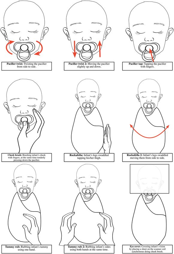
Copeland et al.                                                                                                                         Infant and Child MRI Scanning

  FIGURE 3 | Schematic illustrations of the procedures used to calm the infants if they woke up during the scan in the FinnBrain Study. All procedures can be made by
  carefully reaching inside the scanner bore.

Frontiers in Neuroscience | www.frontiersin.org                                   10                                            July 2021 | Volume 15 | Article 666020
Copeland et al.                                                                                                       Infant and Child MRI Scanning

or silicon), soft shell earmuffs, regular ear protectors or                specify individual factors associated with successful scanning.
combinations of these. Sound-insulating bore liners or foam                Though, some studies reported separate success rates for
inserts were less frequently used. The combination of methods              different groups of the sample, for example, for age groups,
have been demonstrated to be more effective than one method                for sexes or for cases and controls separately. However, these
individually (Tocchio et al., 2015). To assure sufficient noise            rates did not provide reliable information about the effect of
reduction during MRI scanning in newborns, Nordell et al.                  individual variables on the compliance of MRI in children.
(2009) have suggested the combination of dental putty fitted into          Previously Cahoon and Davison (2014) have found that parental
the outer ear canal, earmuffs placed over both ears, and acoustic          expectations and ratings of how well the child normally handles
hood of dampening material placed over the child. However,                 medical procedures were the strongest predictors of MRI
passive noise control methods suffer from limitations such as              compliance, while child attention problems and poor adaptability
discomfort, fitting problems, and very importantly, in some                among children related to non-compliance. Thieba et al. (2018)
cases, insufficient noise reduction. Therefore, active methods like        have shown that higher cognitive and language ability in children
quiet sequences and quiet coils have been developed to perform             may predict success. Finally, there is an evidence that child’s
scanning more quietly (McJury and Shellock, 2000). In contrast             temperament may have an effect on ability to undergo MRI
to methods trying to reduce or eliminate the acoustic noise, a             without sedation (Voepel-Lewis et al., 2000). These subject
new acquisition method (MR Fingerprinting-Music) has been                  specific factors can cause unwanted sample selection during
developed to make the sounds more pleasant by emulating music              different phases of a study (e.g., during recruitment, preparations
(Ma et al., 2016). To our knowledge, there are no studies showing          and imaging) and later on have an effect on interpretation
how the method works with children.                                        of the results. To minimize these kinds of effects, a child’s
   There is a general awareness that motion during acquisition             MRI compliance could be determined beforehand, which might
causes image artifacts, which can lead to unusable data. Based             facilitate the preparation protocol. In the future, this kind of
on the studies reviewed here, motion was the most common                   approach might be helpful. Finally, it is important to note
reason for data exclusion. However, the systematic effects of              that compliance may be related to e.g., temperament features,
motion are poorly known, and often uncontrolled for. This                  developmental stage, and severity of symptoms on clinical
can be problematic especially in research settings, where factors          populations and may thus cause bias in samples.
of interest like age, sex, or disease are usually correlated with             All in all, a lot could be done to enhance the success
both the amount of head motion and structural changes. For                 rates of scans and data quality without any concrete devices,
example, head motion during MRI acquisition has been shown to              by merely paying attention to the environment, atmosphere,
influence estimates of gray matter volume and thickness (Reuter            and suitable communication with the child and parents before,
et al., 2015). Consistent with previous studies and this review,           during, and after scanning. The main goal is to diminish
swaddling or wrapping are the most commonly used techniques                anxiety and distress by creating a comfortable and child-friendly
to restrict motion during infant scanning. These methods are               environment. It is essential to take into account the child’s
simple, low cost, and easily available but unfortunately useless           individual and developmental needs (see Table 2) and tailor
in older age groups. Additionally, special vacuum immobilizer              preparations accordingly. Parent comfort is equally important,
mats, cushions, and foam pads were used to stabilize body and              because it influences directly the child’s feelings. To improve
head position. Older children are often able to watch movies               the communication with the child and family, a child life
or TV shows during acquisition, which has been shown to                    specialist (CLS) can be used in the front-line interaction. For
reduce the head movement. Recently, Greene et al. (2018) have              example, parent and staff satisfaction as well as child pain and
developed this further by testing movies and real-time visual head         distress have been shown to be positively impacted by the
motion feedback simultaneously. Both methods significantly                 child life services in pediatric imaging (Tyson et al., 2014).
reduced movement, but interestingly, no compounding effect of              Finally, positive feedback and thanking the parents for their
combining methods was found. Additionally, these methods can               participation are needed regardless of the scanning success. It
be problematic during fMRI imaging, due to the possible effects            is essential to involve the child and the family in a positive
on functional data (Greene et al., 2018). To improve compliance            overall experience, in any case, but especially if follow-up
and minimize cognitive load during functional imaging, a movie             scans are under consideration. Collecting the feedback from
paradigm, Inscapes, have been conducted (Vanderwal et al., 2015).          parents, and if available from children, may help to improve
However, physical head restraint methods, videos, or behavioral            protocols in the future.
strategies do not completely eliminate motion, thus motion                    MRI methods are constantly developing (Börnert and Norris,
correction later on is needed in any case. These techniques                2020). New techniques such as compressed sensing (Lustig et al.,
become even more crucial with high field MRI systems, such as 7            2007), simultaneous multi-slice imaging (Larkman et al., 2001;
Tesla scanners, which can generate higher levels of acoustic noise,        Breuer et al., 2005) or recently developed image reconstruction
require longer acquisition times, and are more sensitive to motion         algorithms utilizing artificial intelligence can reduce total scan
artifacts (Stucht et al., 2015; Keuken et al., 2018).                      duration significantly. For example, in the case of the protocol
   In addition to preparation methods, individual factors                  from FinnBrain study, duration of PD-T2 TSE and DTI
like subject’s age, sex, culture, medical history, behavioral              sequences could easily be reduced by at least 12 min just by using
characteristics, and parental expectations might have an effect            multi-slice imaging. It is likely that reduced scan time would lead
to MRI procedure compliance. Herein reviewed studies did not               to better success rate of the scans.

Frontiers in Neuroscience | www.frontiersin.org                       11                                      July 2021 | Volume 15 | Article 666020
You can also read