The Molecular Function of PURA and Its Implications in Neurological Diseases
←
→
Page content transcription
If your browser does not render page correctly, please read the page content below
REVIEW
                                                                                                                                              published: 11 March 2021
                                                                                                                                      doi: 10.3389/fgene.2021.638217

                                              The Molecular Function of PURA and
                                              Its Implications in Neurological
                                              Diseases
                                              Lena Molitor 1†, Sabrina Bacher 1†, Sandra Burczyk 2† and Dierk Niessing 1,2*
                                              1
                                               Institute of Structural Biology, Helmholtz Zentrum München – German Research Center for Environmental Health,
                                              Neuherberg, Germany, 2 Institute of Pharmaceutical Biotechnology, Ulm University, Ulm, Germany

                                              In recent years, genome-wide analyses of patients have resulted in the identification of a
                                              number of neurodevelopmental disorders. Several of them are caused by mutations in
                                              genes that encode for RNA-binding proteins. One of these genes is PURA, for which in
                                              2014 mutations have been shown to cause the neurodevelopmental disorder PURA
                                              syndrome. Besides intellectual disability (ID), patients develop a variety of symptoms,
                                              including hypotonia, metabolic abnormalities as well as epileptic seizures. This review
                                              aims to provide a comprehensive assessment of research of the last 30 years on PURA
                             Edited by:
                                              and its recently discovered involvement in neuropathological abnormalities. Being a DNA-
                        Alfredo Brusco,
                 University of Turin, Italy   and RNA-binding protein, PURA has been implicated in transcriptional control as well as
                      Reviewed by:            in cytoplasmic RNA localization. Molecular interactions are described and rated according
                   Antonio Vitobello,         to their validation state as physiological targets. This information will be put into perspective
  INSERM U1231 Lipides, Nutrition,
         and Cancer (LNC), France
                                              with available structural and biophysical insights on PURA’s molecular functions. Two
                        Maurizio Elia,        different knock-out mouse models have been reported with partially contradicting
Oasi Research Institute (IRCCS), Italy
                                              observations. They are compared and put into context with cell biological observations
                    *Correspondence:
                                              and patient-derived information. In addition to PURA syndrome, the PURA protein has
                           Dierk Niessing
              dierk.niessing@uni-ulm.de       been found in pathological, RNA-containing foci of patients with the RNA-repeat expansion
     †
         These authors have contributed       diseases such as fragile X-associated tremor ataxia syndrome (FXTAS) and amyotrophic
                    equally to this work      lateral sclerosis (ALS)/fronto-temporal dementia (FTD) spectrum disorder. We discuss the
                                              potential role of PURA in these neurodegenerative disorders and existing evidence that
                     Specialty section:
           This article was submitted to      PURA might act as a neuroprotective factor. In summary, this review aims at informing
         Genetics of Common and Rare          researchers as well as clinicians on our current knowledge of PURA’s molecular and
                                Diseases,
                 a section of the journal     cellular functions as well as its implications in very different neuronal disorders.
                    Frontiers in Genetics
                                              Keywords: PURA syndrome, amyotrophic lateral sclerosis/fronto-temporal dementia, fragile X-associated tremor
         Received: 21 December 2020           ataxia syndrome, Pur-alpha, PURB, PURG
          Accepted: 09 February 2021
           Published: 11 March 2021

                             Citation:
  Molitor L, Bacher S, Burczyk S and
                                              INTRODUCTION
     Niessing D (2021) The Molecular
Function of PURA and Its Implications
                                              RNA-binding proteins fulfill a number of important regulatory functions for cell differentiation,
             in Neurological Diseases.        cellular asymmetry, and somatic cell activities. In recent years, particular attention has been
            Front. Genet. 12:638217.          paid to the functions of RNA-binding proteins in neuronal cells where their involvement in
    doi: 10.3389/fgene.2021.638217            messenger RNA (mRNA) transport and translational regulation has been linked to memory

Frontiers in Genetics | www.frontiersin.org                                         1                                           March 2021 | Volume 12 | Article 638217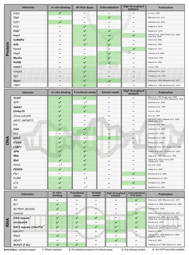
Molitor et al.                                                                                               PURA-Related Functions and Disorders

and learning as well as to neuronal disorders (Tang, 2016;                The crystal structure of PUR repeats I and II from
Holt et al., 2019; Thelen and Kye, 2019). One of them is the              D. melanogaster PURA revealed that two PUR repeats interact
so-called Purine-rich single-stranded DNA (ssDNA)-binding                 with each other to fold into a stable PUR domain (Graebsch
protein alpha (PURA, also referred to as Pur-alpha), which                et al., 2009). With a conserved β-β-β-β-α topology, each of
frequently appears in high-throughput analyses of interaction             the two PUR repeats folded into a four-stranded beta sheet,
networks in neuronal tissue. PURA is co-purifying with a                  followed by a single alpha helix. Such a PUR domain can
multitude of different potential binding partners and has been            either be built from a single peptide chain with a β-β-β-β-
linked by several of these studies to neural pathologies. Despite         α-linker- β-β-β-β-α topology (type I) or from two identical
a significant body of novel insights from recent studies, still           peptides with β-β-β-β-α fold, forming an inter-molecular
very little is known about its cellular and molecular functions.          homodimer (type II; Figures 1B,C; Janowski and Niessing,
Hence, a critical assessment of our current knowledge is                  2020). In both cases, the α-helices swap between both β-β-β-β-α
much needed.                                                              repeats. In PURA, the first and second PUR repeats form a
   The PURA protein was first described to bind to ssDNA                  type I domain, whereas at the C-terminus a PUR domain is
in cell culture and brain extracts (Bergemann and Johnson,                assembled by the third repeats of two different PURA molecules
1992; Bergemann et al., 1992; Haas et al., 1993, 1995). Such              (Figures 1B,C; Graebsch et al., 2009; Weber et al., 2016).
early work provided evidence for the role of PURA in                         PUR repeats and domains have also been found in bacteria
transcriptional control. Only more recently, its function in              (Graebsch et al., 2010). Although almost no protein sequence
RNA-regulated processes has been studied in greater detail,               conservation can be found for instance between the PUR
leading to the altered perception that post-transcriptional control       repeat of PURA from B. burgdorferi and metazoan (identities
constitutes a central function of the PURA protein.                       of 21, 23, and 22% to repeats I, II, and III of human PURA,
   In this review, we provide a detailed analysis of our current          respectively; Figure 1D; Graebsch et al., 2010), its structure
knowledge on this protein and its different cellular roles in             is almost identical to the PUR domain from D. melanogaster
the nucleus and cytoplasm. We combine this with molecular                 (Figures 1E–G). Thus, PUR proteins provide a good example
and structural insights and provide an update of PURA’s role              that the same fold can be achieved from very different
in neuronal disorders, such as Fragile X-associated tremor                sequences (Doolittle, 1994).
ataxia syndrome (FXTAS), the amyotrophic lateral sclerosis                   Albeit showing no sequence conservation to other classes
(ALS), fronto-temporal dementia (FTD) spectrum disorder, and              of nucleic-acid binding proteins, the domain fold of PUR
the PURA syndrome. Together, the available information indicates          domains is found in a large variety of proteins, which all
that the importance of PURA protein for understanding neuronal            belong to the PC4-like family of proteins (Janowski and
disorders is still considerably underestimated.                           Niessing, 2020). They are named after the first high-resolution
                                                                          structure of this fold (PDB ID: 1pcf) from the human replication
                                                                          and transcription cofactor PC4 (Brandsen et al., 1997).
HOMOLOGS OF PURA ARE FOUND IN                                             Interestingly, the proteins of this family contain exclusively
DIFFERENT KINGDOMS OF LIFE                                                PC4-like domains and to some extent unstructured regions
                                                                          but lack any other types of domains. This observation is
To date, homologs of PURA have been described in several                  unusual at least for RNA-binding proteins, which very often
organisms, including metazoans, such as Drosophila melanogaster           contain combinations of different domain types to expand
(Graebsch et al., 2009; Aumiller et al., 2012), Danio rerio               their molecular function (Lunde et al., 2007).
(Penberthy et al., 2004), and Caenorhabditis elegans (termed                 While in most organisms only one PURA gene exists,
PLP-1; Witze et al., 2009), plants such as Arabidopsis thaliana           some species such as D. rerio contain two gene copies
(Tremousaygue et al., 2003), and even in bacteria like Borrelia           (Figure 2A). PURA is also unusual because it belongs to
burgdorferi (Graebsch et al., 2010). Surprisingly, sequence               the 3% of human genes that lack introns (Grzybowska, 2012).
alignments (Altschul et al., 1990) suggest that PURA is lacking           Since alternative splicing of intron-containing genes is an
in fungi.                                                                 important factor to modulate and expand functional properties
   Most of the available studies have been performed with mouse           of a given protein, this limitation indicates that there has
PURA, which is almost identical to its human ortholog (>99%               been evolutionary pressure preventing genomic rearrangements
protein sequence identity; Ma et al., 1994). Early work already           of the PURA gene. This hypothesis is consistent with the
recognized the presence of repetitive sequence elements in the            observation that mutations in almost any part of the PURA
protein, which were defined as class I and class II motifs                protein result in the full spectrum of the human PURA
(Bergemann and Johnson, 1992). However, crystal structure                 syndrome (see below).
determination of PUR domains from D. melanogaster (Figure 1A)                Vertebrate genomes, such as mouse, rat, and humans usually
showed an insufficient correlation of these motifs with the domain        contain two additional paralogs of the PURA gene, termed PURB
folds. Furthermore, no defined function could be assigned to              und PURG. All paralogs contain PUR-repeats as characteristic
them. With improved bioinformatic tools and structural                    protein-sequence elements (Graebsch et al., 2009, 2010; Figure 2B).
information, three so-called PUR repeats were defined that show              Of note, phylogenetic analysis of the PUR protein amino
sequence conservation in all PUR proteins (Graebsch et al., 2009,         acid sequences suggests that PURB emerged prior to PURA
2010) and correlate well with its folded entities (Figures 1A,B).         and PURG (Figure 2A) and thus is the likely founding member

Frontiers in Genetics | www.frontiersin.org                           2                                     March 2021 | Volume 12 | Article 638217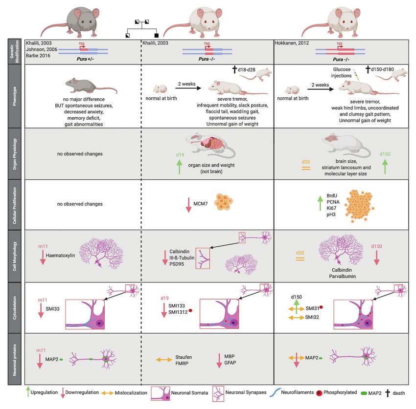
Molitor et al.                                                                                                            PURA-Related Functions and Disorders

                    A                                                             B

                                                                                  C

                    D

                    E                                        F                                   G

  FIGURE 1 | Assessment of structural and sequence conservation between PURA proteins from different species. (A) Cartoon backbone model of the PURA
  DNA-binding domain of Drosophila melanogaster and a view rotated 180°. (B) Schematic illustration of the domain organization of the PURA homodimer.
  (C) Topology diagram of the type I and type II domain of PURA. (D) Amino acid sequence alignment of PURA from selected species with identical amino acids
  highlighted in green. The sequence similarity is notably higher within the PUR repeats and decreases with evolutionary distance. The alignment was executed
  using the MUSCLE algorithm and program MEGAX (Hall, 2013; Mello, 2018). (E) Cartoon backbone model of Borrelia burgdorferi PURA domain forming a type II
  homodimer (C). (F) Superposition of the DNA binding domain of D. melanogaster [shown in (A)] and B. burgdorferi [shown in (E); RMSD = 2.107].
  (G) Superposition of D. melanogaster PURA repeat I and B. burgdorferi PURA (RMSD = 0.293).

Frontiers in Genetics | www.frontiersin.org                                   3                                          March 2021 | Volume 12 | Article 638217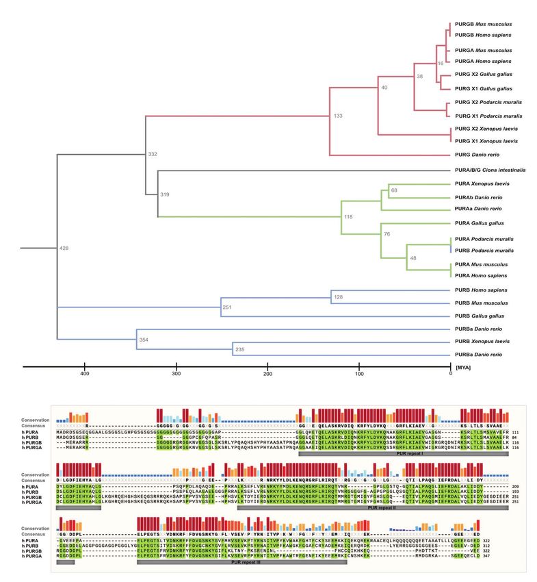
Molitor et al.                                                                                             PURA-Related Functions and Disorders

of the PUR family of proteins. Furthermore, the PURB orthologs           specificity for purines. PURA targets include for instance the
share a higher conservation between species than PURA and                TNF-alpha promoter (Darbinian et al., 2001b), tat-responsive
PURG (Figure 2B).                                                        elements within the TGFb-1 promoter (Thatikunta et al., 1997),
                                                                         and human ferritin gene promoter (FE65; Zambrano et al.,
                                                                         1997; Figure 4). In many studies, the interaction of PURA
MOLECULAR FUNCTIONS OF PURA                                              with promoter regions were shown in vitro, but a quantitative
                                                                         assessment of binding was only made in few exceptions.
PURA was initially found in HeLa cells and described as an               Furthermore, a part of the published DNA targets of PURA
ssDNA binding factor (Bergemann and Johnson, 1992;                       were classified as such exclusively by in vitro binding assays.
Bergemann et al., 1992). Shortly afterwards, it was also isolated        In Figures 3, 4, we defined those targets as “candidates” and
from mouse-brain lysates and shown to bind to a promoter                 distinguish them from targets that have been validated in
region of the myelin basic protein (Haas et al., 1993, 1995).            addition for instance by reporter assays or observed changes
One of the first indications that PURA also interacts with               in knock-out mice (Figure 3; candidate targets are shown with
RNA came from a study with quail embryonic fibroblasts,                  regular letters, validated targets with bold letters). It should
where RNase treatment of nuclear extracts resulted in a loss             be noted that the vast majority of promoter interaction studies
of association of PURA with larger complexes (Herault et al.,            were published more than 15 years ago and were not confirmed
1995). Concurrently, interactions of PURA with the RNAs of               since then. While PURA does bind GGN motifs in vitro in
the HI and JC viruses were also described (Tada and Khalili,             the low micro-molar concentration range, the affinity for a
1992; Chen et al., 1995; Chepenik et al., 1998), indicating that         short sequence within the upstream promoter region MF0677
this protein might act for instance as host factor for HIV               of the cMYC gene is at least an order of magnitude better
propagation (White et al., 2009).                                        (Jurk et al., 1996; Graebsch et al., 2009; Weber et al., 2016).
   In the past, several protein and nucleic-acid targets have            Since this sequence does not exclusively consist of GGN repeats,
been reported (Figures 3, 4). For many of them, PURA-protein             it remains to be shown in what way purines play a role in
fragments bearing internal deletions were created to map                 binding and if a consensus binding motif can be deduced.
respective interaction sites for its binding partners (summarized           One important consideration for the physiological impact
in White et al., 2009; Daniel and Johnson, 2018). Unfortunately,         of nucleic acid binding by PURA is its binding preference for
most of these studies were done before X-ray structures of               either ssDNA or single-stranded (ss) RNA. Surprisingly,
PURA were available and the respective deletions violated                recombinantly expressed PURA from D. melanogaster bound
domain boundaries. As a consequence, such mutant versions                to ssDNA and RNA oligonucleotides with identical sequences
of PURA were most likely unfolded and non-functional. For                with similar equilibrium dissociation constants. Furthermore,
this very reason, these mapped interaction sites within PURA             NMR chemical shift perturbation experiments showed that the
have to be considered with great caution for instance for                same amino acids are affected in PURA upon binding to DNA
proteins, such as HIV-Tat (Krachmarov et al., 1996), Polyomavirus        and RNA (Weber et al., 2016). It indicates that PURA binds
large T-antigen (Gallia et al., 1998), Retinoblastoma protein            both types of nucleic acids in the same way. A co-structure
(Johnson et al., 1995), Cdk2 (Liu et al., 2005), E2F-1 (Darbinian        of the N-terminal PUR domain of D. melanogaster PURA with
et al., 2004), YB1 (Safak et al., 1999), Cdk9 (Darbinian et al.,         a short ssCGG-repeat DNA further confirmed these observations
2001b), and Cyclin T1 (Darbinian et al., 2001b). Furthermore,            by showing that the 3' position of the ribose ring does not
since most reported interactions were detected by methods                contribute to the binding event (Weber et al., 2016). In summary,
that do not allow to distinguish between direct and indirect             these data indicate that PURA binds ssDNA and ssRNA equally
interactions (Figures 3, 4) it remains to be shown whether               well and in a similar way. These in vitro results also suggest
at least some of them represent direct binding events. In                that competition between DNA and RNA could be an important
particular for DNA targets of PURA, a number of cases were               feature for PURA-dependent regulation of gene expression
reported only based on observed in vitro interactions and thus           in cells.
lack a clear confirmation of their physiological relevance.                 Indeed, a recent and comprehensive study provided direct
Figure 4 gives an overview of the reported DNA, RNA, and                 evidence for an RNA-based transcriptional repression of the
protein interactions of PURA, indicating how well each target            muscle myosin-heavy chain (Mhc) gene by PURA and PURB.
has been validated to date.                                              Gorospe and colleagues found that the circular RNA circSamD4
                                                                         binds and sequesters both PUR proteins and thereby prevents
                                                                         them from repressing the transcription of the Mhc gene (Pandey
NUCLEAR TARGETS OF PURA                                                  et al., 2020). This finding is consistent with previous studies
                                                                         showing that PURA and PURB regulate alpha- and beta-myosin
The first publications on PURA suggested that this protein               heavy chains (Gupta et al., 2003; Hariharan et al., 2014).
has a strict requirement for purine-rich sequences on both,              Another study showed that PURA and PURB localized to
ssDNA and RNA level (Bergemann and Johnson, 1992). However,              so-called paraspeckles, which are nuclear membrane-less
this assumption was challenged shortly afterwards (Jurk et al.,          compartments. There, PURA was detected in the interactome
1996). Even to date, the few reported in vitro binding assays            of NEAT1 and MALAT1 nuclear RNAs (West et al., 2014).
with short defined oligonucleotides do not hint at strict sequence       Taken together, independent studies demonstrated that PURA

Frontiers in Genetics | www.frontiersin.org                          4                                    March 2021 | Volume 12 | Article 638217
Molitor et al.                                                                                                                 PURA-Related Functions and Disorders

          A

          B

  FIGURE 2 | Sequence similarities between PUR-family proteins. (A) Phylogenetic tree of the PUR-proteins of selected species. The tree shows evolutionary time
  on the X axis and indicates a differentiation of the PUR proteins between 428 and 319 Million Years Ago (MYA). The tree was inferred using the RelTime method and
  was computed using one calibration constraint. Branch lengths were calculated using the Maximum Likelihood (ML) method and the JTT matrix-based substitution
  model. A discrete Gamma distribution was used to model evolutionary rate differences among sites. The phylogenetic analysis was performed with the program
  MEGAX (Hall, 2013; Mello, 2018). (B) Amino acid sequence alignment of human PURA, PURB, PURG variant A, and PURG variant B. Sequence identity is
  highlighted in green. Whereas human PURA and PURB proteins share 70% sequence conservation, PURG has 48% identity to PURA and thus is more divergent.
  For all paralogs, sequence similarity is notably higher within the PUR repeats. The alignment was executed using the MUSCLE algorithm and program MEGAX.

interacts with nuclear RNA, potentially allowing for direct                           how many more genes are regulated by the PUR protein family
competition between DNA and RNA binding as means for                                  through such RNA-/DNA-competition mechanisms. This can
gene regulation. Future work will be necessary to establish                           be investigated for instance via a high-throughput identification

Frontiers in Genetics | www.frontiersin.org                                       5                                           March 2021 | Volume 12 | Article 638217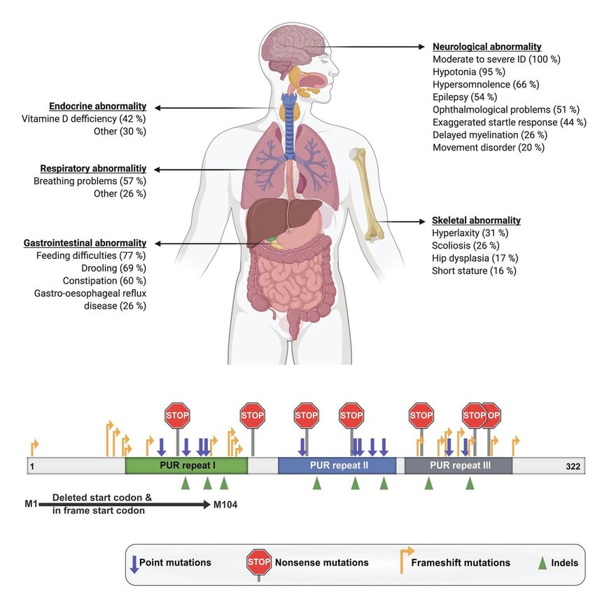
Molitor et al. PURA-Related Functions and Disorders
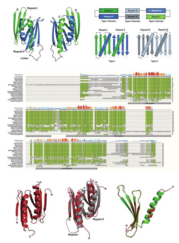
FIGURE 3 | Overview of the molecular pathways of PURA and its known interactions. To date, the only protein interaction of PURA for which experimental
evidence indicates a direct binding event is KIF-5 (marked in bold type). DNA and RNA targets that were confirmed by in vitro binding as well as by functional assays
or direct correlation with observed effects in animal model organisms were considered to be validated targets (marked in bold letters). They include the human CD43
gene promoter (Shelley et al., 2001; Da Silva et al., 2002), FE65 promoter (Zambrano et al., 1997), Gata2 promotor region in zebrafish (Penberthy et al., 2004), MB1
regulatory region of the MBP gene (Haas et al., 1995), Mhc promoter (Gupta et al., 2003; Ji et al., 2007; Pandey et al., 2020), ovine placental lactogen promotor
(Limesand et al., 2004), TGF-ß1 (Thatikunta et al., 1997; Knapp et al., 2006), and the VSM-alpha actin promoter (Knapp et al., 2006). Previously described DNA and
RNA interactions of PURA were considered to be only candidate targets if direct binding had been shown either by in vitro experiments or by high-throughput
analyses, but where further functional verification is lacking. Those candidate targets are displayed in regular type and include the A-ß-PP (Darbinian et al., 2008),
the cMyc upstream promoter region MF0677 (Bergemann and Johnson, 1992; Jurk et al., 1996; Ding et al., 1997; Graebsch et al., 2009; Weber et al., 2016), nAch
receptor (Du et al., 1997), and TNF-alpha (Darbinian et al., 2001b). Although for the latter, a reporter assay has been published, this was done only as duplicate and
lacked statistical analyses. For simplicity reasons, interaction partners shown in this figure constitute a selection. A complete list of all reported interacting partners is
shown in Figure 4.
of direct DNA and RNA targets, followed by systematic motif development, such as Bicoid, Bruno-1, Egalitarian, Oskar, Staufen,
searches. Based on our current limited knowledge it is already and Vasa have been identified already a long time ago (reviewed
clear that the name of the PUR-protein family, Purine-rich in reference Lasko, 2020). Due to the genetic location of PURA
ssDNA-binding protein, is likely to not fully reflect the protein’s on the rather inaccessible fourth Drosophila chromosome, it
nucleic acid-binding features. has not been studied comprehensively and might have escaped
potential detection in systematic genomic approaches. Since to
date no phenotypic data are available in D. melanogaster, it is
CYTOPLASMIC TARGETS OF PURA certainly possible that the biological importance of PURA is
underestimated for this model organism.
Early Drosophila oogenesis and embryogenesis are considered However, the majority of studies were performed in mice,
as prime example for the importance of post-transcriptional where PURA is detected predominantly in the cytoplasm
gene regulation (Lasko, 2020). Indeed, also GFP-tagged PURA (Figure 3; Khalili et al., 2003; Johnson et al., 2006; Hokkanen
is transported together with many RNAs into the oocyte, suggesting et al., 2012; Rossi et al., 2015; Daigle et al., 2016; Mitsumori
a role in RNA-based gene regulation (Aumiller et al., 2012). et al., 2017; Swinnen et al., 2018). Apart from a diffuse
Many other RNA-binding proteins important for early cytoplasmic localization PURA is also reported to localize in
Frontiers in Genetics | www.frontiersin.org 6 March 2021 | Volume 12 | Article 638217Molitor et al. PURA-Related Functions and Disorders
FIGURE 4 | Published interaction partners of the PURA protein. For all “Interactors” (left column) the method used to observe interactions is indicated by a
checkmark and green background in the respective column. Well-validated interaction partners of PURA protein are marked in bold letters. For proteins, interaction
partners were defined in this study as well validated when experimental evidence is available from each of the following two categories: (i) in vitro binding using
(Continued)
Frontiers in Genetics | www.frontiersin.org 7 March 2021 | Volume 12 | Article 638217Molitor et al. PURA-Related Functions and Disorders FIGURE 4 | recombinant proteins or in immunoprecipitation/pull-down experiments from cell lysates; (ii) colocalizing with PURA in cells or identification as PURA interactor in a high throughput proteome analysis. DNA- and RNA-interaction partners are defined as well validated if in vitro binding by PURA as well as functional validation either by a reporter assay or in an animal model has been reported. Protein interaction labeled with “1” was shown to be RNAse resistant. Functional assays labeled with “2” lacked basic statistical analyses (i.e., SD), and hence were ignored as positive score for target validation. Interaction partners labeled with “3” have only been described in a specific disease context. For target labeled with “4” ChIP-seq data have been published (Sun et al., 2020), however without providing the original data for critical assessment. cytoplasmic membrane-less compartments of vertebrates, of the neuronal transport studies to date were performed in including stress granules (Daigle et al., 2016; Markmiller et al., mice (Li et al., 2001; Ohashi et al., 2002; Kanai et al., 2004; 2018) and neuronal transport granules (Figure 3; Li et al., Mitsumori et al., 2017), resulting findings need to be investigated 2001; Ohashi et al., 2002; Kanai et al., 2004; Mitsumori et al., further before any conclusions can be drawn about possible 2017). Furthermore, in C. elegans, the PURA ortholog PLP-1 correlations with human disease-related processes. localizes to posterior germ granules, where it is important for genome maintenance (Vishnupriya et al., 2020). For stress granules, PURA has even been reported to be essential for RNA-REPEAT EXPANSION DISORDERS their formation (Daigle et al., 2016). There and in neuronal RNP granules, PURA co-localizes with other neuronal PURA has been implicated in two different nucleotide-repeat RNA-binding proteins, such as PURB, Staufen, hnRNPU, and expansion disorders: fragile X-associated tremor/ataxia syndrome fragile X mental retardation protein (FMRP; Ross et al., 1997; (FXTAS) and the disease continuum of C9orf72-mediated Ohashi et al., 2002; Kanai et al., 2004; Johnson et al., 2006). amyotrophic lateral sclerosis and fronto-temporal dementia (C9 In neuronal cells, PURA also closely associates with the cargo- ALS/FTD). transporting conventional kinesin KIF5 and myosin Myo5a, which both mediate RNA transport along neurites (Ohashi et al., 2002; Kanai et al., 2004). Upon mGluR5 activation, PURA in Hexanucleotide-Repeat PURA is specifically transported in a Myo5a dependent manner Expansions in C9orf72 RNA Leading to into dendritic spines, where it colocalizes with the synaptic ALS/FTD Spectrum Disorder factor PSD95 (Mitsumori et al., 2017). Furthermore, other Current literature suggests that ALS and FTD are different PURA-associated factors, such as FUS, FMRP, and Stau1 are phenotypic expressions of the same disease origin. Pathological reported to play key roles in the development of long-term expansions of up to thousands of G4C2 repeats in the first depression (LTD; Mitsumori et al., 2017). Stau1 might intron of the C9orf72 gene have been described as the most be important in this context as it is associated with PURA- frequent genetic cause of ALS/FTD (Dejesus-Hernandez positive granules in dendritic shafts (Mitsumori et al., 2017). et al., 2011; Renton et al., 2011). Hallmarks of the disease On the other hand, Stau1 is vastly diminished in PURA positive spectrum include RNA foci and progressive neurodegeneration. granules in dendritic spines, suggesting that it escorts PURA These RNA foci are mainly nuclear and contain G4C2 repeat only before reaching the final destination in spines. sequences as well as a number of proteins including PURA These observations are also consistent with the association (Donnelly et al., 2013; Mizielinska et al., 2013). of PURA and PURB with BC1 RNA in mouse brain cells Three G4C2-repeat-dependent disease pathways have been (Ohashi et al., 2002; Kobayashi et al., 2011). Absence of BC1 proposed. The most obvious potential pathomechanism is the has been implicated in altered glutamatergic transmission and loss-of-function of the C9orf72 gene product through the maladaptive behavior (Briz et al., 2017). Furthermore, it was repeat expansions. However, since C9orf72 knockout mice shown that PURA can associate with MAP2 mRNA in mouse failed to display an ALS/FTD-related motor neuron disease brains and is suggested to be involved in its transport (Johnson phenotype (Lagier-Tourenne et al., 2013; Koppers et al., 2015; et al., 2006). This observation is in line with results from PURA Atanasio et al., 2016; McCauley et al., 2020), this option is knockout mice, where a significant overall reduction of MAP2 rather unlikely. The second and third options are both related protein in whole brain lysates was reported (Hokkanen et al., to the C9orf72 RNA and rather difficult to distinguish 2012). Apart from that, MAP2 was misdistributed to the somata experimentally (discussed in Swinnen et al., 2020). One of of neurons and Purkinje cells. This might indicate a specific these options is based on the observation that Repeat Associated reduction of local MAP2 translation at distal neurites, potentially Non-AUG translation (RAN translation) occurs on G4C2 repeat due to impaired transport of its mRNA (Hokkanen et al., 2012). RNAs, resulting in potentially pathological protein fragments Since most publications showed PURA to be mainly located (Zu et al., 2011). Whether those RAN-derived proteins are in the cytoplasm, it also seems likely that one of PURA’s main solely or partially responsible for the ALS/FTD pathogenesis functions is its cytoplasmic interaction with RNA. Out of all is still heavily discussed. While no functional role of PURA RNA-related processes, neuronal cytoplasmic RNA transport protein in RAN translation has been reported, PURA was has been associated most convincingly with a PURA function found in pathological RNA foci of ALS/FTD patients (Xu so far. Furthermore, this function fits very well with et al., 2013b; Stepto et al., 2014). This indicates an involvement neurodegenerative and neurodevelopmental disorders described of PURA in the third possible disease pathway, i.e., RNA in association with the PURA protein. However, since most toxicity (Swinnen et al., 2018). Frontiers in Genetics | www.frontiersin.org 8 March 2021 | Volume 12 | Article 638217
Molitor et al.                                                                                                 PURA-Related Functions and Disorders

    Since repeat expansion-containing RNAs are readily                      Fragile X-syndrome, which is based on a transcriptional shut-
transcribed, the RNA itself could have neurotoxic properties.               down of the entire FMR1 gene locus. The pathological repeat-
Different disease mechanisms are hypothesized for RNA toxicity              expanded RNAs expressed in FXTAS patients recruit various
to be responsible for the ALS/FTD symptoms. Firstly, the                    proteins including PURA into intra-nuclear inclusions (Iwahashi
recruitment of proteins into RNA foci could result in sequestration         et al., 2006; Jin et al., 2007; Sofola et al., 2007). Described
of proteins such as PURA that are important for neuronal                    hallmarks of FXTAS are a loss of Purkinje cells, RAN-translated
function (Stepto et al., 2014). Secondly, repeat expansions in              FMR-PolyG proteins, and nuclear inclusions (Buijsen et al.,
the C9orf72 RNA could interfere with mRNA-transport granules                2014; Boivin et al., 2018). A well-documented disease mechanism
and thus with neuronal function (Burguete et al., 2015).                    for FXTAS is that the CGG repeat-expanded FMR1 mRNA
    For both scenarios, the identification of proteins binding              sequesters important RNA-binding proteins. This leads to
to nucleotide expansions (G4C2) is essential. Indeed, multiple              disturbed RNA processing very similar to what has been
studies have analyzed these G4C2-interacting proteins and many              described for G4C2 repeat expansions in C9orf72 as cause of
RNA-binding proteins have been identified. The overlap of                   ALS/FTD. Furthermore, CGG-repeat RNA-induced neurotoxicity
proteins that appear in all studies is limited though, probably             can be overcome by simultaneous overexpression of PURA
due to technical differences (Swinnen et al., 2020). In a                   (Jin et al., 2007; Boivin et al., 2018).
comparative analysis of several studies (Donnelly et al., 2013;                As already stated for G4C2 repeats, RNA interactions of
Mori et al., 2013; Xu et al., 2013b; Cooper-Knock et al., 2014;             PURA with CGG repeats were shown by different studies but
Haeusler et al., 2014) with different cell lines, only five proteins        proof for a strict sequence-specificity is still lacking to date.
are found in at least three out of five analyses. These proteins            In fact, one particular RNA oligonucleotide lacking CGG repeats
include HNRNPH3, HNRNPH1, ILF2, MBP, and SFPQ (Haeusler                     even showed 10-fold better binding to PURA in vitro than
et al., 2016). Interestingly, PURA is detected with high significance       CGG repeats of identical size (Weber et al., 2016).
in a study where pull-down experiments with extracts from
mouse cerebellum were performed (Cooper-Knock et al., 2014).
In a second study with mouse brain and spinal cord, PURA                    Potential Mechanisms of Neuroprotection
was again co-purified with G4C2 repeat expansions (Rossi et al.,            by PURA
2015). In contrast, PURA was not identified in pull-down                    In disease models with CGG- or G4C2-repeat expansion-induced
experiments with G4C2 RNA from neuronal SH-SY5Y or                          neurotoxicity, the overexpression of PURA rescues their
HEK293T cells (Mori et al., 2013; Cooper-Knock et al., 2014;                pathological phenotypes. These observations indicate that PURA
Haeusler et al., 2014). Together, these findings suggest that               sequestration might be responsible for neurotoxicity. On the
PURA is recruited to disease-related G4C2 sequences either                  other hand, PURA overexpression is also described to ameliorate
exclusively in the context of functional neuronal tissues or                neurotoxicity of ALS related FUS mutations that are unrelated
only in mice but not in human cells.                                        to RNA repeat expansions (Daigle et al., 2016). Here, PURA
    In cell culture as well as model organisms, neurotoxicity               overexpression reverses cytoplasmic mislocalization of mutant
induced by overexpression of G4C2-repeat RNAs can be overcome               FUS protein and enhances stress granule dynamics. The
by a simultaneous ectopic overexpression of PURA (Xu et al.,                sequestration model is additionally challenged by the fact that
2013b; Swinnen et al., 2018). On one hand, this underlines                  multiple proteins are sequestered to repeat expansions but that
the importance of PURA for ALS/FTD pathogenesis. On the                     the individual overexpression of PURA already reduces
other hand, it also supports the hypothesis that repeat expansions          neurotoxicity (Jin et al., 2007; Xu et al., 2013a; Swinnen et al.,
sequester RNA-binding proteins, leading to a loss of physiological          2018). One would expect that at least some of the other
function of the affected proteins. Conversely, FMRP knock down              sequestered proteins also need to be released in order to restore
mitigates the C9orf72-dependent ALS/FTD phenotype (Burguete                 normal neuronal function. Hence, the sum of these findings
et al., 2015), indicating opposing modes of action for FMRP                 does not provide strong support for a disease-causing
and PURA.                                                                   sequestration of PURA, rather hinting at other disease
    In summary, PURA appears to play an important role as                   mechanisms such as an overall neuroprotective effect of PURA.
G4C2 repeat RNA interactor in a pathological context (Swinnen                  One mechanistic feature of PURA that could be important
et al., 2020). It remains to be shown if the recruitment into               for such a neuroprotective function is its ability to unwind
foci depends on specific repeat binding events by PURA or                   double-stranded nucleic acids in an ATP-independent manner
is based on its rather general interaction with nucleic acids               (Darbinian et al., 2001a; Wortman et al., 2005). This function
when these are present at pathologically high concentrations.               could help PURA to disassemble pathological RNPs and release
                                                                            trapped and mislocalized RNAs and proteins. A recent crystal
                                                                            structure of the N-terminal PUR domain bound to DNA
The Role of PURA in FXTAS                                                   provided a rationale for the molecular principles underlying
Fragile X-associated tremor/ataxia syndrome is a late-onset                 such unwinding events (Weber et al., 2016). The structure
condition that is caused by abnormally high numbers of 55–200               shows that the two nucleic-acid binding β-sheets of a PUR
CGG-repeats in the 5'UTR of the FMR1 gene (Hagerman and                     domain form a wedge-like structure that seem suitable for
Hagerman, 2016; Salcedo-Arellano et al., 2020). Patients with               intercalating between two breathing strands of double-stranded
more than 200 repeats develop a different disorder, termed                  DNA (dsDNA) by contacting one strand and displacement of

Frontiers in Genetics | www.frontiersin.org                             9                                     March 2021 | Volume 12 | Article 638217
Molitor et al. PURA-Related Functions and Disorders
the corresponding complementary strand (Weber et al., 2016). Neither study observed a compensation for differences in
It should be noted that also other members of the PC4-like proliferation by adapted apoptosis (Khalili et al., 2003;
protein family such as the Whirly protein WHY2 from plants Hokkanen et al., 2012). As summarized in Figure 5, both
are able to melt dsDNA (Cappadocia et al., 2010). Since NMR mouse models revealed alterations in the cytoskeleton of
and in vitro-binding data indicated that PURA binds DNA in neurons, in Purkinje cell morphology as well as synapse
the same way as RNA (Weber et al., 2016), it is likely that formation, albeit with numerous inconsistent details at the
PURA also unwinds dsRNA. This feature could help to histological level (Khalili et al., 2003; Hokkanen et al., 2012).
disassemble pathological aggregates caused by tri-nucleotide One common histological finding in both models was the
expansion RNAs. Of note, in Drosophila PURA protein interacts significant reduction of the dendritic protein MAP2 during
with the RNA helicase RM62, which could further support brain development and adolescence of mice (Figure 5). Hokkanen
the aggregate-dissolving function of PURA (Qurashi et al., et al. (2012) additionally described a misdistribution of MAP2 in
2011). Even more, the RM62 with a gain of function mutation somata, leading to a loss of dendritic MAP2 protein.
is able to alleviate repeat-expansion RNA induced neurotoxicity The partially conflicting observations made in the different
even in absence of PURA. In summary, an appealing explanation Pura knock out mouse models could have many reasons;
for the neuroprotective function of PURA may lie in its unusual including differences in the way the knockout mice were
nucleic-acid binding and unwinding features. generated, different genetic backgrounds, and the use of
different diagnostic tools. While Khalili et al. (2003) deleted
418 nucleotides of the Pura ORF directly 3' to the transcription
KNOCKOUT OF Pura IN MICE AND ITS start site (TSS) of embryonics stem (ES) cells derived from
IMPLICATIONS the mouse strain 129, Hokkanen et al. (2012) replaced the
complete ORF of Pura and in addition 1.8 kb of the 3'-flanking
Consistent with an important and potentially neuroprotective sequence by homologous recombination in 129/Ola mouse
function of PURA are the reported phenotypes of Pura knock ES cells (Figure 5, upper panel). Whereas in the first case,
out mouse models and, more recently, symptoms reported for the knock-out strategy and validation is described rather
a genetic human disorder, termed PURA syndrome. briefly, Hokkanen et al. (2012) provide a more detailed
The first evidence for a crucial role of PURA in postnatal description of follow up and validation experiments. One
neurodevelopment was provided in 2003 by Khalili et al. (2003) remarkable example for the influence of different diagnostic
with the targeted disruption of the Pura gene in a mouse tools on their interpretation is their contradicting statements
model. While a second knockout mouse model from an on cell proliferation. While Khalili et al. (2003) initially
independent research group confirmed the main phenotype used a single marker to assess cell proliferation, Hokkanen
(Hokkanen et al., 2012), both studies differed significantly in et al. (2012) re-analyzed proliferation with four different
several aspects (Figure 5). markers, each specific for one cell cycle phase. It should
Both homozygous Pura knockout mice seemed normal at also be considered that in the first published mouse model,
birth but developed a similar neurological phenotype 2 weeks the animals were unable to reach maturity and all observations
after birth. Mice suffered from a continuous and increasingly were made in mice with a maximum age of 19 days (Khalili
severe tremor upon motion, infrequent mobility, spontaneous et al., 2003). In contrast, animals of the second Pura−/−
seizures, and flaccid tail. They were unable to walk normally, model received glucose injections during postnatal days
showing abnormal gait pattern with clumsy and uncoordinated 12–20 to be kept alive. As a consequence, they could be raised
movements and weak hind limbs (Khalili et al., 2003; Hokkanen to adolescence (day 150) and respective observations
et al., 2012). Pura−/− mice displayed feeding problems and be reported (Hokkanen et al., 2012).
did not gain normal weight (Khalili et al., 2003; Hokkanen Based on the mouse model of Khalili et al. (2003),
et al., 2012). Without additional injections of glucose, they heterozygous Pura+/− mice were subsequently also
died within 4 weeks after birth (Hokkanen et al., 2012). characterized (Figure 5). This allowed to assess features
Although the two research groups described a similar overall that more directly relate to patients with heterozygous
phenotype for their knockout mice, their histological analyses mutations in PURA (see below). In contrast to homozygous
differed considerably. Pura knockout mice, almost no obvious morphological
Histological analysis of knockout mice from Khalili et al. (2003) differences could be observed compared to their wild type
revealed that multiple organs, but not the brain, were smaller littermates (Barbe et al., 2016). In heterozygous mice, only
and lighter than those of wild type mice. Their initial the number of neurons and dendrites seemed to be reduced
proliferation assays revealed decreased cellular proliferation and there was a higher prevalence of spontaneous early
in spleen, thymus, and hippocampal as well as cerebellar deaths (Barbe et al., 2016). Moreover, the heterozygous
brain regions. Contrary to this report, the knockout mice Pura animals showed decreased escape to touch, gait
by Hokkanen et al. (2012) showed megalencephaly with a abnormalities, limb and abdominal hypotonia as well as
significant enlargement of the stratum lancunosum and the significant memory deficits (Barbe et al., 2016). Such mouse
molecular layer in the dentate gyrus of the brain. Cerebellum models can potentially provide further insights into
and hippocampus showed higher proliferation rates at some the pathogenesis of the equivalent human monogenetic
time points during development (Hokkanen et al., 2012). neurodevelopmental disorder, PURA syndrome.
Frontiers in Genetics | www.frontiersin.org 10 March 2021 | Volume 12 | Article 638217Molitor et al. PURA-Related Functions and Disorders
FIGURE 5 | Comparison of two published Pura knock-out mouse models. Heterozygous mouse models are indicated by gray fur color of the depicted mice,
homozygouse by white fur color. The deleted genomic region around the transcription start site (TSS) is highlighted by red color. The mouse models are not only
characterized by their clinical phenotype but also by their organ physiology. Specific biomarkers were used to study cellular proliferation, cytoskeleton, and the overall
cell morphology in mouse brain. Proliferation markers: MCM7, BrdU, PCNA, and Ki67. Neurofilament markers: SMI33, SMI32 = non-phosphorylated neurofilament
marker, SMI1312, SMI31 = phosphorylated neurofilament marker, Purkinje cell marker = Calbindin, and Neuronal Marker: Class III-ß-Tubulin. Synapse Marker in CA3
region = PSD95, Neuronal dendritic marker: Parvalbumin, MAP2, Unspecific nuclear staining = Hematoxylin. “d” indicates days, “m” indicates months after birth.
THE NEURODEVELOPMENTAL disability (ID) and other related symptoms (Shimojima et al.,
DISORDER PURA SYNDROME 2011; Hosoki et al., 2012). Patients with this so-called 5q31.3
microdeletion syndrome displayed neurological abnormalities
PURA first became a candidate for neurodevelopmental disorders during postnatal development. In 2014, direct proof was published
when deletions of the genomic region 5q31.2–3, which includes that heterozygous mutations in the PURA gene can lead to
the genes Neuregulin2 (Ring et al., 1999) and PURA neurodevelopmental abnormalities (Hunt et al., 2014;
(Ma et al., 1995), were reported to correlate with intellectual Lalani et al., 2014). Patients with this disorder, named PURA
Frontiers in Genetics | www.frontiersin.org 11 March 2021 | Volume 12 | Article 638217Molitor et al. PURA-Related Functions and Disorders
syndrome, share several symptoms with patients developing the While these first studies on the CNS point toward certain
5q31.3 microdeletion syndrome, suggesting that the lack of PURA morphological abnormalities, to our knowledge, no significant
accounts for many of the clinical features observed in the 5q31.3 work has been undertaken to study the peripheral nervous
microdeletion syndrome (Reijnders et al., 2017). While the 5q31.3 system. For instance, the hypotonic symptoms of patients
microdeletion syndrome was only reported in very few patients could be caused either by insufficient signaling from the
worldwide (Shimojima et al., 2011; Hosoki et al., 2012; Kleffmann central nervous system or by defects in the peripheral nervous
et al., 2012; Brown et al., 2013), the number of patients diagnosed system, including neuro-muscular junctions or even in target
with PURA syndrome is increasing steadily (see below). structures of the peripheral nervous system such as muscle
PURA syndrome is a sporadic monogenetic disorder and cells. Indeed, two recent case studies support a muscular
was first identified via whole exome sequencing. Since the contribution to the symptoms. Whereas the first study reported
initial description of 15 patients with PURA syndrome in 2014 a PURA-syndrome patient with electrical myotonia with
(Hunt et al., 2014; Lalani et al., 2014), a few hundred have hypotonia as clinical symptom (Trau and Pizoli, 2020), the
been diagnosed around the globe (personal communication: second study provides direct evidence for myopathic changes,
PURA Syndrome Foundation). It is therefore classified as a including fiber size variability and fast fiber atrophy as main
rare disorder. Patients are usually diagnosed by gene-panel features (Mroczek et al., 2020).
testing or whole exome sequencing (Reijnders et al., 2018; There is also molecular evidence for a PURA-dependent
Jezela-Stanek et al., 2020). Of note, the latter shows poorer impairment of muscle cells, as PURA and PURB were recently
coverage at the 5'end of the PURA open reading frame (https:// shown to contribute to differentiation of myotubes (Gupta
gnomad.broadinstitute.org/; Karczewski et al., 2020), which is et al., 2003; Ji et al., 2007; Hariharan et al., 2014; Pandey
possibly due to the low sequence complexity and high GC et al., 2020). For a clearer picture, electro-physiological work
content of this region that might interfere with the reverse will have to go hand in hand with other approaches, including
transcription step of this method. This observation suggests postmortem histological analyses of neuronal and muscular
an increased risk of missing disease-causing mutations when tissues as well as functional studies, to understand the respective
whole exome sequencing is used exclusively for diagnosis. contribution of different tissues to the observed symptoms
PURA is now included in many developmental disorder and in patients.
epileptic encephalopathy gene panels, meaning that the diagnosis To date, also the molecular pathomechanisms leading to
of PURA syndrome can be made much more readily than PURA syndrome have not been understood sufficiently.
when the disorder was first described in 2014. Heterozygous dominant mutations like those in PURA are
The disorder is characterized by a mild to moderate usually thought to result in functional haploinsufficiency. Already
developmental delay and ID as well as numerous other clinical in the original description by the United Kingdom-based
manifestations that vary considerably between patients initiative Deciphering Developmental Disorders (DDD) study
(Figure 6A; Reijnders et al., 2018). Nearly all investigated haploinsufficiency was strongly suggested for PURA syndrome
affected individuals suffered from hypotonia, feeding difficulties, (Huang et al., 2010; Deciphering Developmental Disorders
and remained non-verbal. Around half of the patients also Study, 2015). A more direct support for haploinsufficiency as
exhibited gastrointestinal problems such as constipation and disease-causing mechanism is the observation that no correlation
drooling as well as breathing problems, hypersomnolence, is found between the type of mutation and the specific symptoms
ophthalmological abnormality, and epilepsy. Additional reported of patients. A mutation disrupting the start codon of the
symptoms were skeletal and respiratory issues as well as endocrine intron-less PURA gene as well as frameshift mutations at the
abnormalities like Vitamin-D deficiency. Furthermore, one case very 5' end of the open reading frame or various frameshift,
of hypoglycorrhachia (Mayorga et al., 2018), cutis laxa (Cinquina missense, and nonsense mutations located more 3' of the gene
et al., 2020), and symptoms resembling infantile hypotonia all lead to the full spectrum of symptoms (Figure 6B; Reijnders
with psychomotor retardation and characteristic facies (IHPRF; et al., 2018). Most likely all of these mutant PURA proteins
Mishra et al., 2021) have been recently reported. are non-functional and the remaining wild-type allele of PURA
Given the recent discovery of the PURA syndrome in 2014, produces insufficient amounts of protein to fulfill its cellular
only a limited number of systematic clinical studies have been function. Considering that PURA protein forms homodimers
reported to date. MRI brain scans of affected children showed (Figures 1A,B; Graebsch et al., 2009; Weber et al., 2016), a
a frequent dysmaturation of the cortical white matter due to mutant protein that still dimerizes could potentially even form
delayed and decreased myelination (Hunt et al., 2014; Tanaka inactive complexes with the healthy copy of PURA, thereby
et al., 2015; Lee et al., 2018). A recent case study of an leading to a 75% loss of functional PURA dimers.
individual patient also showed an enlargement of the brain Some disorders such as the Rett syndrome show symptoms
stem (Rodriguez-Garcia et al., 2020). It is obvious that more that resemble aspects of the PURA syndrome. This resulted
clinical studies are required to address the functional implications in misdiagnosis of some PURA-syndrome patients prior to
of such morphological differences. Along these lines, a clinical, the availability of genetic testing. Rett syndrome is caused by
EEG-based study has been recently established with support mutations in the MECP2 gene (Tillotson and Bird, 2019;
of the PURA Syndrome Foundation.1 Sandweiss et al., 2020), where disease-causing hot-spot regions
have been described (Lombardi et al., 2015). Furthermore,
https://www.purasyndrome.org/eeg
1
truncation mutations located in more 5' regions of the MECP2
Frontiers in Genetics | www.frontiersin.org 12 March 2021 | Volume 12 | Article 638217Molitor et al. PURA-Related Functions and Disorders
A
B
FIGURE 6 | Clinical manifestations of PURA syndrome and pathogenic mutations. (A) Clinical manifestations in PURA-syndrome patients and their relative
frequencies. (B) Mutations causing PURA syndrome are scattered over most parts of the PURA open reading frame. Information presented in this figure is based on
previously published data (Reijnders et al., 2018).
gene show more severe phenotypes in patients with Rett a few conservative amino-acid exchanges within the folded
syndrome than in patients with more 3'-located mutations domains do not result in PURA syndrome. In contrast, almost
(Lombardi et al., 2015). Also, for the PURA syndrome, an all mutations in folded domains results in the full-blown,
analysis of mutations was performed, in which the location variable spectrum of clinical features observed in PURA
of mutations within the gene was compared with the severity patients. Only few recurrent mutations were reported, the
of symptoms. In contrast to mutations in MECP2, PURA most extreme example is F233del with over a dozen of cases
syndrome-causing mutations are spread almost over the entire (personal communication: PURA Syndrome Foundation).
PURA sequence (Figure 6B; Reijnders et al., 2018). Only Surprisingly, even for those patients with identical mutations,
mutations in the unstructured N- and C-terminal regions and the full variation in penetrance of individual phenotypes could
Frontiers in Genetics | www.frontiersin.org 13 March 2021 | Volume 12 | Article 638217Molitor et al. PURA-Related Functions and Disorders
be observed (Reijnders et al., 2018). This observation points Also, other proteins described to be involved in these RNA
at potential modulators of the function of PURA, such as related processes have been linked to human disorders. Examples
its interacting proteins or different levels of regulation of are different Kinesin motors, Ataxin-2, MBNL, and TDP-43,
downstream targets. In summary, there is no obvious connection among others (Wang et al., 2016; Engel et al., 2020; Kalantari
between the type and location of a given mutation within and Filges, 2020; Zhao et al., 2020). Nevertheless, it should
the open reading frame and its specific clinical phenotype. be taken into account that PURA is ubiquitously expressed in
The observed severe defects by a large range of mutations the human body and might fulfill important functions in a
along most parts of the protein together with structural range of different tissues (Human Protein Atlas, available from
considerations suggest that PURA protein is very sensitive to http://www.proteinatlas.org; Uhlen et al., 2015).
changes. This is indeed reflected in the high sequence conservation Hence, it will not only be necessary to understand which
of this protein (Figure 1D). role PURA plays in neural cells. We also need to thrive for
a better understanding of the role of PURA in non-neural
tissues as this will help to elucidate systemic disease processes
DISCUSSION in patients with PURA syndrome.
PURA syndrome is caused by heterozygous de novo mutations
within the PURA gene. These mutations have been suggested AUTHOR CONTRIBUTIONS
to result in a functional haploinsufficiency leading to a variable
spectrum of phenotypes (Figure 6A; Hunt et al., 2014). When LM, SBa, SBu, and DN designed, discussed and wrote the
comparing the phenotype of the Pura knockout mice with PURA manuscript. All authors contributed to the article and approved
syndrome patients, a number of similarities can be found. For the submitted version.
instance, homozygous knockout mice did not show any phenotype
until 2 weeks postnatal. Subsequently, they developed a continuous
and increasingly severe tremor, weak hind limps, and uncoordinated FUNDING
gait pattern (Khalili et al., 2003; Hokkanen et al., 2012). Also
in patients, similar symptoms were reported (Figure 6A). In This work was supported through a grant to DN by the Deutsche
mice, epileptic seizures have only been described by Khalili et al. Forschungsgemeinschaft (FOR2333), and by the Care-for-
(2003), but not for the other mouse model (Hokkanen et al., Rare Foundation.
2012). Despite these differences, PURA appears to be highly
relevant for neurodevelopment as it can not only be seen in
knock out mouse models but also in patients that contain mutations ACKNOWLEDGMENTS
in one copy of the PURA gene. The association with neuronal
mRNA transport and translation connects PURA to several other We thank the PURA Syndrome Foundation and Melinda
disorders that are described to originate mainly from dysfunctions Anderson for their support. We also thank David Hunt and
of these processes. This includes neurodevelopmental as well as Ralf-Peter Jansen for helpful comments and discussions.
neurodegenerative disorders such as ALS/FTD and FXTAS. Figures 3, 5, 6 were generated using BioRender.com.
REFERENCES binding properties of the encoded protein. Mol. Cell. Biol. 12, 5673–5682.
doi: 10.1128/MCB.12.12.5673
Altschul, S. F., Gish, W., Miller, W., Myers, E. W., and Lipman, D. J. (1990). Boivin, M., Willemsen, R., Hukema, R. K., and Sellier, C. (2018). Potential
Basic local alignment search tool. J. Mol. Biol. 215, 403–410. doi: 10.1016/ pathogenic mechanisms underlying fragile X tremor ataxia syndrome: RAN
S0022-2836(05)80360-2 translation and/or RNA gain-of-function? Eur. J. Med. Genet. 61, 674–679.
Atanasio, A., Decman, V., White, D., Ramos, M., Ikiz, B., Lee, H. C., et al. doi: 10.1016/j.ejmg.2017.11.001
(2016). C9orf72 ablation causes immune dysregulation characterized by Brandsen, J., Werten, S., Van Der Vliet, P. C., Meisterernst, M., Kroon, J., and
leukocyte expansion, autoantibody production and glomerulonephropathy Gros, P. (1997). C-terminal domain of transcription cofactor PC4 reveals
in mice. Sci. Rep. 6:23204. doi: 10.1038/srep23204 dimeric ssDNA binding site. Nat. Struct. Biol. 4, 900–903. doi: 10.1038/
Aumiller, V., Graebsch, A., Kremmer, E., Niessing, D., and Forstemann, K. (2012). nsb1197-900
Drosophila Pur-alpha binds to trinucleotide-repeat containing cellular RNAs Briz, V., Restivo, L., Pasciuto, E., Juczewski, K., Mercaldo, V., Lo, A. C., et al.
and translocates to the early oocyte. RNA Biol. 9, 633–643. doi: 10.4161/rna.19760 (2017). The non-coding RNA BC1 regulates experience-dependent structural
Barbe, M. F., Krueger, J. J., Loomis, R., Otte, J., and Gordon, J. (2016). Memory plasticity and learning. Nat. Commun. 8:293. doi: 10.1038/s41467-017-00311-2
deficits, gait ataxia and neuronal loss in the hippocampus and cerebellum Brown, N., Burgess, T., Forbes, R., Mcgillivray, G., Kornberg, A., Mandelstam, S.,
in mice that are heterozygous for Pur-alpha. Neuroscience 337, 177–190. et al. (2013). 5q31.3 microdeletion syndrome: clinical and molecular
doi: 10.1016/j.neuroscience.2016.09.018 characterization of two further cases. Am. J. Med. Genet. A 161A, 2604–2608.
Bergemann, A. D., and Johnson, E. M. (1992). The HeLa Pur factor binds doi: 10.1002/ajmg.a.36108
single-stranded DNA at a specific element conserved in gene flanking regions Buijsen, R. A., Sellier, C., Severijnen, L. A., Oulad-Abdelghani, M., Verhagen, R. F.,
and origins of DNA replication. Mol. Cell. Biol. 12, 1257–1265. doi: 10.1128/ Berman, R. F., et al. (2014). FMRpolyG-positive inclusions in CNS and
MCB.12.3.1257 non-CNS organs of a fragile X premutation carrier with fragile X-associated
Bergemann, A. D., Ma, Z. W., and Johnson, E. M. (1992). Sequence of cDNA tremor/ataxia syndrome. Acta Neuropathol. Commun. 2:162. doi: 10.1186/
comprising the human pur gene and sequence-specific single-stranded-DNA- s40478-014-0162-2
Frontiers in Genetics | www.frontiersin.org 14 March 2021 | Volume 12 | Article 638217You can also read