Intravital microscopy of lung - München 2021 - Rongrui Na
←
→
Page content transcription
If your browser does not render page correctly, please read the page content below
Intravital microscopy of lung
ischemia reperfusion injury in a
rat model
                          Rongrui Na

           München 2021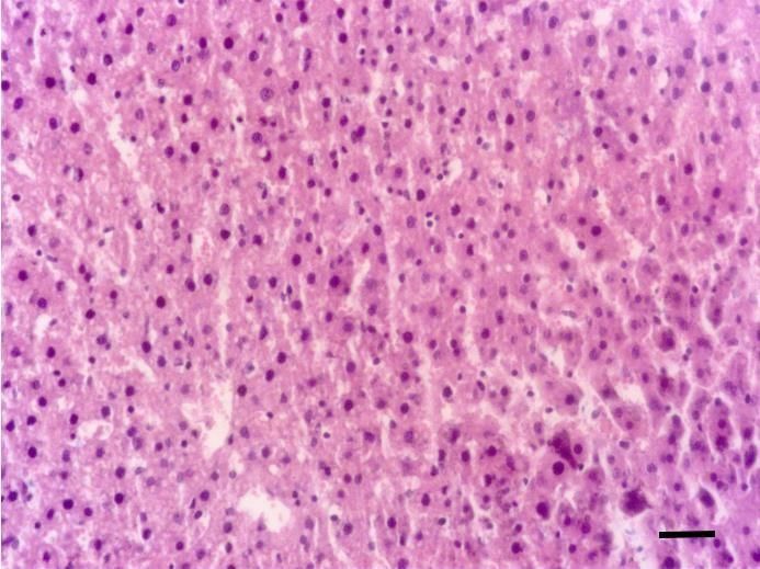
Aus der Herzchirurgischen Klinik und Poliklinik
 Klinik der Ludwig-Maximilians-Universität München Großhadern
            Vorstand: Prof. Dr. med. Christian Hagl

Intravital microscopy of lung ischemia reperfusion
                injury in a rat model

                            Dissertation
             zum Erwerb des Doktorgrades der Medizin
                 an der Medizinischen Fakultät der
             Ludwig-Maximilians-Universität zu München

                           vorgelegt von

                            Rongrui Na

                                aus

                              Sichuan

                                2021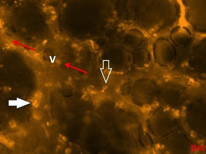
Mit Genehmigung der Medizinischen Fakultät
                             der Universität München

Berichterstatter:                    Prof. Dr. med. René Schramm, PhD

Mitberichterstatter:                 PD Dr. med. habil. Tobias Heer

Dekan:                               Prof. Dr. med. dent. Reinhard Hickel

Tag der mündlichen Prüfung:          14.01.2021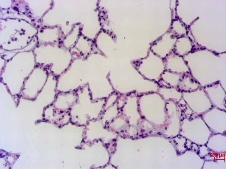
CONTENTS

1.     Abstract                                             1

2.     Zusammenfassung                                      2

3.     Introduction                                         3

3.1.   Lung transplantation                                 3

3.2.   Pulmonary ischemia-reperfusion injury                4

3.3.   Intravital fluorescence microscopy (IVM)            10

4.     Aim of the study                                    14

5.     Materials and Methods                               15

5.1.   Animals                                             15

5.2.   Experimental procedures                             15

5.2.1. Anesthesia                                          15

2.2.2 Microsurgical procedures                             16

5.3.   Intravital fluorescence microscopy                  17

5.3.1. Imaging                                             17

5.3.2. Parameters                                          18

5.4.   Histopathological analyses                          20

5.5.   Statistical analysis                                22

6.     Results                                             23

6.1.   Intravital fluorescence microscopic imaging (IVM)   23

6.2.   Histopatholoic analyses                             36

7.     Discussion                                          43

8.     References                                          51
Abstract

       This thesis establishes and validates an experimental animal model to analyse
leukocyte    recruitment     and     microvascular     dysfunction     in    pulmonary
ischemia/reperfusion injury by intravital fluorescence microscopy in the rat.

     The pleural surface of the rat left lung has been exposed by atraumatic
microsurgical techniques and fluorescence epi-illumination was used to visualize
subpleural pulmonary microcirculation under baseline conditions and during
reperfusion after 30 min of ischemia.

       The results show that this experimental setup bears the required spatial and
temporal resolution to study postischemic leukocyte responses within the pulmonary
microcirculation. All segments of pulmonary microvascular tree can be differentially
analysed, including feeding arterioles, alveolar capillaries and draining postcapillary
venules. The data analyses demonstrate that the relatively mild ischemic insult of 30
min provokes a detectable leukocytic response, i.e. significantly increased leukocyte-
endothelial cell adhesive interactions in postcapillary subpleural pulmonary venules
during reperfusion. Microhemodynamics were not significantly altered during
reperfusion. These results translate into the histopathological analyses, confirming mild
affection of the pulmonary tissue structures after 90 min of reperfusion. Confirming i)
the relatively mild ischemic insult to the lung and ii) virtual absence of trauma by the
microsurgical preparations, remote organ damage was basically absent the heart and
kidney, but only minimally seen in the liver.

       In conclusion, the established experimental setup allows for reproducible
analyses pulmonary ischemia/reperfusion injury on the microcirculatory level in the rat
lung. The use of this basic approach may serve as a reliable basic model for further
studies on pulmonary ischemia/reperfusion injury and experimental allo- and
xenogeneic lung transplantation.

                                                                                            1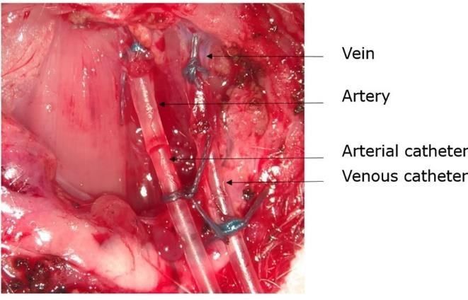
Zusammenfassung

Im Rahmen dieser Promotionsarbeit wird ein tierexperimentelles Modell zur Analyse
von Leukozytenrekrutierung und mikrovaskulärer Dysfunktion während pulmonalem
Ischämie/Reperfusionsschaden mittels intravitaler Fluoreszenzmikroskopie in der
Ratte etabliert und evaluiert.

Die   pleurale    Oberfläche     der   linken   Lunge     wird   durch     atraumatischen
mikrochirurgischen Techniken exponiert. Mittels Fluoreszenz-epi-Illumination wird
die subpleurale Mikrozirkulation der Lunge unter Basisbedingungen und während
Reperfusion nach einer 30 minütigen Ischämie visualisiert.

Die Ergebnisse zeigen, dass der experimentelle Ansatz die notwendige räumliche und
zeitliche Auflösung besitzt, um die postischämische Leukozytenantwort in der
pulmonalen     Mikrozirkulation    darzustellen.   Alle   Segmente       der   pulmonalen
mikrovaskulären Strombahn können differenziert analysiert werden. Dies beinhaltet
zuführe nde Arteriolen, alveoläre Kapillaren und drainierende postkapilläre Venolen.
Die Datenanalyse zeigt, dass der relative milde 30-minütige, ischämische Insult eine
detektierbare Leukozytenantwort provoziert, d.h. significant erhöhte Leukozyten -
Endothelzell Adhäsionen in postkapillären pulmonalen Venolen während Reperfusion.
Die Mikrohämodynamik war nicht significant beeinträchtigt während Reperfusion.
Diese Ergebnisse übersetzen sich in die histopathologische Analysen, eine milde
Affektion der Lungengewebestruktur nach 90 Minuten Reperfusion bestätigen.
Sekundäre Organschäden in Herz und Niere wurden nicht detektiert und waren nur
milde ausgeprägt in der Leber zu finden, was einerseits die relative Milde der
Lungenischämie und andererseits die nahezu atraumati schen mikrochirurgische
Präparationen bestätigt.

Zusammenfassend erlaubt der experimentelle Ansatz eine reproduzierbare Analyse des
Ischämie/Reperfusionsschadens in der Rattenlunge auf mikrovaskulärer Ebene. Dieser
Ansatz kann als zuverlässiges experim entelles Basismodell für weitergehende
Untersuchungen des Ischämie/Reperfusionsschadens in der Lunge, sowie allo - und
xenogener Lungentransplantation dienen.

                                                                                            2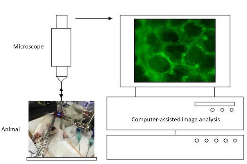
3.     Introduction

3.1.   Lung transplantation

       Lung transplantation has become a valid option in the treatment of end-stage
lung diseases, refractory to any other medical and/or surgical therapy [1]. Since the first
successful clinical transplantation of a donor lung in 1963, the results after lung
transplantation for end-stage lung disease have steadily improved. Most recent data
indicate a median survival after lung transplantation of 6.7 years and a 1-year survival
of 8.9 years [2]. The improved outcome has been achieved by refinements of surgical
techniques, e.g. periprocedural use of extracorporeal circulation [3], and advances in
immunosuppressive therapy, e.g. introduction of calcineurin inhibitors [4, 5]. Despite
all these advances, survival after lung transplantation is still limited and the overall
results are far from perfect.

       Transplanted lung allografts are damaged in the postoperative and long-term
course by several mechanisms (Figure 1). Such include the ischemia/reperfusion injury
mediated by the unspecific innate immunity, as well as acute and chronic rejections
through specific adaptive immune responses [5, 6]. In addition, donor-specific
antibody-mediated rejection episodes may occur, although there is only limited
understanding of when and how this entity of post lung transplant organ injury develops
and works [7]. Regarding the continuous deterioration of graft function post-transplant,
it is a well-established understanding that ischemia/reperfusion injury occurs within the
first days after transplantation, while acute cellular rejection episodes may occur at
different time points longer-term. However, the highest incidence of acute cellular
rejection episodes is within the first 6 to 12 months after lung transplantation [2]. The
detailed mechanisms underlying chronic lung allograft rejection are not completely
understood at present, but it manifests clinically in steadily declining lung function test
parameters and the pathophysiologic correlate is a fibrotic swelling of the terminal
bronchioli, previously designated as the bronchiolitis obliterans syndrome. However,
more recent definitions of the chronic lung allograft dysfunction (CLAD) further
differentiate between more obstructive or restrictive functional and structural
destruction of the transplanted lung allograft [8].

                                                                                              3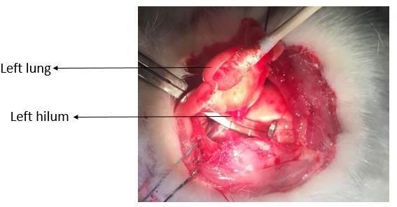
Figure 1: Lung injury after transplantation. The function of a transplanted lung allograft
is at risk during the early postoperative period due to ischemia/reperfusion injury
provided by the innate immune system. The specific and adaptive immune responses
trigger the frequency and intensity of acute rejection episodes as well as chronic
rejection determining the outcome of lung transplantation long-term.

3.2.   Pulmonary ischemia-reperfusion injury

       Clinical aspects

       The magnitude of the injurious net effect following ischemia during reperfusion
primarily depends on and is proportionate to the duration of the anoxic phase following
organ retrieval, during transportation and implantation into the recipient [9, 10]. It has
been well recognized that tolerance of transplanted organs towards ischemia can be
improved by vigorous organ cold preservation, i.e. the organs are perfused in the donor
by a chilled preservation solution at the time of retrieval and are transported on ice to
the recipient operation. This applies also to lung allografts, in which colloidal, buffered
electrolyte solutions are used [11, 12]. However, despite improved procurement
techniques and optimal preservation measures, the ischemic time of a lung allograft
must not be extended over a maximum of 9 to 12 hours [13, 14], because this would
significantly endanger a positive outcome of the transplant procedure.

                                                                                              4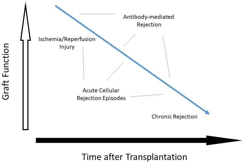
The transplantation of a lung allograft differs to a certain extend from other solid
organ transplantations as the nutritive arterial vasculature is not connected to the
recipients circulation. Only pulmonary arteries carrying deoxygenated blood to the lung
are anastomosed. Drainage of blood is conducted through the pulmonary veins into the
recipients left atrium. Bronchial arteries derived from the systemic blood stream are not
anastomosed [15]. It has been suggested that this reasons, why mainly the bradytroph
bronchial tissue is at risk during severe ischemia/reperfusion injury of the lung after
transplantation, potentially leading to impaired healing of the anastomosed bronchi or
even necrotising disruption of this fragile airway connection [15, 16].

       Ischemia/reperfusion injury has been considered to manifest clinically as the
primary graft dysfunction (PGD) after lung transplantation. It has a reported incidence
of between 10%-30% and is a major cause of death in early post-transplantation days
[17-19]. According to the 34th report of the registry of the International Society for
Heart and Lung Transplantation (ISHLT), PGD is one major reason of death within the
first 30 days of transplantation [4]. The ISHLT proposed a clinical definition of PGD
and that is the presence of hypoxemia and radiographic appearance of diffuse
pulmonary opacities in post-transplant chest X-rays without other identifiable causes
within the first 72 hours after surgery. Its severity is judged clinically depending on the
ratio of arterial oxygen fraction (PaO2) per inspired oxygen fraction (FiO2) [20]. It
remains under discussion whether machine perfusion techniques during transportation
of the allograft and the corresponding limitation of the anoxic phase may blunt
ischemia/reperfusion injury after transplantation and thereby improve postoperative
outcomes [21]. There are some indicator suggesting that such techniques may reduce
the incidence of post-transplant PGD. This however, may not impact on long-term
outcomes [22].

       Pathophysiologic aspects

       The absence of nutritive arterial perfusion with oxygenated blood during
ischemia leads to a depression of mitochondrial energy production and a depletion of
adenosine triphosphate. This affects the intracellular ion homeostasis and cellular
membrane permeability. Cellular necrosis and activation of apoptotic cell signals may

                                                                                              5
follow [23]. In addition, the fall in cellular energy content activates self-propagating
deleterious cascades [23]. It is directly related to the production of reactive oxygen
species (ROS) and reactive nitrogen species (RNS). Their production is induced during
ischemia [24] and will increase rapidly during reperfusion [25]. Among all the potential
sources and the formation pathways of ROS and RNS within cells, xanthine oxidases,
nicotinamide adenine dinucleotide phosphate (NADPH) oxidases, and nitric oxide
synthases (NOS) have been investigated [26].
Reperfusion of a transplanted allograft starts with the release of blood flow from
the donor pulmonary trunc, passing the pulmonary arterial anastomosis into the grafts’
pulmonary arterial vascular tree. The start of reperfusion is the restoration of blood flow
in a previously anoxic graft, but it is at the same time the onset of microvascular
perfusion failure within the grafted tissue [6]. The so-called “no reflow phenomenon”
exacerbates hypoxia/anoxia in the grafted tissue despite the macroscopic restitution of
the grafts blood circulation. The phenomenon describes the post-ischemic collapse of
the graft’s microcirculation due to endothelial cell swelling, hemoconcentration,
narrowing of nutritive capillaries and interstitial edema formation [27-30].
Ischemia/reperfusion injury of a transplanted graft is an inflammatory response, an
unspecific reaction rather than a cognitive response. The inflammatory response during
ischemia reperfusion is a sterile process, which is activated by sterile cell death or injury.
It has been confirmed that receptors, such as Toll-like receptors (TLRs) and DAPMs
play a central roles in such immune response [31, 32]. When ligands bind to TLRs, cell
signal pathways, such as NF-kB, mitogen-activated protein kinase (MAPK) and type I
interferon pathways are activated, leading to the formation of pro-inflammatory
mediators, such as cytokines and chemokines [33]. Neutrophils, monocytes and
macrophages exert complementary functions during inflammation, also in the lung [34].
It is thought that, after stimulation, tissue resident macrophages and sentinel monocytes
release chemokines and pro-inflammatory cytokines to recruit circulating white blood
cells, primarily polymorphonuclear leukocytes, into the inflamed tissue compartment.
Neutrophil infiltration and granule release is followed by monocyte recruitment in a
secondary phase.
The early activation of alveolar macrophages via TLR-4 is thought to play a
critical role in the development of lung ischemia/reperfusion injury [35, 36]. It has
been shown that there was a significant increase in the number of alveolar macrophages
6in bronchi-alveolar lavage fluid (BALF) after one hour hypoxia [37]. Using a murine
lung transplantation model, it was shown that depletion of donor macrophages
attenuated ischemia/reperfusion injury [38]. Suppressing of TLR-4 signalling inhibited
the activation of alveolar macrophage as well and lung ischemia/reperfusion injury was
almost completely attenuated [39]. Due to oxidative stress, alveolar macrophages
produce monocyte chemoattractant protein-1 (MCP-1) and macrophage inflammatory
protein-2 (MIP-2) which contribute to infiltration of neutrophils and lead to pulmonary
oedema [40]. Beside this, macrophages can release tumour necrosis factor-α (TNF-α),
one of the pro-inflammation cytokines which can lead to acute lung damage directly
[41]. In a rabbit lung ischemia/reperfusion injury model, TNF-α rose continuously
during reperfusion and by attenuation TNF-α, the alveolar cell apoptosis and acute lung
injury could be inhibited [42].
Also monocytes have been suggested to be involved in pulmonary
ischemia/reperfusion injury. Classical monocytes are considered to be pro-
inflammatory, since they can produce pro-inflammatory cytokines and differentiate into
macrophages and dendritic cells [43]. In a model of lung ischemia/reperfusion injury,
classical monocytes were found to be mobilized from their natural reservoir in the
spleen, to enter into pulmonary tissue and provoke subsequent neutrophilic infiltration
into the lung by releasing IL-1β [44]. Non-classical monocytes are thought to regulate
wound healing and the resolution of inflammation. As such, they may be important
mediators in lung injury as well. It was shown in a murine lung transplantation model
that the depletion of donor intravascular non-classical monocytes, both
pharmacologically and genetically, attenuated lung graft injury [45]. However, the
precise role of non-classical monocytes, particularly in lung ischemia/reperfusion
injury is still unclear [44].
The recruitment of circulating polymorphonuclear leukocytes (PMNLs), i.e.
neutrophils, is a paramount feature of ischemia/reperfusion injury. This has been
studied extensively in various organs such as the liver, striated muscle, intestine, kidney
and the heart [46-49].
There has been conflicting data on the role of neutrophils in pulmonary
ischemia/reperfusion injury. Neutrophils were initially thought to be unnecessary in
lung ischemia/reperfusion injury [50, 51]. However, it was postulated that lung
7ischemia/reperfusion injury has distinct phases [52, 53]. There seems to be an early
phase, which is independent of neutrophils, while neutrophilic infiltration occurs later
and is associated with increased pulmonary vascular permeability. Obviously, the
activation of the pulmonary microvascular endothelium is a crucial part for the
recruitment of neutrophils during lung ischemia/reperfusion injury. It is induced by
TNF-α, IL-1β and will lead to an up-regulation of certain adhesion molecules on the
luminal surface of the pulmonary vascular endothelium [54]. The recruitment of blood-
borne white blood cells into the lung has been suggested to follow the same pattern of
neutrophil recruitment in other organs (Figure 2). Neutrophils are thought to roll along
the activated pulmonary endothelial surface orchestrated by a cytokine gradient and
mediated by adhesion molecules of the selectin family, i.e. P- and/or L-selectin on
neutrophils binding to E-selectin on the pulmonary endothelial surface. Further
activation of the rolling cell will slow it down and more firm attachment is then
mediated by the integrin family o f adhesion molecules. The final step is the
transendothelial migration. It is unknown, however, whether the transmigrating cell
passes the endothelial lining between or directly through the endothelial cells [54]. The
number of infiltrated neutrophils correlates to the severity of tissue damage [55]. By
reducing the alveolar infiltration of neutrophils in a rat lung transplantation model, the
gas exchange after surgery was improved [56]. It was thought that neutrophils exert
lung ischemia/reperfusion injury by releasing elastase and granular proteins [57].
Neutrophil extracellular trap is an extracellular complex, including DNA, histones and
neutrophil granular proteins [58]. Caudrillier et al found that neutrophil extracellular
traps were created during lung transfusion and related to acute lung injury [59]. It was
further shown that the neutrophil extracellular traps accumulated and contribute to
primary graft dysfunction after lung transplantation as well [60].
8Figure 2: Schematic sequence of leukocyte recruitment in a microvessel. The
extravasation of circulating leukocytes during inflammation may be defined as a
sequential cascade of events including (A) free flowing, B) rolling, C) activation, D)
firm adhesion and, finally, E) transmigration of inflammatory cells. Blood flow is from
left to right. (The use if this figure has kindly been granted by Rene Schramm, Heart
and Diabetes Center NRW, Bad Oeynhausen, Germany)
Despite these lines of evidence strongly suggesting that circulating leukocyte
recruitment from the pulmonary vasculature is a key feature in lung
ischemia/reperfusion injury, it has to be noted that the underlying evidence has been
gathered only indirectly. Direct visualization of the pulmonary microvasculature during
reperfusion has not been performed. It has to be considered also that the so- called
marginated pool of leukocytes, i.e. a large number of granulocytes residing in the lung
under normal physiological conditions, may have affected the results of previous
studies [61-63].
Ischemia/reperfusion injury can affect platelet function and the coagulation
system due to ROS induced increases in extracellular adenosine di-phosphate
concentrations and endothelial cell injury induced activation of the von Willebrand
factor (vWF) and thrombin [64-66]. Activated platelets may also interact directly with
endothelial cells via selectins, which has been shown to cause microvascular
constriction, aggravate postischemic injury and lead to thrombus formation [10]. P-
selection can regulate platelet-endothelium interaction and promote platelet-leukocyte
interaction during ischemia/reperfusion injury [67]. Finally, activated platelets form
microthrombi perpetuating endothelial cell injury and microvascular dysfunction [68].
9Microvascular permeability is increased because of hypoxia during reperfusion,
triggered by the rapid production of ROS, complement activation, leucocyte-
endothelial cell adhesion and platelet-leucocyte aggregation [6, 69]. In lungs, it presents
as the alveolar-capillary barrier dysfunction which can lead to pulmonary edema and
ultimately induces respiratory failure [70]. Vascular endothelial growth factor and
angiopoietin-2 are considered as mediators of vascular permeability in lung
ischemia/reperfusion injury [71, 72]. Several approaches may attenuate this alveolar-
capillary dysfunction. For example, dimethylthiourea reduces postischemic
microvascular dysfunction via its hydroxyl radical scavenger effects [73]. Hypoxia-
inducible factor 1α (HIF-1α) controls endothelial function, but is downregulated in lung
ischemia reperfusion injury. Stabilizing it can prevent endothelial dysfunction [74].
Sphingosine 1-phosphate (S1P) controls endothelial cell tight junctions and
supplementation with this compound before reperfusion inhibits inflammatory cytokine
production and improves oxygenation in murine lung ischemia/reperfusion [75].
Antibody targeting of αvβ5, an integrins that regulates endothelial permeability, also
improved the postischemic oxygenation in a murine lung transplantation model [76].
3.3. Intravital fluorescence microscopy (IVM)
The principle of fluorescence microscopy is that a distinct fluorescent dye can
be rayed with light of a defined wavelength (excitation wavelength) and in response
emits a quantum of light with a corresponding, different wavelength (emission
wavelength) (Figure 3). This phenomenon is based on the fact that the excitation light
transports energy. Distinct electrons on the fluorescent dye molecule adsorb photons
and are thereby lifted to another energy level. This is an unstable condition and these
electrons fall back into their original, lower energetic level immediately. This fall back
liberates another quantum of energy, which is emitted as light. Because the liberated
energy content is somewhat smaller, the emitted light quantum has a longer wavelength.
10Figure 3: Schematic illustration of the excitation of a fluorescent dye on the atomic
level. The schematic images show a molecule with a central atomic nucleus (large
central black spot) encircled by three electron energy levels (blue circles). A) depicts
the excitation of a resting electron on the second level (open spot) with fluorescent blue
light. The concomitant energy transfer lifts the electron up to a higher energy level
(filled spot). B) depicts the fact that the electron immediately loses this additional
energy, falls back to a lower level and the originating slightly lower energy is emitted
as fluorescent green light.
Epi-illumination microscopy uses filter sets, which allow to differentiate
between excitation and emission light (Figure 4). The light source (herein a mercury
lamp) of the microscope emits light through an excitation filter, which absorbs the wide
spectrum of light letting pass exclusively light of the defined excitation wave length.
This virtually single-coloured light hits onto the fluorescent dye and provokes the
emission of lower energetic excitation light, which is reflected back to a filter letting
pass only light of the defined excitation wavelength. Reflected light of the excitation
wavelength is absorbed. The resulting image is constructed of the residual emited light.
11Figure 4: Schematic illustration of the principle of an epi-illumination fluorescence
microscope. A: Light source; B: Filter for Excitation light; C: Beam splitter; D:
Experiment object with fluorescent dye; E: Filter for Emission light; F: Camera
IVM in experimental research on ischemia/reperfusion injury
IVM has been utilized for the analyses of cell-cell interactions and
microvascular dysfunction in a large array of experimental models. The use of this
technique has allowed to understand the stepwise mechanisms of the leukocyte
extravasation process during inflammation. For example, in hepatic injury, selectin-
mediated leukocyte rolling was found to be a prerequisite for subsequent integrin-
mediated firm adhesion of leukocytes [77]. Such fundamental and distinct roles of P-
selectin and the integrin lymphocyte function associated antigen (LFA)-1 were directly
visualized during in the postischemic microcirculation of the murine colon [78].
Postischemic vasoreactivity could be demonstrated in in the intestinal microcirculation
as well [29]. In the murine striated cremaster muscle preparation, it was demonstrated
12that chemokines regulate selectin-dependent neutrophil trafficking in a mast cell-
controlled manner [79].
IVM of the thoracic organ microcirculation has been challenging, since
tremendous physiologic movements elicited by the beating heart and the ventilated
lungs have to be counterbalanced. Nevertheless, distinct experimental models have
been elaborated to circumvent such hurdles and both the subepicardial coronary
microcirculation of the heart and the subpleural pulmonary microcirculation of the lung
could be visualized even in small animals as the mouse [80, 81]. In both latter organs,
the sequential leukocyte extravasation cascade has been visualized. However,
pulmonary ischemia/reperfusion injury has not been investigated so far by IVM in a
rodent model.
134. Aim of the study
Detailed understanding of the pathophysiological mechanisms underlying
pulmonary ischemia/reperfusion injury are key to improve outcomes after lung
transplantation clinically. Although circulating white blood cell recruitment and
microvascular dysfunction during pulmonary ischemia/reperfusion injury have been
investigated previously, evidence on the underlying mechanisms have been gathered
mostly indirectly, experimentally and clinically. Further, results obtained from cell
cultures and isolated organ perfusion experiments may not resemble the complex
interplay of pathophysiological processes as in a live individual.
The aim of the present study was it therefore to establish and validate an
experimental approach which allows to study leukocyte recruitment and microvascular
dysfunction within the pulmonary microcirculation during ischemia/reperfusion injury
in a rodent model by means of intravital fluorescence microscopy. The experimental
setup should also allow to monitor potential secondary organ dysfunction in the heart,
liver and kidney by histopathological means. The experimental setup should be
reproducible and provide an experimental basis for studies on experimental lung
transplantation.
145. Materials and Methods
5.1. Animals
Female Lewis rats (Charles River) weighing 220-240g were used. The animals
had free access to standard pellet food and tap water and were kept in 12h light and 12h
dark cycles. All experiments were approved by the governmental ethical committee for
animal experimentation and performed as the legislation on the protection of animals.
Animals were sacrificed immediately after the experiment in deep anaesthesia through
exsanguination.
5.2. Experimental procedures
5.2.1. Anesthesia
Global anaesthesia was induced by intramuscular injection of a mixture of
medetomidine (150 µg/kg body weight, Domitor), midazolam (2 mg /kg body weight,
Dormicum) and fentanyl (5 µg/kg body weight, Fentanyl-Janssen) into the right
quadriceps femoris muscle.
The anesthetized animals were placed in a supine position for subsequent
endotracheal intubation with a 16G catheter (Introcan Safety, Braun Melsungen AG,
Melsungen, Germany) under direct vision. The catheter was then connected to small
animal ventilator (Small animal ventilator KTR-5, Harvard Apparatus, Canada).
Correct endotracheal intubation was additionally verified by synchronic expansion of
the thoracic cage. The basic settings for the ventilator were settled to 75 strokes per
minute with an inspiration/expiration ratio of 1 / 2. The air flow was adjusted to 0.5
liters / min with a positive end-expiratory pressure of 9 cm H2O without additional
oxygen supply. The respirator settings were adjusted throughout the experimentations
based on arterial blood gas analyses.
Central catheter implantation was performed as previously described [82].
Briefly, after intubation, an incision was made on the right paramedian aspect of the
neck. of the rat, then the right external jugular vein and the right common carotid artery
were exposed gently. The two vessels were secured proximally and distally by ordinary
silk ordinary silk suture. Two central 20G catheters (BD Microlance, Becton Dickinson
15S.A. Fraga, Huesca, Spain) were then inserted into the two vessels, flushed with saline
and secured by the silk sutures (Figure 5).
Figure 5: Central catheterization of the right jugular vein and common carotid artery.
2.2.2 Microsurgical procedures
The lateral thoracotomy approach to the lung has been described in a murine
model previously by Gielis et al [83]. After placement of the animal from the supine
into the right lateral position, the chest wall was shaved gently. The incision was made
beginning at the medial sternal side of the left rib cage and was followed along the 5th
intercostal space directed to the spine. After disconnecting the endotracheal tube from
the respirator, great care was taken when blunt penetration of the intercostal muscular
layer was performed in order to avoid any alteration of the visceral pleural surface of
the lung. Respiration was continued immediately after the thoracic cavity has been
opened safely. The rib cage was spread open by placing two 1-0 holding sutures
(Prolene, Polypropylen, Ethicon, USA) around the adjacent upper and lower ribs and
pulling them gently towards the cranial and caudal directions. The lung tissue was
moistened by temperature (37°C) saline regularly throughout the entire
experimentation. A moistened cotton stick was used to gently move the pulmonary
16tissue taking great care to not tough the ventral aspect of the lung, i.e. the region of
interest for subsequent intravital fluorescence microscopy. The pulmonary ligament
was cut off in order to get a better access to the lung hilum. Before clamping of the
hilum for induction of ischemia, 125 I.U. of heparin (Rotexmedica, Trittau, Germany)
was administered intravenously (i.v.). Clamping was performed by use of an ordinary
microvascular vessel clamp (Figure 6). The lung and pleural cavity was kept moistened
with temperature saline (37°C) throughout the entire experiments.
Figure 6: Macroscopic image of the surgical field during clamping of the left
pulmonary hilum, i.e. during ischemia.
5.3. Intravital fluorescence microscopy
5.3.1. Imaging
The microsurgically equipped and prepared animals were transferred to the
intravital microscope stage. Intravital fluorescence labelling of circulating leukocytes
was established by i.v. injection of 0.5 ml of Rhodamine 6G (Molecular weight 479 Da;
0.1 mg/ml, Sigma Chemicals). For plasma labelling, an injection of Fluoresceine Iso-
Thyo-Cyanate (FITC)-Dextran (Molecular weight 150 kDa; 2 mg/ml, Sigma Chemicals)
was injected via the central venous catheter. The moistened left lung surface was
carefully covered with a horizontally positioned cover glass slip, which was fixed to a
micromanipulator. The hydrostatic forces of the moistening saline attached the lung
surface under the cover glass slip (Figure 7). The subpleural pulmonary
17microcirculation of the left lung was visualized by a modified microscope (Zeiss,
Oberkochen, Germany), which provides blue- (excitation wave length 490nm) and
green-light (excitation wavelength 530nm) epi-illumination and is equipped with a
filter set (Filter set 62HE, Colibri, Zeiss, Germany) to visualize the corresponding
emission wave lengths of 510nm and 560nm, respectively. Water immersion lenses
with a 10- and 20-fold magnification were used. All microscopic images were televised
and recorded onto a computer-assisted hard disc drive.
Figure 7: Schematic experimental setup for intravital fluorescence microscopy (IVM)
of the rat pulmonary microcirculation. Animals were placed in a right-sided position on
the microscope stage. Microscopic images were transferred to a computer-assisted
image analysis system and saved onto a hard disc for subsequent off-line quantification
of experimental parameters.
5.3.2. Parameters
Intravital fluorescence microscopy of the left lung subpleural pulmonary
microcirculation was performed at three different time points, i.e. before induction of
ischemia (T0), at 30min of Reperfusion (T1) and after 90 min of reperfusion (T2). At
18least four different areas were analysed at each individual time point for at least 30
seconds.
Leukocyte endothelial cell adhesive interactions
Firm leukocyte adhesion was defined as the number of intravascular leukocytes
firmly attached for >15 seconds to the pulmonary microvascular endothelial cell lining
of pulmonary arterioles and venules, given in cells / mm² endothelial surface.
Leukocyte rolling was defined as the number of intravascular leukocytes loosely
interacting with and rolling along the endothelial lining passing a reference point within
pulmonary arterioles and venules during the observation period of 30 seconds, given in
cells / min.
Alveolar leukocyte recruitment
The numbers of Rhodamine 6G-stained leukocytes within alveolar spaces were
counted and are given as cells per high power field (HPF) [aU].
Macromolecular leakage
The extravascular fluorescence intensity was measured within alveolar spaces
after intravenous injection of the fluorescent dye FITC-dextrane at T0, T1, and T2. To
account for different animal body weights and circulating blood volumes the individual
measures at T1 and T2 were normalized by the intra-individual measurements at T0.
Distance of air-filled alveolar spaces
The distance of air-filled alveolar spaces is an indirect measure for interstitial
pulmonary edema formation and is given in µm.
Microvascular diameters
The diameters of pulmonary terminal arterioles, alveolar capillaries and
draining postcapillary venules are given in µm.
193.4. Histopathological analyses
After completion of the experimental procedures and intravital microscopic
analyses, i.e. after 90 min of pulmonary reperfusion (T2), both lungs, the heart, the
kidneys and the livers were harvested for subsequent histopathological examination.
The lungs were harvested by ligation and transection in inspiration. The other organs
were harvested by transection of incoming and outgoing vessels. All organs were fixed
in 4% paraformaldehyde overnight and were then stored in 80% ethanol until further
processing.
For routine hematoxylin-eosin staining, organ tissues were dehydrated in an
ascending concentration of ethanol series and the ethanol was removed by aromatic
hydrocarbon xylene. Afterwards all organs were embedded in paraffin wax and cut into
4 μm thick layers with a microtome (Leica Biosystems, Germany) and fixed on a glass
slide. The sections on the glass slips were dewaxed in a descending concentration of
ethanol series and stained by haematoxylin and eosin in an ascending concentration of
ethanol series. The basophilic structures were stained blue with haematoxylin, the
acidophilic structures were stained red with eosin.
The evaluation of the histological specimens were carried out by use of a light
microscope (Zeiss, Oberkochen, Germany), which was connected to a digital camera
(Zeiss, Oberkochen, Germany) and a corresponding computer-assisted software, the
Axiovision (Zeiss, Oberkochen, Germany). 25 fields per slide were evaluated in each
organ in 200× high-power-fields (HPFs).
Pulmonary injury was semi-quantitatively analysed by using a lung injury score
system, previously published by the American Thoracic Society Workshop [84] (Table
1). Secondary organ damage to the kidney and liver were analysed quantifying the areas
of tubular necrosis per HPF [85] and by use of the previously published Suzuki Score
system (Table 2) [86], respectively.
20Score per field
Parameter 0 1 2
A. Neutrophils in the alveolar
space None 1-5 >5
B. Neutrophils in the interstitial
space None 1-5 >5
C. Hyaline membranes None 1 >1
D. Proteinaceous debris in the air
space None 1 >1
E. Alveolar septal thickening 4x
Final score= [(20 ×A) + (14 ×B) + (7 × C) + (7 × D) + (2 × E)]/( number of fields ×
100)
Table 1: The Lung Damage Score for quantification of structural pulmonary injury.
Adopted from [84].
Score Congestion Vacuolization Necrosis
0 None None None
1 Minimal Minimal Minimal
2 Mild Mild Mild
3 Moderate Moderate Moderate
4 Severe Severe Severe
The level of damage was defined by planimetry in each high power field. Minimal = 0
- 10%, Mild= 11% - 30%, Moderate= 31% -60%, Severe= over 61%
Table 2: The Suzuki Score system for quantification of structural liver injury. Adopted
from [86].
215.5. Statistical analysis
Data are given as mean values ± standard error of the mean (SEM) and n
represents the number of animals per experimental group and/or time point. Data was
analysed using one-way analyses of variance (ANOVA) and corresponding post-hoc
testing utilizing statistic computer software packages (SPSS 22, IBM, USA; SigmaStat,
Jandel Scientific, USA). A value of probability of less than 0.05 was considered to
indicate a statistically significant difference.
226. Results
6.1. Intravital fluorescence microscopic imaging (IVM)
During the learning curve, six animal preparations did not allow for the planned
IVM observations. These animals had to be sacrificed because of technical failure,
uncontrolled respiratory acidosis or insufficient exposure for intravital imaging. In a
total of 8 animals, successful surgical preparation was followed by intravital
fluorescence microscopic observation (IVM) of the pulmonary microcirculation before
ischemia and at 30 min (T1) as well as 90 min of reperfusion (T2).
The entire pulmonary microvascular tree could be visualized after contrast
enhancement with FITC-Dextran including feeding pulmonary arterioles, alveolar
capillaries and draining pulmonary venules (Figures 8-10). Pulmonary arterioles were
differentiated from venules by the peripheral branching of blood flow and visible
pulsatility. Venules were draining the blood from peripheral branches and did not show
any pulsatility. Bronchial arteries could not be differentiated.
23Figure 8: Intravital microscopic image of the pulmonary microcirculation in the left
lung upper lobe. Contrast enhancement after intravenous injection of FITC-Dextran at
90 min of reperfusion (T2). Blue arrows indicate the distributing and divergent blood
flow of the central arteriole into alveolar capillaries. Scale represents 20 µm.
24Figure 9: Intravital microscopic image of the pulmonary microcirculation in the left
lung upper lobe. Contrast enhancement after intravenous injection of FITC-Dextran
prior to induction of ischemia (T0). The asterisk indicates an exemplary alveolar space
surrounded by alveolar capillaries indicated by “C”. The red arrows indicates the blood
flow in a draining pulmonary venule. Scale represents 20 µm.
25Figure 10: Intravital microscopic image of the pulmonary microcirculation in the left
lung upper lobe. Contrast enhancement after intravenous injection of FITC-Dextran
prior to induction of ischemia (T0). Red arrows indicate the collecting blood flow from
alveolar capillaries into a draining venule (V). Scale represents 20 µm.
26Contrast enhancement with Rhodamine-6G allowed to visualize intravascular
leukocytes and occasionally thrombocytes. Leukocytes were differentiated from
thrombocytes based on their size difference. Visualized leukocytes were presumably
granulocytes as reflected by their irregularly and granulated nuclear staining (Figure
11).
Figure 11: Intravital microscopic image of the pulmonary microcirculation in the left
lung upper lobe. Contrast enhancement after intravenous injection of Rhodamine 6G at
90 min of reperfusion (T2). Red arrows indicate the collecting blood flow in a draining
venule (V). The open arrow indicates an adherent leukocyte. The filled arrow indicates
a group of intensely stained small thrombocytes. Scale represents 20 µm.
27IVM – Parameters
Leukocyte endothelial cell adhesive interactions
IVM allowed for quantitative and qualitative analyses of leukocyte endothelial-
cell adhesive interactions in the pulmonary microcirculation of the left lung surface.
Rhodamine-6G stained leukocytes were interacting with the pulmonary vascular
endothelium only in post-capillary venules. Virtually no adhesive interactions were
observed in arterioles at any time point. In addition, interacting leukocytes were found
to be firmly attached to the post-capillary venular endothelium (Figure 11). Rolling
interactions were just occasionally found.
Figure 12: Firm leukocyte adhesion in post-capillary venules of the left lung
pulmonary microcirculation. Data are given as mean values ± standard error of the
mean and n represents the number of animals at the different time points, i.e. prior to
induction of ischemia (T0), at 30 min of reperfusion (T1) and at 90 min of reperfusion
(T2). The asterisk indicates a statistically significant difference when compared to T0
(PFirm leukocyte adhesion was increasing from 117.14 ±10.12 cells/mm 2 at T0 (prior to
induction of ischemia) to 199.37 ±15.03 cells/mm 2 at T1 (30min of reperfusion) and
294.76 ±13.31 cells/mm 2 at T2 (90 min of reperfusion). The difference between firm
leukocyte adhesion at T0 and T2 was statistically significant (P=0.005; Figure 12).
Alveolar leukocyte recruitment
The numbers of Rhodamine-6G stained leukocytes within the alveolar spaces
were 1.88 ± 0.17 [aU] at T0 (prior to induction of ischemia), 3.71 ± 0.21 [aU] at T1
(30min of reperfusion) and 7.8 ± 0.49 [aU] at T2 (9 0 min of reperfusion). There were
statistically significant differences between the alveolar space leukocyte counts
between T0 and T2 (P=0.002) as well as between T1 and T2. (P=0.033; Figure 13).
Figure 13: Numbers of leukocytes within alveolar spaces the left lung. Data are given
as mean values ±standard error of the mean and n represents the number of animals at
the different time points, i.e. prior to induction of ischemia (T0), at 30 min of
reperfusion (T1) and at 90 min of reperfusion (T2). * indicates a statistically significant
difference when compared to T0 (PMacromolecular leakage
The extravascular fluorescence intensity was measured within alveolar spaces
after intravenous injection of the fluorescent dye FITC-dextrane at T0, T1, and T2. To
account for different animal body weights and circulating blood volumes the individual
measures at T1 and T2 were normalized by the intra-individual measurements at T0.
The T1/T0 and T2/T0 ratios were 1.231 ± 0.018 [aU] and 1.408 ± 0.045 [aU]. The
difference between the ratios is statistically not significant (P=0.167; Figure 14).
Figure 14: Macromolecular leakage in the left lung pulmonary microcirculation. To
account for different animal body weights and circulating blood volumes the individual
measurements of extravascular fluorescence intensity at T1 and T2 were normalized by
the intra-individual measurements at T0. Data are given as mean values ±standard error
of the mean and n represents the number of animals at the different time points.
30Distance of air-filled alveolar spaces
The distance of air-filled alveolar spaces is an indirect measure for interstitial
pulmonary edema formation. The mean distances between air-filled alveolar spaces
were 13.12 ±0.33 µm at T0, 14.71 ±0.32 µm at T1 and 15.78 ±0.29 µm at T2. The
differences between the individual time points were statistically not significant
(P=0.055; Figure 15).
Figure 15: Distances between air-filled alveolar spaces given in µm. Data are given as
mean values ±standard error of the mean and n represents the number of animals at the
different time points, i.e. prior to induction of ischemia (T0), at 30 min of reperfusion
(T1) and at 90 min of reperfusion (T2).
31Microvascular diameters
The diameters of terminal pulmonary arterioles in the left lung surface, i.e. the
final, capillary feeding arteriolar branches, did not relevantly change over the
observation period. Arteriolar diameters were 15.81 ±0.67 µm prior to induction of
ischemia (T0), 14.46 ±0.94 µm at 30 min of reperfusion (T1) and 14.17 ±0.67 µm at
90 min of reperfusion (T2; Figure 16). The differences were statistically not significant.
Figure 16: Diameters of terminal pulmonary arterioles. Data are given as mean values
± standard error of the mean and n represents the number of animals at the different
time points, i.e. prior to induction of ischemia (T0), at 30 min of reperfusion (T1) and
at 90 min of reperfusion (T2).
32In addition to terminal arterioles, the diameters of third grade arterioles, i.e. two
bifurcations further upstream of terminal arterioles (Figure 17), were analysed. The
mean diameters of third grade arterioles in the left lung surface did not relevantly
change over the observation period. Mean diameters of third grade arterioles were 23.78
±2.63 µm prior to induction of ischemia (T0), 21.96 ±1.28 µm at 30 min of reperfusion
(T1) and 19.61 ± 1.70 µm at 90 min of reperfusion (T2; Figure 18). The differences
were statistically not significant.
Figure 17: Intravital microscopic image of the pulmonary microcirculation in the left
lung upper lobe. Contrast enhancement after intravenous injection of FITC-Dextran at
90 min of reperfusion (T2). The diameter of the central pulmonary arteriolar vessel is
measured two bifurcations upstream the capillary branching level, i.e. in third grade
arterioles.
33Figure 18: Diameters of third grade pulmonary arterioles. Data are given as mean
values ± standard error of the mean and n represents the number of animals at the
different time points, i.e. prior to induction of ischemia (T0), at 30 min of reperfusion
(T1) and at 90 min of reperfusion (T2).
34The alveolar capillary diameter in the left lung surface did not relevantly change
over the observation period. Mean diameters of pulmonary alveolar capillaries were
5.30 ± 0.13 µm prior to induction of ischemia (T0), 5.72 ± 0.18 µm at 30 min of
reperfusion (T1) and 5.63 ± 0.11 µm at 90 min of reperfusion (T2; Figure 19). The
differences were statistically not significant.
Figure 19: Diameters of alveolar capillaries. Data are given as mean values ±standard
error of the mean and n represents the number of animals at the different time points,
i.e. prior to induction of ischemia (T0), at 30 min of reperfusion (T1) and at 90 min of
reperfusion (T2).
35The diameters of postcapillary pulmonary venules in the left lung surface did
not relevantly change over the observation period. Postcapillary venular diameters were
20.04 ±0.85 µm prior to induction of ischemia (T0), 18.47 ±0.71 µm at 30 min of
reperfusion (T1) and 16.51 ±0.62 µm at 90 min of reperfusion (T2; Figure 20). The
differences were statistically not significant.
Figure 20: Diameters of post-capillary pulmonary venules. Data are given as mean
values ± standard error of the mean and n represents the number of animals at the
different time points, i.e. prior to induction of ischemia (T0), at 30 min of reperfusion
(T1) and at 90 min of reperfusion (T2).
6.2. Histopatholoic analyses
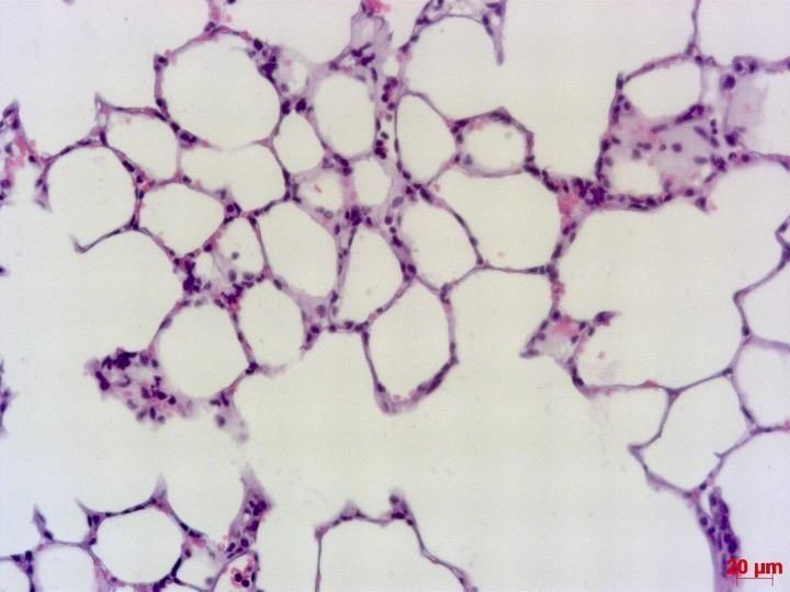
Histopathology of the lungs
The histopathologic analysis of lung tissue specimens harvested after 90 min of
reperfusion (at T2), indicated mild damage to the pulmonary parenchymal tissue
architecture. Figure 21 shows a high power field of a histopathologic specimen of a left
lung harvested at T2 with mild alveolar septal thickening and some leukocytic
infiltrations. As an internal control, Figure 22 shows a high power field of a
histopathologic specimen of a right lung harvested at T2.
36Figure 21: Exemplary histological specimen of a left lung, harvested after 90min of
reperfusion (T2). Routine hematoxylin-eosin staining. Scale bar represents
approximately 20µm.
Figure 22: Exemplary histological specimen of a right lung, harvested after 90min of
reperfusion (T2). Routine hematoxylin-eosin staining. Scale bar represents
approximately 20µm.
37The mean lung damage scores were 0.73 ± 0.03 [aU] and 0.61 ± 0.08 [aU] in left and right lung samples harvested at 90 min of reperfusion (T2), respectively. The difference was statistically significant (Figure 23; P=0.024). Figure 23: Lung injury Scores in left and right lung tissue specimens harvested after left-sided 30 min of ischemia and 90 min of reperfusion (T2). Data are given as mean values ± standard error of the mean and n represen ts the number of experiments. The asterisk indicates a statistically significant difference (P
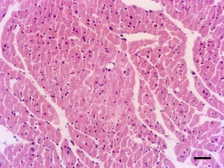
Histopathology of the heart

       The histopathologic analysis of cardiac tissue specimens harvested after 90 min
of pulmonary reperfusion (at T2) showed a normal architecture of the myocardium
(Figure 24), indicating no relevant effect of pulmonary ischemia/reperfusion injury on
the myocardium at this time point.

Figure 24: Histological specimen of the heart, harvested after 90min of pulmonary
reperfusion (T2). Routine hematoxylin-eosin staining. Scale bar represents
approximately 20µm.

                                                                                         39
Histopathology of the kidney
The histopathologic analysis of kidney tissue specimens harvested after 90 min
of pulmonary reperfusion (at T2) showed a vastly normal architecture (Figure 25),
indicating no relevant effect of pulmonary ischemia/reperfusion injury on the kidney
microarchitecture at this time point.
Figure 25: Histological specimen of a kidney harvested after 90min of pulmonary
reperfusion (T2) showing relatively undisturbed tubular cellular architecture. Routine
hematoxylin-eosin staining.
40Histopathology of the liver
The Suzuki Score [86]was used to semi-quantitatively quantify the secondary
organ damage in the liver following pulmonary ischemia/reperfusion injury.
Hepatocyte necrosis was not found in none of the liver specimens analysed. Instead,
there were specimens with areas of hepatocyte congestion and vacuolization (Figure
26).
Figure 26: Histological specimen of a liver harvested after 90min of pulmonary
reperfusion (T2) showing intense hepatocytic vacuolization. Routine hematoxylin-
eosin staining. Scale bar represents approx. 20 micrometers.
41Semi-quantitative analyses revealed a mean Suzuki Score for hepatocytic
congestion and vacuolization of 1.64 ± 0.20 [aU] and 0.82 ± 0.31 [aU], respectively
(Figure 27).
Figure 27: Secondary organ damage in the liver after pulmonary ischemia/reperfusion.
Semi-quantitative histopathologic analysis was performed using the Suzuki-Score [ref],
assessing hepatocytic congestion and vacuolization in liver specimens harvested at 90
min of pulmonary reperfusion (T2). Data are given as mean values ± standard error of
the mean (n=7).
427. Discussion
This study established an experimental approach to analyse cellular interactions
and microcirculatory dysfunction during pulmonary ischemia/reperfusion injury by
intravital fluorescence microscopy in the rat. While the experimentation of large
experimental animals is appealing also in view of mimicking the human situation, it is
challenging because of the demanding logistic efforts (housing, anaesthesia etc.). Small
animal models are therefore preferred. Moreover, molecular targeting by genetic
knock-out or knock-in techniques or pharmaco-immunological measures such as
specific antibody treatment are well established and dramatically cheaper in rodents.
Nevertheless, surgical preparation of the experimental animals and transplantation of
solid organs for example in rodents is seemingly different from the larger experimental
animal and human situations demanding meticulous microsurgical training and
experience [87].
Experimental studies on pulmonary ischemia/reperfusion injury have been
carried out in a large variety of animal models, e.g. in mice, rats, rabbits, dogs and sheep
[36, 52, 70, 88, 89]. Some of these studies also investigated pulmonary
ischemia/reperfusion injury and experimental lung transplantation. For example, early
experimental works on lung transplantation analysed the effects of distinct lung graft
inflation statuses during preservation [90-93]. Such insights in the relationship between
graft inflation during procurement as well as transportation and post-transplant lung
graft function have led to the generally accepted and practiced concept of clinical lung
transplantation, harvesting and transporting lung allografts in a mild to moderately
inflated state.
Many of the experimental studies on pulmonary ischemia/reperfusion injury
intended to focus on microvascular dysfunction. A large variety of experiments
indirectly assessed for example post-ischemic neutrophil accumulation in the lung by
quantification of myeloperoxidase tissue content. Intravital fluorescence microscopy
(IVM) has been extensively used and is a well established experimental and clinical
means to observe and analyse microcirculatory phenomena under physiological and
pathophysiological conditions in a large variety of organs [29, 77, 80, 94-96]. IVM of
the thoracic organs is challenging because of the constant need for counterbalancing
43the movements of the ventilated lung and the pulsations of the beating heart [97].
Kreisel and co-workers have used a veterinary glue to attach a cover glass onto the lung
surface and found that neutrophil extravasation was monocyte-dependent in a
challenging murine lung transplantation model [98]. However, the glue itself and/or the
shear forces exerted by the moving lung after sticking to the glass slip may provoke
distinct inflammatory responses affecting the pulmonary microcirculation apart from
the desired pathophysiological setting of interest. Bennewitz et al. stabilized the lung
surface by use of a specific suction system and analysed neutrophil and red blood cell
trafficking in the pulmonary microcirculation in a model of sickle cell disease in mice
[99]. Just as in the “glued lung”, there is some concern about potential extra shear force-
induced “ventilator injury” in this approach [100]. Tabuchi et al. established a plateau
phase at the end of the inspiration phase yielding a lung motion stop for 300ms before
continuation with the respiratory cycle and thereby analysed the murine pulmonary
microcirculation [101]. The herein used method also intended to establish an approach
minimizing shear stress to the lung surface and excluding other toxic side effects
potentially elicited by a glue. Access of the rat’s left lung subpleural pulmonary
microcirculation for IVM was established as previously published in a murine model
[102]. We used a cover glass slip that was fixed horizontally over the exposed lung
surface by means of a micromanipulator. The level of the cover glass defined a stable
focus level for the microscopic observations. The lung was attached to the lower side
of the glass solely by hydrostatic forces after moistening its surface with warm
physiological saline solution supported by a positive end-expiratory pressure level of
9mmHg [102]. Only horizontal excursions of the lung during inspiration and expiration
were observed, but these did not hinder continuous visualization of the subpleural
pulmonary microcirculation. Pulsations of the beating heart were found to be negligible
choosing a target area in the lower lobe of the left lung. Supporting this technique, we
had established this approach for IVM of the coronary microcirculation in rodents
before [80, 103]
Despite all the technical advances, the use of IVM for studies on physiological
and pathophysiological microcirculatory phenomena has some shortcomings.
Obviously, there is a certain amount of surgical trauma that cannot be prevented for the
exposure of the lung surface. It can only be minimized by refined microsurgical
techniques. Direct manipulation of the target tissue must be omitted and the organs
44You can also read