Structure and analysis of nanobody binding to the human ASIC1a ion channel
←
→
Page content transcription
If your browser does not render page correctly, please read the page content below
RESEARCH ARTICLE

                                       Structure and analysis of nanobody
                                       binding to the human ASIC1a ion channel
                                       Yangyu Wu1, Zhuyuan Chen1, Fred J Sigworth2, Cecilia M Canessa1,2*
                                       1
                                        Basic Sciences Department, Tsinghua University School of Medicine, Beijing, China;
                                       2
                                        Cellular and Molecular Physiology, Yale University School of Medicine, New Haven,
                                       United States

                                       Abstract ASIC1a is a proton-gated sodium channel involved in modulation of pain, fear,
                                       addiction, and ischemia-induced neuronal injury. We report isolation and characterization of alpaca-
                                       derived nanobodies (Nbs) that specifically target human ASIC1a. Cryo-electron microscopy of the
                                       human ASIC1a channel at pH 7.4 in complex with one of these, Nb.C1, yielded a structure at 2.9 Å
                                       resolution. It is revealed that Nb.C1 binds to a site overlapping with that of the Texas coral snake
                                       toxin (MitTx1) and the black mamba venom Mambalgin-1; however, the Nb.C1-binding site does
                                       not overlap with that of the inhibitory tarantula toxin psalmotoxin-1 (PcTx1). Fusion of Nb.C1 with
                                       PcTx1 in a single polypeptide markedly enhances the potency of PcTx1, whereas competition of
                                       Nb.C1 and MitTx1 for binding reduces channel activation by the toxin. Thus, Nb.C1 is a molecular
                                       tool for biochemical and structural studies of hASIC1a; a potential antidote to the pain-inducing
                                       component of coral snake bite; and a candidate to potentiate PcTx1-mediated inhibition of
                                       hASIC1a in vivo for therapeutic applications.

                                       Introduction
                                       ASICs are proton-activated sodium channels present in most neurons of the central and peripheral
                                       nervous systems (Krishtal and Pidoplichko, 1981; Waldmann et al., 1997). There are four ASIC
*For correspondence:                   genes (ASIC1-4) and six isoforms in the human genome (Kellenberger and Schild, 2002). The most
cecilia.canessa@yale.edu               abundant and broadly expressed subunit is ASIC1a; its deletion in the mouse genome eliminates
                                       most of the proton-induced currents mediated by ASICs (Wemmie et al., 2002). Association of
Competing interests: The
                                       three pore-forming subunits, either as homotrimers or heterotrimers, forms functional channels. All
authors declare that no
                                       ASIC isoforms share a common structure consisting of two transmembrane domains (TMDs) (TM1
competing interests exist.
                                       and TM2), cytosolic amino- and carboxy-termini with the amino-terminus forming a reentrant loop
Funding: See page 17                   into the lower pore (Yoder and Gouaux, 2020), and a large extracellular domain (ECD) that adopts
Received: 01 February 2021             a fist-like conformation with distinct subdomains. These structural features were unveiled by the first
Accepted: 13 July 2021                 crystal structure (Jasti et al., 2007), which was obtained from chicken ASIC (cASIC1) in the desensi-
Published: 28 July 2021                tized conformation at low pH. Subsequently, structures of cASIC1 in the open (Baconguis et al.,
                                       2014), closed (Yoder et al., 2018), desensitized at high and low pH (Gonzales et al., 2009;
Reviewing editor: Randy B
Stockbridge, University of
                                       Yoder and Gouaux, 2020), and in complex with various toxins have been resolved (Dawson et al.,
Michigan, United States                2012; Baconguis and Gouaux, 2012; Baconguis et al., 2014). Recently, structures of a human ASIC
                                       subunit, hASIC1a, have been obtained alone and in complex with Mambalgin-1 toxin (Sun et al.,
    Copyright Wu et al. This article
                                       2020).
is distributed under the terms of
                                           ASICs are the target of many polypeptide toxins that induce significant functional changes (Cris-
the Creative Commons
Attribution License, which             tofori-Armstrong and Rash, 2017). Of the three most studied, two are antagonists and one is an
permits unrestricted use and           activator of ASIC channels. PcTx1 is a 40-residue peptide from the venom of the tarantula Psalmo-
redistribution provided that the       poeus cambridgei (Escoubas et al., 2000) that inhibits hASIC1a (IC50 ~3 nM when conditioned at
original author and source are         pH 7.2). It has been investigated as an analgesic and neuroprotective agent from ischemic injury of
credited.                              the brain (Mazzuca et al., 2007; Xiong et al., 2004). Mambalgin-1 is a 78-residue three-finger toxin

Wu et al. eLife 2021;10:e67115. DOI: https://doi.org/10.7554/eLife.67115                                                               1 of 20
Research article Structural Biology and Molecular Biophysics
from the venom of the black mamba snake Dendroaspis polypepsis that rapidly and reversibly inhib-
its ASIC1a in neurons (Diochot et al., 2012). Because Mambalgin-1 exhibits strong analgesic effects,
it has raised interest as a potential treatment for chronic pain and as an alternative to opioids
(Diochot et al., 2016). Third, the toxin MitTx, found in the venom of the Texas coral snake Micrurus
tener tener, functions as a potent, persistent, and selective agonist for ASICs (Bohlen et al., 2011).
It is a heterodimer of an a-subunit (60 a.a.) and b-subunit (119 a.a.).
These toxins bind primarily to the thumb domain of the ECD of ASICs. Structures of each of these
toxins in complex with cASIC1 have been solved, and in the recent study of hASIC1a by Sun et al.,
2020 a cryo-electron microscopy (cryo-EM) structure of hASIC1a bound to Mambalgin-1 at pH 7.4
was solved at 3.9 Å resolution.
To address present challenges in analysis of hASIC1a, we have developed nanobodies (Nbs) with
high specificity and affinity to human ASIC1a. These versatile molecules are derived from the vari-
able domain (VHH) of single-domain antibodies produced in camelids (Hamers-Casterman et al.,
1993) and in cartilaginous fishes (Stanfield et al., 2004). We present a set of Nbs for uses in novel
applications that extend to the possibility of examining structure and function of hASIC1a in vivo
and in vitro and as potential therapies of pathologic conditions involving hASIC1a.
Results
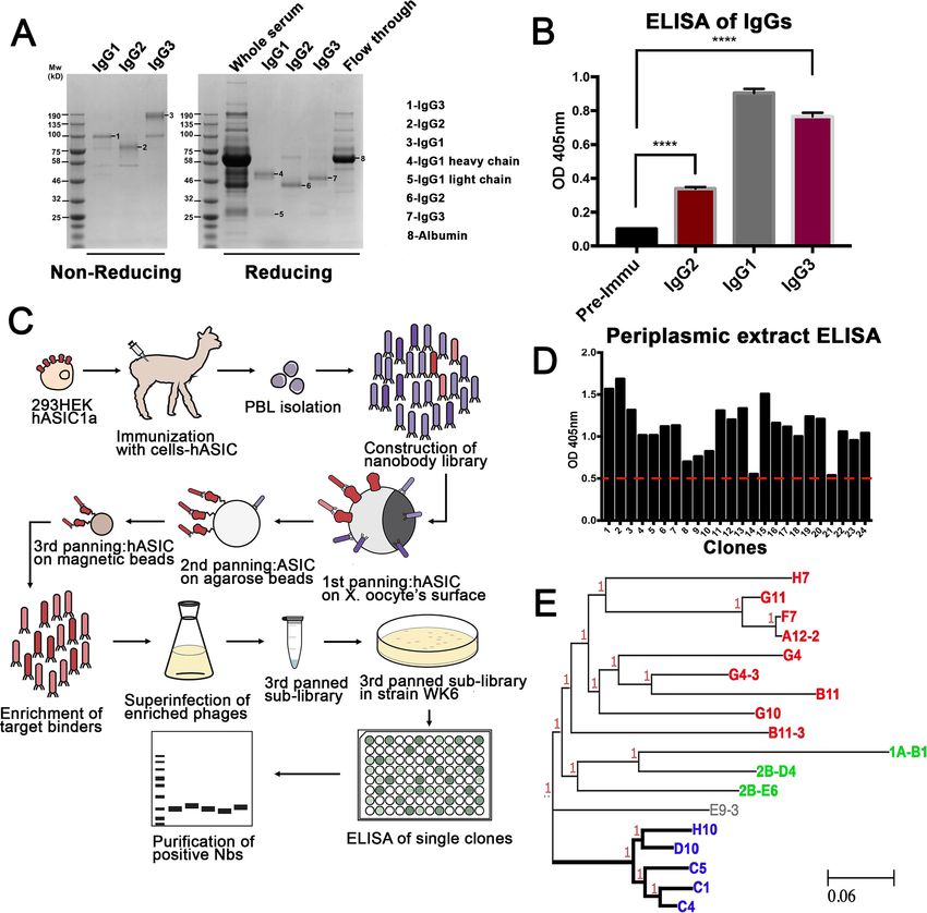
Generation of Nbs specific to human ASIC1a
An alpaca was immunized through the injection of 293 T cells expressing full-length hASIC1a on a 9-
week immunization schedule (Figure 1—figure supplement 1A). Sera collected post-immunization
contained conventional IgG1 and single-domain IgG2/3 antibodies that bind to hASIC1a, as ana-
lyzed by ELISA (Figure 1A–B). A phage display library was constructed from mRNA isolated from
peripheral lymphocytes (titer of approximately 109 independent clones) (Pardon et al., 2014; Fig-
ure 1—figure supplement 1B–E). To select Nbs that recognize hASIC1a in native conformation and
to minimize isolation of nonspecific binders, three successive panning protocols were used: the first
panning was conducted on Xenopus oocytes injected with hASIC1a cRNA while the second and third
used recombinant hASIC1a protein bound to agarose and magnetic beads, respectively (Figure 1C).
Phagemids recovered from the third panning were used for expression and isolation of Nbs: 600
were tested by ELISA. We considered a clone to be positive if the ELISA intensity was above a
threshold that eliminated about two-thirds of the clones. Representative ELISA results are shown in
Source data 1. Approximately 200 clones were selected for sequencing of DNA. The sequencing
result indicated that many of ELISA positive clones were identical or had one or two amino acid dif-
ferences, which is consistent with efficient enrichment for high affinity clones obtained by our screen-
ing strategy. DNA sequences of final candidates separated into three main groups, as shown in a
phylogeny tree (Figure 1E) and in the protein alignment of Figure 1—figure supplement 2. All
these clones were further examined by immunofluorescence of cells transfected with hASIC1a. From
all the Nbs tested, the group consisting of C1-4-5, D10, and H10 produced strong signals and low
background. A different group (1A-B1, 2B-D4, and 2B-E60) required permeabilization of cells for
labeling, suggesting that the recognized epitopes are intracellular; this was confirmed in immunocy-
tochemistry of permeabilized cells.
Nb.C1 stabilizes and prevents aggregation of hASIC1a
With the exception of the one recent cryo-EM study of hASIC1a (Sun et al., 2020), all structural
information about ASIC channels has been obtained from the chicken isoform. Although it shares
approximately 89% sequence identity with hASIC1a, the chicken isoform differs in functional proper-
ties and response to toxins and other compounds (Saez et al., 2011; Alijevic and Kellenberger,
2012; Smith and Gonzales, 2014). Thus, when considering the development of therapeutics, the
structure of the human channel is preferred. In the past, challenges working with hASIC1a arose
from the tendency of hASIC1a to give low yields and to aggregate, making it difficult to obtain high-
quality protein preparations suitable for structural analysis.
One of the goals of this work was to explore whether an Nb with high affinity to hASIC1a would
overcome these problems. Among the best binders initially screened, Nb.C1was selected on the
basis of high-affinity, low background, and absence of modification of channel function.
Wu et al. eLife 2021;10:e67115. DOI: https://doi.org/10.7554/eLife.67115 2 of 20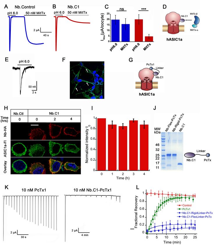
Research article                                                                                  Structural Biology and Molecular Biophysics

Figure 1. Generation of nanobodies (Nbs) specific to hASIC1a. (A) Fractionation of IgG (immunoglobulin G) classes from serum after completion of
immunization schedule of alpaca. (B) Each fraction was tested for antibodies against hASIC1a by ELISA. All three immunoglobulin fractions, including
single-domain antibodies Ig2 and IgG3 shown in red columns, are significantly higher than the pre-immune serum, t-test p-value < 0.001. (C) Overall
method for generation of a phage display library, panning strategy for selection of highly reactive phages, and final purification of Nbs. (D) Example of
ELISA results from 24 out of 600 selected clones. Only clones with signal above the red-dashed line were selected for further characterization. (E) The
DNA of those clones was sequenced and analyzed by similarity. A phylogenetic tree made with those clones shows that they distribute into three
groups. Thick lines mark the branch encoding Nbs with high reactivity and specificity. Nb C1 was chosen for further studies.
The online version of this article includes the following figure supplement(s) for figure 1:
Figure supplement 1. Construction of phage library.
Figure supplement 2. Amino acid sequence of highly reactive nanobodies to hASIC1a.

Wu et al. eLife 2021;10:e67115. DOI: https://doi.org/10.7554/eLife.67115                                                                           3 of 20
Research article                                                                             Structural Biology and Molecular Biophysics

                                    Subsequently, Nb.C1 was added to large-scale preparations of crude membranes from 293 F cells
                                    expressing an affinity-tagged construct (functional hASIC1a comprising amino acids 12–478 with a
                                    Strep Tag II in the N-terminus) prior to solubilization with DDM detergent. After affinity purification,
                                    the hASIC1a-Nb.C1 complex was pure and monodispersed (Figure 2A). This contrasts with the pro-
                                    tein aggregates observed in size exclusion chromatography (SEC) of solubilized hASIC1a alone (Fig-
                                    ure 2—figure supplement 1). In the complex, proteins from the peak of the SEC separated by gel
                                    electrophoresis showed bands for hASIC1a and Nb.C1 consistent with a stoichiometric ratio of 1:1
                                    (Figure 2B). The solubilized hASIC1a-Nb.C1 complex at 3.8 mg/mL was used to make cryo-EM sam-
                                    ples. Images were obtained on a Krios microscope (Thermo Fisher Scientific) using an energy filter
                                    and electron-counting camera. Some 2D classes of picked particles are shown in Figure 2C, and fur-
                                    ther processing described in Figure 2—figure supplement 2 and Table 1 led to a map of hASIC1a-
                                    Nb.C1 complex (Figure 2D).
                                        We determined the cryo-EM structure of hASIC1a-Nb.C1 complex at pH 7.4 with estimated reso-
                                    lution of 2.9 Å (Figure 3; Figure 3—figure supplement 1; Table 1). In agreement with other ASIC
                                    structures, hASIC1a is a trimer with each subunit ECD resembling the architecture of a hand. At pH
                                    7.4 the thumb domain is away from the finger producing an expanded acidic pocket as observed
                                    previously in cASIC1 (Yoder et al., 2018) and hASIC1a (Sun et al., 2020). The TMD shows a closed
                                    pore and the domain-swapped TM2 helixes that define the GAS belt of the pore. We also observed
                                    protein densities in the EM map inside the lower pore that were weak, presumably from disorder.
                                    When the map was filtered to 7 Å resolution (Figure 3—figure supplement 2) density for a reen-
                                    trant loop with two short helices was observed, consistent with the loop described by Yoder and
                                    Gouaux, 2020. In cASIC1 the reentrant loops, one from each subunit, form the lower
                                    ion permeation pathway. In each loop, the short linker between the helices Re-1 and Re-2 contains

Figure 2. Structural determination of human ASIC1a in complex with Nb.C1. (A) Size exclusion chromatography (SEC) purification of the hASIC1a-Nb.
C1 complex elutes as a single monodispersed peak. (B) Coomassie blue-stained SDS-PAGE shows two bands corresponding to the molecular weights
of hASIC1a and Nb.C1, indicating stable association of the complex that persists after SEC. (C) Representative 2D classes of hASIC1a-Nb.C1 complex
particles show distribution in various orientations. The extracellular domain (ECD) and transmembrane domain (TMD) can be readily distinguished as
well as Nb.C1 attached to the ECD. (D) Representative view of the 3D density map shows the Nb.C1 in complex with hASIC1a. The three hASIC1a
subunits are shown in green, red, and blue; Nb.C1s are shown in dark gray. Lipids are seen attached to the TMD (light gray).
The online version of this article includes the following figure supplement(s) for figure 2:
Figure supplement 1. Representative examples of size exclusion chromatography (SEC) profiles of hASIC1a purified in 1% dodecylmaltoside (DDM) or
1% Fos-choline14 in the absence of nanobody.
Figure supplement 2. Cryo-electron microscopy (cryo-EM) data processing pipeline for hASIC1a-Nb complex at pH 7.4.

Wu et al. eLife 2021;10:e67115. DOI: https://doi.org/10.7554/eLife.67115                                                                    4 of 20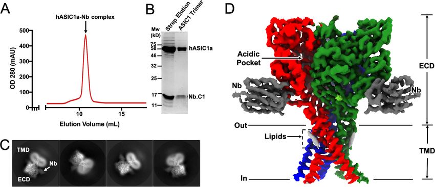
Research article                                                                      Structural Biology and Molecular Biophysics

                                   Table 1. Cryo-electron microscopy (cryo-EM) data collection, refinement, and validation statistics.
                                   Data collection and processing                                hASIC1a-Nb
                                   Magnification                                                 105,000
                                   Voltage (kV)                                                  300
                                   Electron exposure (e/Å2)                                     45.3
                                   Defocus range (mm)                                                1.0 to   2.0
                                   Pixel size (Å)                                               0.83
                                   Symmetry imposed                                              C3
                                   Initial particle images (no.)                                 1,287,029
                                   Final particle images (no.)                                   84,500
                                   Map resolution (Å)                                           2.86
                                   FSC threshold                                                 0.143
                                   Refinement
                                   Initial model used (PDB code)                                 6VTL
                                   Model resolution (Å)                                         3.7
                                   FSC threshold                                                 0.5
                                   Map sharpening B factor (Å)                                      15
                                   Model composition
                                   Non-hydrogen atoms                                            4000
                                   Protein residues                                              540
                                   Ligands                                                       2
                                   Bonds (RMSD)
                                   Length (Å) (# > 4s)                                          0.012
                                   Angles (˚) (# > 4s)                                           0.93
                                   MolProbity score                                              1.69
                                   Clash score                                                   6.87
                                   Ramachandran plot (%)                                         Ramachandran plot (%)
                                   Outliers                                                      0.00
                                   Allowed                                                       3.17
                                   Favored                                                       96.83
                                   Rama-Z (Ramachandran plot Z-score, RMSD)                      Rama-Z (Ramachandran plot Z-score, RMSD)
                                   Whole (N = 536)                                               1.31 (0.36)
                                   Helix (N = 136)                                               0.35 (0.44)
                                   Sheet (N = 103)                                               1.59 (0.45)
                                   Loop (N = 297)                                                0.77 (0.37)
                                   Rotamer outliers (%)                                          0.00
                                   Cb outliers (%)                                               0.00

                                   the highly conserved HG motif (His28, Gly29) that, in combination with the GAS belt, is involved in
                                   channel gating and selectivity.
                                      Three Nbs are observed bound to the trimeric channel, each in contact with a subunit at the end
                                   of the a4-helix and the extended loop of the thumb domain (Figure 3A). The most noticeable struc-
                                   tural difference between hASIC1a and cASIC1 is in this long loop that extends down from the a4-
                                   helix to the tip of the thumb (Figure 3B). The loop is longer and more twisted in hASIC1a because it
                                   has two extra amino acids D298 and L299 (DL) that are absent in cASIC1 and in all other known
                                   ASIC isoforms. These two amino acids are essential for binding of Nb.C1 as their deletion from
                                   hASIC1a eliminates the signal in immunofluorescence microscopy. Channels that do not encode

Wu et al. eLife 2021;10:e67115. DOI: https://doi.org/10.7554/eLife.67115                                                             5 of 20
Research article Structural Biology and Molecular Biophysics Figure 3. Cryo-electron microscopy (cryo-EM) structure of hASIC1a-Nb.C1 complex in the closed conformation. (A) Overall structure of hASIC1a-Nb complex in side and top views. Trimeric hASIC1a subunits are shown in red, green, and blue. Nanobodies Nb.C1 attached to the thumb domain of each hASIC1a subunit are shown in dark gray. General location of the overlapping binding sites of MitTx and Mambalgin-1 is indicated by the dashed rectangle (1) while the binding site of PcTx-1 is indicated by dashed rectangle (2). (B) Superposition of hASIC1a secondary structure (red) with cASIC1 (6vtl) (light blue) shows substantial differences only in the extended loop of thumb domain. (C) Detailed interactions between hASIC1a and Nb.C1 are Asp296-Tyr108 (lower left panel), Asp298-Arg35 and Arg105 (lower middle panel), Arg317-Phe58 (lower right panel). Map densities shown as a mesh. The negatively charged residues are in red, positively charged residues in blue, and aromatic residues are in green. The online version of this article includes the following figure supplement(s) for figure 3: Figure supplement 1. Cryo-electron microscopy (cryo-EM) imaging of hASIC1a-Nb complex at pH 7.4 and 4 mM Ca2+. Figure supplement 2. Intracellular densities in the hASIC1a-Nb electron microscopy (EM) map. Figure supplement 3. Immunoreactivity of HA-tagged Nb.C1 with various species and isoforms of ASIC. Figure supplement 4. Structural comparison of hASIC1-Nb.C1 complex with hASIC1 at high pH. Figure supplement 5. Functional characterization of hASIC1a-Nb.C1 and channels with or without DL residues. Wu et al. eLife 2021;10:e67115. DOI: https://doi.org/10.7554/eLife.67115 6 of 20
Research article                                                                      Structural Biology and Molecular Biophysics

                                   those two residues such as cASIC1, mASIC1a, mASIC2a, and mASIC3 are also not recognized by
                                   Nb.C1 in IF experiments (Figure 3—figure supplement 3). The residues involved in interactions with
                                   Nb.C1 are shown in Figure 3C. Two Asp residues in the extended loop of the thumb are involved:
                                   D296 interacts with Nb-Y108, and D298 with Nb-R35 and Nb R105. In addition, R317 at the end of
                                   a4 interacts with Nb-F58. None of these interactions would be possible in cASIC1. The loop in the
                                   hASIC1a structure of Sun et al., 2020 is poorly resolved, but in the current structure is well defined
                                   likely because of stabilization provided by Nb.C1, as shown in Figure 3—figure supplement 4.
                                       Of note, extensive functional analysis of hASIC1a bound to Nb.C1 showed that Nb binding did
                                   not change the channel’s properties. The average magnitude of current, rate of desensitization, mid-
                                   point pH of activation (pH50a), and of steady-state desensitization (pH50ssd) were all unchanged (Fig-
                                   ure 3—figure supplement 5A–B). The presence of the DL motif itself, however, affects the pH
                                   dependence of activation and desensitization. DL lies within a five-amino-acid stretch of hASIC1a
                                   which, when mutated, confers the altered pH sensitivity of the mouse isoform on hASIC1a
                                   (Sherwood and Askwith, 2008). Further, we compared the effects of the DL motif on hASIC1a and
                                   cASIC1. Deletion of DL in hASIC1a produces a small but significant left shift of the pH50ssd (from
                                   7.11±0.02 to 7.18±0.02), while insertion of DL into cASIC1 produces the opposite effect (from
                                   7.45±0.01 to 7.38±0.01) (Figure 3—figure supplement 5C–D).
                                       To visualize the binding sites of MitTx, PcTx1, and Mambalgin-1 and compare them to the bind-
                                   ing site of Nb.C1, our structure of the hASIC1a-Nb.C1 complex was superimposed on chicken or
                                   human subunits bound to each of the three toxins, and shown in orthogonal views (Figure 4). The
                                   superimposition of cASIC1-MitTx1 in the open conformation shows the a-subunit of the toxin inter-
                                   acting with the tip of the thumb close to the membrane bilayer, and with the extended chain that
                                   connects a5 to the b10 strand. Meanwhile, the b-subunit forms contacts with a4 and a5 of the
                                   thumb, extensively overlapping with the binding site of PcTx1 but not with that of Nb.C1. Though
                                   the binding sites of Nb.C1 and MitTx subunits are different, the bulky scaffold of the Nb produces
                                   steric hindrance to binding of the a-subunit, marked in the figure by a dashed square. In contrast,
                                   Nb.C1 and PcTx1 bind at distinct and well-separated sites, preventing mutual interference when
                                   both peptides bind simultaneously to the surface of the channel (Figure 4B).
                                       Mambalgin-1 and Nb.C1 share common interactions: residue D298 of hASIC1 interacts with K8 in
                                   Mambalgin-1, and hASIC1 residues at the end of a4 interact with both polypeptides. The dashed
                                   square indicates the area of clashes between Nb.C1 and toxin (Figure 4C). Although we did not test
                                   here the effect of Nb.C1 on Malganbin-1 binding, the results predict a decrease in the functional
                                   effects mediated by Malganbin-1.

                                   Nb.C1 antagonizes binding of MitTx to hASIC1a
                                   The predicted overlap of Nb.C1 with the MitTx a-subunit binding site raises the possibility that Nb.
                                   C1 could interfere with the action of MitTx on hASIC1a. To that end, oocytes expressing hASIC1a
                                   were incubated with 50 nM of Nb.C1 for 15 min; they were then exposed to 50 nM of MitTx at pH
                                   7.4 while measuring currents with two-electrode voltage clamp. MitTx-induced currents were
                                   decreased in oocytes pretreated with the Nb (Figure 5A–D), consistent with the Nb.C1 interfering
                                   with toxin binding to hASIC1a.

                                   Increased potency of PcTx1 by tethering to Nb.C1
                                   PcTx1 functions as an inhibitor of ASIC1a by shifting the steady-state desensitization toward more
                                   alkaline pH (Chen et al., 2005; Saez et al., 2011). Two crystal structures of cASIC1 with PcTx1 show
                                   that the toxin binds to the a5-helix of the thumb, at the interface with the palm domain of the adja-
                                   cent subunit (Dawson et al., 2012; Baconguis and Gouaux, 2012). The apparent EC50 of PcTx1 for
                                   hASIC1a has been reported in the range 0.9–3.7 nM and the time constant of recovery from inhibi-
                                   tion is reported as 125 s (Chen et al., 2021) or 87 s (Chassagnon et al., 2017). We hypothesized
                                   that slowing the recovery from inhibition would be advantageous for in vivo applications of PcTx1
                                   either as a potential pain suppressor or for protecting neurons from ischemia of the brain. That aim
                                   could be achieved by fusing Nb.C1 to PcTx1 in a single polypeptide provided that the Nb exhibits
                                   slower dissociation than the toxin and that the fusion does not interfere with binding to hASIC1a. To
                                   verify binding we first used SH-SY5Y cells, which are derived from a human neuroblastoma and

Wu et al. eLife 2021;10:e67115. DOI: https://doi.org/10.7554/eLife.67115                                                             7 of 20
Research article Structural Biology and Molecular Biophysics
Figure 4. Structural comparison of hASIC1a-Nb.C1 complex to toxin-bound ASICs. Two side, top and bottom views of superimposed structures of
hASIC1a-NbC1 complex (red) with (A) MitTx-bound to chicken ASIC1 (4ntw) in open conformation (orange). In side views, the threefold axis of the
channel is indicated by a dashed vertical line; in top and bottom views it is indicated by dotted triangles. (B) PcTx1-bound chicken ASIC1 (3s3x) (gray).
(C) Mambalgin-1-bound human ASIC1 (7ctf) (blue). Only one subunit is shown for simplicity. Surface clashes are indicated by dashed rectangles. Nb.C1,
MitTx- a, MitTx- b, PcTx1, Mambalgin-1 are shown as red, orange, light-orange, light-purple, marine respectively.
express endogenous hASIC1a (Xiong et al., 2012). An Nb.C1-PcTx1 fusion construct produced a
fluorescence signal that decorated the plasma membrane of SH-SY5Y (Figure 5E–G).
The time course of dissociation of Nb.C1 from hASIC1a was followed in transfected HEK293 cells
using the following protocol. Live cells were incubated with 10 nM Nb.C1 for 30 min at 18˚C. After
several washes to remove the unbound Nb.C1, cells were followed for four intervals of 1 hr duration.
Wu et al. eLife 2021;10:e67115. DOI: https://doi.org/10.7554/eLife.67115 8 of 20Research article Structural Biology and Molecular Biophysics Figure 5. Effects of Nb.C1 on MitTx and PcTx1 binding to hASIC1a. (A) Representative currents of an oocyte expressing hASIC1a activated with pH 6.0 followed by a second activation with 50 nM MitTx at pH 7.4. (B) Same experiment after pre-incubation of the oocyte with 50 nM Nb.C1 for 15 min. (C) Summary of the peak currents from pH 6.0 and MitTx activations. In this and all traces, the conditioning pH is 7.4. The bars represent the mean±SD of currents, n=8 Nb control and n=6 Nb.C1. Asterisks indicate statistical significance in t-test, p
Research article                                                                                  Structural Biology and Molecular Biophysics

Figure 5 continued
Nb.C1 associated with hASIC1a may interfere with MitTx binding. (E) Whole-cell patch clamp of SH-SY5Y cells activated with pH 6.0 generates typical
hASIC1a currents. Proton-induced currents are inhibited by PcTx and amiloride. (F) Immunofluorescence confocal image of SH-SY5Y cells incubated
with Nb.C1-PcTx1-HA fusion and anti-HA antibody (green) shows cells decorated on the periphery. Nuclei were stained with DAPI (blue). Scale bar, 5
mm. (G) Cartoon representation showing the Nb.C1-PcTx1 polypeptide binding to two distinct sites on the surface of hASIC1a, accounting for a
possible mechanism of toxin potentiation. (H) Confocal images of live HEK-293 cells transfected with hASCIC1a-Flag on coverslips incubated with Nb.
C1-HA for 30 min and followed for 0, 1, 2, 3, and 4 hr at 18˚C in DMEM containing HEPES. Three of the five time points are shown. At each 1 hr interval,
all cells were washed except for the one dish of cells removed for fixation. All cells were processed for immunofluorescence with HA and Flag
monoclonals to visualize Nb.C1-HA and hASIC1a-Flag, respectively. Nb.C1-HA labels only the cell surface whereas hASIC1a distributes in the plasma
membrane and intracellular endoplasmic reticulum and perinuclear membrane. Scale bar, 5 mm. (I) Quantification of fluorescence intensity of Nb.C1
(red channel) normalized to time 0 hr (t0). For each time point 300 cells were analyzed. Columns are the mean ± SEM. (J) Coomassie blue SDS-PAGE of
purified fusion proteins (Nb.C1-FlexLinker-PcTx and Nb.C1-RigidLinker-PcTx) and Nb.C1 alone. On the right a cartoon representation of the fusion
proteins. (K) Representative examples of oocytes expressing hASIC1a exposed to 10 nM of PcTx1 or 10 mM of Nb.C1-Rigid-PcTx1 fusion for 60 s prior
to serial activations with a change of pH from 7.35 to 6.0. Cells remained in the perfusion chamber throughout the experiment. (L) Time course of
recovery of acid-induced currents in control (no pretreatment), and pretreatment with PcTx1, Nb.C1-Flex-PcTx, or Nb.C1-Rigid-PcTx1. Preconditioning
pH 7.35, activation pH 6.0. Data were fit with a single exponential a 1 e t=t where t is 220 s for PcTx, 350 and 880 s for Nb.C1-Flex-PcTx and Nb.C1-


Rigid-PcTx; a = 0.90 for PcTx, and 0.16 and 0.14 for the fusions, respectively. Data points represent the mean ± SD of 7–12 cells. Values of currents from
each cell are shown in Source data 3.

                                     At each of the four time points, a group of cells was fixed and processed for IF or washed again until
                                     the next time point. Confocal images showed no decay of the Nb.C1 signal during the course of the
                                     experiment indicating that the Nb.C1 remains bound to the channel with no significant decay
                                     through the 4 hr observation period (Figure 5H–I).
                                         To estimate the Nb binding affinity, we first measured binding of Nb.1C to immobilized, deter-
                                     gent-solubilized hASIC1a protein using surface plasmon resonance. From experiments with two dif-
                                     ferent Nb and hASIC1a protein preparations, global fits to association time courses with Nb
                                     concentrations from 0.4 to 100 nM yielded association rate constants kon ranging from 6.8104 to
                                     2.2105 M 1 s 1. Unfortunately, the dissociation time courses (measured for up to 1 hr) from these
                                     experiments were too brief to observe the full dissociation time course, but a rate constant
                                                      5   1          1
                                     koff <
                                          ~ 1.410 s (0.05 hr ) can be estimated from the live-cell binding data of Figure 5I. Taking
                                     the smallest kon value as a lower bound on association rate yields the equilibrium constant kd
Research article Structural Biology and Molecular Biophysics
to further our understanding of these channels. To that end, we sought to develop Nbs specific to
hASIC1a. This strategy exploits the capacity of the immune system to produce a large variety of
small polypeptides against a single target, each with unique properties that can be selected for spe-
cific applications.
Here, we show that one Nb (Nb.C1) isolated from a phage display library prevents aggregation
and stabilizes hASIC1a, markedly improving the quality of the sample preparation for cryo-EM stud-
ies. We obtained the structure of hASIC1-Nb.C1 complex in the closed conformation at 2.9 Å resolu-
tion, indicating that the Nb has potential toward attaining high-resolution structural information of
hASIC1a in additional conformations. The growing use of Nbs in structural biology stems from the
fact that they can stabilize native conformations of proteins, in particular membrane proteins, facili-
tating structural analysis by crystallization or cryo-EM.
The Nb also constitutes a new tool to accelerate the development of therapeutic agents targeted
to hASIC1a. The cryo-EM structure of the hASIC1a-Nb.C1 complex reveals that Nb.C1 binds to the
thumb domain of hASIC1a to an epitope in the extended loop of the thumb that contains two extra
amino acids (D298 and L299) unique to hASIC1a. These residues determine the high specificity
toward hASIC1a and their absence explains the lack of reactivity with other species or isoforms
(cASIC1, mASIC1, ASIC2, or ASIC3). Nb.C1 binding also spans to the end of the a4-helix. This is a
region on the surface of the ECD that is also recognized by various toxin polypeptides with func-
tional activity toward many ASIC isoforms. Specifically, it overlaps with the binding site of Mambal-
gin-1 (Sun et al., 2020) and the binding site of the a-subunit of MitTx. This last toxin is the
component of the Texas coral snake venom that produces severe pain (Greene et al., 2020) by
activating hASIC1a in peripheral neurons (Bohlen et al., 2011). In these two instances, most of the
binding interference with the Nb is due to steric hindrance produced by the scaffold of the Nb,
which is large (16.28 kDa) compared to the size of Mambalgin-1 (6.55 kDa) and MitTx a-subunit (7
kDa). Therefore, Nb.C1 could serve as a competitive antagonist to MitTx and as a potential antidote
for the pain-producing component of the snake bite. Nbs offer practical advantages over currently
available antivenoms usually raised in sheep or horses (Yang et al., 2017) because Nbs can be pro-
duced in bacteria in large amounts and at low cost.
The Nb.C1-binding site is distinct from that of the inhibitory toxin PcTx. The lack of competition
for binding sites and steric interference but yet close proximity of the two binding sites offers the
possibility of using Nb.C1 as a carrier for PcTx1 to increase potency and decrease off-target effects
of the toxin. While the effect of PcTx1 alone is rapidly reversible, a fusion protein incorporating Nb.
C1 achieves inhibition of ~84–87% that persists for more than 30 min in functional studies and
remains bound for more than 4 hr on the surface of live cells according to IF assays. The changes in
toxin kinetics are consistent with the Nb having a much slower koff rate than that of the toxin. Further
optimization of the sequence of the linker, mainly to minimize or eliminate cleavage, could increase
even more the fractional current inhibited by the Nb.C1-PctX fusion peptide. Tethering of PcTx to
the Nb also would direct the toxin to bind preferentially to the subunit ASIC1a increasing specificity,
which is beneficial for in vivo applications such as the amelioration of ischemic damage to the brain
(McCarthy et al., 2015; Yang et al., 2011; Xiong et al., 2004).
Why does Nb.C1 bind to sites overlapping with those of toxins (Figure 4) but itself does not alter
channel function? We note that Nb.C1 does not interact with the a5-helix of the thumb, while the
three toxins in consideration all bind to a5 and these interactions change the conformation of a5 in
a toxin-specific manner. Many lines of evidence support the notion that displacement of a5 is an
essential component of channel gating. Mutations in a5 (Jasti et al., 2007; Vullo et al., 2017), for-
mation of cysteine bonds that restrain movement of helix (Yoder et al., 2018; Chen et al., 2021),
and binding of polypeptides that produce small but significant displacements are maneuvers that
alter gating by lowering pH50a, abolishing currents, inducing desensitization or causing channel
opening. We posit that the absence of Nb.C1 contacts with a5 is the most likely explanation of why
Nb.C1 does not change function of hASIC1a. Interactions of Nb.C1 are limited to a short segment
of the extended loop of the thumb that projects down to TM1 and a segment of the a4-helix of the
thumb. Comparison of our structure with the hASIC1a structure of Sun et al., 2020 shows that Nb.
C1 binding produces a very small movement of a4 and negligible perturbation of a5 (Figure 3—fig-
ure supplement 4).
Wu et al. eLife 2021;10:e67115. DOI: https://doi.org/10.7554/eLife.67115 11 of 20Research article Structural Biology and Molecular Biophysics
In summary, we show examples of uses of Nb.C1 isolated from an hASIC1a-specific phage display
library to advance structural and functional studies of the human channel, and as means to increase
or attenuate effects of ASIC-specific toxins with potential therapeutic applications.
Materials and methods
Key resources table
Reagent type
(species) or Source or
resource Designation reference Identifiers Additional information
Gene (Homo ASIC1a GenBank NCBI Ref Seq:
sapiens) NP_001086.2
Strain, strain TG1 Lucigen Cat#: 60502 Electrocompetent cells
background
(Escherichia coli)
Strain, strain WK6 ATCC 47078 Thermo Fisher Scientific Expression of
background nanobody proteins
(Escherichia coli)
Strain, strain DH5a Max efficiency Cat#: 18258012 Electrocompetent cells
background DH5a
(Escherichia coli)
Cell line HEK293T ATCC 47078 ATCC 47078
(Homo sapiens)
Cell line FreeStyle 293 F cells Thermo Fisher Cat#: R79007
(Homo sapiens) Scientific
Cell line SH-SY5Y ATTC ATTC CRL-2266
(Homo sapiens)
Recombinant pADL-22c Antibody Design Cat#: PD0110 Phagemid for
DNA reagent Labs construction of
nanobody library
Recombinant CM13 Helper phage Antibody Cat#: PH020L Rescue
DNA reagent Design Labs phagemid library
Recombinant pcDNA3.1 Invitrogen Cat#: V790-20 Vector
DNA reagent
Antibody Goat anti-llama NOVUS Cat#: NB7242 Detection of anti-ASIC1a
polyclonal antibody HRP antibodies in alpaca
serum (1/1000)
Antibody Anti-HA Cell Signaling Cat#: 3724T IF (1/1000)
rabbit monoclonal C29F4
Antibody Anti-Flag mouse monoclonal M2 Sigma-Aldrich Cat#: F1804 IF (1/1000)
Antibody Anti-M13 g8p Antibody Cat#: AS003-100 For phage ELISA
antibody Design Labs (1/5000)
HRP
mouse monoclonal
Peptide, PcTx1 Alome Cat#: STP-200
recombinant
protein
Peptide, Alpha/beta MitTx Alome Cat#: M-100
recombinant
protein
Polypeptide, Alpaca nanobodies This study Isolated from phage display
recombinant library of immunized
proteins alpaca with hASIC1a
Commercial QuickChange mutagenesis Agilent Cat#: 200521 Mutagenesis of DNA
assay or kit Technologies
Commercial ProtoScrript II New Cat#: E6560L Synthesis of single
assay or kit First strand England Biolabs strand DNA
cDNA
Continued on next page
Wu et al. eLife 2021;10:e67115. DOI: https://doi.org/10.7554/eLife.67115 12 of 20Research article Structural Biology and Molecular Biophysics
Continued
Reagent type
(species) or Source or
resource Designation reference Identifiers Additional information
Chemical Pierce anti-HA magnetic beads Thermo Fisher Cat#: 88837 Affinity purification of
compound, drug Scientific HA-tag proteins
Chemical Monoclonal Anti-HA agarose Sigma-Aldrich Cat#: A2095 Affinity purification
compound, drug of HA-tag proteins
Chemical Strep Tactin Resin IBA Cat#: 2-1201-002 Affinity purification
compound, drug of Strep-tag proteins
Chemical Ni-NTA Agarose Qiagen Cat#: 30210 Affinity purification of
compound, drug nanobodies from
periplasm
Chemical Cholesterol Anatrace Cat#: CH210
compound, drug Hemisuccinate tris
Software, MotionCor2 DOI: 10.1038/ RRID:SCR_016499 http://msg.ucsf.
algorithm nmeth.4193 edu/em/software/
motioncor2.html
Software, Gctf DOI: 10.1016/j. RRID:SCR_016500 https://www.mrc-lmb.
algorithm jsb.2015.11.003 cam.ac.uk/kzhang/Gctf/
Software, RELION 3.1 DOI: 10.7554/ RRID:SCR_016274 http://www2.mrclmb.
algorithm eLife.42166 cam.ac.uk/relion;
Software, PHENIX RRID:SCR_014224 https://www.
algorithm phenixonline.org;
Software, Coot DOI: 10.1107/S0907 RRID:SCR_014222 https://www2.mrc-
algorithm 444910007493 lmb.cam.ac.uk/
DOI: 10.1107/S0907 personal/pemsley/coot/
444910007493
Software, MolProbity DOI: 10.1107/ RRID:SCR_014226 RRID:SCR_014226
algorithm S0907444909042073
Software, Pymol PyMOL Molecular RRID:SCR_000305 RRID:SCR_000305
algorithm Graphics System,
Schrodinger, LLC
Software, UCSF Chimera DOI: 10.1002/ jcc. RRID:SCR_004097 http://plato.cgl.
algorithm 20084 ucsf.edu/chimera/
Software, UCSF ChimeraX DOI: 10.1002/ pro. RRID:SCR_015872 http://cgl.ucsf.
algorithm 3235 edu/chimerax/
Software, CCP-EM DOI: 10.1002/ pro. https://www.
algorithm 3235 ccpem.ac.uk/
Software, DemoPIcker This study https://github.com/fsigworth/aEM
algorithm CodeRepository/tree/master/
Teaching/PartPickingDemo,
(Sigworth, 2021;
copy archived
at swh:1:dir:2cdf6a8a6b19d8b
e1408954f51bf9d81e44edb11)
Other Series S Sensor Chip CM5 Cytiva Cat#: 29104988 For Biacore
(GE) instrument
Alpaca immunization
A male alpaca was immunized with intact HEK293T cells (ATCC CRL-11268) transfected with
hASIC1a according to the schedule shown in Figure 1—figure supplement 1. Antigen expressing
cells and adjuvant were injected subcutaneously in adjacent sites to preserve the native conforma-
tion of hASIC1a. Blood was obtained before the first injection, and 1 week after the third and fifth
injections. Figure 1 depicts the subsequent steps of construction and screening of the phage display
library. Immunization and bleeding of alpaca were conducted with the assistance of a veterinarian,
and protocols were approved by IACUC of Tsinghua University (protocol number 07749). The
Wu et al. eLife 2021;10:e67115. DOI: https://doi.org/10.7554/eLife.67115 13 of 20Research article Structural Biology and Molecular Biophysics
Association for Assessment and Accreditation of Laboratory Animal Care International (AALAC) has
accredited Tsinghua University veterinarians and facilities.
Isolation of alpaca IgGs
One-hundred microliter of protein G agarose beads (Sigma) were added to 0.5 mL of alpaca serum
and incubated for 1 hr followed by three washes with phosphate buffer saline (PBS). The IgG3 frac-
tion bound to the beads was eluted with 0.5 mL of 150 mM NaCl, 0.58% acetic acid, pH 3.5, and
immediately neutralized with 1 M Tris-HCl pH 8.0. Beads were washed and the IgG1 fraction still
bound to the G agarose was eluted with lower pH: 0.5 mL of 100 mM glycine-HCl pH 2.7 followed
by neutralization. The initial flow through from protein G beads was incubated with 50 mL protein A
beads for 1 hr. After washes with PBS, the IgG2 fraction was eluted with 0.5 mL of 150 mM NaCl,
0.58% acetic acid pH 4.5 and neutralized to pH 8.0. Alpaca IgG2/3 represent the single-domain anti-
bodies (Maass et al., 2007).
Construction of phage displayed Nb library
Five days after the final immunization, peripheral blood lymphocytes were isolated from 100 mL of
whole blood using Accuspin-Histopaque System (Sigma). Total RNA was extracted with Trizol (Invi-
trogen); 30 mg of total RNA were used for synthesis of single strand DNA primed with oligo-dT and
using Superscript III kit (Life Technologies). The variable region of the heavy chain from IgG2/IgG3
(VHH) domains was amplified by nested PCR. The first PCR was conducted with a pair of primers
specific for alpaca annealing to the leader sequence and to IgG CH2 domain. The amplified IgG2/3
dsDNA was gel extracted from the first PCR product by cutting the 700 bp band. The second PCR
(18 cycles) was conducted with a pair of primers specific to alpaca IgG FR1 region and IgG2/3 hinge
region (Figure 1—figure supplement 1B). SfiI restriction sites, generating different sticky ends,
were introduced by PCR for cloning into a pADL-22c phagemid vector (Antibody Design Labs). A
total of 30 ligation reactions were pooled and electroporated into TG1 competent Escherichia coli
(Lucigen); 18 clones were randomly picked to examine the efficacy of dsDNA insertion (Figure 1—
figure supplement 1D). A library of 1109 individual transformants was superinfected with CM13
helper phage (Antibody Design Labs) after TG1 F-pilus induction. VHH-domain-displaying bacterio-
phages were produced overnight by shaking the bacterial culture at 37˚C supplemented with IPTG
to induce expression of Nb fragments. Phages were isolated from the medium of an overnight cul-
ture by two successive precipitation steps with 4% PEG-8000 in 500 mM NaCl. Phages displaying
Nb were dissolved in 1 mL PBS followed by selection for binding to hASIC1a.
Library screening with a customized panning protocol
Panning was conducted using a modified multi-antigen presenting system that maximizes capturing
high-affinity native epitope binders. For the first round of panning, Xenopus oocytes expressing high
levels of hASIC1a at the plasma membrane were used for selection. Cells were pre-incubated with
blocking solution (100 mM NaCl, 3 mM KCl, 2 mM CaCl2, 10 mM HEPES pH 7.5, and 2% skimmed
milk) and incubated with phage in the same solution without milk. For the second and third round of
panning, affinity-purified hASIC1a bound to either Strep Tactin XT Agarose beads or magnetic
beads (IBA) were used for selection. Incubation and washing buffer (150 mM NaCl, 2 mL CaCl2, 10
mM HEPES pH 7.4, 0.04% DDM ± 2% milk). After three rounds of panning, binding phages were
pooled and a sub-library from the third panning was transformed into WK6 –a strain of E. coli for Nb
expression. A sample of single clones was examined by extraction of crude periplasm proteins and
tested by ELISA. Positive clones were sequenced followed by Nb purification.
ELISA
Affinity-purified hASIC1a protein (5 mg/mL) was coated onto 96-well microtiter plates overnight at 4˚
C and blocked with 5% skimmed milk. For alpaca IgGs ELISA, different concentrations of IgG1,
IgG2, and IgG3 were added and incubated at room temperature (RT) for 2 hr. After three washes,
goat anti-llama IgG conjugated with HRP (NOVUS) was added and enzyme reaction was detected
with peroxidase substrate ABTS and quantified at 405 nm in a microplate reader. Non-coated or
bovine serum albumin-coated wells served as controls. For phage ELISA, hASIC1a-coated plates
were incubated with the generated phage library as well as the panned phage sub-libraries in
Wu et al. eLife 2021;10:e67115. DOI: https://doi.org/10.7554/eLife.67115 14 of 20Research article Structural Biology and Molecular Biophysics
various phage particle concentrations. Goat anti-M13 monoclonal antibody HRP-conjugated (GE
Healthcare) was used for detecting Nb enrichment after consecutive rounds of panning. For periplas-
mic extract ELISA, single-clone periplasmic extracts from the third panned sub-library were pre-
pared. In brief, individual bacterial clones were cultured in deep-square-96-well plates (Corning) and
grown to exponential phase. Nb production was induced by IPTG and the incubation continued
overnight at 28˚C. Plates were centrifuged and bacterial pellets were resuspended in 100 mL of TES
buffer (in mM): 200 Tris-HCl pH 8.0, 0.5 EDTA, 500 sucrose on a vibrating platform at 2000 rpm for
1 hr; 100 mL of ddH2O was added to each well and returned to vibrating platform for 1 hr. Plates
were centrifuged and the supernatants – containing Nbs – were recovered and examined by ELISA.
Single-clone periplasmic extracts were added to coated ELISA plates. Mouse anti-HA monoclonal
antibody (Santa Cruz) and goat anti-mouse-HRP were added to recognize and detect the bound
Nbs. Non-coated wells or an irrelevant Nb were used as controls.
Nb purification
The phagemid vector pADL-22c encoding a bacterial periplasmic secretion leader sequence and
amber stop codon was used for Nb purification. A 30 mL LB overnight culture was diluted 1:1000
into 800 mL fresh Terrific Broth in the presence of 100 mg/mL ampicillin. When the culture reached
an OD600 of 0.8, Nb expression was induced by 1 mM IPTG and the temperature was decreased to
28˚C for 16 hr. Bacteria were harvested by centrifugation and the pellet was resuspended in 20 mL
of TES buffer. Periplasmic protein extraction was conducted by osmotic shock as indicated above.
After centrifugation, the Nbs were affinity-purified with Ni-NTA agarose beads (Qiagen) and eluted
with imidazole (Pardon et al., 2014).
hASIC1a expression and purification
Fully functional hASIC1a comprising amino acids 12–478 was tagged in the N-terminus with StrepII
tag. The construct was expressed in HEK293F cells (Invitrogen) cultured in suspension. Expression
level of hASIC1a was estimated 48–72 hr post-transfection by immunofluorescence using StrepII tag
monoclonal (Abcam). One liter of cell culture was routinely used for isolation of hASIC1a. Crude
membrane pellets were resuspended in lysis buffer (50 mM HEPES pH 7.4, 150 mM NaCl, 5 mM
CaCl2). For hASIC1a-Nb complex purification, crude membranes were first incubated with 3–4 mg of
purified Nbs, protease inhibitor cocktail (Roche) under slow agitation at 4˚C for 3 hr to allow binding
of the Nb. The suspension was then treated with 1% DDM (Anatrace). After clarifying the homoge-
nate by ultracentrifugation, hASIC1a was affinity-purified with Strep Tactin XT agarose beads (IBA)
to achieve high degree of purity and to remove free Nbs. Elution was conducted with the addition
of 50 mM D-biotin, samples were concentrated to a volume of 0.8 mL using a 50 kDa cutoff Centri-
con (Millipore). Samples were injected to a size exclusion column (Superdex 200 Increase 10/200 GL,
GE Healthcare). Fractions containing the hASIC1a-Nb.C1 micelle complex were pooled and concen-
trated to 3.8 mg/mL.
Cryo-EM specimen preparation, data acquisition, and processing of
hASIC1a-Nb complex
Quantifoil holey carbon grids (R1.2/1.3 300 mesh Au) were glow-discharged with carbon side facing
up for 1 min at 15 mA. Human ASIC1a-Nb affinity-purified protein at pH 7.4 was subjected to SEC
(running buffer 20 mM HEPES pH 7.4, 150 mM NaCl, 5 mM CaCl2) and immediately concentrated to
3.8 mg/mL for grid preparation. A 3 mL droplet of sample was applied to the carbon side of each
grid. Grids were blotted and plunge-frozen using a Vitrobot apparatus (Thermo Fisher Scientific)
with the chamber at 18˚C and 100% humidity. In total, 9039 micrographs were collected on Titan
Krios microscopes (Thermo Fisher Scientific) operated at 300 keV. Images were collected using Seri-
alEM (Schorb et al., 2019) with an image shift pattern of 3 three holes, with one shot per hole.
The detector was a Gatan K3 camera positioned after an energy filter (20 eV slit width). Recording
was in super-resolution mode with a binned pixel size (equal to the physical pixel size) of 0.83 Å and
dose-fractionated to 28 frames for a total exposure time of 1.4 s and a total dose of 45.3 e/Å2. Raw
cryo-EM movies were motion-corrected using UCSF MotionCor2 (Zheng et al., 2017) and CTF esti-
mation was performed using Gctf (Zhang, 2016). Particle picking utilized a simple adversarial-tem-
plate-based program DemoPicker written by FJS. Extracted particles were subjected to reference-
Wu et al. eLife 2021;10:e67115. DOI: https://doi.org/10.7554/eLife.67115 15 of 20Research article Structural Biology and Molecular Biophysics
free 2D classification in RELION 3.1 and non-particles were removed. Rounds of 3D classification
and refinement (C3 symmetry) were processed in RELION 3.1, using a cASIC1 cryo-EM map
(emd_7009) as reference (Yoder and Gouaux, 2020). CTF refinement and Bayesian polishing were
applied to further improve the resolution.
Model building and refinement of hASIC1-Nb
Using a rigid-body fitting program, Molrep, the cryo-EM structure of the resting channel (PDB 6VTL)
(Yoder and Gouaux, 2020) was docked into the cryo-EM density map. Docked models were used
as templates for iterative rounds of manual model building in Coot (Emsley et al., 2010). For the
Nb structure, a crystal structure of Nb (PDB 5IVO) was docked into the cryo-EM map by UCSF Chi-
mera (Goddard et al., 2007) and was built in Coot. Additional real space refinement was performed
using Refmac 5 (Murshudov et al., 2011) and Phenix (Liebschner et al., 2019).
Immunofluorescence microscopy
HEK293T (ATCC CRL-11268) or SH-SY5Y (ATTC CRL-2266) were authenticated by the presence of
low-pH induced currents sensitive to 50mM amiloride using patch clamp. Mycoplasma infection was
ruled out by a commercial PCR assay (EZ-PCR Mycoplasma detection kit from Biological Industries,
#20-700-20). Cells seeded onto glass coverslips treated with poly-L-lysine were either non-trans-
fected or transfected with hASIC1a in pCDNA3.1 vector with Lipofectamine. For surface labeling of
hASIC1a, 18–20 hr post-transfection, anti-hASIC1a Nbs were added to the culture medium in a con-
centration of 1 nM and cells were placed on ice to inhibit endocytosis. After 1 hr of incubation, cells
were rinsed three times with PBS and fixed with 4% paraformaldehyde prepared in PBS for 30 min
at 37˚C. Cells were further permeabilized with buffer containing 1%Triton-X100 for 30 min. Mouse
anti-Flag (Sigma) primary antibodies were added for 1 hr at RT. After three washes, Alexa Flour goat
594 and/or Alexa Flour 488 (Invitrogen) were added for 1 hr. Both primary and secondary antibody
incubations were conducted in the presence of 5% normal goat serum to decrease background.
DAPI was added to visualize nuclei. In chase experiments, Nb.C1 (1 nM) was incubated for 30 min,
washed three times with PBS followed by a chase of 0, 1, 2, 3, and 4 hr at RT. At each time point,
cells were fixed and processed for immunofluorescence. Coverslips were laid on glass slides with
mounting solution (VECTASHIELD, Vector Laboratories H-1000). Images were captured with a Nikon
confocal fluorescence microscope A1RMP LSM and analyzed using NIS Viewer 3.2. For visualizing
intracellular hASIC1, cells were first fixed and permeabilized with 1% Triton-X100 before adding
Nbs. The rest of the protocol was the same as described above. The human neuronal cell line SH-
SY5Y (ATCC CRL-2266) was seeded on coverslips and treated with 10 nM staurosporin for 48 hr to
induce cell differentiation. Cells were fixed, permeabilized prior to conducting immunofluorescence
with Nb.C1-PcTx1 fusion as the primary antibody; the rest of the procedure was the same as above.
Production of recombinant Nb.C1-PcTx1 polypeptides
The cDNA of Nb.C1 was modified first by introducing the DNA sequence of PcTx1 in the carboxyl-
terminus of the Nb prior to the 6 His and HA epitopes using the unique restriction sites XhoI and
XbaI. In a second step, a predicted flexible (KLGGGSGGSAGSAAGGSGSGGEFGGGGSLE) or more
rigid (GGGSGAEAAAKAEAEAKAEAAAKGGGGSG) linker was introduced using another pair of
unique restriction sites: HindIII-XhoI, located upstream the PcTx1 cloning site. Plasmid was trans-
formed into BL21(DE3) competent E. coli by heat shock. The previous protocol for purification of
Nbs was followed with the following modifications. Cells were kept at 18˚C during induction. Prote-
ase inhibitors and 1 mM b-mercaptoethanol were added to solutions of periplasmic protein purifica-
tion. After affinity purification with Ni-NTA resin, proteins were concentrated using a Millipore
concentrator and then added x2 Redox buffer (5 mL: 1.8 mg reduced glutathione added to 2.4 mg
of oxidized glutathione resuspended in TN buffer without imidazole). His and HA tags were not
removed. Peptides were further purified according to the protocol by Saez et al., 2017. Protein
quantification was by absorbance at 280 nm using NanoDrop.
Two-electrode voltage clamp of Xenopus laevis oocytes
Oocytes were injected with 5 ng of in vitro synthesized hASIC1a cRNA using the kit (mMESSAGEm-
MACHINE T7, Thermo Fisher Scientific). Whole-cell currents were measured using a two-electrode
Wu et al. eLife 2021;10:e67115. DOI: https://doi.org/10.7554/eLife.67115 16 of 20Research article Structural Biology and Molecular Biophysics
voltage clamp (Oocyte-Clamp OC-725C, Warner Instrument Corp.) with PowerLab 8/35 (ADInstru-
ments) running LabChart Prosoftware. Electrode resistance was 0.5–1 MW when filled with 3 M KCl.
Cells were placed in a fast-exchange perfusion chamber,Research article Structural Biology and Molecular Biophysics
Software, Formal analysis, Supervision, Validation, Writing - review and editing; Cecilia M Canessa,
Conceptualization, Supervision, Funding acquisition, Project administration, Writing - review and
editing
Author ORCIDs
Yangyu Wu https://orcid.org/0000-0001-8064-6132
Fred J Sigworth https://orcid.org/0000-0002-7178-8494
Cecilia M Canessa https://orcid.org/0000-0001-7316-5082
Ethics
Animal experimentation: The study was carried out in accordance with the recommendations of the
Institutional Animal Care Committee of Shanxi Agricultural University, China, where the alpaca was
kept through the immunization protocol (protocol # 07990) The use of Xenopus laevis oocytes was
approved by the Institutional Animal Care Committee of Tsinghua University, China (protocol
#125154). Our group did not conduct frog surgeries, we received fragments of ovaries from the
amphibian facility. The Association for Assessment and Accreditation of Laboratory Animal Care
International (AALAC) has accredited Tsinghua amphibian animal facility.
Decision letter and Author response
Decision letter https://doi.org/10.7554/eLife.67115.sa1
Author response https://doi.org/10.7554/eLife.67115.sa2
Additional files
Supplementary files
. Source data 1. ELISA screening 96-well plates of ELISA signal from screening of nanobodies recov-
ered from third panning of library. In blue are indicated the clones with highest values.
. Source data 2. Values of oocyte currents elicited by changes in pH for activation and steady-state
desensitization (SSD). Peak currents (mA/oocyte) elicited with solutions with the indicated pH mea-
sured with two-electrode voltage clamp. pHa = pH of activation, pHssd=pH of steady-state
desensitization.
. Source data 3. Acid-induced currents (pH 6.0) in oocytes pretreated with nanobody (Nb). C1-Flexi-
ble linker-PcTx or Nb.C1-Rigid linker-PcTx shown if Figure 5L.
. Transparent reporting form
Data availability
All data generated in this study are included in the MN and supporting files.
References
Alijevic O, Kellenberger S. 2012. Subtype-specific modulation of Acid-sensing ion channel (ASIC) Function by 2-
Guanidine-4-methylquinazoline. Journal of Biological Chemistry 287:36059–36070. DOI: https://doi.org/10.
1074/jbc.M112.360487
Baconguis I, Bohlen CJ, Goehring A, Julius D, Gouaux E. 2014. X-ray structure of acid-sensing ion channel 1-
snake toxin complex reveals open state of a na(+)-selective channel. Cell 156:717–729. DOI: https://doi.org/10.
1016/j.cell.2014.01.011, PMID: 24507937
Baconguis I, Gouaux E. 2012. Structural plasticity and dynamic selectivity of acid-sensing ion channel–spider
toxin complexes. Nature 489:400–405. DOI: https://doi.org/10.1038/nature11375
Bohlen CJ, Chesler AT, Sharif-Naeini R, Medzihradszky KF, Zhou S, King D, Sánchez EE, Burlingame AL, Basbaum
AI, Julius D. 2011. A heteromeric Texas coral snake toxin targets acid-sensing ion channels to produce pain.
Nature 479:410–414 . DOI: https://doi.org/10.1038/nature10607
Chassagnon IR, McCarthy CA, Chin YK-Y, Pineda SS, Keramidas A, Mobli M, Pham V, De Silva TM, Lynch JW,
Widdop RE, Rash LD, King GF. 2017. Potent neuroprotection after stroke afforded by a double-knot spider-
venom peptide that inhibits acid-sensing ion channel 1a. PNAS 114:3750–3755. DOI: https://doi.org/10.1073/
pnas.1614728114
Wu et al. eLife 2021;10:e67115. DOI: https://doi.org/10.7554/eLife.67115 18 of 20Research article Structural Biology and Molecular Biophysics
Chen X, Kalbacher H, Gru€nder S. 2005. The tarantula toxin psalmotoxin 1 inhibits Acid-sensing ion channel (ASIC)
1a by increasing its apparent H+ affinity. Journal of General Physiology 126:71–79. DOI: https://doi.org/10.
1085/jgp.200509303
Chen Z, Kuenze G, Meiler J, Canessa CM. 2021. An arginine residue in the outer segment of hASIC1a TM1
affects both proton affinity and channel desensitization. Journal of General Physiology 153:e202012802.
DOI: https://doi.org/10.1085/jgp.202012802, PMID: 33851970
Cristofori-Armstrong B, Rash LD. 2017. Acid-sensing ion channel (ASIC) structure and function: insights from
spider, snake and sea Anemone venoms. Neuropharmacology 127:173–184. DOI: https://doi.org/10.1016/j.
neuropharm.2017.04.042, PMID: 28457973
Dawson RJP, Benz J, Stohler P, Tetaz T, Joseph C, Huber S, Schmid G, Hügin D, Pflimlin P, Trube G, Rudolph
MG, Hennig M, Ruf A. 2012. Structure of the Acid-sensing ion channel 1 in complex with the gating modifier
Psalmotoxin 1. Nature Communications 3:936. DOI: https://doi.org/10.1038/ncomms1917
Diochot S, Baron A, Salinas M, Douguet D, Scarzello S, Dabert-Gay A-S, Debayle D, Friend V, Alloui A, Lazdunski
M, Lingueglia E. 2012. Black mamba venom peptides target acid-sensing ion channels to abolish pain. Nature
490:552–555. DOI: https://doi.org/10.1038/nature11494
Diochot S, Alloui A, Rodrigues P, Dauvois M, Friend V, Aissouni Y, Eschalier A, Lingueglia E, Baron A. 2016.
Analgesic effects of mambalgin peptide inhibitors of acid-sensing ion channels in inflammatory and neuropathic
pain. Pain 157:552–559. DOI: https://doi.org/10.1097/j.pain.0000000000000397
Emsley P, Lohkamp B, Scott WG, Cowtan K. 2010. Features and development of coot. Acta Crystallographica.
Section D, Biological Crystallography 66:486–501. DOI: https://doi.org/10.1107/S0907444910007493
Escoubas P, De Weille JR, Lecoq A, Diochot S, Waldmann R, Champigny G, Moinier D, Ménez A, Lazdunski M.
2000. Isolation of a tarantula toxin specific for a class of Proton-gated na+ channels. Journal of Biological
Chemistry 275:25116–25121. DOI: https://doi.org/10.1074/jbc.M003643200
Goddard TD, Huang CC, Ferrin TE. 2007. Visualizing density maps with UCSF Chimera. Journal of Structural
Biology 157:281–287. DOI: https://doi.org/10.1016/j.jsb.2006.06.010
Gonzales EB, Kawate T, Gouaux E. 2009. Pore architecture and ion sites in acid-sensing ion channels and P2X
receptors. Nature 460:599–604. DOI: https://doi.org/10.1038/nature08218
Greene S, Ruha AM, Campleman S, Brent J, Wax P. 2020. Epidemiology, clinical features, and management of
texas coral snake (Micrurus tener) Envenomations reported to the north american snakebite registry. Journal of
Medical Toxicology 14:51–56. DOI: https://doi.org/10.1007/s13181-020-00806-3
Hamers-Casterman C, Atarhouch T, Muyldermans S, Robinson G, Hammers C, Songa EB, Bendahman N,
Hammers R. 1993. Naturally occurring antibodies devoid of light chains. Nature 363:446–448. DOI: https://doi.
org/10.1038/363446a0
Jasti J, Furukawa H, Gonzales EB, Gouaux E. 2007. Structure of acid-sensing ion channel 1 at 1.9 Å resolution
and low pH. Nature 449:316–323. DOI: https://doi.org/10.1038/nature06163
Kellenberger S, Schild L. 2002. Epithelial sodium channel/degenerin family of ion channels: a variety of functions
for a shared structure. Physiological Reviews 82:735–767. DOI: https://doi.org/10.1152/physrev.00007.2002,
PMID: 12087134
Krishtal OA, Pidoplichko VI. 1981. A receptor for protons in the membrane of sensory neurons may participate in
nociception. Neuroscience Letters 24:243–246. DOI: https://doi.org/10.1016/0306-4522(81)90105-6
Liebschner D, Afonine PV, Baker ML, Bunkóczi G, Chen VB, Croll TI, Hintze B, Hung L-W, Jain S, McCoy AJ,
Moriarty NW, Oeffner RD, Poon BK, Prisant MG, Read RJ, Richardson JS, Richardson DC, Sammito MD,
Sobolev OV, Stockwell DH, et al. 2019. Macromolecular structure determination using X-rays, neutrons and
electrons: recent developments in Phenix . Acta Crystallographica Section D Structural Biology 75:861–877.
DOI: https://doi.org/10.1107/S2059798319011471
Maass DR, Sepulveda J, Pernthaner A, Shoemaker CB. 2007. Alpaca (Lama pacos) as a convenient source of
recombinant camelid heavy chain antibodies (VHHs). Journal of Immunological Methods 324:13–25.
DOI: https://doi.org/10.1016/j.jim.2007.04.008
Mazzuca M, Heurteaux C, Alloui A, Diochot S, Baron A, Voilley N, Blondeau N, Escoubas P, Gélot A, Cupo A,
Zimmer A, Zimmer AM, Eschalier A, Lazdunski M. 2007. A tarantula peptide against pain via ASIC1a channels
and opioid mechanisms. Nature Neuroscience 10:943–945. DOI: https://doi.org/10.1038/nn1940
McCarthy CA, Rash LD, Chassagnon IR, King GF, Widdop RE. 2015. PcTx1 affords neuroprotection in a
conscious model of stroke in hypertensive rats via selective inhibition of ASIC1a. Neuropharmacology 99:650–
657. DOI: https://doi.org/10.1016/j.neuropharm.2015.08.040
Murshudov GN, Skubák P, Lebedev AA, Pannu NS, Steiner RA, Nicholls RA, Winn MD, Long F, Vagin AA. 2011.
REFMAC 5 for the refinement of macromolecular crystal structures. Acta Crystallographica Section D Biological
Crystallography 67:355–367. DOI: https://doi.org/10.1107/S0907444911001314
Pardon E, Laeremans T, Triest S, Rasmussen SGF, Wohlkönig A, Ruf A, Muyldermans S, Hol WGJ, Kobilka BK,
Steyaert J. 2014. A general protocol for the generation of Nanobodies for structural biology. Nature Protocols
9:674–693. DOI: https://doi.org/10.1038/nprot.2014.039
Saez NJ, Mobli M, Bieri M, Chassagnon IR, Malde AK, Gamsjaeger R, Mark AE, Gooley PR, Rash LD, King GF.
2011. A dynamic pharmacophore drives the interaction between Psalmotoxin-1 and the putative drug target
acid-sensing ion channel 1a. Molecular Pharmacology 80:796–808. DOI: https://doi.org/10.1124/mol.111.
072207, PMID: 21825095
Saez NJ, Cristofori-Armstrong B, Anangi R, King GF. 2017. A strategy for production of correctly folded
Disulfide-Rich peptides in the periplasm of E. coli. Methods in Molecular Biology 1586:155–180. DOI: https://
doi.org/10.1007/978-1-4939-6887-9_10, PMID: 28470604
Wu et al. eLife 2021;10:e67115. DOI: https://doi.org/10.7554/eLife.67115 19 of 20You can also read