A Physiologically Informed Strategy to Effectively Open, Stabilize, and Protect the Acutely Injured Lung - Frontiers
←
→
Page content transcription
If your browser does not render page correctly, please read the page content below
REVIEW
                                                                                                                                                 published: 19 March 2020
                                                                                                                                           doi: 10.3389/fphys.2020.00227

                                                A Physiologically Informed Strategy
                                                to Effectively Open, Stabilize, and
                                                Protect the Acutely Injured Lung
                                                Gary F. Nieman 1 , Hassan Al-Khalisy 1,2 , Michaela Kollisch-Singule 3 , Joshua Satalin 1* ,
                                                Sarah Blair 1 , Girish Trikha 1,2 , Penny Andrews 4 , Maria Madden 4 , Louis A. Gatto 1,5 and
                                                Nader M. Habashi 4
                                                1
                                                 Department of Surgery, SUNY Upstate Medical University, Syracuse, NY, United States, 2 Department of Medicine, SUNY
                                                Upstate Medical University, Syracuse, NY, United States, 3 Department of Pediatric Surgery, Arkansas Children’s Hospital,
                                                Little Rock, AR, United States, 4 Department of Trauma Critical Care Medicine, R Adams Cowley Shock Trauma Center,
                                                University of Maryland School of Medicine, Baltimore, MD, United States, 5 Department of Biological Sciences, SUNY
                                                Cortland, Cortland, NY, United States

                                                Acute respiratory distress syndrome (ARDS) causes a heterogeneous lung injury and
                                                remains a serious medical problem, with one of the only treatments being supportive
                                                care in the form of mechanical ventilation. It is very difficult, however, to mechanically
                                                ventilate the heterogeneously damaged lung without causing secondary ventilator-
                                                induced lung injury (VILI). The acutely injured lung becomes time and pressure
                                                dependent, meaning that it takes more time and pressure to open the lung, and
                                                it recollapses more quickly and at higher pressure. Current protective ventilation
                        Edited by:
                                                strategies, ARDSnet low tidal volume (LVt) and the open lung approach (OLA), have
                     Lars Knudsen,
  Hannover Medical School, Germany              been unsuccessful at further reducing ARDS mortality. We postulate that this is because
                        Reviewed by:            the LVt strategy is constrained to ventilating a lung with a heterogeneous mix of
                            Bela Suki,          normal and focalized injured tissue, and the OLA, although designed to fully open and
      Boston University, United States
             Katharine L. Hamlington,           stabilize the lung, is often unsuccessful at doing so. In this review we analyzed the
      University of Colorado Anschutz           pathophysiology of ARDS that renders the lung susceptible to VILI. We also analyzed
      Medical Campus, United States
                                                the alterations in alveolar and alveolar duct mechanics that occur in the acutely injured
                 *Correspondence:
                                                lung and discussed how these alterations are a key mechanism driving VILI. Our analysis
                       Joshua Satalin
                SatalinJ@upstate.edu            suggests that the time component of each mechanical breath, at both inspiration
                                                and expiration, is critical to normalize alveolar mechanics and protect the lung from
                   Specialty section:
         This article was submitted to
                                                VILI. Animal studies and a meta-analysis have suggested that the time-controlled
               Respiratory Physiology,          adaptive ventilation (TCAV) method, using the airway pressure release ventilation mode,
               a section of the journal         eliminates the constraints of ventilating a lung with heterogeneous injury, since it is
                Frontiers in Physiology
                                                highly effective at opening and stabilizing the time- and pressure-dependent lung. In
         Received: 25 October 2019
        Accepted: 27 February 2020              animal studies it has been shown that by “casting open” the acutely injured lung
          Published: 19 March 2020              with TCAV we can (1) reestablish normal expiratory lung volume as assessed by
                             Citation:          direct observation of subpleural alveoli; (2) return normal parenchymal microanatomical
             Nieman GF, Al-Khalisy H,
Kollisch-Singule M, Satalin J, Blair S,
                                                structural support, known as alveolar interdependence and parenchymal tethering, as
    Trikha G, Andrews P, Madden M,              assessed by morphometric analysis of lung histology; (3) facilitate regeneration of normal
 Gatto LA and Habashi NM (2020) A               surfactant function measured as increases in surfactant proteins A and B; and (4)
    Physiologically Informed Strategy
         to Effectively Open, Stabilize,        significantly increase lung compliance, which reduces the pathologic impact of driving
and Protect the Acutely Injured Lung.           pressure and mechanical power at any given tidal volume.
                Front. Physiol. 11:227.
      doi: 10.3389/fphys.2020.00227             Keywords: ARDS, VILI (ventilator induced lung injury), mechanical ventilalion, APRV, alveolar mechanics

Frontiers in Physiology | www.frontiersin.org                                          1                                              March 2020 | Volume 11 | Article 227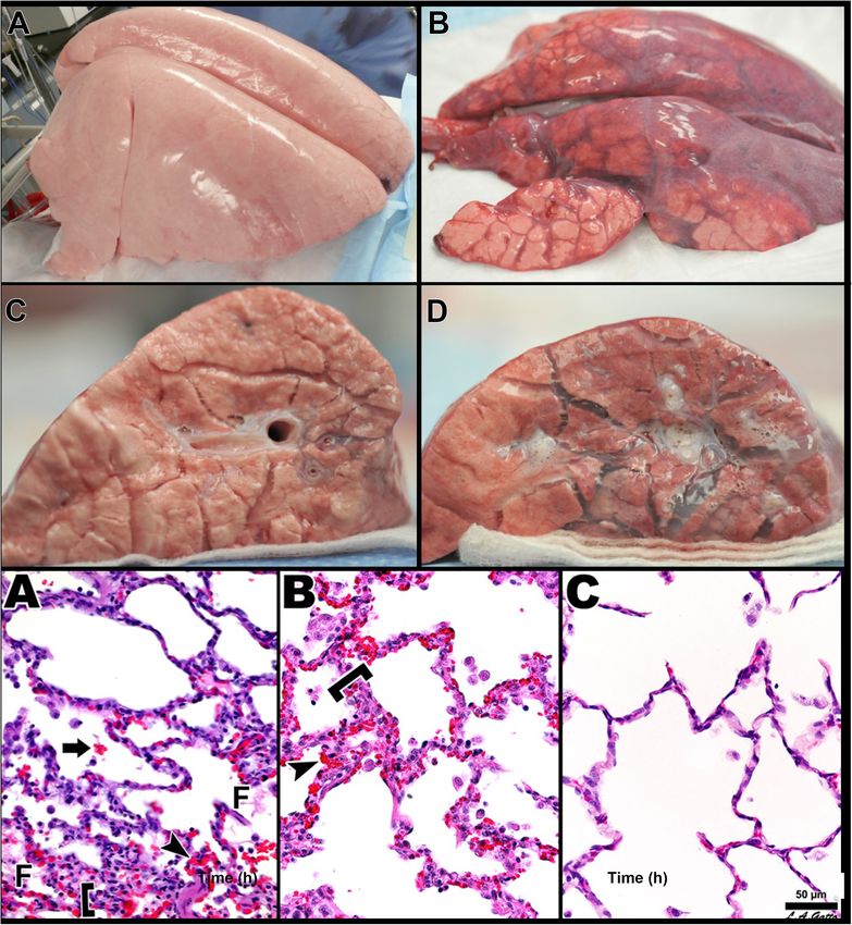
Nieman et al. Protecting the Acutely Injured Lung
INTRODUCTION inflated at end-expiration—functional residual capacity (FRC)—
and is referred to as the “baby lung” (Gattinoni and Pesenti,
Acute respiratory distress syndrome (ARDS) was initially thought 2005). The second compartment consists of alveoli in the
to be a lethal double pneumonia and was identified as a dependent areas that are collapsed and/or edema filled. The third
syndrome by Ashbaugh et al. (1967). Unfortunately, in the compartment consists of alveoli that remain in the transition
50 years since ARDS was identified, only a few treatments zone between healthy and unstable, due to loss of surfactant
have been used, with the mainstay being supportive in the function, and that open and collapse with every breath.
form of mechanical ventilation (Slutsky and Ranieri, 2013). Consequently, the ARDSnet low-Vt and plateau pressure
However, mechanical ventilation constrained to the limitations (Pplat) strategy is constrained to ventilating this heterogeneous
of a heterogeneously injured lung can cause unintended tissue lung tissue without causing VILI using a three-tiered approach:
damage, referred to as ventilator-induced lung injury (VILI), (1) protect the baby lung by not overdistending the compliant
which can significantly increase mortality (∼40%) as compared tissue that is open at FRC, (2) rest the dependent collapsed and
to lung protective ventilation (∼31%) (Acute Respiratory Distress edema-filled tissue by keeping it out of the ventilatory cycle,
Syndrome Network, 2000). Initial randomized controlled trials and (3) stabilize the tissue in between by applying positive end-
(RCTs) attempting to reduce VILI by lowering tidal volume expiratory pressure (PEEP), usually adjusted by oxygenation
(Vt) failed to reduce mortality (Stewart et al., 1998; Brower (Figure 2) (Acute Respiratory Distress Syndrome Network, 2000;
et al., 1999). It was not until the ARDS Network (ARDSnet) Del Sorbo et al., 2017).
conducted the seminal ARMA study, published in 2000, that a
reduction in mortality was shown (Acute Respiratory Distress Problems With Protecting the Baby Lung
Syndrome Network, 2000). However, most (Phua et al., 2009; Since the baby lung is believed to be a small volume of
Villar et al., 2011; Caser et al., 2014; Bellani et al., 2016; Laffey normal tissue (Gattinoni and Pesenti, 2005), its overdistension
et al., 2016; Villar et al., 2016; Maca et al., 2017; Raymondos has been postulated to be a primary VILI mechanism (Brower
et al., 2017; Rezoagli et al., 2017; Fan et al., 2018; McNicholas et al., 2004). However, in studies in which normal lungs were
et al., 2018; Pham et al., 2019; Shen et al., 2019) but not all subjected to excessively high airway pressures (>30 cm H2 O)
(Brun-Buisson et al., 2004; Fan et al., 2005; Putensen et al., and strain (2.5 ratio), overdistension-induced VILI did not
2009; Petrucci and De Feo, 2013; Shen et al., 2019) of the recent occur as long as this excessive strain was nearly static. This
statistical- and meta-analyses have shown that ARDS mortality suggests that overdistension of normal tissue with high volume
has not been reduced below the 31% “gold standard” of the and pressure is not a primary VILI mechanism unless there
2000 ARMA study but rather remains unacceptably high at is also a large dynamic strain (Seah et al., 2011; Protti et al.,
∼40% (Figure 1). Despite these disappointing results, the low-Vt 2013b; Jain et al., 2017). It is possible that the baby lung is
ARDSnet method is still recommended as the standard-of-care injured not by overdistension of the normal tissue, but rather by
protective ventilation strategy for ARDS patients (Fan et al., 2017, the recruiting and recollapse of unstable tissue in the adjacent
2018; Papazian et al., 2019). collapsed regions (Gattinoni et al., 1995). Thus, lowering Vt
Since outcome data for ARDS patients has not improved for and Pplat to reduce overdistension in the open lung tissue,
almost 20 years, it is imperative to (1) ascertain the mechanisms which is surrounded by a large volume of collapsed and unstable
of dynamic alveolar and alveolar duct volume change during tissue of very low compliance, may not reduce VILI. Regional
mechanical ventilation (elastic, viscous, or viscoelastic), (2) instability and inflammation occur throughout the entire lung,
characterize ARDS-induced changes in alveolar mechanics (i.e., including in tissue that appears to be normal on computed
the dynamic change in alveolar size and shape during mechanical tomography (CT) scan or chest X-ray, and serve as pathologic
ventilation) (Grune et al., 2019) that drive VILI-induced tissue focal points from which VILI-induced tissue damage expands
damage, (3) identify the role of airway pressure and the duration (Wellman et al., 2014, 2016; Cereda et al., 2016b, 2017). This
at both inspiration and expiration on alveolar mechanics in the suggests that to protect the normal lung tissue regional instability
acutely injured lung (Kollisch-Singule et al., 2014a, 2018), and must be eliminated.
(4) use this knowledge to develop novel ventilation strategies to However, others have shown that overdistension is a major
better reduce VILI and protect the lung. Although pulmonary component of VILI pathophysiology in injured lungs (Guldner
inflammation (biotrauma) also plays a critical role in ARDS and et al., 2016). Guldner et al. (2016), in a porcine ARDS
VILI pathogenesis, the focus of this review will be the mechanical model, showed that extreme conditions of overdistension
injury to tissue caused during ventilation. resulted in more lung inflammation than did extreme lung
collapse, suggesting that static stress and strain are major VILI
mechanisms. Although this study clearly showed that volutrauma
CONSTRAINTS OF VENTILATING THE increased inflammation, histopathology was not measured, and
ACUTELY INJURED LUNG there was no difference in pulmonary edema as measured by lung
weight. Thus, it is not clear whether the increase in inflammation
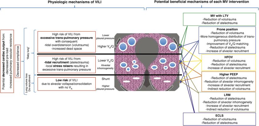
The current concept is that ARDS causes heterogeneous injury caused any lung pathology. In addition, recalculation of the data
in three general lung compartments that are layered by gravity showed that the average mechanical power in the volutrauma
(Figure 2). The first compartment, or the non-dependent area, group was 17.12 J/min, more than double that of the atelectrauma
contains a small number of normal compliant alveoli that remain group (7.13 J/min) (Tonetti et al., 2017). Others have also
Frontiers in Physiology | www.frontiersin.org 2 March 2020 | Volume 11 | Article 227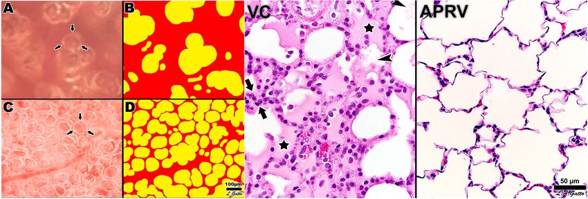
Nieman et al.                                                                                                                            Protecting the Acutely Injured Lung

  FIGURE 1 | (A) Hospital mortality (%) in the main epidemiological studies for all three American-European Consensus Conference (AECC) classifications of acute
  respiratory distress syndrome (ARDS – mild, moderate, and severe) and (B) mortality (%) for only moderate and severe ARDS. The mean and standard deviation for
  mortality in all of the studies following the 2000 ARMA study (Acute Respiratory Distress Syndrome Network, 2000) is 45.4 ± 9.5 (Rezoagli et al., 2017). Permissions
  obtained from AME Publishing Company, License ID 1017423-1.

  FIGURE 2 | Gravity dependent three-compartment model of acute respiratory distress syndrome (ARDS) pathology: Left – Physiological mechanisms of
  ventilator-induced lung injury (VILI) and Right – Potential beneficial mechanisms of various protective ventilation strategies to minimize VILI. Open alveoli are shown
  as blue circles similar in size (top), unstable alveoli as smaller circles of various sizes (middle), and collapsed alveoli as solid white lines (bottom). The ARDSnet low
  tidal volume (LVt) method is designed to protect compliant open alveoli in the non-dependent lung and rest the collapsed tissue in the dependent portion of the lung
  by keeping it unventilated. Positive end-expiratory pressure (PEEP) is added in an attempt to stabilize the alveoli in between (Acute Respiratory Distress Syndrome
  Network, 2000). High-frequency oscillatory ventilation (HFOV) and lung recruitment maneuvers (LRMs) have been shown ineffective in reducing ARDS mortality
  (Brower et al., 2004; Meade et al., 2008; Mercat et al., 2008; Ferguson et al., 2013; Young et al., 2013; Cavalcanti et al., 2017; Hodgson et al., 2019). Prone position
  has been shown effective at reducing mortality (Guerin et al., 2013) by a mechanism of reducing regional alveolar strain and inflammation (Motta-Ribeiro et al., 2018;
  Xin et al., 2018). VILI, ventilator-induced lung injury; MV, mechanical ventilation; LTV, low tidal volume and inspiratory pressure; PEEP, positive end-expiratory
  pressure; ECLS, extracorporeal life support; HFOV, high-frequency oscillatory ventilation; LRM, lung recruitment maneuver; Q, perfusion; VA , alveolar ventilation;
  VA /Q, ventilation/perfusion ratio; baby lung, functional residual capacity (FRC) (Del Sorbo et al., 2017). Permissions to publish obtained from ATS.

Frontiers in Physiology | www.frontiersin.org                                         3                                                March 2020 | Volume 11 | Article 227
Nieman et al.                                                                                                       Protecting the Acutely Injured Lung

shown that increasing airway pressure in an acutely injured lung           Caser et al., 2014; Bellani et al., 2016; Laffey et al., 2016; Villar
will cause a rapid progression of injury in a “rich-get-richer”            et al., 2016; Maca et al., 2017; Raymondos et al., 2017; Rezoagli
power-law fashion, supporting the findings in the Guldner study            et al., 2017; Fan et al., 2018; McNicholas et al., 2018; Pham
(Hamlington et al., 2018). Combined, these studies suggest that            et al., 2019). By allowing the lung to remain heterogeneously
high static stress and strain are associated with volutrauma in            collapsed, the protect, rest, and stabilize method is unintendedly
acutely injured lung tissue but not in normal lung tissue.                 preserving the constraints of ventilating the heterogeneously
                                                                           injured lung, which is nearly impossible to do without causing
Problems With Resting the Collapsed                                        some degree of VILI.
Lung
When the lung is allowed to collapse below normal FRC,                     Open Lung Approach (OLA) as a
atelectatic, and edema-filled resting tissue (1) does not exchange         Protective Strategy
gas; (2) is susceptible to the development of pneumonia (Huynh             The goal of the open lung approach (OLA) is to eliminate
et al., 2019; Li Bassi et al., 2019); (3) will become fibrotic if          the constraints of ventilating a heterogeneously injured lung
not reopened (Burkhardt, 1989; Cabrera-Benitez et al., 2014;               by normalizing all three pathologic compartments (Figure 2).
Lutz et al., 2015); (4) initiates patient-ventilator dyssynchrony,         The aim is to reinflate the collapsed tissue using a recruitment
which is caused by the firing of mechanical stretch, PO2 , PCO2 ,          maneuver (RM) and to keep it open by using an appropriate
and pH receptors (Solomon et al., 2000; Manning and Mahler,                level of PEEP. If the entire lung could be recruited and recollapse
2001; Widdicombe, 2001; Mellott et al., 2009; Burki and Lee,               prevented, the main VILI mechanical mechanisms (dynamic
2010; Yu, 2016; Yoshida et al., 2017) and which is associated              strain and overdistension of alveolar walls in areas of stress-
with high mortality (Blanch et al., 2015); and (5) creates a               focus) would be eliminated (Nieman et al., 2017b). An RM is an
stress-focus in the adjacent open alveoli and alveolar ducts,              acute event performed by raising the airway pressure (30–40 cm
greatly amplifying the forces applied to these parenchymal tissues         H2 O) and holding it for ∼40 s (Fan et al., 2008) or by greatly
during tidal ventilation (Mead et al., 1970; Gattinoni et al., 2012;       increasing PEEP (25 cm H2 O) and combining it with 15 cm H2 O
Cressoni et al., 2014; Makiyama et al., 2014; Retamal et al.,              of driving pressure above the PEEP (Borges et al., 2006). In the
2014). It has been shown that letting the lung rest in obese               latter strategy, PEEP is increased in 5 cm H2 O increments up
bariatric surgery patients is associated with worse oxygenation,           to 45 cm H2 O until the lung fully recruits, which was confirmed
longer post-anesthesia care unit stay, and more post-operative             when PaO2 + PaCO2 > 400 mmHg (Borges et al., 2006).
pulmonary complications as compared with patients in which the                 Following the RM, PEEP is titrated downward to find the lung
atelectatic resting lung was opened and ventilated (Talab et al.,          recollapse point (usually by a sharp fall in lung compliance), and
2009). Although, the phrase resting the lung sounds protective,            then PEEP is set 2 cm H2 O above this collapse pressure, following
the lung is not meant to function in a deflated state, and as              a second RM. However, multiple RCTs testing the OLA in ARDS
listed above, such a state is associated with numerous pathologies.        patients have failed to show significant benefits over standard
If the lung is to be rested, meaning that parenchymal tissue is            of care (Brower et al., 2004; Meade et al., 2008; Mercat et al.,
to be kept from being damaged by mechanical ventilation, the               2008; Cavalcanti et al., 2017; Hodgson et al., 2019). Reasons for
better strategy would be to rest it in the natural inflated state          these failures include the following: (1) timing of OLA application
(Nieman et al., 2018).                                                     [early (Borges et al., 2006) vs. late (Gattinoni et al., 2006)] (2) one-
                                                                           size-fits-all RM strategies, (3) PEEP set inappropriately to keep
Problems With Stabilizing the Lung                                         the recruited lung open, (4) recruiting pressures insufficient to
There is no consensus on how best to set PEEP to effectively               open all of the lung, (5) a patient population of responders (lung
stabilize lung tissue (Coruh and Luks, 2014; Gattinoni et al., 2017;       recruits) and non-responders (lung does not recruit) (Gattinoni
Nieman et al., 2017a; Bergez et al., 2019). The current ARDSnet            et al., 2006), and (6) application of OLA not as a continuous
method for setting PEEP uses a sliding scale of oxygenation                treatment but rather as a one-time event with a long time period
(Acute Respiratory Distress Syndrome Network, 2000), but                   before a second application or with no second application at all
increased oxygenation does not correlate well with an increase             (Goligher et al., 2017; Lu et al., 2017; Bhattacharjee et al., 2018;
in alveolar stability (Andrews et al., 2015), a key VILI mechanism         Cui et al., 2019; Hodgson et al., 2019; Kang et al., 2019; van der
(Wellman et al., 2014, 2016; Cereda et al., 2016b, 2017). Many             Zee and Gommers, 2019; Zheng et al., 2019). Most (Bhattacharjee
methods have been used in an attempt to titrate the PEEP to                et al., 2018; Cui et al., 2019; Hodgson et al., 2019; Kang et al., 2019;
stabilize lung tissue. These methods include using dead space,             Zheng et al., 2019) but not all (Goligher et al., 2017; Lu et al.,
lung stress and strain, lung compliance, CT, pressure-volume               2017) meta-analyses have shown no decrease in ARDS-related
curve inflection points, and electrical impedance tomography,              mortality associated with the OLA.
but there is no current bedside technique to determine whether                 To further reduce ARDS mortality and acute lung
the set PEEP has actually stabilized the lung (Nieman et al.,              injury, two pathologic processes must be understood: (1)
2017a). The above problems with the ARDSnet protect, rest,                 the pathophysiology of ARDS that predisposes the lung to
and stabilize method may partially explain the lack of improved            a secondary VILI and (2) the mechanisms of VILI in the
outcome in ARDS mortality over the last 20 years (Figure 1)                microenvironment (i.e., the terminal airspaces, alveoli, and
(Brun-Buisson et al., 2004; Phua et al., 2009; Villar et al., 2011;        alveolar ducts). This knowledge informs the design of a

Frontiers in Physiology | www.frontiersin.org                          4                                          March 2020 | Volume 11 | Article 227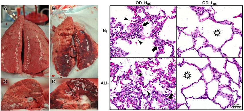
Nieman et al.                                                                                                                               Protecting the Acutely Injured Lung

  FIGURE 3 | The pathologic tetrad of acute respiratory distress syndrome (ARDS). Alveolar walls contain pulmonary capillaries (red circles) and are lined with a liquid
  hypophase (blue layer inside each alveolus), with an intact pulmonary surfactant layer (small blue ball with tail) layered on the hypophase. The systemic inflammatory
  response syndrome (SIRS) secondary to sepsis, trauma, burns, pneumonia, and so on increases pulmonary capillary permeability. Endothelial leakage: increased
  microvascular permeability allowing pulmonary edema to move into the alveolus (black arrows and tan edema blebs) (Martin and Brigham, 2012). Surfactant
  deactivation: the continuous layer of pulmonary surfactant molecules is disrupted as the edema blebs expand causing surfactant deactivation (surfactant sluffing off
  into the alveolar space). Edema usurping surfactant from the alveolar surface, the proteins in the edema fluid deactivating the surfactant (Taeusch et al., 2005), and
  improper mechanical ventilation (Albert, 2012) causing further surfactant disruption all combine to exacerbate surfactant loss. Alveolar edema: increased capillary
  permeability (Martin and Brigham, 2012) and high alveolar surface tension combine to flood alveoli with edema fluid (tan). Recruitment/derecruitment (R/D): loss
  of surfactant function results in increased alveolar surface tension causing loss of alveolar stability (i.e., causing alveolar R/D with each breath). Alveoli in the top
  frame of R/D are fully inflated but collapse during expiration in the bottom R/D frame. Alveolar R/D, known as atelectrauma, is another key VILI mechanism (Cressoni
  et al., 2017). Stress-focus: edema-filled or collapsed alveoli adjacent to air-filled alveoli create a stress-focus causing the alveolar wall to bend toward the fluid-filled
  alveolus (green arrow), which can cause stress failure at the alveolar wall (Perlman et al., 2011). Stress-focus is another key mechanism of VILI (Perlman et al., 2011;
  Chen et al., 2014; Makiyama et al., 2014; Retamal et al., 2014). Thus, the pathologic tetrad sets up a vicious cycle of high microvascular permeability → edema →
  surfactant deactivation → high alveolar surface tension → more edema → alveolar R/D → further increase in microvascular permeability → severe ARDS (Nieman
  and Bredenberg, 1985).

protective mechanical breath that will allow the lung to heal                              lung tissue instability and collapse. In addition, surfactant
by eliminating the constraints present when ventilating a                                  secretions from type II cells would be inhibited in collapsed
heterogeneously injured lung (Nieman et al., 2018).                                        areas that are not being stretched during ventilation (Wirtz
                                                                                           and Dobbs, 1990; Majumdar et al., 2012). Reduced surfactant
                                                                                           secretion would exacerbate and perpetuate the already reduced
ARDS PATHOPHYSIOLOGY THAT                                                                  surfactant function caused by alveolar flooding with edema.
PREDISPOSES THE LUNG TO VILI
ARDS Is a Pathologic Tetrad                                                                VILI MECHANISMS: HETEROGENEOUS
To investigate the relationship between ARDS and VILI,                                     ALVEOLAR INSTABILITY AND
we need to understand the pathology of acute lung injury.                                  COLLAPSE
Although ARDS is a complex syndrome, it features four well-
accepted central components (Thompson et al., 2017) known                                  The hallmark of ARDS is a heterogeneous lung injury
as the “pathologic tetrad” (Figure 3) (Nieman et al., 2018).                               encompassing normal, collapsed, edematous, and unstable tissues
The components of the tetrad include increased pulmonary                                   (Figure 2). This pathology alters pulmonary microanatomy
capillary permeability (Figure 3, Endothelial Leakage), which if                           and dynamic alveolar inflation physiology, generating three
unchecked will lead to loss of surfactant function (Figure 3,                              basic VILI mechanisms: volutrauma (overdistension of airways),
Surfactant Deactivation) (Lewis et al., 1993). The resultant                               atelectrauma (R/D of alveoli), and biotrauma (inflammation)
high alveolar surface tension will exacerbate the permeability-                            (Thompson et al., 2017). From an engineering perspective,
induced increase in alveolar flooding with edema fluid (Figure 3,                          volutrauma is caused by excessive static strain and atelectrauma
Alveolar Edema) (Nieman and Bredenberg, 1985). Surfactant                                  by excessive dynamic strain (Seah et al., 2011; Protti et al.,
dysfunction will alter alveolar mechanics, resulting in alveolar                           2013a,b, 2014). In this review we do not discuss biotrauma but
recruitment/derecruitment (R/D) with each breath (Figure 3,                                rather focus on the unintentional mechanical damage to the
R/D) (Schiller et al., 2001). Each component of the tetrad has a                           pulmonary parenchyma caused during mechanical ventilation.
profound impact on alveolar mechanics. Surfactant deactivation                                Mechanical ventilation of the acutely injured lung with altered
sets the stage for a secondary VILI by promoting heterogeneous                             alveolar opening and collapse time constants can cause tissue

Frontiers in Physiology | www.frontiersin.org                                          5                                                  March 2020 | Volume 11 | Article 227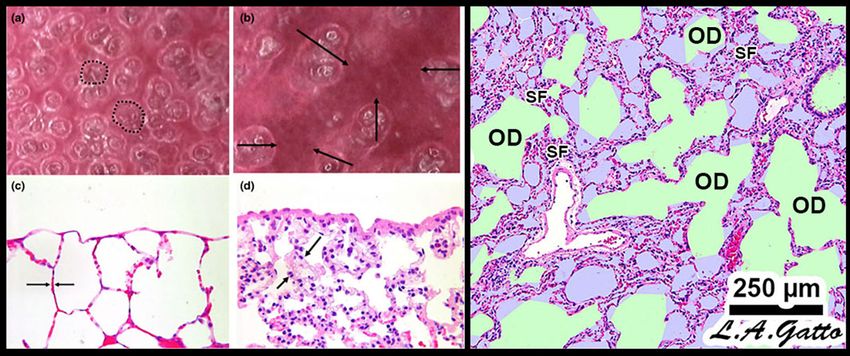
Nieman et al.                                                                                                                           Protecting the Acutely Injured Lung

  FIGURE 4 | Mechanical mechanisms of ventilator-induced lung injury (VILI) in the microenvironment: Left – In vivo subpleural alveoli in an acutely injured rat lung at
  inspiration (a) and expiration (b). Lung histology in the same rat lung injury model fixed at inspiration (c) and expiration (d) (Pavone et al., 2007). Alveolar
  recruitment/derecruitment (R/D) causing atelectrauma can be seen as fully recruited alveoli filling the microscopic field (white circles, two of which are highlighted
  with black dotted line) (a) that collapse during expiration (red atelectatic areas highlighted by arrows) (b). Histology shows open alveoli at inspiration with a shared
  alveolar wall highlighted by arrows (c) that collapse with expiration by alveolar wall folding, identified by arrows. Right – Stress-focus (SF) in areas with collapsed
  alveoli. SF adjacent to open alveolar ducts, causing alveolar duct overdistension (OD). SF-induced OD causes excessive mechanical stress and strain on alveolar
  duct walls, resulting in damage to pulmonary parenchymal cells (Ghadiali and Gaver, 2008). Loss of surfactant function results in alveolar instability (Figure 3, R/D).
  Histology at exhalation in a rat Tween-induced surfactant deactivation model with a tidal volume of 6 ml/kg and a PEEP of 5 cm H2 O. Note the heterogeneous
  collapse of alveoli causing areas of stress-focus (SF). Adjacent to these areas of SF are overdistended (OD) alveolar ducts (Kollisch-Singule et al., 2014b).
  Summary – R/D causes excessive normal or “peeling” stress as the adhered alveolar walls peel apart during inflation; both R/D and SF cause OD in adjacent open
  stable tissue. This excessive stress and strain on alveolar walls results in lung parenchymal cell death and is a major VILI mechanism. Permissions obtained to reuse
  panels (a–d) (Pavone et al., 2007). Springer Nature license number 4766000130978.

stress-failure by the following mechanisms: (1) alveolar R/D-                            damage to both of these tissues (Figure 5). Further support for the
induced excessive stress on the epithelial cells as the alveolar walls                   contention that volutrauma of normal lung tissue is not a primary
in apposition peel apart (Bilek et al., 2003), (2) stress-focusing in                    VILI mechanism comes from the Bates group, who showed that
areas of open alveoli adjacent to collapsed or edema fill alveoli                        4 h of mechanical ventilation in mice with high static strain
(Gattinoni et al., 2012; Cressoni et al., 2014; Retamal et al.,                          was not associated with lung injury, but when it was combined
2014), and (3) collapsed tissue stretching and overdistending the                        with high dynamic strain, it caused VILI-induced tissue damage
shared walls of patent alveoli, as alveolar walls are interconnected                     (Seah et al., 2011).
(Figure 4) (Nieman et al., 2017b). Theoretically, if it were possible                       Protti et al. (2014) showed that a large dynamic strain
to minimize or prevent all of the above VILI mechanisms, ARDS                            (atelectrauma) is much more harmful to the normal lung than
associated mortality would be significantly reduced.                                     a large static strain (volutrauma), and when combined, the two
   In normal pigs ventilated for 54 h, Protti et al. (2013b)                             work additively or synergistically to greatly accelerate tissue
examined the impact of high static strain and several levels of                          damage (Seah et al., 2011; Hamlington et al., 2016; Ruhl et al.,
dynamic strain. They demonstrated that high static strain, using                         2019). In addition, the heterogeneous injury caused by ARDS
elevated PEEP and minimal Vt, caused little damage, but when                             establishes many areas of stress-focus between the open tissue
PEEP was reduced, producing a high dynamic strain, it caused                             and collapsed or unstable tissue and have been shown to double
pulmonary edema and death. In subsequent work they showed                                the stress and strain calculated for the entire lung (Cressoni
that high static strain did not merely act as a dam to prevent                           et al., 2014). The impact that areas of stress-focus have on the
edema formation by altering the Starling forces (i.e., increasing                        forces generated on alveolar walls during ventilation was first
the pulmonary interstitial pressure) (Effros and Parker, 2009) but                       described by Mead et al. (1970) and was more recently analyzed
rather preserved the integrity of the alveolar-capillary membrane                        by Makiyama et al. (2014) using computer-simulated alveolar
(Figure 3, Endothelial Leakage) (Protti et al., 2013a). These                            walls. This latter group demonstrated that a Pplat at the upper
findings were supported by Jain et al. (2017) using a porcine                            end of the “safe” level (30 cm H2 O) could result in a local stress-
heterogeneous lung injury model. They subjected two study                                focusing in individual alveolar walls of up to 48 cm H2 O and
groups to high static strain (Pplat = 40 cm H2 O) that was believed                      could concentrate stress in an individual alveolar wall as much
to be more than sufficient to cause volutrauma-induced VILI                              as 16-fold (Figure 6).
(Acute Respiratory Distress Syndrome Network, 2000; Sahetya                                 In summary, ARDS results in a heterogeneous loss of
et al., 2017). Following heterogeneous lung injury, the animals                          surfactant function (Figure 3, Surfactant Deactivation) and
in the second group were also subjected to high dynamic strain.                          airway flooding (Figure 3, Alveolar Edema) that cause the lung
High static strain did not injure normal open tissue (baby lung),                        to become time and pressure dependent, which means that the
nor did it exacerbate acutely injured tissue. However, combining                         lung will collapse in a relatively short time at atmospheric
high static strain with high dynamic strain caused significant                           pressure and will require a longer period of time to open

Frontiers in Physiology | www.frontiersin.org                                        6                                                March 2020 | Volume 11 | Article 227
Nieman et al. Protecting the Acutely Injured Lung
FIGURE 5 | Heterogeneous Tween Injury in pigs ventilated for 6 h. A bronchoscope was used to deliver Tween to the dependent areas of the diaphragmatic or
caudal lung lobes. Thus, the upper lobes would be normal homogeneously inflated tissue (i.e., would simulate the baby lung), and the dependent areas of the caudal
lobe would model acutely injured tissue that would be either collapsed or unstable during tidal ventilation. Using this injury model we can determine whether a
ventilation strategy protects either normal tissue (NT ) or acutely injured lung tissue (ALIT ), protects neither, or protects both. Two groups were studied. Both groups
were subjected to a high static strain (Pplat = 40 cm H2 O) hypothesized to be sufficient to cause overdistension (OD)-induced VILI to the baby lung (Acute
Respiratory Distress Syndrome Network, 2000). One group was also subject to high dynamic strain (HDS ) and the other to low dynamic strain (LDS ). The dynamic
strain was adjusted using the airway pressure release ventilation (APRV) mode by changing the expiratory duration, which changed tidal volume size. Left – Gross
photographs of the whole lung (A,B) and cut surface (C,D) of the diaphragmatic lung lobe. The LDS group (A,C) showed minimal damage in both NT and ALIT lung
tissue (i.e., no dark red hepatized atelectasis). This was in contrast to the HDS group, in which there was severe injury in both the NT and ALIT lung tissues (B,D).
Right – In the OD + HDS group (OD + HDS ), widespread histopathology typical of ARDS was seen, with inflammatory cell inflation (arrows) and fibrin deposits
(arrowheads) in both the NT and ALIT lung tissue. In the OD + LDS group, minimal histopathology was seen, and alveoli remained open (stars) in both NT and ALIT
lung tissue. These data support Protti et al.’s (2013b) work and demonstrate that normal lung tissue is highly resistant to static strain-induced volutrauma. In
addition, this study showed that acutely injured lung tissue is also resistant to volutrauma as long as dynamic strain remains low. Both normal and acutely injured
lung tissue are highly susceptible to high alveolar R/D-induced VILI when under high inflation pressure (Jain et al., 2017). These data support the rapid progression of
lung injury in a power-law fashion when high static and dynamic strain are combined (Hamlington et al., 2018).
even at high inflation pressures (Takahashi et al., 2015). heterogeneously injured lung. The better strategy would be
Mechanical ventilation can exacerbate the initial ARDS-induced to eliminate these constraints by opening and stabilizing the
inflammatory injury (Figure 3, Endothelial Leakage, Surfactant acutely injured lung or, better yet, by applying a protective
Deactivation, and Alveolar Edema) by generating excessive stress ventilation strategy early and never letting the lung collapse
and strain on alveolar and alveolar duct walls resulting from a (Satalin et al., 2018). Gattinoni and Pesenti stressed that in
collapsing and reopening of alveoli and heterogeneous areas of a patient with ARDS the Vt should not be set by body
stress-focusing in open tissue adjacent to collapsed or edema- weight (Vt/kg ratio) but rather by the size of the remaining
filled tissue (Figure 4). normal open tissue at FRC: the baby lung (Vt/baby lung
It is important to note that parenchymal overdistension volume ratio) (Gattinoni and Pesenti, 2005). They hypothesized
following acute lung injury is a regional phenomenon and occurs that the baby lung was the small amount of open tissue
only in open alveoli and alveolar ducts that are adjacent to at end-expiration (i.e., FRC) surrounded by a large volume
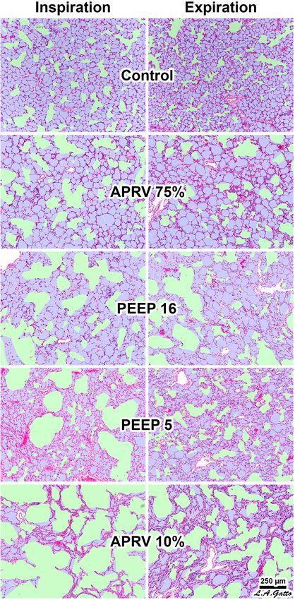
the unstable or collapsed tissue (Figure 7, PEEP 16, PEEP 5, of collapsed tissue with very low compliance. Since the
APRV 10%); it does not occur in homogeneously inflated acutely specific elastance [Espec = transpulmonary pressure (Ptp)/Vt
injured lung tissue (Figure 7, APRV 75%) (Nieman et al., 2017b). x baby lung volume, which is the airway pressure at which
It has been shown that combining high pressure with alveolar expiratory lung volume or FRC or baby lung volume doubles
instability greatly exacerbates tissue tearing in a rich-get-richer in size (i.e., when Vt/baby lung volume = 1)] if the baby
fashion (i.e., the larger the initial tear in the epithelial membrane lung is postulated to be normal, there would be a greater
the more that this tear will be expanded by increased airway potential for overdistension in normal lung tissue because
pressure) (Figure 8) (Hamlington et al., 2016; Ruhl et al., 2019). of the high compliance at any given level of static strain
(Gattinoni and Pesenti, 2005). Both the potential of excessive
stress (Ptp) to cause volutrauma and of excessive strain (Vt/end-
VENTILATING WITHIN THE expiratory lung volume) to cause atelectrauma are linked with the
CONSTRAINTS OF AN ACUTELY following equation:
INJURED LUNG
Ptp (stress) = Espec × (Vt/baby lung volume).
The ARDSnet method is a logical physiologically based lung
ventilation strategy within the constraints of protecting a (Eq. 1) (Gattinoni and Pesenti, 2005) (1)
Frontiers in Physiology | www.frontiersin.org 7 March 2020 | Volume 11 | Article 227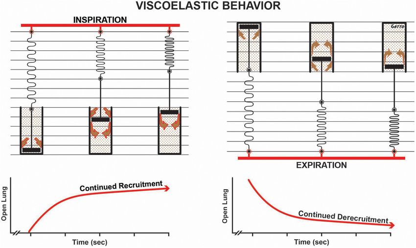
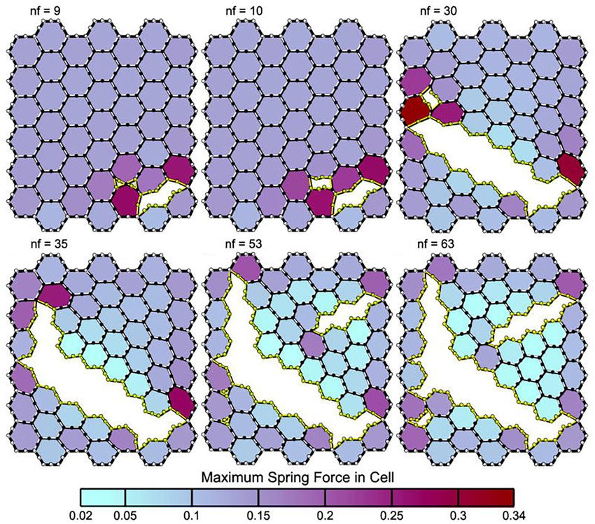
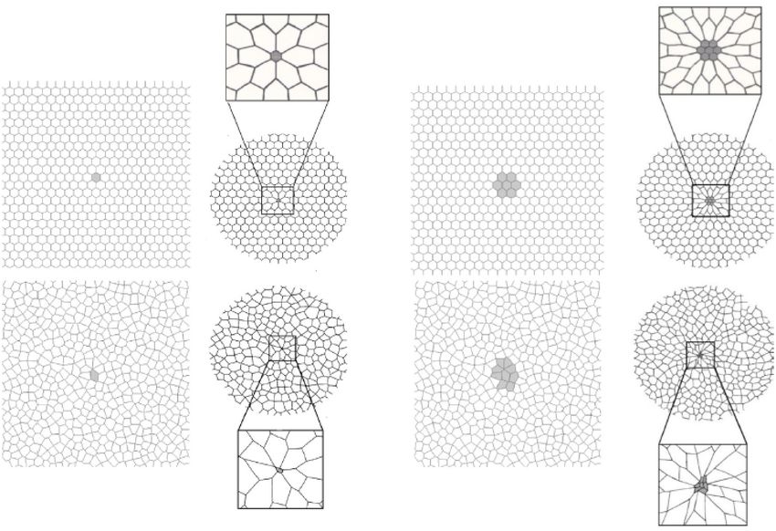
Nieman et al.                                                                                                                          Protecting the Acutely Injured Lung

  FIGURE 6 | A two-dimensional finite element computational model of
  interconnected alveolar walls to study the impact of areas of stress-focus (S-F)
  on stress–strain relationships. Both a hexagonal honeycomb (top) and Voroni
  honeycombs (bottom) were studied with areas of S-F created by increasing
  the stiff regions in one (A) or nine (B) cells. These stiff regions simulate
  collapsed alveoli adjacent to open alveoli that create an S-F in acute
  respiratory distress syndrome (ARDS) patients (Figure 4) (Cressoni et al.,
  2014). The entire honeycomb structure was expanded and exposed to strains
  of 15, 30, 45, and 55% above the resting geometry. Some of the alveolar
  walls in the Voroni honeycomb were exposed to a S-F ∼16 times greater than
  that applied to uniformly expanded areas. This suggests that ventilation
  pressures considered safe in ARDS patients (
Nieman et al.                                                                                                                           Protecting the Acutely Injured Lung

  FIGURE 7 | Continued
                                                                                              Animal studies have shown that the OLA can protect the
  expiration in PEEP16. In the PEEP5 group alveolar ducts were relatively                 lung from VILI even if 1P is not reduced (Tojo et al., 2018).
  normal size at expiration but greatly overdistended at inspiration with areas of        Recruiting the lung with PEEP has been shown to reduce
  alveolar collapse (i.e., high dynamic strain) at both inspiration and expiration.       tissue damage secondary to spontaneous breathing (SB) by
  APRV 10% resulted in highly overdistended alveolar ducts at both inspiration            two mechanisms: (1) the intensity of the SB is reduced via
  and expiration with large areas of collapsed alveoli. APRV 75% (i.e., the TCAV
  method) resulted in the smallest increase in alveolar duct size, with uniformly
                                                                                          neuromechanical uncoupling, and (2) the reduced amount of
  open, homogeneously ventilated alveoli that were closest to those seen in the           atelectatic tissue decreases the volume of stress-focus areas
  Control group (Kollisch-Singule et al., 2014b). Permissions obtained from               (Morais et al., 2018). Other studies using animal models have
  Elsevier. License 4732510649425.                                                        also shown the physiologic and pathologic benefit of opening the
                                                                                          acutely injured lung (Faridy et al., 1966; Webb and Tierney, 1974;
                                                                                          Dreyfuss et al., 1988; Muscedere et al., 1994; Nakazawa et al.,
                                                                                          2007; Nieman et al., 2015; Magalhaes et al., 2018). Additionally,
in the adjacent patent alveoli and that alveolar size actually                            animal studies have shown that to successfully implement the
decreases with increased airway pressure (i.e., PEEP) if lung tissue                      OLA, the RM and subsequent PEEP level must be applied
is recruited by a mechanism of gas redistribution (Cereda et al.,                         properly, or the approach may actually increase lung damage.
2011, 2013, 2016a). It has also been shown that gas redistribution                        Farias et al. (2005) showed that an RM increased lung pathology
in the microenvironment is not only pressure dependent but also                           if PEEP was not set sufficiently high to prevent recollapse of the
time dependent (i.e., the longer the pressure is applied, the better                      newly opened tissue, a finding that is supported by direct in vivo
the gas redistribution) (Figure 7) (Kollisch-Singule et al., 2014b).                      observation of subpleural alveoli (Halter et al., 2003). In light of

  FIGURE 8 | Panels (A–F) depict leak progression caused by an applied stretch force. A single node fails (nf) at each time point with nf indicating the number of
  nodes that have failed in each panel. Computational model of an epithelial monolayer to simulate leak progression due to overdistension. Leak progression in a
  45-cell (hexagon) network caused by applied stretch (i.e., Vt). After the force required to initiate the leak was reached, the leak area increased at a constant rate as
  the force increased further (Hamlington et al., 2016). Atelectrauma caused the initial tears, after which volutrauma expanded those tears. The tears progressed in a
  rich-get-richer mechanism in which the likelihood of a tear getting larger increased with the size of the initial tear. This mechanism explains why atelectrauma appears
  to be essential to the initiation of VILI in a normal lung, and why atelectrauma and volutrauma act synergistically once VILI is underway (Hamlington et al., 2018).
  Permissions obtained from Springer Nature. License 4699410825602.

Frontiers in Physiology | www.frontiersin.org                                         9                                               March 2020 | Volume 11 | Article 227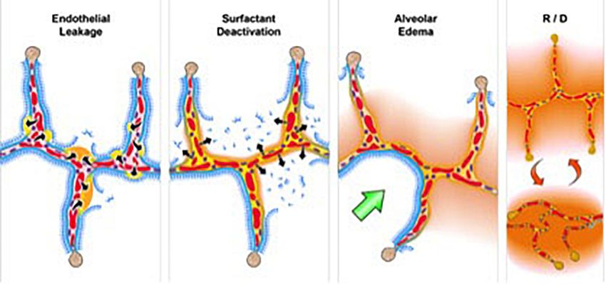
Nieman et al.                                                                                                                            Protecting the Acutely Injured Lung

  FIGURE 9 | Viscoelastic behavior of alveolar recruitment and derecruitment described using a spring and dashpot model. During inspiration the spring is rapidly
  stretched, whereas the dash in the pot moves more slowly as fluid squeezes around it (brown arrows), in response to the applied force (i.e., tidal volume – red bar).
  The effect of this viscoelastic behavior is continual alveolar recruitment as long as inspiration is held (open lung/time curve). Viscoelastic behavior during expiration
  shows a rapid collapse of the spring and a much slower collapse of the dash in the pot. This is shown in the open lung/time curve as continuous alveolar collapse or
  derecruitment over time. Viscoelastic alveolar opening and collapse suggests that the longer inspiration is held, the more alveoli are recruited, and the shorter the
  expiratory time, the fewer the alveoli that collapse.

this evidence, discarding the OLA does not seem logical, since                            inspiration and expiration, must be analyzed (Kollisch-Singule
it is unclear whether the RCTs testing the OLA actually opened                            et al., 2014a). The current OLA ventilation strategies do not
and stabilized the lung. A better strategy might be to identify                           consider the pathophysiological changes that occur in alveolar
ventilation strategies that are most likely to accomplish the goals                       opening and collapse time constants. Attempting to recruit large
of the OLA (Sahetya and Brower, 2017).                                                    volumes of collapsed lung with a single RM over a very brief
                                                                                          period of time is often not physiologically possible depending
                                                                                          on the degree of surfactant damage and edema. A more effective
A PHYSIOLOGICALLY INFORMED                                                                strategy may be a ventilation method that “nudges” the lung open
STRATEGY TO EFFECTIVELY OPEN AND                                                          over an extended period of time (6–24 h) using a high mean
STABILIZE THE LUNG                                                                        airway pressure held for most of each breath. Likewise, the very
                                                                                          fast alveolar collapse time constants are not considered when
Surfactant deactivation and edema flooding with acute lung                                setting PEEP with expiratory durations in the 2- to 3-s range.
injury cause alveolar collapse; the result is very “sticky” and                           A more effective strategy may be to use a ventilator method
does not reopen easily (Figure 3) (Crotti et al., 2001; Gattinoni                         with a very brief (≤0.5 s) expiratory time while maintaining
et al., 2003). This tissue has a very long opening time constant,                         a level of time-controlled PEEP (TC-PEEP). Greatly limiting
and thus it will take an extended period of time at any given                             the expiratory duration (≤0.5 s) keeps the lung from emptying
pressure to recruit these surfactant deficient and edematous                              completely, maintaining a TC-PEEP that is related to expiratory
tissues. Once the collapsed alveoli are recruited, the opposite                           duration and the collapse time constant of the lung. Thus, for
problem develops; the newly opened alveoli have a very brief                              any given collapse time constant, the shorter the expiratory
collapse time constant (Neumann et al., 1998a,b, 2000). Thus,                             time, the higher the TC-PEEP and vice versa. In addition, since
alveoli collapse quickly (≤0.5 s) once a critical collapse airway                         alveolar collapse is viscoelastic in nature (Figure 9), the very
pressure is reached (i.e., collapse is time and pressure dependent)                       short expiratory duration would work synergistically with TC-
(Kollisch-Singule et al., 2014a,b).                                                       PEEP to prevent alveolar collapse – alveoli simply would not
    To effectively implement the OLA, the entire mechanical                               have sufficient time to derecruit (Nieman et al., 2019). Analysis
breath pattern (MBP ), including all airway volumes, flows,                               of the change in alveolar opening and collapse time constants
pressure, rates, and the times during which they apply during                             with acute lung injury suggests that the time component of the

Frontiers in Physiology | www.frontiersin.org                                        10                                               March 2020 | Volume 11 | Article 227
Nieman et al. Protecting the Acutely Injured Lung
MBP (Kollisch-Singule et al., 2014a) can be used to improve the amplify energy dissipation locally. Regional instability acts as a
ability to recruit and stabilize acutely injured lung tissue, which is stress-focus, and once a microstrain threshold is reached, the
necessary to successfully implement the OLA. unrecovered energy will cause a rapid progression of lung tissue
In addition to the time component of MBP , the dynamic injury in a power-law fashion (Hamlington et al., 2018).
physiology of alveolar R/D must also be understood to design
a mechanical breath for the OLA. Alveoli and alveolar ducts
inflate and deflate as a viscoelastic system (Denny and Schroter,
2000; Escolar and Escolar, 2004; Farias et al., 2005; Carvalho ELIMINATING CONSTRAINTS OF
and Zin, 2011; Suki and Bates, 2011; Nieman et al., 2017b). VENTILATING THE ACUTELY INJURED
The model most used to analyze viscoelastic behavior is the LUNG
spring and dashpot (Figure 9). The most important thing to
know about a viscoelastic system, in relation to lung opening It was recently shown in ARDS patients that low Vt and Pplat
and collapse, is that there is a time lag from when the force do not correlate well with reduced patient mortality, whereas low
(inspiratory pressure) is applied until alveoli begins to open and 1P correlated strongly with improved survival (Amato et al.,
a lag between when force is removed (expiratory pressure) and 2015). The critical lesson is that it is not the settings dialed
when alveoli begin to collapse. Thus, the longer the inspiratory into the ventilator (i.e., Vt, Pplat, and so on) that are key to
time, the more lung tissue that will be recruited, and the shorter improved survival, but rather the impact of these settings on lung
the expiratory time, the less lung tissue that will be allowed physiology measured as a change in 1P (Kollisch-Singule et al.,
to recollapse (Figure 9). A collapsed airway inflates after the 2014a,b, 2015b).
opening threshold pressure is reached, and the pressure then Using the ARDSnet method, the Vt was set at 6 cc/kg,
propagates down the airway, inflating more airways and alveoli. the Pplat was set at < 30 cm H2 O, and PEEP was set on a
This process progresses in an avalanche manner with power-law sliding oxygenation scale; the mechanism of improved mortality
distributions of both the size of and intervals between avalanches was assumed to be due to these parameters. The Amato study
(Suki et al., 1994; Alencar et al., 2002). It has been postulated clearly demonstrated that the mechanism of increased survival
that as the lung opens, the increase in parenchymal tethering was not these desired setting values, because none correlated
of airways (Broche et al., 2017) and alveolar interdependence with outcome (Amato et al., 2015). Lower 1P, on the other
improves lung function as a power-law function (Nieman et al., hand, strongly correlated with reduced mortality. Respiratory
2019). These new perspectives inform the quest for novel system compliance (CRS ) was used to calculate driving pressure
protective ventilation strategies. Indeed, our work in translational (1P = Vt/CRS ), which was shown to decrease in patients
animal models and a meta-analysis of data on surgical intensive who survived, reflecting a desirable change in lung physiology
care unit (SICU) patients has shown that our time-controlled caused by recruitment.
adaptive ventilation (TCAV) method, using airway pressure The ARDSnet method assumes that the constraints of
release ventilation (APRV) mode, is highly effective at keeping ventilating a heterogeneously collapsed lung are unavoidable,
the lung open and stable, significantly reducing morbidity in in which case the Ptp is high due to the high Espec value and
translational animal models and reducing the ARDS incidence low baby lung (FRC) volume. However, if the entire lung could
and mortality rates of SICU patients at high risk of developing be fully recruited, these constraints would be eliminated, Espec
ARDS (Roy et al., 2012; Andrews et al., 2013; Emr et al., 2013; Roy reduced, and FRC increased, resulting in a significant reduction
S.K. et al., 2013; Kollisch-Singule et al., 2014a,b, 2015a,b, 2017; in the applied stress (i.e., Ptp) for any given Vt. Rahaman used
Nieman et al., Nieman et al., 2015, 2017a,b, 2018; Smith et al., a stress equation similar to Eq. 1 to analyze the mathematics
2015; Jain et al., 2016, 2017; Satalin et al., 2018; Silva et al., 2018; of VILI (Rahaman, 2017). He concluded that stress increases
Mahajan et al., 2019). for any given Vt and PEEP with an increase in respiratory rate
Others have shown the importance of ventilation time on lung (RR). This conclusion assumes that the clinician is constrained
mechanics. Saddy et al. (2013) calculated a pressure-time product in ventilating a heterogeneously collapsed lung such that neither
(PTP)/breath as the integral of the change in esophageal pressure FRC (i.e., the size of the baby lung) nor Espec can be changed.
over time. They found that, when using biphasic positive airway When these conditions are true, the conclusion that Vt, PEEP, and
pressure, PTP significantly increased when the rate of time-cycled RR must be kept low to reduce stress is correct. Conversely, our
control breaths was at 50 breaths/min as compared to when it perspective is the better strategy is to treat the lung by reinflating
was 100 or 75 breaths/min. The energetics of ventilation also the collapsed tissue, increase FRC (eliminating the baby lung),
contain components of ventilator time. Power is defined as work and decreasing Espec . This combination will reduce lung stress
per unit time and is thus equal to pressure x (volume/time) (Ptp) during ventilation even at higher Vt, PEEP, and RR (Eq. 1).
(Marini, 2018). Most of the energy applied to the lung during Of course the strategy of “casting” the broken lung open
inflation is accounted for in elastic storage and airway resistance. until it heals would be clinically effective only with a ventilation
It is postulated that the damage due to power is caused by the strategy that could perform such a feat (Nieman et al., 2018).
energy that is dissipated and unrecovered during exhalation. Unfortunately, the current OLA strategies have not been shown
In addition, it is not just the power but also the changes in effective at opening and stabilizing the lung (Bhattacharjee et al.,
the microenvironment that result in tissue damage. Regional 2018; Cui et al., 2019; Hodgson et al., 2019; Kang et al., 2019;
alveolar instability can develop during ARDS and can greatly Zheng et al., 2019), and the recent ART trial showed an increase in
Frontiers in Physiology | www.frontiersin.org 11 March 2020 | Volume 11 | Article 227Nieman et al. Protecting the Acutely Injured Lung
FIGURE 10 | Left – Subpleural alveoli seen using in vivo microscopy in a rat hemorrhagic shock-induced ARDS model ventilated with volume cycled ventilation (VC;
A,B) or airway pressure release ventilation (APRV) using the time controlled adaptive ventilation (TCAV) method (APRV; C,D). Individual alveoli are shown by arrows.
Inflated alveoli were color coded yellow, and alveolar number, size, and surface area were measured by computer image analysis in each group. TCAV significantly
improved alveolar patency and stability as compared with the VC group. Right – The APRV group delivered using the TCAV method stabilized alveoli that was
associated with a significant reduction in lung histopathology as evidenced by open alveoli and reduced intra-alveolar edema (purple areas) as compared with the
collapsed and edema-filled alveoli (stars), fibrinous deposits in the air compartment (arrowheads), and white cell infiltration (between arrows) in the VC group (Roy
S.K. et al., 2013). Permissions obtained from Wolters Kluwer Health, Inc. License 4699411295724.
mortality using the OLA (Brower et al., 2004; Meade et al., 2008; (Roy et al., 2012; Emr et al., 2013; Roy S.K. et al., 2013; Roy S. et al.,
Mercat et al., 2008; Cavalcanti et al., 2017). 2013; Kollisch-Singule et al., 2014a,b, 2015a,b, 2017; Smith et al.,
2015; Silva et al., 2018) and in a meta-analysis of data on SICU
patients (Andrews et al., 2013). In addition, the TCAV method
THE TCAV METHOD TO OPEN AND is the primary ventilator strategy used at the R Adam Cowley
STABILIZE THE ACUTELY INJURED Shock Trauma Center in Baltimore, with well over 1,000,000 h
LUNG of ventilator time. Below, we discuss the results from our animal
data and our clinical statistical analysis as evidence for the TCAV
Our TCAV method using the APRV mode has been discussed method’s mechanisms and efficacy.
in detail elsewhere (Habashi, 2005; Jain et al., 2016; Nieman In rat VILI and hemorrhagic shock models, it was shown
et al., 2018, 2019; Kollisch-Singule et al., 2019). Briefly, TCAV that ventilation for 6 h using the TCAV method was superior to
consists of an extended (4–5 s) open valve continuous positive volume-controlled ventilation (VCV; Vt 10 ml/kg, PEEP 0.5 cm
airway pressure (CPAP) phase with a very short (≤0.5 s) release H2 O) at preventing the development of ARDS and that lung
phase. The inspiratory:expiratory ratio is ∼10:1. The open protection was associated with stabilization of alveoli (Figure 10)
valve allows the patient to spontaneously breath (inspiration or (Emr et al., 2013; Roy S.K. et al., 2013). The TCAV method
expiration) with little resistance. Tidal volume (Vt) is not set was also shown to be lung protective in a preterm piglet model
but rather is a product of the CPAP level and lung compliance. of infant respiratory distress syndrome (Kollisch-Singule et al.,
A heterogeneously collapsed lung will have a very low Vt because 2017). These data were supported by mechanistic studies showing
the compliance will be low, but as the lung gradually recruits over the ability of the TCAV method to normalize alveolar and alveolar
time, the compliance will increase and so will the Vt. Since the Vt duct microanatomy (Kollisch-Singule et al., 2014b) (Figure 7)
is set based on lung compliance, a high Vt will never be delivered and to reduce dynamic alveolar strain (Kollisch-Singule et al.,
to a non-compliant collapsed lung using the TCAV method. 2014a, 2015b; Smith et al., 2015). We developed a 48-hr
Thus, the Vt size is personalized and adaptive as the patient’s clinically applicable, high-fidelity, porcine peritoneal sepsis (PS)
lung gets better or worse, directed by changes in lung compliance. plus gut ischemia/reperfusion (I/R), multiple organ dysfunction
The extended CPAP time will gradually nudge viscoelastic alveoli syndrome (MODS) and ARDS model. In three studies using
open over several hours until the lung is fully inflated. The newly this clinically applicable model, we demonstrated that the TCAV
recruited alveoli will be prevented from recollapse through the method was superior to VC or the ARDSnet method at blocking
use of a very short expiratory duration (release phase). Expiratory progressive acute lung injury and preventing ARDS development
time is very short (≤0.5 s), and thus the lung is reinflated (CPAP (Figure 11) (Roy et al., 2012; Roy S. et al., 2013; Kollisch-Singule
phase) before it has time to completely empty, maintaining a et al., 2015a). In addition, we have shown that surfactant proteins
TC-PEEP. The very short expiratory time is not sufficient for A and B are both better preserved with the TCAV as compared
viscoelastic alveoli to collapse and when combined with the with the ARDSnet method, suggesting that there is sufficient
TC-PEEP is highly effective at preventing alveolar derecruitment. lung volume change with TCAV to preserve stretch-induced
Our group has investigated the physiological impact of the surfactant release (Roy et al., 2012; Emr et al., 2013; Roy S.K. et al.,
TCAV method in both mechanistic and efficacy animal studies 2013; Roy S. et al., 2013; Kollisch-Singule et al., 2015a). Lastly, in a
Frontiers in Physiology | www.frontiersin.org 12 March 2020 | Volume 11 | Article 227Nieman et al. Protecting the Acutely Injured Lung FIGURE 11 | Three groups of pigs were ventilated for 48 h with a clinically applicable, high-fidelity porcine peritoneal sepsis (PS) and gut ischemia/reperfusion (I/R) acute respiratory distress syndrome (ARDS) model: (1) Control group without PS + I/R injury ventilated with a tidal volume (Vt) of 10 cc/kg and a positive end-expiratory pressure (PEEP) of 5 cm H2 O. (2) ARDSnet group using a low-Vt ventilation method, applied following PS + I/R injury at the time point at which the animal reached the oxygen saturation limit listed in the ARDSnet protocol, with PEEP and FiO2 adjusted by oxygenation (Acute Respiratory Distress Syndrome Network, 2000). (3) Time-controlled adaptive ventilation (TCAV) method using the airway pressure release ventilation (APRV) mode applied immediately after PS + I/R injury (Habashi, 2005; Jain et al., 2016). Upper – Gross lung photographs after 48 h of ventilation following PS + I/R injury at necropsy (Control not shown). In the TCAV group (A,B), lungs inflated fully to near total lung capacity at 25 cm H2 O without any gross atelectasis. The cut surface of the diaphragmatic lobe also showed no interstitial or airway edema and no atelectasis. Lungs in the ARDSnet group (C,D) also inflated to 25 cm H2 O showed low lung volume with heterogeneous collapse and atelectasis and were wet and “boggy.” The cut surface showed both interstitial and airway edema. Lower – Photomicrographs of representative lung sections of specimens from the Control (A), ARDSnet (B), and TCAV (C) groups each at 40× magnification. F, fibrinous deposit in the air compartment; arrow, blood in alveolus; arrowhead, congested alveolar capillary; bracket, thickened alveolar wall. (A) Control: animals received 48 h of mechanical ventilation without PS + I/R injury. Specimen shows typical early acute lung injury pathology including fibrinous deposits, blood in alveolus, congested capillaries, and thickened alveolar walls. (B) ARDSnet: animals received PS + I/R injury, and LVt ventilation was applied after the onset of acute lung injury (i.e., PaO2 /FiO2 ratio
You can also read