The RNA-Binding Proteins SRP14 and HMGB3 Control HIV-1 Tat mRNA Processing and Translation During HIV-1 Latency - Frontiers
←
→
Page content transcription
If your browser does not render page correctly, please read the page content below
ORIGINAL RESEARCH
                                                                                                                                                   published: 14 June 2021
                                                                                                                                          doi: 10.3389/fgene.2021.680725

                                              The RNA-Binding Proteins SRP14
                                              and HMGB3 Control HIV-1 Tat mRNA
                                              Processing and Translation During
                                              HIV-1 Latency
                                              Georges Khoury 1† , Michelle Y. Lee 1† , Sri H. Ramarathinam 2 , James McMahon 3 ,
                                              Anthony W. Purcell 2 , Secondo Sonza 1 , Sharon R. Lewin 3,4,5 and Damian F. J. Purcell 1*
                                              1
                                               Department of Microbiology and Immunology, Peter Doherty Institute for Infection and Immunity, University of Melbourne,
                                              Melbourne, VIC, Australia, 2 Infection and Immunity Program, Department of Biochemistry and Molecular Biology,
                                              Biomedicine Discovery Institute, Monash University, Clayton, VIC, Australia, 3 Victorian Infectious Diseases Service, The Royal
                                              Melbourne Hospital at the Peter Doherty Institute for Infection and Immunity, Melbourne, VIC, Australia, 4 Department
                           Edited by:
                                              of Infectious Diseases, The University of Melbourne at the Peter Doherty Institute for Infection and Immunity, Melbourne, VIC,
                      Jane E. A. Reid,
                                              Australia, 5 Department of Infectious Diseases, Alfred Hospital and Monash University, Melbourne, VIC, Australia
         Australian National University,
                              Australia
                        Reviewed by:          HIV-1 Tat protein is essential for virus production. RNA-binding proteins that facilitate Tat
                     Fatah Kashanchi,
                                              production may be absent or downregulated in resting CD4+ T-cells, the main reservoir
            George Mason University,
                          United States       of latent HIV in people with HIV (PWH) on antiretroviral therapy (ART). In this study, we
              Elton J. R. Vasconcelos,        examined the role of Tat RNA-binding proteins on the expression of Tat and control
 University of Leeds, United Kingdom
                                              of latent and productive infection. Affinity purification coupled with mass spectrometry
                   *Correspondence:
                   Damian F. J. Purcell
                                              analysis was used to detect binding partners of MS2-tagged tat mRNA in a T cell-line
                  dfjp@unimelb.edu.au         model of HIV latency. The effect of knockdown and overexpression of the proteins of
                    † Present address:        interest on Tat transactivation and translation was assessed by luciferase-based reporter
                   Georges Khoury and
                                              assays and infections with a dual color HIV reporter virus. Out of the 243 interactions
                         Michelle Y. Lee,
                Division of Microbiology      identified, knockdown of SRP14 (Signal Recognition Particle 14) negatively affected tat
     and Immunology, Yerkes National          mRNA processing and translation as well as Tat-mediated transactivation, which led
     Primate Research Center, Emory
 University, Atlanta, GA, United States
                                              to an increase in latent infection. On the other hand, knockdown of HMGB3 (High
                                              Mobility Group Box 3) resulted in an increase in Tat transactivation and translation as well
                    Specialty section:        as an increase in productive infection. Footprinting experiments revealed that SRP14
          This article was submitted to
                                   RNA,
                                              and HMGB3 proteins bind to TIM-TAM, a conserved RNA sequence-structure in tat
                a section of the journal      mRNA that functions as a Tat IRES modulator of tat mRNA. Overexpression of SRP14
                   Frontiers in Genetics
                                              in resting CD4+ T-cells from patients on ART was sufficient to reverse HIV-1 latency and
           Received: 15 March 2021
                                              induce virus production. The role of SRP14 and HMGB3 proteins in controlling HIV Tat
            Accepted: 17 May 2021
           Published: 14 June 2021            expression during latency will be further assessed as potential drug targets.
                            Citation:         Keywords: HIV-1, latency, tat mRNA, SRP14, HMGB3, mRNA processing, translation
                   Khoury G, Lee MY,
     Ramarathinam SH, McMahon J,
  Purcell AW, Sonza S, Lewin SR and
 Purcell DFJ (2021) The RNA-Binding
                                              INTRODUCTION
Proteins SRP14 and HMGB3 Control
          HIV-1 Tat mRNA Processing
                                              The persistence of a reservoir of resting CD4+ T-cells harboring silent provirus is the major
and Translation During HIV-1 Latency.         impediment toward attaining a cure for HIV-1 (Deeks et al., 2016). Despite two decades of
            Front. Genet. 12:680725.          intensive investigation, the mechanisms contributing toward establishment and maintenance of
    doi: 10.3389/fgene.2021.680725            latent infection in vivo remain incompletely understood (Khoury et al., 2018a). To date, blocks

Frontiers in Genetics | www.frontiersin.org                                           1                                             June 2021 | Volume 12 | Article 680725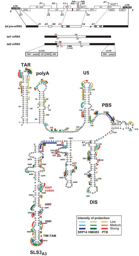
Khoury et al.                                                                                Post-Transcriptional Control of Tat Expression During Latency

to the initiation of transcription have been the most widely                model (Saleh et al., 2011; Spina et al., 2013) infected with virus
studied and targeted for reversal of latent infection in clinical           carrying mutated TIM-TAM was affected after treatment with
trials (Ait-Ammar et al., 2020). In these trials, reactivation of           PMA/PHA (Phorbol Myristate Acetate/PHytohAemagglutinin).
virus in the form of cell-associated HIV-1 RNA is detected but              The TIM-TAM was also shown to exhibit the properties of an
decay of the reservoir is almost never induced (Zerbato et al.,             internal ribosome entry site (IRES), RNA structures that facilitate
2019). Successful production of HIV-1 protein would be required             translation initiation in a cap-independent manner.
to prime the immune response after reactivation of virus, and                   Proteins required for processing and translation of tat mRNAs
results of these trials suggest that additional blocks are present in       may be differentially expressed in cells carrying latent provirus
the latently infected cell that prevent complete processing of RNA          and in cells undergoing productive infection. The recent study
or translation to occur. The production of multiply spliced (MS)            by Moron-Lopez et al. (2020) showed that human splice factors
mRNA is required for successful production of HIV-1 virions,                were differentially expressed between unstimulated and activated
and these transcripts are not always detected after treatment with          cells from ART-suppressed individuals. Activation of the splice
latency-reversing agents (Pasternak and Berkhout, 2018). We                 acceptor 3 (SA3) site in the HIV-1 genome is required for
recently found that production of MS RNA is a better indication             production of tat mRNA, the use of which is tightly regulated
of virus reactivation ex vivo (Zerbato et al., 2021). In addition, a        by splicing silencers and enhancers (Saliou et al., 2009). In
recent study showed that latent infection in blood CD4+ T cells             addition to translation initiation factors, auxiliary proteins
from HIV-1 infected individuals on suppressive therapy was due              unique to a particular IRES element may affect IRES function
to blocks in the post-initiation stages of transcription, including         positively or negatively (Lozano and Martínez-Salas, 2015).
elongation, RNA-capping and splicing (Yukl et al., 2018).                   Several proteins, including HNRNPA1, DDX3, hRIP and HuR
    The HIV-1 regulatory protein, Tat, is essential for successful          (Monette et al., 2009; Rivas-Aravena et al., 2009; Liu et al.,
transcription of the HIV-1 genome and virus production in                   2011) have been reported to impact the function of the HIV-1
natural infection (Ott et al., 2011). Whilst transcription initiation       50 UTR IRES element.
is a Tat-independent process, Tat is required for efficient                     In this study, we used affinity purification coupled mass
elongation of transcription as the association of the negative              spectrometry analysis to identify cellular RNA-binding proteins
factors DSIF (DRB Sensitivity-Inducing Factor) and NELF                     that interact with tat mRNA during productive and latent
(Negative ELongation Factor) with the RNA polymerase II causes              infection. Using a principal component analysis-based scoring
promoter-proximal pausing (Ping and Rana, 2001). Tat liberates              system, a short-list of thirteen proteins were chosen for follow-
its co-factor, the positive transcription elongation factor-b (P-           up investigation. Knockdown (KD) and overexpression assays
TEFb), from its sequestration in the inactive 7SK snRNP complex             were used to investigate the roles of these proteins in latent
(Krueger et al., 2010; Muniz et al., 2010), to associate with and           and productive infection of HIV-1 with a dual-fluorescent
phosphorylate DSIF and the carboxy-terminus of the stalled RNA              reporter virus. The effect of KD of these proteins on the various
polymerase II resulting in release of the transcriptional complex           stages of Tat expression—Tat mRNA splicing, Tat translation
(Ping and Rana, 2001). Notably, Tat is also involved in the                 and Tat transactivation—were explored using luminescence-
other post-transcriptional processes of RNA polyadenylation and             based reporter systems. The affinity purification approach and
splicing (Chiu et al., 2001, 2002; Schapira et al., 2003; Jablonski         downstream validation assay systems allowed identification of
et al., 2010) where additional blocks to HIV-1 transcription in             Tat-RNA binding proteins that are differentially expressed in
latent infection have been suggested. There are multiple pieces             resting and activated CD4+ T-cells isolated from people living
of evidence supporting the importance of Tat in diverting the               with HIV on ART and are potential druggable targets for
integrated provirus away from latent infection. Fluctuations in             modulation of HIV-1 latency.
the levels of Tat protein are a strong indicator of whether a cell
will enter latency (Weinberger et al., 2005; Razooky et al., 2015).
Nuclear retention of the multiply spliced RNAs that encode Tat or           MATERIALS AND METHODS
the presence of low levels of Tat protein contribute to maintaining
the cells in a latent state (Lassen et al., 2006). Exogenous Tat            RNP Purification by MS2 Selection
delivered into latently infected cells inhibited proviral entry             Affinity
into latency, whilst established latency can be reversed by Tat             HIV 50 UTRtat1-Tat (NL4-3, nt 455–743; 5777-6044; 8369–8414)
(Donahue et al., 2012). Over time on suppressive therapy,                   and Tat (nt 5,830–6,044; 8369–8414) fragments were amplified
mutations detrimental to Tat function accumulate, contributing              and cloned into NheI and EcoRI restriction sites of pcDNA3.1(-
to the persistence of latent provirus (Yukl et al., 2009).                  )::MS2 plasmid, upstream of 3× MS2 stem-loops (BamHI-
    We recently characterized the presence of an RNA regulatory             KpnI). RNAs were generated by run-off transcription with T7
element underlying Tat-encoding sequence, which we termed                   MEGAscript kit (Promega) using KpnI linearized plasmids. DNA
TIM-TAM (Tat IRES modulator of tat mRNA) (Khoury et al.,                    templates were digested with RQ1 RNase-Free DNase (Promega)
2020). We showed that TIM-TAM is involved in regulating latent              and RNA were recovered by lithium chloride precipitation then
infection, as viruses carrying a silent mutation disrupting the             dissolved in MilliQ water. J-Lat cells [clone 6.3, from NIH AIDS
secondary structure of TIM-TAM resulted in a restriction in                 Reagent Program, (Jordan et al., 2003)] were expanded in RPMI
establishment of latency in primary CD4+ T-cells. Furthermore,              supplemented with 10% FBS to 108 cells per sample, left untreated
reactivation of latent HIV-1 from the CCL19 primary cell                    or stimulated with TNF-α (20 ng/mL) for 24 h. Activation of virus

Frontiers in Genetics | www.frontiersin.org                             2                                         June 2021 | Volume 12 | Article 680725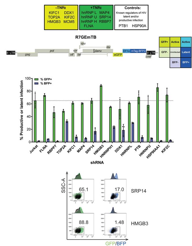
Khoury et al.                                                                            Post-Transcriptional Control of Tat Expression During Latency

expression was validated by flow cytometry (GFP+ expression)             enzyme digestion, instrument-specific settings for TripleTOF
and p24CA -ELISA as previously described (Khoury et al., 2020).          5600 + (MS tolerance 0.05 Da, MS/MS tolerance 0.1 Da, charge
Cells were then collected and lyzed with ice-cold lysis buffer           state + 2– + 5), biological modification probabilistic features
(50 mM Tris-HCl pH 8, 1 mM EDTA, 150 mM NaCl, 0.1% IgePal)               on, thorough ID algorithm, and detected protein threshold
supplemented with protease inhibitors (Roche) for 30 min at 4◦ C.        0.05. Mass Spectrometry Interaction Statistics (MiST) (Jäger
Protein cell extracts were dialyzed before use against buffer D 1×       et al., 2011) analysis was conducted to sort proteins based
[Hepes KOH pH 7.9 20 mM, KCl 100 mM, glycerol 20%, EDTA                  on specificity, reproducibility and abundance over the three
0.2 mM, DTT 0.5 mM) + MgCl2 3 mM + protease inhibitor                    replicates. The mass spectrometry proteomics data have been
tablet (Roche)] for 2 h at 4◦ C, followed by centrifugation for          deposited to the ProteomeXchange Consortium via the PRIDE
10 min at 1,700 × g at 4◦ C. Five hundred pmol of MS2-tagged             (Perez-Riverol et al., 2019) partner repository with the dataset
RNAs into 100 µl buffer D 1× were denatured by 10 min heating            identifier PXD025782.
at 65◦ C, followed by slow cooling at room temperature with
addition of 7.75 µl of 62.5 mM MgCl2 to a final concentration            Generation of Knockdown RFP+ Jurkat
of 4.5 mM MgCl2 . After 10 min incubation at room temperature,           Cell Lines
RNAs were incubated with a fivefold molar excess of purified             Three or four Sherwood UltramiR shRNA viral particles
MBP-MS2 fusion protein at 4◦ C for 20 min. The RNA-MS2:MS2-              (106 –107 TU/mL) against each of the 15 protein targets of
MBP complexes formed were incubated with amylose beads (200              interest were obtained from TransOmic Technologies. The pZIP
µl, GE Healthcare) equilibrated in buffer D for 2 h at 4◦ C.             (SFFV) shRNA-mir lentivectors constitutively express the short
After three washes with 500 µl of buffer D, 1 mg of protein              hairpin RNA (shRNA-mir), puromycin selection marker and red
extract supplemented with 5 µM of yeast tRNAs (Sigma-Aldrich)            fluorescent protein (RFP) driven by the Spleen Focus Forming
was added. After 20 min of incubation at 4◦ C with constant              Virus (SFFV) promoter. Jurkat cells (2.1×10⁵ ) were transduced
agitation, three successive washes were performed in Buffer D            with 100 µL of pooled pseudoviruses for each protein target
and RNP complexes eluted twice by incubation for 20 min at               separately at a MOI of 2.5. Spinoculation was conducted at 1,200
room temperature with 200 µl of Buffer D containing 10 mM                × g for 2 h at 23◦ C in a flat-bottom 96-well plate. Cells were
maltose. Half of the eluted RNP complexes were processed                 incubated at 37◦ C for 72 h then sorted on RFP+ expression on
in solution for mass spectrometry analyses. For western-blot             an Astrios cell sorter (Beckman Coulter). Bulk populations were
analysis, 10% of the eluted material was used. Experiments were          then kept in culture under 0.7 µg/mL puromycin selection. Single
repeated three independent times using different batches of RNA          clones expressing high levels of RFP+ cells were selected from
and protein lysate.                                                      the bulk population on the BD FACSAria III. Bulk and single
                                                                         clones were maintained under constant puromycin selection
Mass Spectrometry Analysis                                               then surviving clones were expanded. Cells were collected for
TCEP (10 mM, Thermo Fisher Scientific) was added to the                  RT-qPCR analysis as well as immunoblotting.
eluted sample to reduce the cysteine bonds in proteins, and
heated for 20 min at 60◦ C followed by alkylation of cysteines           Cloning of Tat Expression Reporter
using 25 mM Iodoacetamide (Sigma-Aldrich) for 20 min at RT
in the dark. Samples were subjected to proteolytic cleavage by           Constructs
addition of 1 µg of trypsin protease from porcine pancreas               For the Tat/GH1 constructs, HIV-1 sequences were derived
per 100 µg of protein and incubated overnight at 37◦ C under             from pNL4-3 (M. Martin, NIH, Bethesda, MD, United States),
agitation. Reactions were stopped by addition of formic acid and         GH1 sequences were amplified from pØhGH (Nichols Institute
the resultant peptides were concentrated using C18-packed tips           Diagnostics), LucF from pGL4.13[luc2/SV40] (Promega), and
(BondElut, Agilent/Varian). The peptides were subject to online          LucR from pGL4.73[hRluc/SV40] (Promega). These segments
trapping using a PepMap100 trap column at 15 µL/min followed             were assembled by SOE-PCR and cloned into the pcDNA3.1-
by separation on PepMap 100 C18 nanocolumn (50 cm × 75µm)                (Invitrogen) backbone via use of the Xho1 and XbaI sites. The
using a 30-min gradient of Buffer B (80% ACN 0.1% Formic acid)           NT5C3 splicing constructs were cloned into the pcDNA3.1-
over Buffer A (0.1% Formic acid). The online separated peptides          backbone through cleavage of NhoI and NheI where the NT5C3
were analyzed using a Q-Exactive plus mass spectrometer                  cellular sequences were amplified from HeLa cell genomic DNA
(Thermo Fisher Scientific, Bremen, Germany). The survey scans            and the other components derived as for the Tat/GH1 constructs.
were acquired at 70,000 resolution from 375 to 1,800 m/z, the ion
accumulation target was set to 3e6 with maximum injection time           DNA Transfection of KD RFP + Cells and
of 120 ms. A total of 12 most intense ions (with charge more             Luciferase Analysis
than 2) were sequentially isolated and fragmented by higher-             Bulk populations or single clones of the KD RFP+ Jurkat cell
energy collisional dissociation (HCD) set to 27%, at a resolution        lines (6×10⁴ cells) were plated into a round-bottom 96-well
of 17,500, target of 1e5 ions and maximum injection time of              plate. Media was completely removed and cells were transfected
120 ms. To identify protein groups, data acquired was converted          with 1.6 µg reporter constructs using DMRIE-C reagent (1:2.5,
into mgf and searched against Swissprot human database (version          Invitrogen) in 62.5 µL Opti-MEM (Gibco). For transactivation
2016_12) using ProteinPilot software (v4.0, SCIEX) with the              assays, 200 ng of Tat WT and 1 µg of Tat hGH reporter construct
following search parameters: Iodoacetamide alkylation, trypsin           with 300 ng of both LTR-lucFirefly and lucRenilla. For translation

Frontiers in Genetics | www.frontiersin.org                          3                                        June 2021 | Volume 12 | Article 680725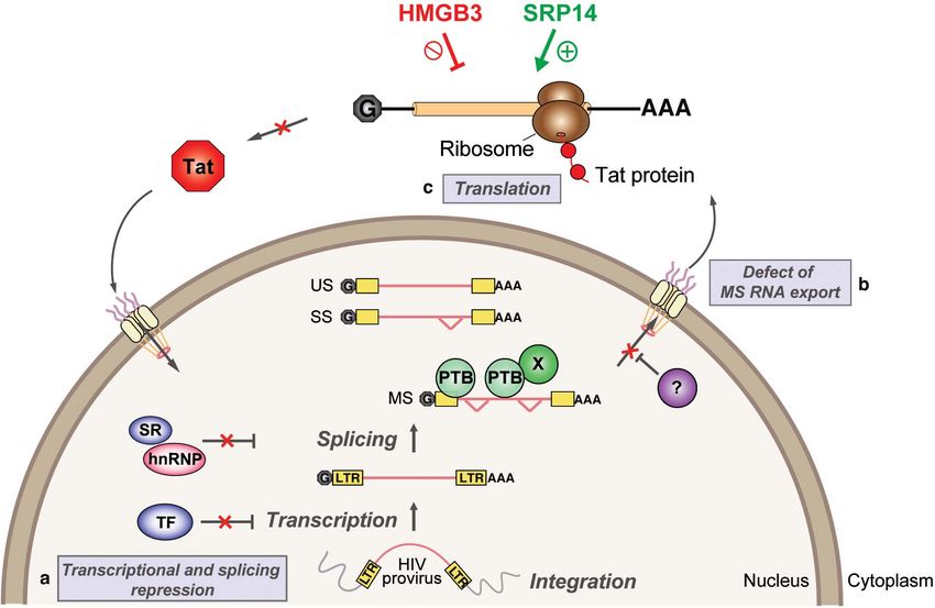
Khoury et al.                                                                              Post-Transcriptional Control of Tat Expression During Latency

assays, 300 ng of Tat-Cap or IRES LucF reporter constructs were            and Ethics Committees from the University of Melbourne (15-
used with 300 ng of lucRenilla for normalization. For splicing             09VIC-03 and 17-08VIC-01). All HIV-1 seronegative donors
assays, 300 ng of Tat+ or Tat- reporter constructs with 300 ng             were recruited by the Red Cross Blood Bank (Melbourne,
of LTR-lucFirefly. Three hours post-transfection, DNA:DMRIE-               Australia) and provided written informed consent for the use of
C mix was removed and media replenished. Twenty-four hours                 their blood products for the research. The use of blood samples
post-transfection, cells were collected and luciferase activity was        from people living with HIV was approved by the Alfred Hospital
measured following lysis in 40 µL of 1X passive lysis buffer               (HREC214/15) for the study entitled Large volume peripheral
using a FLUOstar plate reader with the dual-luciferase reporter            blood mononuclear cells (PBMCs) collection by leukapheresis to
assay (Promega).                                                           define HIV persistence in HIV-infected adults. All participants
                                                                           provided informed consent and the protocol was approved by the
Virus Production and Transduction                                          local Institutional Review Board.
R7GEmTB dual color reporter virus was produced by replacing
mCherry into R7GEmC [obtained from NIH AIDS Reagent                        Participant Details
Program, (Calvanese et al., 2013)] with mtagBFP2 fluorescent               PBMCs from people living with HIV on ART with a viral
protein. SRP14 and PTB cDNA were cloned into pInducer10                    load < 20 copies/mL for ≥3 years were collected by leukapheresis
lentivector (Meerbrey et al., 2011) by replacing tRFP-shRNA                (Alfred Hospital, Melbourne, Australia; Supplementary
cassette with protein ORF-T2A-mtagBFP2. Viral stocks were                  Table 1) and stored in liquid nitrogen. Resting CD4+ T-cells
generated by transfecting the proviral constructs into HEK 293T            (purity > 95%) were isolated from PBMCs by negative selection
cells with Lipofectamine 2000 (Invitrogen) in serum free media             using CD4+ T-cell isolation kit (Miltenyi Biotec) supplemented
(Opti-MEM, Gibco). Supernatants were collected after 72 h,                 with anti-CD69 (clone L78, BD) and anti-HLA-DR (clone 2-O6).
clarified by centrifugation and 0.45 µm filtration to clear cell           CD4+ T-cells were activated with anti-CD3/CD28 (coated
debris. Particles were concentrated using microcon centrifugal             α-CD3 clone OKT3, BD, 1 µg/mL and soluble α-CD28 clone
filter device (30K, Merck Millipore) or pelleted by overlaying             L293, BD, 0.5 µg/mL) for 48 h at 37◦ C.
supernatant on a cushion of 20% (w/v) sucrose in TNE buffer
(10 mM Tris-HCl pH 8, 1 mM EDTA, 150 mM NaCl) in Ultra                     Western-Blot
Clear Thinwall tubes and centrifugation at 24,200 rpm for 2 h at           Resting and activated CD4+ T-cells isolated from patients
4◦ C (Beckman SW28/SW41Ti rotor). Viral particle pellets were              under ART (30×106 cells) were lysed for 30 min on ice using
resuspended into Opti-MEM and stored at –80◦ C. Virus titres               ice-cold IGEPAL cell lysis buffer (Tris-HCl pH 8.8 50 mM,
were quantified by measuring p24CA levels by capture ELISA                 NaCl 150 mM, IGEPAL 1%, EDTA 1 mM) supplemented
as previously described (Khoury et al., 2020) and titration into           with protease inhibitor cocktail (CompleteTM , Mini, EDTA-free
TZM-bl cells (Sarzotti-Kelsoe et al., 2014). KD RFP+ Jurkat                Protease Inhibitor Cocktail, Roche). Cell lysate was cleared and
cells and CD4+ T-cells (106 cells) were infected with 40 and               quantified using the Bio-Rad Protein Assay Reagent (Bio-Rad).
100 ng R7GEmTB virus (+Env 92HT593.1), respectively. Cells                 Equal amounts of each sample (20 µg) were loaded on 15%
were spinoculated at 1,200 × g for 2 h at 23◦ C in a 96 well               SDS-PAGE, transferred to nitrocellulose membrane. Blots were
flat-bottom plate and incubated at 37◦ C for 3 days. Cells were            probed with anti-SRP14 (B-3, sc-377012, Santa Cruz) at 1:10,
collected, washed and resuspended in 100 µL of 1X PBS then                 anti-HMGB3 (clone 546519, R&D) 1:1,000, anti-PTB (clone 7,
loaded onto cover slips that had been pre-coated with poly-L-              #325000, Invitrogen) at 1:500 and anti-GAPDH (14C10, Cell
Lysine 0.01% (Sigma-Aldrich). Cells were incubated for 1 h on the          Signaling) at 1:1,000. Antibodies were detected using either goat
coverslip then rinsed with PBS 1X and fixed with 2% PFA. Cells             anti-rabbit (Invitrogen, #656120) or goat anti-mouse IgG (H + L)
were treated with glycine 0.2 M for 10 min at room temperature             HRP (Invitrogen, #626520) at 1/5,000 and developed with
then rinsed with PBS 1X and H2 O. Coverslips were inverted on a            SuperSignal West Pico Chemiluminescent Substrate (Thermo
microscope slide containing ProLong Gold Antifade Mountant                 Fisher Scientifc). Images were visualized using an MF-ChemiBis
(Thermo Fisher Scientific). Images were acquired on LSM710                 3.2 imaging system (DNR).
confocal microscope using Zeiss Zen software. J-Lat 10.6, 8.4 and
A2 (obtained from the NIH AIDS reagent program) were infected              Quantitative Real-Time PCR Analysis
via spinoculation at 1,500 × g for 2 h at 23◦ C followed by 1 h            RNA from resting and activated CD4+ T-cells isolated from
incubation at 37◦ C before replenishing the culture with fresh             patients under ART (15×106 cells) and bulk and individual
media without or with 5 µg/mL doxycycline. Two days post-                  clone of interest for the KD RFP + Jurkat cells as well as
infection, cells were washed and stained with Near-IR Live/Dead            untransduced Jurkats were extracted using TRIzol following
fixable dead cell staining (Invitrogen). Finally, cells were washed,       the manufacturer’s protocol. RNA (500 ng) was DNase-treated
fixed with 1% formaldehyde and acquired using a Fortessa flow              with RQ1 DNase (Promega) and reverse transcribed using 4 U
cytometry instrument (BD Bioscience). Analysis was performed               OmniScript (Qiagen) with dNTP (0.5 mM), random hexamers
using FlowJo Software, version 10.4.2.                                     (2 µM), oligo(dT)15 (1 µM), and RNasin (10 U) in 30 µL
                                                                           reactions for 1 h at 37◦ C. Real-time PCR was performed on CFX
Ethics                                                                     Connect real-time PCR detection system (Bio-Rad) using Fast
The studies involving the use of blood samples from HIV negative           SYBR Green Master Mix (Applied Biosystems), forward/reverse
donors were reviewed and approved by the Human Research                    primers (Supplementary Table 2) at 500 nM final concentration,

Frontiers in Genetics | www.frontiersin.org                            4                                        June 2021 | Volume 12 | Article 680725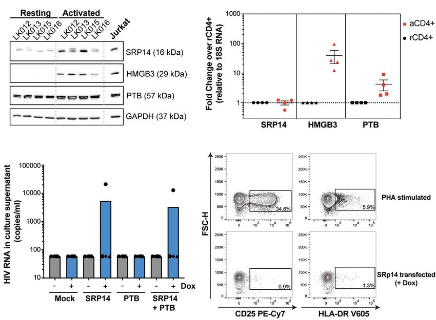
Khoury et al.                                                                             Post-Transcriptional Control of Tat Expression During Latency

2 µL of undiluted cDNA in reactions with a final volume of                were purified by affinity purification using Ni-NTA affinity resin
20 µL. Each sample was assayed in duplicate and normalized                (Qiagen) coupled to an Äkta pure FPLC (GE Healthcare). The
on GAPDH content and untransduced Jurkat cells for KD                     bound proteins were eluted with a linear imidazole gradient
RFP + Jurkats or 18S content and activated CD4+ T-cells for               (20 mM to 500 mM). Proteins were buffer exchanged into
primary CD4+ T-cells. Cycling conditions used were 95◦ C for              buffer D (20 mM HEPES–KOH pH 7.9, 100 mM KCl, 0.2 mM
10 min for activation of the DNA polymerase followed by 45                EDTA, 3 mM MgCl2 , 0.5 mM DTT, 20% glycerol) using PD-
cycles of denaturation at 95◦ C for 3 s and annealing/extension at        10 desalting columns (GE Healthcare) followed by concentration
60◦ C for 20 s. Melt curve analysis was performed each time with          using 3, 10, and 30 k cutoff amicon columns (Merck) for
a starting temperature of 60◦ C, increasing to 95◦ C with 0.5◦ C          SRP14, HMGB3 and PTB, respectively. Proteins concentration
increments every 0.05 s. Data were analyzed on CFX Manager 3.0            was determined using UV spectroscopy at 280 nm. Purity
software employing 2−11Ct to determine fold changes.                      was confirmed by 12.5% SDS-PAGE and western-blot analysis
                                                                          using protein specific antibodies and anti-His HRP conjugated
T-Cell Electroporation                                                    antibody (clone J099B12, BioLegend) at 1:500.
Electroporation of resting CD4+ T-cells was performed using
an Amaxa human T-cell Nucleofector kit (Lonza). Purified                  Footprinting Assays
resting CD4+ T-cells (5.3×10⁶ ) were re-suspended in 100 µl               Footprinting analysis was performed by SHAPE as previously
nucleofector solution (4.5:1 ratio of human T-cell nucleofector           described (Mortimer and Weeks, 2007). Briefly, tat2 RNA was
solution:supplement) and transfected with 1.2 µg DNA per 106              generated by run-off transcription using Sp6 MEGAscript kit
cell using Amaxa nucleofector program U-014 for high viability.           (Promega) and pSP65::tat2 construct linearized with ClaI. In vitro
Cells were then transferred into a 12-well plate containing pre-          transcribed tat2 RNA (1 pmol) was probed in 3× folding
equilibrated media, followed by half-media change 6 h post-               buffer (333 mM HEPES–KOH pH 8, 333 mM NaCl, 33.3 mM
nucleofection. Cells were maintained for 48 h at 37◦ C in media           MgCl2 ) in the presence or absence of SRP14, HMGB3 or PTB
supplemented with 1% FBS and 1 U/mL IL-2, with or without                 recombinant protein at 5, 10 and 20 protein/RNA molar ratio
5 µg/mL doxycycline. After 2 days, cells were collected and               (0.41–1.6 µM). RNP complexes were formed by incubation for
stained for CD25 (PE-Cy7, clone 2A3, BD) and HLA-DR (V605,                20 min at room temperature then probed with 1-methyl-7-
clone L243 BioLegend) activation markers in comparison to                 nitroisatoic anhydride (1M7, 65 mM in DMSO) or DMSO for
PHA stimulated (10 µg/mL) cells. Culture supernatant was also             4 min at 37◦ C, then recovered by ethanol precipitation. Primer
harvested and viral RNA was isolated using QIAamp Viral                   extension was conducted as described previously with 0.4 µM
RNA Mini Kit (Qiagen) following the manufacturer’s protocol.              fluorescently labeled odp3102 primer (6-FAM or HEX, Sigma-
vRNA was DNase treated (2 U/µg RNA, Promega) and reversed                 Aldrich, Supplementary Table 2). The dideoxy sequencing
transcribed using Omniscript as described above. HIV RNA                  reactions were generated using unmodified RNA, labeled primers
levels were assessed using droplet digital PCR (ddPCR) using pol          (PET or NED, Applied Biosystems) and 0.5 mM ddGTP.
primers/probe (Supplementary Table 2). Thermal cycling was                cDNAs were recovered by ethanol precipitation and separated
conducted as follows: 95◦ C for 10 min, 40 cycles of 94◦ C for 30 s       by capillary electrophoresis with LIZ500 size standard (ABI
and 60◦ C for 60 s, followed by 98◦ C for 10 min (ramp rate 2◦ C/s        3130, AGRF). Data was processed using the QuShape software
for each step) on a C1000 Touch Thermal cycler (Bio-Rad). The             (Karabiber et al., 2013). Protections induced by SRP14, HMGB3
droplets were subsequently read on a QX200 droplet reader (Bio-           or PTB binding were indicated by a reduction in the normalized
Rad) and the data were analyzed with Quanta-Soft 1.7.4 software.          SHAPE reactivities.
The limit of detection of our assay was of 58 copies/mL.

Recombinant Proteins                                                      RESULTS
Production and purification of MBP-MS2 protein was done
as previously described (Deckert et al., 2006). cDNA encoding             Detection of 243 Putative Tat-RNA and
SRP14, HMGB3 and PTB were cloned into NheI and HindIII                    Cellular Protein Interactions
restriction sites of pET28a + bacterial vector (Novagen) allowing         To explore MS RNA binding partners during latency, we
the expression of N-terminally 6×-His tagged proteins. All                initiated a proteomic approach based on affinity chromatography
recombinant proteins were expressed in E. coli BL21 (DE3)                 purification of RNA-protein complexes (Maenner et al., 2010;
Codon + bacteria grown in LB/Kanamycin and induced by                     Bar et al., 2011) formed upon incubation of in vitro transcribed
addition of 0.1 mM IPTG overnight at 37◦ C (for PTB and                   tat RNA with protein lysate, followed by protein identification
SRP14) or 30◦ C (for HMGB3). Cells were sonicated in lysis buffer         by mass spectrometry. An overview of the processes is shown
containing 20 mM potassium phosphate pH 7.4 (pH to 6 for                  in Figure 1A. Protein lysates were prepared from the HIV-
HMGB3), 1M KCl, 1 mM DTT, 20 mM imidazole and protease                    1 latently infected T cell line, J-Lat6.3 where the cells were
inhibitor cocktail (Roche), then treated with 1.5 mg/mL lysozyme          either left untreated (latent infection) or activated with TNF-
(from chicken egg white, Sigma-Aldrich) for 20 min on ice.                α (productive infection). The RNA of interest, 50 UTRtat1-Tat
Soluble proteins were purified by polyethyleneimine (0.4% final)          and Tat with three binding sites for the MS2 coat protein fused
followed by ammonium sulfate precipitation (30% for SRP14                 at their 30 -ends were used as baits. In parallel, a control RNA
and PTB; 40% then 60% precipitation for HMGB3). Proteins                  that only contained the three MS2 binding sites was used. RNA

Frontiers in Genetics | www.frontiersin.org                           5                                        June 2021 | Volume 12 | Article 680725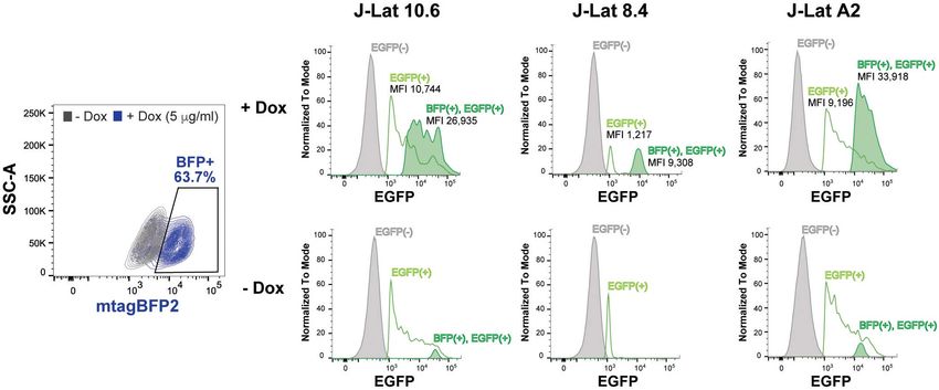
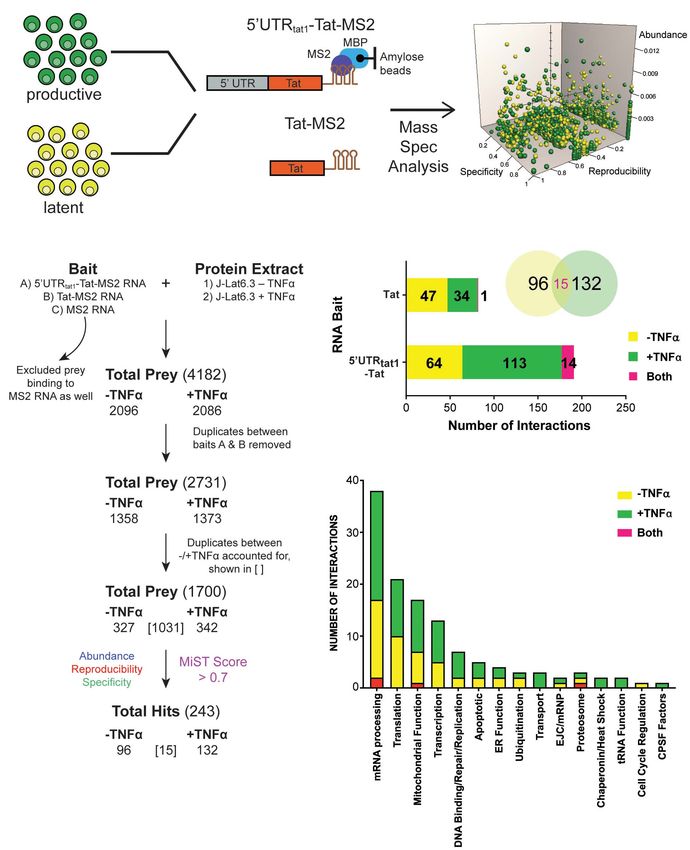
Khoury et al.                                                                                               Post-Transcriptional Control of Tat Expression During Latency

  FIGURE 1 | Enrichment in mRNA processing and translation factors assembling on tat mRNA. (A) Representation of the affinity purification strategy used for
  isolation of cellular RNA binding proteins that interact with tat mRNA. Whole cell lysates were prepared from the latently infected T-cells, J-Lat clone 6.3 where
  untreated cells were used as the latent sample and TNF-α activated cells were used as the productively infected sample. Cellular proteins that interact with tat mRNA
  were pulled down through MBP-MS2 affinity purification using two RNA baits that contained Tat exon 2 with or without the 50 UTR from tat1 mRNA and fused to
  three MS2 sequences. Eluted RNA-protein complexes were analyzed by mass spectrometry. MiST analysis was used to determine the most biologically relevant
  interactions after combining data from three replicates. (B) The filtering steps applied to refine the mass spectrometry data. Prey binding to the control bait, MS2
  RNA were excluded, followed by removal of duplicate prey captured by baits A (50 UTRtat-Tat) and B (Tat RNA). Prey were then defined as derived from
  uniquely –TNFa or +TNFa lysates or from both. Lastly, MiST analysis was applied to score interactions and a confidence threshold of 0.7 was used to refine the list of
  candidates for relevant Tat mRNA: cellular protein interactions. (C) Breakdown of the 243 proteins selected by MiST analysis by bait and lysate. (D) Annotation of the
  243 proteins based on GO biological process.

Frontiers in Genetics | www.frontiersin.org                                         6                                             June 2021 | Volume 12 | Article 680725
Khoury et al.                                                                              Post-Transcriptional Control of Tat Expression During Latency

retention to amylose beads was mediated by the MS2-MBP                    on HIV-1 infection, we used a single-round dual-fluorescent
fusion protein containing the MS2 coat protein RNA binding                reporter virus (DuoFluo, R7GEmTB) based on the R7GEmC
domain and the E. coli maltose binding protein (MBP). RNP                 backbone described in Calvanese et al. (2013). In our DuoFluo
complexes were eluted by maltose and the protein composition              virus, HIV-1 50 -LTR controls eGFP expression indicative of
of the purified RNPs were analyzed by mass spectrometry.                  productive infection and mTagBFP2, controlled by the EF1α
The proteins identified by MS were quantitatively scored using            promotor, is the marker of latent infection (Figure 2B). The
the mass spectrometry interaction statistics (MiST) platform              fluorescent phenotype of cells and corresponding profile of HIV-
devised by Jäger et al. (2011).                                           1 infection are shown in the grid (Figure 2B). We confirmed
    Several filtering steps described in Figure 1B were applied to        the ability of the eGFP/mTagBFP2 expressing dual-fluorescent
refine the protein preys obtained by MBP-MS2 pull-down for                reporter virus to identify the presence of latently (blue, BFP+ )
generation of a list of 1,700 unique proteins where duplicates            and productively (green, GFP+ or cyan, GFP+ BFP+ ) infected
between the different conditions were accounted for. These 1,700          Jurkat and primary CD4+ T-cells by fluorescence microscopy
proteins were quantitatively scored based on their abundance,             after infection with R7GEmTB (Supplementary Figure 3).
reproducibility and specificity and 243 putative Tat RNA:protein              All bulk populations of the KD RFP+ Jurkat cells were
interactions were identified using a confidence threshold of 0.7          infected with the DuoFluo virus and collected for flow cytometry
for biological relevance (Figure 1B).                                     analysis after 72 h. Levels of eGFP and mTagBFP2 expression
    As expected, when assigned to their respective bait (Figure 1C,       were assessed by gating on the RFP+ population and compared
bar graph) or protein lysate (Figure 1C, Venn Diagram), we                against DuoFluo infected untransduced RFP- parental Jurkat
observed a larger number of proteins interacting with the longer          cells. An increase in productive infection was detected after
RNA bait (191 proteins interacting with the 50 UTRtat1-Tat-MS2            knockdown of FLNA, HMGB3, PTBP1, HSP90AA1, and KIF2C
vs. 82 with Tat-MS2). Moreover, a higher number of interactions           (Figure 2C, green bars). On the other hand, a dramatic increase
were detected with the lysates prepared from activated T-cells            in latent infection was seen after knockdown of TOP2A, SRP14,
(147 proteins for +TFNα vs. 111 proteins for −TNFα). Analysis             HNRNPH1, DDX1, and HNRNPL (Figure 2C, blue bars). The
of the 243 proteins by broad gene ontology terms (Spliceosome             increase in latent infection after knockdown of TOP2A and
database, Cvitkovic and Jurica, 2013) showed that the top two             HNRNPH1 was coupled with a decrease in productive infection.
overrepresented annotated biological processes were mRNA                      PTB, known to facilitate the export of multiply spliced (MS)
processing and translation (Figure 1D). Analysis of the GO                mRNAs to the cytoplasm (Lassen et al., 2006), appears to play a
molecular function also showed a predominance of RNA-binding              role in controlling latent and productive infection, as increases in
proteins (Supplementary Figure 1).                                        both forms of infection were observed after knockdown of the
                                                                          protein (productive: 70.9%, latent: 7.61%). However, the effect
                                                                          on latent infection was smaller than expected. Interestingly, two
Knockdown of Tat RNA Binding Proteins                                     genes, SRP14 and HMGB3 have not been previously reported
Affect Latent and Productive Infection of                                 to play a role in the regulation of HIV-1 replication and
HIV-1                                                                     their knockdown here had very marked effects on latent and
From the 243 proteins identified through MS2 chromatography               productive infection, respectively. Knockdown of SRP14 had no
affinity purification, thirteen proteins were selected to follow          effect on productive infection but increased dramatically the
through with validation studies (Figure 2A and Supplementary              percentage of cells entering into latency (17.0% vs. Jurkat 1.71%,
Table 3). The main criteria for selection were the MiST score,            Figure 2D). In contrast, knockdown of HMGB3 had no effect
the role of the protein in cellular pathways and the novelty of           on latent infection, but increased the percentage of productively
the protein in the context of regulation of HIV-1 latent infection.       infected cells (88.8% vs. Jurkat 65.2%, Figure 2D). These data
Two additional proteins, PTB1 and HSP90A were selected as                 suggest that SRP14 is a negative regulator of latent infection,
controls as both proteins have previously been shown to be                whilst HMGB3 is a negative regulator of productive infection.
involved in regulation of HIV-1 latency (Lassen et al., 2006;
Anderson et al., 2014).
    For further refinement of the list of candidates for in-depth         Knockdown of SRP14 and HMGB3
investigation, we assessed the effect of knockdown of the proteins        Strongly Modulates Splicing at SA3
on HIV-1 latent and productive infection. Jurkat T-cells were             As a strong block to multiply splicing of HIV-1 mRNA was
transduced with shRNA expressing lentivectors targeting each              recently characterized in CD4+ T-cells isolated from patients
of the fifteen protein targets for knockdown (KD). MCM5                   under ART (Yukl et al., 2018), we examined the role of
KD induced high cell death hence MCM5 was excluded from                   knockdown of the RNA binding proteins on tat mRNA splicing
further investigation. Successfully transduced cells were sorted          using an HIV-1 splicing reporter, which harbors SD1 50 ss and
into a bulk population by RFP+ expression (Supplementary                  SA3, SA4a,b,c and SA5 30 ss (Ropers et al., 2004). In this splicing
Figure 2A) where the degree of knockdown was heterogeneous                reporter construct, Tat-exon1,2 (nt 1-839/5590-6044 NL4-3) was
between individual cells. Success of the knockdown of the                 placed in the context of a human gene to recapitulate HIV
gene targets in the bulk Jurkat cell populations were confirmed           integration in latently infected cells. The context of HIV-1
by western blot and densitometry analysis (Supplementary                  integration in the latent cell line, ACH2 cells (Clouse et al., 1989,
Figure 2B). To assess the outcome of protein knockdown                    Folks et al., 1989) was used as the basis of the design hence NT5C3

Frontiers in Genetics | www.frontiersin.org                           7                                         June 2021 | Volume 12 | Article 680725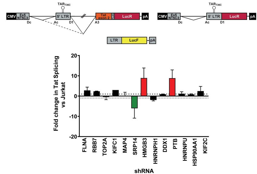
Khoury et al.                                                                                                 Post-Transcriptional Control of Tat Expression During Latency

  FIGURE 2 | SRP14 and HMGB3 knockdown impact HIV latent and productive infection. (A) Short-list of 13 proteins chosen for follow-up validation and two control
  proteins, PTB and HSP90A that are known regulators of HIV-1 replication. (B) Genome of the single round DuoFluo virus [R7/E-/GFP-EF1α-mTagBFP2 (R7GEmTB)]
  used in this study. The fluorescence profiles that correspond to latent or productive infection are shown in the grid. (C) Bulk populations of RFP+ shRNA knockdown
  (KD) Jurkats were infected with the single round DuoFluo virus pseudotyped with the dual-tropic 92HT593.1 envelope, and collected 48 h later for flow cytometry
  analysis. Percentage of productive or latently infected cells after knockdown of the specific protein is shown, with productive infection in green and latent infection in
  blue. Data shown are means of three independent experiments ± SEM. (D) Scatter plots highlighting the shift in GFP+ or BFP+ populations after infection of
  RFP+ SRP14 (top) or HMGB3 (bottom) KD Jurkats with DuoFluo virus. Black population represent infected, untransduced Jurkat cells, while green/blue populations
  represent infected SRP14/HMGB3 KD RFP+ Jurkats. Values represent the percentage of GFP+ or BFP+ cells in KD RFP+ Jurkat cells.

Frontiers in Genetics | www.frontiersin.org                                           8                                              June 2021 | Volume 12 | Article 680725
Khoury et al. Post-Transcriptional Control of Tat Expression During Latency
exon 5 and intron 5 were incorporated upstream of the HIV-1 the effects of these proteins on latent and productive infection,
50 -LTR (Figure 3A). Renilla luciferase (LucR) was introduced suggesting an important role of these proteins in Tat mRNA
at the 30 end of Tat exon 2, thus an increase in LucR would processing and regulation of HIV-1 infection.
indicate splicing at SA3, SA4a,b,c or SA5. Co-transfection of
the splicing reporter with an LTR-LucF reporter cassette allows
Firefly luciferase to be used as a specific readout of splicing at SA3 Knockdown of SRP14 and HMGB3
and Tat production. The effect of cellular proteins on use of SA3 Impacts Tat Expression and Function
in the Tat+ splicing reporter were compared to a matched Tat- Major blocks of HIV transcription and translation have been
splicing reporter lacking both the HIV introns and Tat exon 2 reported during latency (Khoury et al., 2018a). Due to the
(Figure 3A). An increase in LucR expression in the Tat- context central role of Tat protein in promoting HIV transcription and
shows the promotion of splicing between the cellular splice site, post-transcriptional events, this warranted a deeper investigation
SDc site at the 30 end of NT5C3 exon 5 and the SAc upstream of of the role of SRP14, HMGB3 and PTB on Tat expression.
the 50 -LTR. One caveat of using bulk populations of the KD RFP+
Bulk populations of the KD RFP + Jurkat cells were co- Jurkat cells is the large clonal variation. To circumvent this
transfected with the Tat+ or Tat- splicing reporter and the LTR- obstacle, single clones were sorted following 12 days of
LucF cassette and harvested for luciferase analysis 24 h later. puromycin selection and assessed through expression of RFP
The LucF/LucR ratio was calculated for both the Tat+ and Tat- by flow cytometry (Supplementary Figure 2C). To examine
contexts in response to the knockdown of each protein target. KD efficiency in the various clones, changes in mRNA levels
Fold changes in Tat splicing over transfected RFP- untransduced compared to untreated Jurkat T-cells was determined by RT-
Jurkat cells were reported in Figure 3B. Knockdown of SRP14 qPCR. Importantly, we observed across all single clones tested
decreased splicing at SA3 by sixfold compared to untransduced a significant reduction in the levels of SRP14, HMGB3 and PTB
Jurkats, whereas knockdown of HMGB3 and PTB increased the mRNA compared to untransduced Jurkats (FC vs. Jurkat ≥50%,
use of SA3 by 8.9- and 8.8-fold, respectively (Figure 3B). None Supplementary Figure 2D).
of the other ten proteins of interest affected splicing at SA3. The Next, bulk and single SRP14 (B5, C10, and G4), HMGB3
effect of SRP14 and HMGB3 KD on use of SA3 is consistent with (C2, D4, E7, and G9) or PTB (C10, C11, D5, and D8) shRNA
FIGURE 3 | SRP14 and HMGB3 knockdown influence tat mRNA splicing. (A) Schematic representation of the NT5C3-Tat splicing reporter construct (Tat+), the
corresponding Tat- control and the transactivation reporter cassette (LTR-LucF). The splicing patterns that lead to SA3 activation and Tat expression are indicated.
RFP+ shRNA KD Jurkats were co-transfected with either the Tat+ or Tat– splicing reporter vectors and LTR-LucF, and 24 h later harvested for luciferase readout.
(B) The fold changes in Tat splicing (Luciferase Firefly/Renilla ratio for Tat+ vs. Tat– cells) over untransduced Jurkat cells were determined. Data represent mean of
three independent experiments ± SEM. D, donor; A, acceptor splice site.
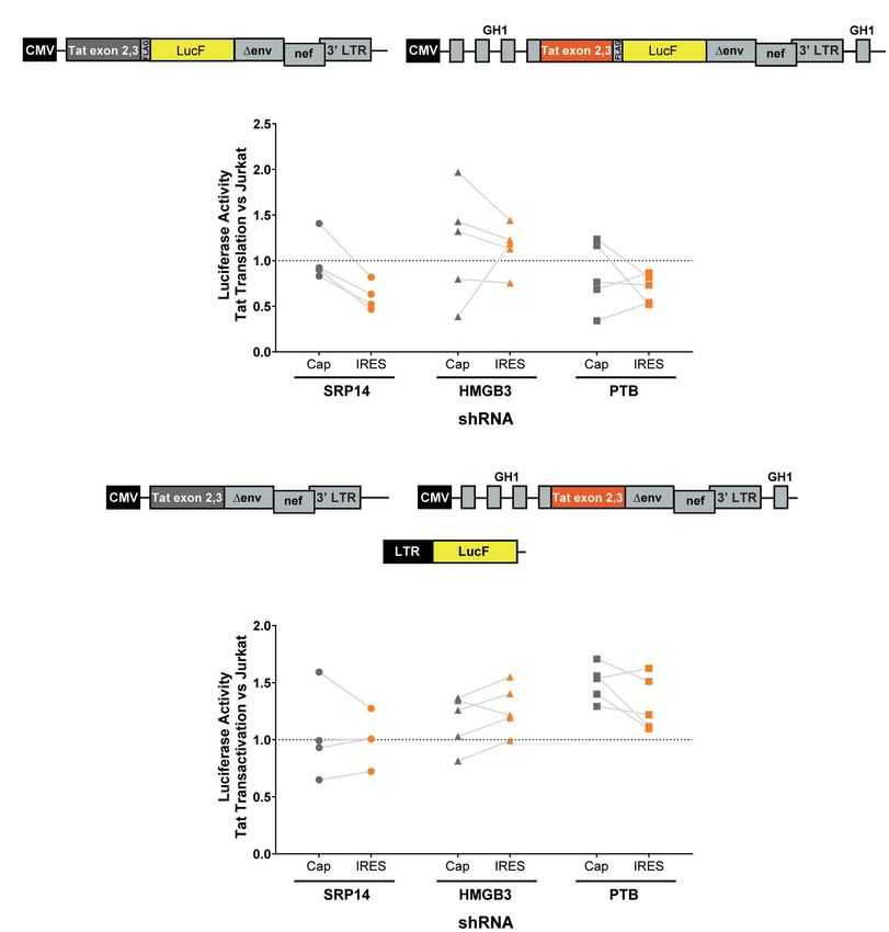
Frontiers in Genetics | www.frontiersin.org 9 June 2021 | Volume 12 | Article 680725Khoury et al. Post-Transcriptional Control of Tat Expression During Latency KD clones were transfected with Tat expression constructs to In our IRES-dependent Tat translation expression cassette, the assess the effect on Tat transactivation and translation. We Tat encoding exons have been incorporated into the human previously characterized a highly conserved element underlying growth hormone gene (GH1), where readthrough transcription the Tat open reading frame, named TIM-TAM (for Tat IRES and alternative splicing would allow low level expression of GH1- modulator of tat mRNA) and characterized its role in controlling Tat protein through an IRES-dependent mechanism (Figure 4A). Tat translation through cap- and IRES-dependent mechanisms In the control construct, Tat was placed under the control of a (Khoury et al., 2020). Moreover, we developed a model system cytomegalovirus (CMV) promoter allowing Tat expression in a to assess Tat cap and IRES translation (Nguyen et al., 2019). cap-dependent manner. In both contexts, Tat was cloned in phase FIGURE 4 | SRP14 and HMGB3 knockdown control Tat -cap and -IRES dependent translation. Diagrams depicting Tat reporter constructs used to study Tat cap- (left) or IRES- (right) dependent translation (A) and transactivation (B). Luciferase Firefly expression, which is produced in fusion with Tat (in A) or under the control of the 50 -LTR (in B), is used as a readout for Tat translation and transactivation, respectively. Bulk populations or single clones selected for knockdown of SRP14, HMGB3 or PTBP1 were transfected with Tat cap or IRES expression constructs to assess the effect on Tat translation (A) or transactivation (B). Cells were harvested for luciferase analysis 24 h later. Firefly luciferase activity (LucF) was normalized on Renilla luciferase activity (LucR) and shown as a fold change over untransduced Jurkats. Data shows mean of two independent experiments with single clones and bulk populations. CMV, cytomegalovirus promoter; GH1, human growth hormone gene. Frontiers in Genetics | www.frontiersin.org 10 June 2021 | Volume 12 | Article 680725
Khoury et al.                                                                               Post-Transcriptional Control of Tat Expression During Latency

with LucF hence lucF expression is a marker of Tat translation.             TIM-TAM and its bordering sequences. Strong protections were
Bulk population and single clones of the KD RFP+ Jurkat cells               also observed in the 50 untranslated region, more specifically on
were transfected with cap or IRES Tat-LucF expression constructs            the TAR, PBS, and DIS elements (Figure 5B). Upon increasing
and harvested for luciferase analysis 24 h later. Fold changes in           SRP14, HMGB3, and PTB concentration, protections of TIM-
luciferase activity were then calculated in comparison to RFP-              TAM were reinforced and new ones were detected on the Tat
untransduced Jurkats. In the SRP14 KD RFP + Jurkats, Tat                    start codon and in the vicinity of SA3. Altogether, these data are
translation was reduced from the cap-dependent context in the               consistent with direct binding of SRP14, HMGB3 as well as PTB
bulk population and 2 of 3 single clones and from the IRES-                 to MS RNA highlighting a potential role of these RNA binding
dependent context in the bulk population and all three single               proteins in controlling Tat expression during latent infection.
clones (Figure 4A). In contrast, HMGB3 knockdown induced
an increase in Tat translation from the cap-dependent context
for the bulk population and 2 of 4 single clones, and in the                SRP14 Reactivates HIV-1 Latently
bulk population and 3 of 4 single clones for the IRES-dependent             Infected Cells and Virus Production
context (Figure 4A). Lastly, knockdown of PTB resulted in a                 To assess the role of SRP14 in controlling latent infection, we
decrease in Tat translation in 2 of 4 single clones transfected with        tested the effect of its overexpression on virus reactivation in
Tat-lucF construct, as well as all 4 single clones and the bulk             a T-cell line model of latent infection, using the J-Lat 10.6, 8.4
population transfected with Tat IRES construct (Figure 4A).                 and A2 clones. J-Lats are Jurkat derived cells containing one
   We next investigated the effect of SRP14 and HMGB3                       stably integrated, but transcriptionally silenced full-length HIV-
KD on Tat transactivation by using a modified expression                    1 genome with GFP in place of the nef gene (Jordan et al.,
cassette system, where Tat is translated through an IRES-                   2003). J-Lat cells were transduced with pInducer-SRP14-T2A-
or cap-dependent pathway and the luminescence readout is                    mtagBFP2 lentiviral vectors and cultured in the absence or
induced by Tat transactivation of an HIV-1 LTR-LucF reporter                presence of doxycycline (+Dox) for 2 days. Representative scatter
cassette. The effect on Tat transactivation after knockdown of              plot highlighting mtagBFP2 expression following treatment
SRP14 was variable across the single clones for both cap- and               of transduced cells with 5 µg/mL doxycycline is shown in
IRES-Tat translation, however, there was a clear increase in                Figure 6A. SRP14 expression was validated by western-blot
Tat transactivation for 3 out of 4 Tat-cap transfected single               analysis (data not shown). The different J-Lat clones exhibit
clones and 4 out of 4 Tat-IRES transfected single clones after              variable levels of basal GFP expression. Upon doxycycline
knockdown of HMGB3 compared to untransduced Jurkats                         treatment, we observed an increase in GFP expression in all
(Figure 4B). Knockdown of PTBP1 resulted in an increase in Tat              T-cell lines. Indeed, a significant increase in mean fluorescence
transactivation for all single clones transfected with Tat-Cap and          intensity (MFI) was detected for BFP+ GFP+ cells vs. GFP+ cells
Tat-IRES (Figure 4B). These data demonstrate a role of SRp14                alone (Figure 6B).
and HMGB3 in controlling HIV-1 latent and productive infection                 To determine whether differences in RNA binding protein
in Tat-dependent manner.                                                    levels in resting and activated CD4+ T-cells might be involved
                                                                            in the observed effect on HIV expression, we measured SRP14,
                                                                            HMGB3 and PTB protein and RNA levels in resting and
SRP14 and HMGB3 Binds in the Vicinity                                       α-CD3/CD28 stimulated CD4+ T-cells from patients living with
of Tat Start Codon                                                          HIV on ART. Western-blot analysis indicated low expression
Whilst SRP14 and HMGB3 were detected in the RNP complexes                   levels of SRP14 and HMGB3 in resting cells, and upon
(Supplementary Figure 4), further investigation was required to             stimulation a 2.5- and 18.8-fold increase in SRP14 and HMGB3
assess direct interaction of these proteins with tat mRNA. To               protein level was detected, respectively (Figure 7A). Similar
delineate SRP14, HMGB3, and PTB binding sites on multiply                   results were observed with RT-qPCR analysis of HMGB3 mRNA
spliced RNA, we performed footprinting assays coupled to                    levels between resting and activated CD4+ T-cells as we detected
SHAPE (selective 20 hydroxyl acylation analyzed by primer                   a 28.5-fold increase in HMGB3 mRNA expression following
extension) analysis. We have recently determined tat1 and tat2              CD4+ T-cell activation (Figure 7B). However, no changes in
mRNA folding using enzymatic and chemical probing and                       SRP14 mRNA levels were seen in response to T-cell activation.
identified a highly conserved sequence-structure within MS                  Interestingly, while a 4.4-fold increase in PTB mRNA expression
RNA, TIM-TAM (Khoury et al., 2020). TIM-TAM forms the                       was observed, no significant change in PTB protein expression
apical part of an irregular stem-loop structure SLS3A3 that                 was detected following stimulation of CD4+ T-cells.
harbors the Tat start codon. TIM-TAM controls the timing                       PTB was identified as an HIV RNA binding protein that
and level of Tat translation during the early and late phases of            induces virus reactivation and release of replication competent
infection, while promoting latent infection and virus reactivation.         virus in resting CD4+ T-cells from patient on ART (Lassen
Footprinting assays were performed on RNP complexes formed                  et al., 2006). To examine whether SRP14 might also act as a
by tat2 transcript (Figure 5A) and recombinant proteins at three            positive factor for HIV-1 gene expression, resting CD4+ T-cells
different [RNA]/[protein] ratios 5, 10, and 20. Normalized shape            isolated from people living with HIV on ART were electroporated
reactivities and probing data were used to determine the binding            with SRP14 or PTB Dox-inducible expression constructs alone
sites of SRP14, HMGB3, and PTB. At the lowest [RNA]/[protein]               or in combination using an Amaxa nucleofector. After 48 h,
ratio, protections were mainly detected on SLS3A3 including                 virion release into the culture supernatant was assessed using

Frontiers in Genetics | www.frontiersin.org                            11                                        June 2021 | Volume 12 | Article 680725
Khoury et al. Post-Transcriptional Control of Tat Expression During Latency FIGURE 5 | Interaction of SRP14, HMGB3 and PTB with tat mRNA. (A) Schematic representation of HIV-1 proviral genome, tat pre-mRNA and multiple splice events leading to tat1 and tat2 mRNAs production during the early phases of HIV-1 infection. D represent donor splice sites, A represent acceptor splice sites, 50 UTR/30 UTR indicate 50 /30 untranslated regions. The hybridisation site used for primer extension with O-548 (also known as Odp3102) is indicated by black arrow. (B) Probing of SRP14, HMGB3, and PTB binding sites on tat2 mRNA. Tat2 transcript (1 pmol) was modified with 1M7 (65 mM) in the absence or presence of increasing concentration (0.41—0.83—1.6 µM) of recombinant proteins. Conditions of modification are given in Materials and Methods. Protections generated by SRP14, HMBG3 and PTB recombinant proteins are indicated on the secondary structure model of tat2 mRNA by blue, green and red lines, respectively. Pale, medium, and dark colors indicate the intensity of protections (low, medium and strong protections). Numbering of nucleotides and positions of the cis regulatory elements are given in reference to HIV-1 BRU (K02013). The start codon of Tat protein is circled. Frontiers in Genetics | www.frontiersin.org 12 June 2021 | Volume 12 | Article 680725
Khoury et al.                                                                                            Post-Transcriptional Control of Tat Expression During Latency

  FIGURE 6 | SRP14 expression reactivates latently infected T-cell lines. (A) J-Lat cells were transduced with SRP14-T2A-mtagBFP2 lentivector and cultured with
  (+Dox) or without (–Dox) doxycycline at 5 µg/mL. Cells were analyzed for mtagBFP2 expression by flow cytometry 72 h post-infection. (B) Histogram depicting
  EGFP+ expression in latently infected T-cells following transduction with SRP14-T2-mtagBFP2. Virus reactivation in J-Lat 10.6, 8.4 and A2 clones is shown by an
  increase in mean fluorescence intensity (MFI) of BFP(+) EGFP(+) cells in comparison to EGFP( - ) and EGFP(+) cells.

an RT-ddPCR assay. One out of the four patient CD4+ T-cells                           that showed a series of blocks to HIV proximal elongation,
electroporated with SRP14 and SRP14+ PTB presented an                                 distal transcription/polyadenylation and splicing preventing HIV
upregulation of virus production upon doxycycline treatment                           expression in CD4+ T-cells from blood of HIV infected patients
(Figure 7C). Virus production following SRP14 overexpression                          on ART (Yukl et al., 2018). Prior studies have shown that MS
was not coupled with an increase in cell activation as only 0.9%                      RNA encoding Tat protein inhibits the establishment of HIV
and 1.3% of SRP14 transfected cells expressed middle (CD25) and                       latency (Donahue et al., 2012). When present, Tat activates virus
late (HLA-DR) activation markers, respectively (Figure 7D).                           replication at a higher rate than any of the known LRAs (Razooky
                                                                                      et al., 2015; Khoury et al., 2018b) by potently inducing HIV
                                                                                      transcription and splicing (Khoury et al., 2018b). Moreover, by
DISCUSSION                                                                            controlling its own production at the splicing (Jablonski et al.,
                                                                                      2010) and translational levels (Charnay et al., 2009, Khoury et al.,
We have characterized tat RNA:cellular protein interactions                           2020), Tat acts as a switch for productive and latent HIV infection
differentially expressed between productive and latent infection.                     (Khoury et al., 2020).
Out of the 243 proteins identified by mass spectrometry, multiple                        siRNA knockdown of TOP2A in Sup T1 cells (Balakrishna
cellular factors were investigated for their putative roles in the                    et al., 2013), MAP4 in TZM-bl, HEK 293T and HeLa P4.2 cells
control of Tat expression and viral replication. A consistent effect                  (Brass et al., 2008; König et al., 2008; Gallo and Hope, 2012),
on Tat expression and HIV-1 replication was exerted by both                           HNRNPH1 in 293T cells (König et al., 2008), DDX1 in HeLa
SRP14 and HMGB3, where SRP14 acts as a positive regulator of                          cells (Edgcomb et al., 2012) and HNRNPU in HeLa P4/R5 cells
Tat expression and negative regulator of latent infection while                       (Zhou et al., 2008) inhibit HIV-1 replication, corroborating our
HMGB3 acts as a negative regulator of Tat expression and                              findings following shRNA KD of these proteins in Jurkat cells.
negative regulator of productive infection (Figure 8). However,                       Furthermore, an enrichment in mRNA processing proteins was
the exact mechanisms exerted by SRP14 and HMGB3 on the                                observed in two previous HIV pull-down assays that used HIV-
pathways of Tat expression have not been determined. It should                        1 50 leader sequence and unspliced RNA (Vallejos et al., 2011;
be noted that knockdown of SRP14 and HMGB3 affected to a                              Knoener et al., 2017). Although the pull-down methods and
larger degree Tat expression in the IRES context. This suggests                       cell types used in both these studies were distinct, common
that SRP14 and HMGB3 proteins are acting through the Tat IRES                         proteins with our tat RNA pull-down assay were identified
pathway, by directly interacting with TIM-TAM or by acting as                         such as HNRNPL, HNRNPU, SRP14, and TERA that were
scaffolds for other RNA-binding proteins.                                             isolated from HeLa cells arrested at G2/M with the HIV-
   In a recent study, Yukl’s group identified HIV multiple                            1 50 UTRgag (Vallejos et al., 2011), as well as PPIA, GSTP1,
splicing as a common block in three primary cell models                               STIP1, PHB, NUDC, FLNB, FUBP1, DEK, MAP4, CLIC1, and
of latent infection and in peripheral CD4+ T-cells isolated                           CD47 (Supplementary Table 4) identified from Jurkat cells
from HIV infected ART suppressed individuals (Moron-Lopez                             infected with NL4-3 and unspliced HIV-1 RNA-cellular protein
et al., 2020), confirming previous findings from the same group                       complexes (Knoener et al., 2017).

Frontiers in Genetics | www.frontiersin.org                                      13                                           June 2021 | Volume 12 | Article 680725
You can also read