Histone H3 lysine 4 methyltransferases and demethylases in self-renewal and differentiation of stem cells
←
→
Page content transcription
If your browser does not render page correctly, please read the page content below
Gu and Lee Cell & Bioscience 2013, 3:39

                                                                                                                              Cell & Bioscience
http://www.cellandbioscience.com/content/3/1/39

 REVIEW                                                                                                                                       Open Access

Histone H3 lysine 4 methyltransferases and
demethylases in self-renewal and differentiation
of stem cells
Bingnan Gu and Min Gyu Lee*

  Abstract
  Epigenetic mechanisms are fundamental to understanding the regulatory networks of gene expression that govern
  stem cell maintenance and differentiation. Methylated histone H3 lysine 4 (H3K4) has emerged as a key epigenetic
  signal for gene transcription; it is dynamically modulated by several specific H3K4 methyltransferases and
  demethylases. Recent studies have described new epigenetic mechanisms by which H3K4 methylation modifiers
  control self-renewal and lineage commitments of stem cells. Such advances in stem cell biology would have a
  high impact on the research fields of cancer stem cell and regenerative medicine. In this review, we discuss the
  recent progress in understanding the roles of H3K4 methylation modifiers in regulating embryonic and adult stem
  cells’ fates.
  Keywords: Histone methylation, H3K4, Methyltransferase, Demethylase, Stem cell, Self-renewal, Differentiation

Introduction                                                                           Histone lysine methylation has been widely accepted
Stem cells have long-term self-renewing activity and can                            as a key epigenetic modification. Unlike acetylation, the
commit to multiple cell types upon differentiation                                  methylation does not change the charge of lysine resi-
signals. Since Yamanaka and colleagues demonstrated                                 dues and thus has a minimal direct effect on DNA-
that the four DNA-binding transcription factors Oct4,                               histone association. Rather, the different methylation
Sox2, c-Myc, and Klf4 transform fibroblasts into a type                             status of specific histone lysines can serve as a unique
of pluripotent cells known as induced pluripotent stem                              platform for recruiting methylation “reader” proteins
cells, the importance of transcription factors in cellular                          that activate or repress genes’ transcriptional activity. In
reprogramming has been more recognized [1]. However,                                general, histone H3 lysine 4 (H3K4), H3K36, and H3K79
because the reprogramming efficiency of these four                                  methylation are gene activation marks, whereas H3K9,
factors is low, it is evident that additional layers of co-                         H3K27, and H4K20 methylation are gene-repressive
regulatory mechanisms exist besides transcription factor-                           modifications [4].
driven regulation [2]. In fact, a recent study demonstrated                            Histone lysine methylation is generated by a battery of
that the histone modification and DNA methylation                                   histone methyltransferases (HMTs) that transfer the me-
profiles differ in one-third of the genome between human                            thyl group from S-adenosylmethionine to specific lysine
embryonic stem (ES) cells and primary fibroblasts [3],                              residues. For example, H3K4 methylation is mediated by
indicating that such remarkable epigenetic difference may                           several SET [Su(var)3-9, Enhancer of zeste, Trithorax]
serve as a major molecular mechanism in determining                                 domain-containing methyltransferases, including mixed
cellular characteristics of these two cell types. Notably, the                      lineage leukemia 1–5 (MLL1−5), SET1A/B, SET7/9, SET
functions of epigenetic modifiers in stem cell fate decision                        and MYND domain-containing protein 1–3 (SMYD1−3),
have been intensively studied.                                                      Absent, Small, or Homeotic 1-like (ASH1L), SET domain
                                                                                    and Mariner transposase fusion gene (SETMAR), and PR
                                                                                    domain zinc finger protein 9 (PRDM9) [5-24]. Methylated
* Correspondence: mglee@mdanderson.org                                              lysines exist in three forms: mono-, di- and tri-methylation
Department of Molecular and Cellular Oncology, The University of Texas MD           (me1, me2, and me3).
Anderson Cancer Center, 1515 Holcombe Blvd., Houston, TX 77030, USA

                                       © 2013 Gu and Lee; licensee BioMed Central Ltd. This is an open access article distributed under the terms of the Creative
                                       Commons Attribution License (http://creativecommons.org/licenses/by/2.0), which permits unrestricted use, distribution, and
                                       reproduction in any medium, provided the original work is properly cited.
Gu and Lee Cell & Bioscience 2013, 3:39                                                                             Page 2 of 14
http://www.cellandbioscience.com/content/3/1/39

  Similar to other histone modifications, histone methy-         have higher H3K4me3 occupancy at the promoter of the
lation can be reversed by histone demethylases (HDMs).           pluripotent gene Oct4 [46]. In agreement with this, glo-
The first identified lysine-specific demethylase 1 [LSD1;        bal decreases in H3K4me3 levels occur during retinoic
also known as FAD-binding protein BRAF35-HDAC                    acid (RA)-induced differentiation of mouse ES cells [47].
complex, 110 kDa subunit (BHC110) and Lysine-specific            In addition, there are dynamic changes in H3K4me3
demethylase 1A (KDM1A)], together with LSD2, belongs             profiles at specific sets of genes during ES cell differenti-
to the polyamine oxidase family. LSD1 and LSD2                   ation. Such global and local changes in H3K4me3 pro-
remove methyl groups from di- and monomethylated                 files are partly because levels of H3K4me3-regulatory
H3K4 but are unable to demethylate trimethylated                 factors [e.g., WD repeat-containing protein 5 (WDR5),
H3K4 [25-28]. LSD1 was reported to also have H3K9                MLL1 and MLL3] are modulated [47]. It is believed that
demethylation activity [29]. Subsequently, many Jumonji          higher H3K4me3 levels allow the ES cell genome to be
(JmjC) domain-containing histone demethylases have               more open and transcriptionally permissive by recruiting
been discovered. In particular, the JARID1 family of             chromatin-modifying factors. Therefore, unique H3K4me3
histone demethylases (JARID1A−D) can erase H3K4me3               profiles at pluripotent and differentiation-specific genes
and H3K4me2 [30-35].                                             may be key determinants of cellular identity.
  In this review, we summarize the recent progress in               Most H3K4me3-containing promoters are also occupied
understanding the functions of H3K4 methyltransferases           by H3K9/H3K14 acetylation [41]. In transcriptionally
and demethylases in modulating stem cells’ fates.                active genes, H3K36me3 and H3K79me2 are significantly
                                                                 enriched downstream of H3K4me3-containing promoters:
H3K4 methylation                                                 H3K36me3 peaks toward the 3′ end of genes in gene bod-
H3K4me3 occupies as many as 75% of all human gene                ies, whereas H3K79me2 is located toward the 5′ end [41].
promoters in several cell types (e.g., ES cells), indicating     Therefore, H3K4me3 likely cooperates with other histone
that it plays a critical role in mammalian gene expression       marks for gene activation. The combinatorial arrangement
[36,37]. In fact, H3K4me3 is required to induce critical         of H3K4me3 and other histone marks may support, at
developmental genes in animals, including Drosophila and         least in part, the “histone code” hypothesis [48].
several mammals, and is important for animal embryonic              H3K4me2 decorates genomic regions independently of
development [38]. H3K4me3 levels are positively corre-           H3K4me3, although most of it overlaps with H3K4me3
lated with gene expression levels [39,40] (Figure 1A).           near the transcription start sites [49]. H3K4me2 may
  Although H3K4me3 is clearly associated with actively           have an antagonistic effect on DNA methylation [50].
transcribed genes, however, studies have demonstrated            Monomethylated H3K4 (H3K4me1) also co-occupies
that H3K4me3 is localized around the transcription initi-        regions near the start sites with H3K4me3. Apart
ation sites of numerous unexpressed genes in human ES            from the transcription start sites, H3K4me1, together
cells, primary hepatocytes, and several other cell types         with H3K27 acetylation, specifies enhancer regions
[36,37,41]. In particular, it frequently co-resides with the     [51,52]. In summary, H3K4me1, H3K4me2 and H3K4me3
repressive mark H3K27me3 in the promoters of critical            have a commonality for gene activation, although their
differentiation-specific genes [e.g., Homeobox (HOX)             subsets play distinct roles in modulating chromatin
gene clusters] that are transcriptionally inactive in ES         function.
cells [36,37,42,43] (Figure 1B). It has been proposed that
the “bivalent” domains, composed of H3K4me3 and                  H3K4 methyltransferases
H3K27me3, may maintain differentiation-specific gene             Some H3K4 methyltransferases are well conserved in
promoters in a repressive status in self-renewing stem           different species. In yeast, the Set1 complex, also called
cells but be poised for prompt gene activation upon              Complex of Proteins Associated with Set1 (COMPASS),
differentiation stimuli [42]. Consistent with this, many         catalyzes the mono-, di- and trimethylation of H3K4
bivalent genes have increased H3K4me3 levels and de-             [5,8]. The protein complex is composed of the catalytic
creased H3K27me3 levels while being transcriptionally            component of Set1 and seven other regulatory subunits
activated during differentiation. Interestingly, recent          (Cps60, Cps50, Cps40, Cps35, Cps30, Cps25, and Cps15)
studies demonstrated that most bivalent domains are              that are essential for full enzyme activity [38] (Table 1).
occupied by LSD1 [44,45], indicating that it plays a role in     In Drosophila, there are three Set1 homologs: dSet1,
maintaining low levels of dimethylated H3K4 (H3K4me2)            Trithorax (Trx), and Trithorax-related (Trr). The dele-
that are often co-localized with H3K4me3. For these              tion of any of their genes results in lethality in flies, indi-
reasons, H3K4me3 is classified as a chromatin landmark           cating that their target genes may not be redundant. In
for transcriptionally active or poised genes in ES cells [41].   particular, loss of dSet1, but not Trx or Trr, leads to a
  Compared with mouse thymocytes, mouse ES cells                 global reduction of H3K4me2/3, suggesting that Trx and
contain higher levels of total genomic H3K4me3 and               Trr have more specialized functions [38]. Human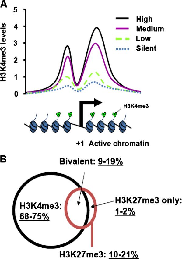
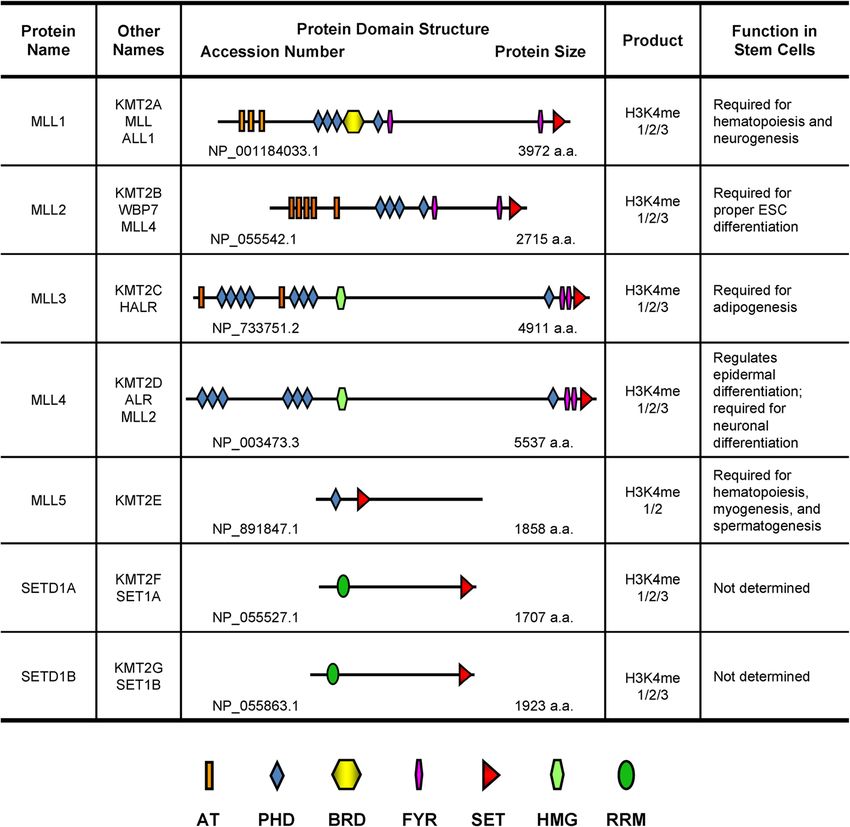
Gu and Lee Cell & Bioscience 2013, 3:39                                                                                          Page 3 of 14
http://www.cellandbioscience.com/content/3/1/39

 Figure 1 H3K4me3 marks actively transcribed and poised gene promoters in mammals. (A) The genome-wide correlation of mRNA
 expression levels (High, Medium, Low, and Silent) with H3K4me3 levels at human gene promoters. Note that a dip of H3K4me3 levels may be
 associated with the nucleosome-free region around the transcriptional start site (TSS). Adapted from [39]. (B) The Venn diagram showing the
 percentage of genes that have H3K4me3 and/or H3K27me3 in their promoters in mouse and human ES cells. All percentages are based on about
 total 18,000 genes. The “bivalent” denotes the promoters that contain both H3K4me3 and H3K27me3 marks. Adapted from [36,37,43].

SET1A, SET1B, and MLL1−4 are yeast Set1 homologs                        conserved in yeast and flies [38] (Table 1). Several studies
and are related to dSet1 (the counterpart of SET1A and                  have demonstrated that these core subunits are indispens-
SET1B), Trx (the counterpart of MLL1 and MLL2), and                     able for the enzyme activity of methyltransferases and bio-
Trr (the counterpart of MLL3 and MLL4) in Drosophila.                   logical functions [53-55]. In addition to common core
Other SET domain-containing histone methyltransferases                  subunits, there are unique subunits in the individual
that methylate H3K4 but are not closely related to yeast                H3K4 methyltransferase complexes: WDR82 and CXXC
Set1/COMPASS have also been identified and include                      finger protein 1 (CFP1) in the SET1 complex; Multiple
MLL5, SET7 (also called SET9), SMYD1-3, SETMAR, and                     endocrine neoplasia type 1 (MENIN) and PC4 and SFRS1-
PRDM9 [6,15,24].                                                        interacting protein 1 (PSIP1) in MLL1 and 2 complex;
  SET1A/1B and MLL1−4 are present in multi-protein                      Host cell factor 1/2 (HCF1/2) in SET1, MLL1, and MLL2
complexes and share common core subunits, such as                       complexes; and PAX transcription activation domain
WDR5, Retinoblastoma-binding protein 5 (RBBP5),                         interacting protein 1 (PTIP), PTIP-associated protein 1
ASH2L, and Dumpy-30 (DPY-30), which are also highly                     (PA1), Nuclear receptor coactivator 6 (NCOA6), and
Gu and Lee Cell & Bioscience 2013, 3:39                                                                                        Page 4 of 14
http://www.cellandbioscience.com/content/3/1/39

Table 1 Subunit composition of H3K4 methyltransferase complexes in yeast and human
Yeast SET1        Human SET1A          Human SET1B          Human MLL1          Human MLL2     Human MLL3     Human MLL4     Human MLL5*
    SET1               SET1A                SET1B                MLL1              MLL2           MLL3           MLL4             Mll5
Cps60/Bre2            ASH2L                 ASH2L               ASH2L             ASH2L           ASH2L          ASH2L           HCF1
Cps50/Swd1             RBBP5                RBBP5                RBBP5            RBBP5           RBBP5          RBBP5            OGT
Cps30/Swd3             WDR5                 WDR5                 WDR5             WDR5            WDR5           WDR5            STK38
Cpd25/Sdc1            DPY-30               DPY-30               DPY-30            DPY-30         DPY-30          DPY-30         PPP1CA
Cps40/Spp1             CFP1                 CFP1                                                                                PPP1CB
Cps35/Swd2            WDR82                WDR82                                                                                PPP1CC
Cps15/Shg1                              BOD1/BOD1L                                                                               ACTB
                      HCF1/2               HCF1/2               HCF1/2            HCF1/2         NCOA6           NCOA6
                                                                MENIN             MENIN            UTX            UTX
                                                                                   PSIP1           PTIP           PTIP
                                                                                                   PA1            PA1
* The subunits of MLL5 are not related to those of SET1, SET1A/B, and MLL1−4.

Ubiquitously transcribed X chromosome tetratricopeptide                           MLL2 (also called MLL4 and KMT2B) has a similar
repeat protein (UTX) in the MLL3 and MLL4 complexes                             protein domain structure to that of MLL1 and was
[12,16,19,22,56-63] (Table 1). These subunits may play                          found to be the MLL1 paralog [74]. Like Mll1, Mll2 is
important roles in recruiting H3K4 methyltransferases                           widely expressed during development and in adult tis-
to specific genes and integrating additional histone-                           sues. Mll2-null mice die before embryonic day E11.5,
modifying capacities (see below).                                               with drastically reduced expression of Hoxb2 and Hoxb5
                                                                                [75]. However, Mll2 may be only required briefly for de-
MLL1 and MLL2                                                                   velopment, because it appears to be dispensable for
MLL1 (also known as MLL and KMT2A) was initially                                mouse development after E11.5 [76]. Mll2−/− ES cells
cloned from acute myeloid and lymphoid leukemia that                            maintain pluripotency, have increased apoptotic activity,
contain frequent MLL1 chromosomal fusions and trans-                            and undergo skewed cellular differentiation along three
locations [64-66]. The MLL1 gene encodes a protein of                           germ layers [77]. Therefore, Mll1 and Mll2 are unlikely
3,972 amino acids; this protein contains several highly                         redundant for gene regulation during early embryonic
conserved functional domains, including the N-terminal                          development. In support with this notion, the pheno-
AT-hook DNA binding domains, Plant homeo domains                                types of Mll1 and Mll2 knockout mice are different in
(PHD), a Bromo domain, and the catalytic SET domain                             adult tissues. For example, hematopoietic-specific loss of
(Figure 2). Inside cells, MLL1 protein is cleaved into                          Mll1 showed defects in hematopoiesis [71,72], whereas
MLL-N (320 kDa) and MLL-C (180 kDa) by Taspase I;                               Mll2 loss did not show any aberrant blood profiles and
these two large fragments dimerize through FY-rich mo-                          notable pathology [76].
tifs to form the functional MLL complex in vivo [67,68].
   Homozygous deletion of Mll1 is embryonic lethal;                             MLL3 and MLL4
Mll1+/− mice display retarded growth and hematopoietic                          MLL3 (also called HALR/KMT2C) and MLL4 (alias
defects [69,70]. Specifically, expression of the key develop-                   ALR/KMT2D) are mammalian counterparts of Drosophila
mental genes, including Hoxa7 and Hoxc9, were shifted                           Trr and were co-purified as transcriptional coactivator
from the anterior boundaries toward the posterior regions                       complexes [14,78-80]. MLL3 and MLL4 associate with nu-
in Mll1+/− embryos and were lost in Mll1−/− mice [69]. In                       clear hormone receptors in both Drosophila and mam-
addition, recent studies using a tissue-specific knockout                       mals. For example, the MLL3/MLL4 complex is recruited
mouse model revealed that Mll1 is essential for sustaining                      to HOXC6 gene and activates its transcription in an estro-
adult hematopoiesis [71,72]. Mll1 is not required for sur-                      gen receptor-dependent manner [79]. Frequent somatic
vival, proliferation, and differentiation of subventricular                     loss-of-function mutations have been identified in MLL3
zone neural stem cells but plays an essential role in                           and MLL4 genes in human cancers, including colorectal
neurogenesis in the postnatal mouse brain [73]. Mechanis-                       cancer, non-Hodgkin B-cell lymphoma, and medulloblas-
tically, Mll1 directly occupies the promoter of Distal-less                     toma [81-85]. Consistently, a recent study reported that
homeobox 2 (Dlx2), a critical regulator of neurogenesis,                        trr gene product suppresses cell growth in Drosophila eye
and is required to resolve the poised bivalent state to the                     imaginal discs. Of interest, trr mutation markedly reduced
actively transcribed status with predominant H3K4me3                            H3K4 monomethylation levels without significantly chan-
during neurogenesis of neural stem cells [73].                                  ging H3K4 di- and trimethylation levels [86], in agreement
Gu and Lee Cell & Bioscience 2013, 3:39 Page 5 of 14 http://www.cellandbioscience.com/content/3/1/39 Figure 2 Protein domain architectures and stem cell function of MLL/SET1 H3K4 methyltransferases. AT: AT-hook DNA binding domain; PHD: Plant Homeo Domain; BRD: Bromodomain; FYR: FY-rich domain; SET: Su(var)3-9, Enhancer of zeste, Trithorax domain; HMG: High Mobility Group domain; RRM: RNA Recognition Motif. with earlier findings that Trr is a major H3K4 mono- gene-repressive mark, blocks the binding activity of methyltransferase for Drosophila enhancers [87]. Mll3 MLL4′s PHD4-6. Consistent with this, knockdown of the homozygous mutant mice, which have an in-frame dele- protein arginine methyltransferase 7, which is involved in tion of a 61-aa catalytic core of the SET domain, exhibited generation of H4R3me2s, increases MLL4 occupancy and reduced white adipose tissue, stunted growth, and slow H3K4me3 levels at the MLL4 target gene promoters and cellular doubling rate [88,89]. During epidermal differenti- enhances the MLL4-dependent neural differentiation pro- ation, the MLL4 complex is recruited to differentiation- gram. Therefore, these results revealed that the trans-tail related genes via the transcription factor GRHL3/GET1 regulation of MLL4-catalyzed H3K4me3 by protein argin- and collaboratively activates the epidermal progenitor dif- ine methyltransferase 7-controlled H4R3me2s serves as a ferentiation program [90]. novel epigenetic mechanism underlying neuronal differen- Recently, we found that MLL4 is essential for the tiation of human stem cells. neuronal differentiation of human NT2/D1 stem cells [91]. Mechanistically, the neuron-specific gene NESTIN MLL5 and key developmental genes HOXA1–3 are activated by Independent studies have demonstrated that MLL5 is re- MLL4 during RA-induced differentiation. Intriguingly, quired for hematopoiesis [92-94]. Moreover, MLL5 pro- the tandem PHD4-6 of seven PHD motifs in MLL4 motes myogenic differentiation by controlling expression (Figure 2) specifically recognized unmethylated or asym- of cell cycle genes (e.g., Cyclin A2) and myogentic regu- metrically dimethylated histone H4 Arg 3 (H4R3me0 or lator genes (e.g., Myogenin) [95]. Mll5 knockout male H4R3me2a) and is required for MLL4′s nucleosomal mice are sterile, at least in part because of deregulated methyltransferase activity and MLL4-mediated differenti- expression of genes that are required for terminal differen- ation. H4R3 symmetric dimethylation (H4R3me2s), a tiation during spermatogenesis [96]. Of interest, although
Gu and Lee Cell & Bioscience 2013, 3:39                                                                         Page 6 of 14
http://www.cellandbioscience.com/content/3/1/39

MLL5 was reported to be inactive [92,95], GlcNAcylation         target genes. Intriguingly, SET7 antagonizes Suv39h1-
of MLL5 greatly increased MLL5′s enzymatic activity to-         mediated H3-K9 methylation at the myogenic differenti-
wards H3K4me1/2 and facilitated RA-induced granulo-             ation gene promoters [109].
poiesis in human HL60 promyelocytes [24].
                                                                SMYD1−3
SET1A and SET1B                                                 Smyd1 (also called Bop) is essential for mouse cardiac
Human SET1A and SET1B have an N-terminal RNA                    differentiation [110]. Consistently, knockdown of Smyd1
recognition motif and a C-terminal enzymatic SET do-            in zebrafish embryos results in defective skeletal and car-
main (Figure 2). The SET1A complex was purified as a            diac muscle differentiation; this cannot be rescued by
multi-protein complex that associates with CFP1 [19].           the Smyd1 catalytic mutant, which lacks H3K4 methyl-
CFP1 is required for stem cell differentiation and inter-       transferase activity [21]. SMYD2 methylates H3K4 and
acts with unmethylated CpGs via its zinc finger domain          H3K36, as well as tumor-suppressor proteins such as
CXXC [97]. Interestingly, Cfp1−/− ES cells displayed            p53 and Retinoblastoma protein (pRB) [23,111-113].
aberrant H3K4me3 peaks at numerous ectopic sites                Specifically, SMYD2-mediated monomethylation of p53
(i.e., distinct regions outside annotated CpG islands),         K370 attenuates the interaction of p53 with p53 target
suggesting that CFP1 recruits the SET1 complex to               promoters and consequently antagonizes p53-dependent
CpG island-containing promoters and consequently                transcriptional regulation [112]. Unlike SMYD1, cardiac-
prevents it from generating H3K4me3 to inappropriate            specific knockout of Smyd2 has no phenotype during
chromatin locations [19,98,99].                                 mouse heart development [114]. SMYD3 is a methyl-
   A protein sequence analysis revealed that SET1A              transferase for both H3K4 and H4K5 [15,115]. It is
shares 39% identity with a SET domain protein named             overexpressed in colorectal and hepatocellular cancers
SET1B [22]. Although both proteins associate with a             and promotes cell proliferation [15]. During zebrafish
similar set of non-catalytic subunits, a confocal micros-       embryogenesis, SMYD3 appears to be important for car-
copy analysis revealed that SET1A and SET1B exhibit             diac and skeletal muscle development [116].
distinct subnuclear localizations in euchromatin regions;
thus, this suggests that each protein regulates a unique        SETMAR
group of target genes [22].                                     SETMAR (also called METNASE) encodes a chimeric
                                                                protein that contains an N-terminal SET domain and a
ASH1L                                                           C-terminal mariner transposase domain [117] (Figure 3).
ASH1L (also called Ash1) is the human homolog of                The function of SETMAR in stem cells remains un-
Ash1, a Drosophila Trithorax group protein that is es-          known. However, SETMAR-catalyzed methylation of
sential for expression of several HOX genes. Some re-           H3K4 and H3K36 may lead to an open chromatin struc-
ports have indicated that ASH1L primarily acts as a             ture, which may facilitate its transposase-dependent pro-
H3K4 methyltransferase [13,100,101], whereas others             cesses, such as foreign DNA integration and DNA
have reported that human ASH1L specifically mono- and           double-strand break repair [20].
dimethylates H3K36 [102-104]. ASH1L cooperates with
MLL1 in HOX gene activation and is required for the             PRDM9
myelomonocytic lineage differentiation of hematopoietic         PRDM9 (also called MEISETZ) is a PR/SET domain-
stem cells [105]. Of interest, a mutation of the SET            dependent histone methyltransferase that is required for
domain of ASH1L did not decrease HOX gene expression,           meiotic prophase progression [18]. Deletion of the
suggesting that ASH1L’s catalytic activity is dispensable for   Prdm9 gene attenuates H3K4me3 levels, resulting in de-
hematopoietic stem cell differentiation [105].                  fective chromosome pairing, impaired sex body forma-
                                                                tion, damaged meiotic progression, and sterility in both
SET7/9                                                          sexes of mice [18]. Mechanistically, Prdm9 binds to
SET7 (or called SET9) is an H3K4 mono- and di-                  13-base pair DNA elements via its C2H2 zinc fingers.
methytransferase [6,106-108]. SET7 expression is upregu-        During early meiosis, this binding event may link
lated during myoblast differentiation [109]. Specifically,      Prdm9-catalyzed H3K4me3 to mammalian meiotic re-
SET7 interacts with Myoblast determination protein 1            combination hotspots that contain the 13-nucleotide
(MyoD), a central transcriptional factor for myogenic gene      DNA elements [118-120].
expression, and is indispensable for MyoD-mediated
muscle differentiation. Knockdown of SET7 impaired the          Subunits of H3K4 methyltransferases
association of MyoD with the promoter and enhancer re-          WDR5, a core subunit of the SET1 and MLL1−4 com-
gions of the myogenic genes (e.g., Myogenin) and reduced        plexes, plays an important role in ES cell self-renewal
gene expression by decreasing H3K4me1 levels at its             and somatic cell reprogramming [47]. WDR5 is highly
Gu and Lee Cell & Bioscience 2013, 3:39 Page 7 of 14 http://www.cellandbioscience.com/content/3/1/39 Figure 3 Protein domain architectures and stem cell function of other H3K4 methyltransferases and core subunits. AT: AT-hook DNA binding domain; AWS: Associated With SET domain; SET: Su(var)3-9, Enhancer of zeste, Trithorax domain; BRD: Bromodomain; PHD: Plant Homeo Domain; BAH: Bromo Adjacent Homology domain; MYND: Myeloid, Nervy, and DEAF-1 domain; MT: Mariner Transposase domain; KRAB: Krüppel Associated Box domain; C2H2: C2H2-type zinc finger; WD: WD40 repeat; SPRY: SplA and Ryanodine domain. expressed in ES cells and downregulated upon differenti- cells. ASH2L knockdown resulted in elevated expression ation. Knockdown of WDR5 resulted in loss of ES cell of mesodermal lineage differentiation genes [121]. self-renewal and decreased the generation of induced DPY-30 and RBBP5 are other core components of the pluripotent stem cells [47]. WDR5 interacts with OCT4 SET1/MLL methyltransferases. In contrast to ASH2L and activates transcription of the self-renewal factors, and WDR5, DPY-30 and RBBP5 were not required for such as OCT4 and NANOG, in ES cells. Moreover, ES cell self-renewal [53]. DPY-30 or RBBP5 knockdown WDR5, together with OCT4, NANOG and SOX2, regu- reduces global and neuronal gene-specific H3K4me3 lates the self-renewal-regulatory network [47]. Similarly, levels, resulting in inefficient RA-induced neural differ- ASH2L is required for the pluripotency of mouse ES entiation of mouse ES cells.
Gu and Lee Cell & Bioscience 2013, 3:39                                                                        Page 8 of 14
http://www.cellandbioscience.com/content/3/1/39

   Differing biological outcomes for ASH2L and WDR5            domain (AOD). The AOD is divided by an insertion
from DPY-30 and RBBP5 are surprising because these             known as the tower domain (Figure 4). LSD1 alone
four proteins are core components of the same SET1/            demethylates H3K4me2/1 on histones but not nucleo-
MLL1−4 methyltransferases. These unexpected findings           somes, while the association of Co-REST with LSD1 al-
might be explained by the following possibilities. Besides     lows LSD1 to demethylate nucleosomal H3K4 [26,27,126].
the known SET1/MLL1−4 complexes, some of these                    Numerous studies in ES cells and neural stem cells
subunits may be present in other complexes in the same         strongly suggest that LSD1 is a key histone methyla-
cells so that they may exert different biological functions    tion modifier in transcriptional regulation for stem cell
from SET1/MLL1−4 complexes. In fact, gel filtration            fate determination. Lsd1-null mice are embryonic lethal
analysis of ES cell nuclear extracts showed that elution       around E6.5, and Lsd1-deficient mouse ES cells demon-
profiles of WDR5/OCT4 did not overlap with those of            strate increased cell death and impaired differentiation,
WDR5/ASH2L/RBBP5, suggesting that WDR5 also be-                such as embryoid body formation defects [127-129].
longs to another new complex containing OCT4 [47].             Similar to mouse ES cells, LSD1 is required for neural
Another possible scenario is that cellular levels of some      stem cell proliferation; it is recruited by the nuclear re-
core subunits and H3K4 methyltransferases may be               ceptor TLX to repress negative cell cycle regulators,
dynamically changed between ES cells and differen-             including p21, in neural stem cells [130]. Interesting-
tiated cells. Such changes might allow certain H3K4            ly, LSD1 is indispensable for differentiation of several
methyltransferase complexes to be dominant over the            cell types, including skeletal muscles and adipocytes
others or lead to formation of new functional complexes,       [131,132]. In mouse ES cells, LSD1 demethylates and
subsequently affecting expression of stemness genes and        stabilizes DNA methyltransferase 1 (DNMT1), and Lsd1
differentiation-specific genes. In support with this, during   deletion results in progressive loss of DNA methylation
ES cell differentiation, ASH2L and WDR5 levels are down-       [128]. Moreover, LSD1 and its associated nucleosome re-
regulated whereas MLL1 and MLL3 are up-regulated               modeling and histone deacetylase (NuRD) complex are
[47,121]. In addition, some H3K4 methyltransferase             recruited to Oct4-occupied enhancers at active stemness
complexes may have non-redundant cellular function             genes in ES cells, but the repression activities of LSD1-
by regulating their unique target genes in a cell type-        NuRD may be antagonized by histone acetyltransferases
specific manner, as mentioned earlier. Future studies          (e.g., p300). During mouse ES cell differentiation, Oct4
are required to further understand the distinct roles          and acetyltransferase levels are down-regulated, and
of the SET1/MLL complexes.                                     LSD1-NuRD decommissions active enhancers by remov-
                                                               ing H3K4me1 while promoting cellular differentiation
H3K4 demethylases                                              [45]. In contrast to the above stem cell studies, seem-
The reversibility of histone methylation was not clear         ingly conflicting results regarding the role of LSD1 in ES
until the discovery of the first histone demethylase LSD1      cells have been reported. Knockdown of LSD1 induces
in 2004 [25]. Subsequently, a new class of JmjC-domain-        differentiation in human ES cells, which is correlated
containing proteins was identified that can demethyl-          with de-repression of developmental genes with elevated
ate methylated lysine residues in histones. The F-box          H3K4me2/3 levels [44]. In addition, Lsd1−/− ES cells had
and leucine-rich repeat protein (FBXL11, also known as         a strong potential to generate extraembryonic tissues
KDM2A) is the first identified JmjC domain-containing          from the embryoid body [133].
demethylase that removes methyl groups from H3K36me2/             LSD2 (AOF1 or KDM1B) was recently identified
1 [122]. The catalytic JmjC domain requires iron and α-        as a homolog of LSD1; it demethylates H3K4me2/1
ketoglutarate as cofactors to hydroxylate methyl groups        like LSD1 [28,134-136]. Interestingly, unlike LSD1,
[123]. Among this class of demethylases, JARID1A−D (or         LSD2 has no tower domain in the AOD region, but con-
KDM5A−D) proteins specifically remove the methyl group         tains unique N-terminal zinc fingers, including C4H2C2
from H3K4me2/3. NO66, a bifunctional lysine-specific           and CW-type zinc fingers, which are required for
demethylase and histidyl-hydroxylase, can demethylate          demethylase activity [136,137] (Figure 4). A genome-
H3K4me/ H3K36me and hydroxylate a histidyl group of            wide mapping analysis revealed that LSD2 primarily
the non-histone protein Rpl8 [124,125]. Not surprisingly,      resides in the intragenic regions of actively expressed
the LSD family (LSD1 and LSD2) and JARID1 family of            genes [28]. LSD2 may activate its target genes, possibly
H3K4 demethylases play important roles in gene transcrip-      via its association with transcriptional elongation factors
tion in stem cell homeostasis.                                 [28]. Lsd2 is not essential for mouse development. How-
                                                               ever, the DNA methylation of several imprinted genes is
LSD1 and LSD2                                                  lost in oocytes from lsd2-deleted females [135]. Conse-
LSD1 protein contains an N-terminal SWIRM domain               quently, the embryos derived from these oocytes
and a long C-terminal FAD-dependent amine oxidase              exhibited biallelic expression or silencing (i.e., loss of
Gu and Lee Cell & Bioscience 2013, 3:39 Page 9 of 14
http://www.cellandbioscience.com/content/3/1/39
Figure 4 Protein domain architectures and stem cell function of H3K4 demethylases. SWIRM: SWI3, RSC8 and MOIRA domain; AOD-N:
Amine Oxidase Domain-N terminal; TOWER: LSD1 tower domain; AOD-C: Amine Oxidase Domain-C terminal; C4H2C2: C4H2C2-type zinc finger;
ZF_CW: CW-type zinc finger; AOD: Amine Oxidase Domain; JmjN: Jumonji N domain; ARID: AT-rich interactive domain; PHD: Plant Homeo
Domain; JmjC: Jumonji C domain; C5HC2: C5HC2-type zinc finger.
monoallelic expression) of the affected imprinted genes repressive complex 2 (PRC2), which enzymatically gener-
and died before mid-gestation [135]. The molecular ates the repressive mark H3K27me3 for silencing of many
mechanism underlying the functional link between H3K4 differentiation-specific genes in ES cells [140]. A genome-
demethylation and DNA methylation for expression of wide chromatin immunoprecipitation (ChIP)-on-chip ana-
imprinted genes remains to be investigated. lysis revealed that RBP2 colocalizes on a subset of PRC2
target gene promoters in mouse ES cells. However, the
JARID1A interaction of RBP2 with PRC2 may not be strong, be-
JARID1A (RBP2 or KDM5A) was identified as a binding cause the mass spectrometric analysis revealed that affinity
partner of pRB protein in early 1990 [138]. RBP2 con- eluates of the PRC2 component EED, which were purified
tains a highly conserved JmjC domain and was found as from ES cell extracts, did not contain RBP2 [141]. Beshiri
a specific H3K4me3/2 demethylase [30,139] (Figure 4). et al. recently demonstrated that RBP2 augments the re-
Rbp2−/− mice are viable and display mild phenotypic de- pressive effects of the pRB-related protein p130 and E2F4
fects in expansion of hematopoietic stem cells and mye- on cell cycle genes during stem cell differentiation via
loid progenitors. The weak phenotype of Rbp2−/− mice H3K4me3 demethylation [142]. Interestingly, RBP2 in-
suggests that other JARID1 family proteins may compen- hibits osteogenic differentiation of human adipose-derived
sate the loss of Rbp2 [139]. stroma cells [143]. RBP2 interacts with Runt-related tran-
During ES cell differentiation, RBP2 is dissociated from scription factor 2 (RUNX2), a transcriptional factor that
HOX genes, resulting in increased H3K4me3 levels and is required for osteogenic differentiation. Subsequently,
gene activation [30]. Consistently, Pasini et al. reported RBP2 represses RUNX2 target genes, including Alkaline
that RBP2 associates with the important Polycomb phosphatase, Osteocalcin, and Osterix [143].Gu and Lee Cell & Bioscience 2013, 3:39 Page 10 of 14
http://www.cellandbioscience.com/content/3/1/39
JARID1B and X-linked mental retardation (XLMR), suggesting that
JARID1B (PLU1 or KDM5B) was shown to be over- it has important functions in the human kidneys and brain
expressed in breast cancer cell lines [144]. As a member [150,151]. Indeed, SMCX is highly expressed in brain dur-
of the JARID1 family, PLU1 catalyzes the demethylation ing zebrafish development and is required for neuron sur-
of H3K4me2/3. Its full activity requires JmjN, ARID, vival [31]. Moreover, SMCX knockdown reduces dendritic
PHD1, and C5HC2 zinc finger in addition to the catalytic length of rat primary neurons, which cannot be rescued
domain JmjC [30,34] (Figure 4). Consistent with the re- by its XLMR-patient mutants with reduced demethylase
sult of earlier studies, knockdown of PLU1 reduced activity [31]. Therefore, SMCX may play an important role
MCF7 breast cancer cell proliferation and concomitantly in neuronal development. In addition, Outchkourov et al.
upregulated expression of the Breast cancer1, early onset reported that SMCX may interact with the transcriptional
(BRCA1), Caveolin 1 (CAV1), and HOXA5 genes as a re- factors c-MYC and ELK1 to regulate gene expression in
sult of increased H3K4me3 levels on their promoters mouse ES cells [152].
[34]. However, PLU1′s role in ES cell self-renewal and JARID1D requires multiple domains, including ARID,
differentiation is controversial. Xie et al. reported that JmjC, and C5HC2 zinc finger, for its full demethylase ac-
PLU1 is a downstream target of the pluripotent factor tivity towards H3K4me3/2 [32] (Figure 4). JARID1D in-
Nanog and is required for ES cell self-renewal [145]. teracts with RING6A/MBLR, a polycomb-like protein
PLU1 interacts with the chromodomain protein MRG15 with homology to Mel18 and Bmi1 proteins [153]. This
and is recruited to H3K36me3-containing sites within interaction stimulates JARID1D’s enzyme activity in vitro;
gene bodies of self-renewal-associated genes via MRG15. the protein complex mediates H3K4me3 demethylation at
Knockdown of PLU1 or MRG15 increased intragenic the Engrailed 2 gene promoter and is required for
H3K4me3 that produces cryptic intragenic transcription Engrailed 2 gene repression [32]. However, JARID1D’s bio-
and inhibited the transcriptional elongation [145]. An- logical role in stem cells is largely unknown. Given its
other study showed that constitutive overexpression of localization on the Y-chromosome, it will be interesting to
PLU1 blocked neural terminal differentiation [146]. On determine whether JARID1D plays a role in male-specific
the contrary, Schmitz et al. has provided evidence that gene expression in vivo.
PLU1 is required for the neural differentiation of ES
cells but is dispensable for self-renewal [147]. Using a NO66
genome-wide ChIP-sequencing analysis, they found that NO66 has been reported to demethylate H3K4me3/2/1
PLU1 predominantly localizes on the transcription start and H3K36me3/2 [124] and to catalyze histidyl hydroxyl-
sites of target genes, over 50% of which are also occu- ation of the 60S ribosomal protein Rpl8 [125]. This en-
pied by Polycomb group proteins. PLU1-depleted ES zyme inhibits osteoblast differentiation [124]. Specifically,
cells fail to differentiate into the neural lineage, which it directly interacts with Osterix, an osteoblast-specific
correlates with the inappropriate depression of stem and transcription factor, and represses Osterix target gene ex-
germ cell genes [147]. These findings are further sup- pression [124]. In addition, NO66 plays a role in mouse
ported by their recent research in Plu1 knockout mice, ES cell differentiation [154]. During this process, it is
which have the phenotype of neonatal lethality and recruited to stemness genes (e.g., Oct4 and Nanog) via the
neural defects [148]. The discrepancies in these studies PHD finger protein 19 (PHF19), which interacts with the
regarding the role of PLU1 in ES cell homeostasis are H3K27 methyltransferase complex PRC2; NO66-PHF19-
not entirely clear. However, Schmitz et al. indicated that PRC2 represses gene expression by reducing H3K36me3
their PLU1 localization data were obtained using a better and increasing H3K27me3 [154].
PLU1 antibody and that the unimportance of PLU1 in
ES cell self-renewal was confirmed by both a lentiviral Conclusions
shRNA knockdown method and a genetic deletion Stem cells are indistinguishable from somatic cells at the
approach. genomic level. In contrast, there are remarkable differ-
ences in epigenomes that may be represented by cova-
JARID1C and JARID1D lent and noncovalent modifications of histones and
Compared with RBP2 and PLU1, much less is known DNA. As reviewed herein, specific epigenetic modifiers,
about the biological function of JARID1C (SMCX or such as H3K4 methylation modifiers, may play funda-
KDM5C) and JARID1D (SMCY or KDM5D). Both mental roles in orchestrating cellular epigenomes whose
demethylases have similar domain structures and con- genomic sequences are identical. Consistent with this,
tain a conserved and functional JmjC domain that is re- many H3K4 methylation modifiers and their components
sponsible for demethylating H3K4me2/3 [30-32]. SMCX are required for ES cell self-renewal or differentiation.
is an X-chromosome gene that escapes from X inacti- In addition, some of them cooperate with transcription
vation [149] and is often mutated in renal tumors factors for efficient somatic cell reprogramming. ForGu and Lee Cell & Bioscience 2013, 3:39 Page 11 of 14
http://www.cellandbioscience.com/content/3/1/39
example, WDR5 is required for the efficient generation of 8. Roguev A, Schaft D, Shevchenko A, Pijnappel WW, Wilm M, Aasland R,
pluripotent stem cells that were induced by Oct4, Sox2, Stewart AF: The Saccharomyces cerevisiae Set1 complex includes an Ash2
homologue and methylates histone 3 lysine 4. EMBO J 2001, 20:7137–7148.
c-Myc, and Klf4 [47]. Therefore, the epigenetic modi- 9. Bryk M, Briggs SD, Strahl BD, Curcio MJ, Allis CD, Winston F: Evidence that
fiers, with the transcription factor network, may establish Set1, a factor required for methylation of histone H3, regulates rDNA
epigenomes in a coordinate manner. silencing in S. cerevisiae by a Sir2-independent mechanism. Curr Biol
2002, 12:165–170.
Recently, small molecule inhibitors against specific 10. Krogan NJ, Dover J, Khorrami S, Greenblatt JF, Schneider J, Johnston M,
histone methyltransferases, including LSD1 inhibitors, Shilatifard A: COMPASS, a histone H3 (Lysine 4) methyltransferase
have been developed by several pharmaceutical compan- required for telomeric silencing of gene expression. J Biol Chem 2002,
277:10753–10755.
ies, although their specificities and efficacies require im- 11. Nakamura T, Mori T, Tada S, Krajewski W, Rozovskaia T, Wassell R, Dubois G,
provement [155]. Certain inhibitors, alone or combined, Mazo A, Croce CM, Canaani E: ALL-1 is a histone methyltransferase that
may increase somatic reprogramming efficiency or drive assembles a supercomplex of proteins involved in transcriptional
regulation. Mol Cell 2002, 10:1119–1128.
somatic reprogramming, perhaps providing new avenues 12. Wysocka J, Myers MP, Laherty CD, Eisenman RN, Herr W: Human Sin3
for personalized therapeutic interventions using stem deacetylase and trithorax-related Set1/Ash2 histone H3-K4
cells. With regard to the roles of histone modifiers in methyltransferase are tethered together selectively by the cell-
proliferation factor HCF-1. Genes Dev 2003, 17:896–911.
stem cell maintenance and differentiation, many more 13. Byrd KN, Shearn A: ASH1, a Drosophila trithorax group protein, is
new exciting findings are expected. We predict that required for methylation of lysine 4 residues on histone H3. Proc Natl
our current and future knowledge about stem cell self- Acad Sci USA 2003, 100:11535–11540.
14. Goo YH, Sohn YC, Kim DH, Kim SW, Kang MJ, Jung DJ, Kwak E, Barlev NA,
renewal and lineage commitment will be highly relevant Berger SL, Chow VT, et al: Activating signal cointegrator 2 belongs to a
to cancer stem cell studies, because stem cells and can- novel steady-state complex that contains a subset of trithorax group
cer stem cells share several characteristics, such as high proteins. Mol Cell Biol 2003, 23:140–149.
15. Hamamoto R, Furukawa Y, Morita M, Iimura Y, Silva FP, Li M, Yagyu R,
degrees of self-renewal and differentiation [156]. We be-
Nakamura Y: SMYD3 encodes a histone methyltransferase involved in the
lieve that a new era of stem cell epigenetics has begun. proliferation of cancer cells. Nat Cell Biol 2004, 6:731–740.
16. Yokoyama A, Wang Z, Wysocka J, Sanyal M, Aufiero DJ, Kitabayashi I, Herr
Competing interests W, Cleary ML: Leukemia proto-oncoprotein MLL forms a SET1-like histone
The authors declare that they have no competing interests. methyltransferase complex with menin to regulate Hox gene expression.
Mol Cell Biol 2004, 24:5639–5649.
Authors’ contributions 17. Dou Y, Milne TA, Tackett AJ, Smith ER, Fukuda A, Wysocka J, Allis CD, Chait
BG prepared the initial draft of the paper. MGL initiated and modified the BT, Hess JL, Roeder RG: Physical association and coordinate function of
manuscript. Both authors read and approved the final manuscript. the H3 K4 methyltransferase MLL1 and the H4 K16 acetyltransferase
MOF. Cell 2005, 121:873–885.
Acknowledgements 18. Hayashi K, Yoshida K, Matsui Y: A histone H3 methyltransferase controls
We sincerely apologize for not citing and reviewing many relevant articles epigenetic events required for meiotic prophase. Nature 2005, 438:374–378.
due to space limitation. We are grateful to our laboratory members for their 19. Lee JH, Skalnik DG: CpG-binding protein (CXXC finger protein 1) is a
helpful suggestions and comments and Ms. Ann Sutton for the manuscript component of the mammalian Set1 histone H3-Lys4 methyltransferase
editing. This work was supported by grants from the NIH (R01 GM095659, complex, the analogue of the yeast Set1/COMPASS complex. J Biol Chem
R01 CA157919, and CCSG 5 P30 CA0166672 35), Cancer Prevention and 2005, 280:41725–41731.
Research Institute of Texas (RP110183), and the Center for Cancer Epigenetics 20. Lee SH, Oshige M, Durant ST, Rasila KK, Williamson EA, Ramsey H, Kwan L,
at the University of Texas MD Anderson Cancer Center. Nickoloff JA, Hromas R: The SET domain protein Metnase mediates
foreign DNA integration and links integration to nonhomologous end-
Received: 17 June 2013 Accepted: 27 July 2013 joining repair. Proc Natl Acad Sci USA 2005, 102:18075–18080.
Published: 9 October 2013 21. Tan X, Rotllant J, Li H, De Deyne P, Du SJ: SmyD1, a histone methyltransferase,
is required for myofibril organization and muscle contraction in zebrafish
References embryos. Proc Natl Acad Sci USA 2006, 103:2713–2718.
1. Takahashi K, Yamanaka S: Induction of pluripotent stem cells from mouse 22. Lee JH, Tate CM, You JS, Skalnik DG: Identification and characterization of
embryonic and adult fibroblast cultures by defined factors. Cell 2006, the human Set1B histone H3-Lys4 methyltransferase complex. J Biol
126:663–676. Chem 2007, 282:13419–13428.
2. Meissner A: Epigenetic modifications in pluripotent and differentiated 23. Abu-Farha M, Lambert JP, Al-Madhoun AS, Elisma F, Skerjanc IS, Figeys D:
cells. Nat Biotechnol 2010, 28:1079–1088. The tale of two domains: proteomics and genomics analysis of SMYD2,
3. Hawkins RD, Hon GC, Lee LK, Ngo Q, Lister R, Pelizzola M, Edsall LE, Kuan S, a new histone methyltransferase. Mol Cell Proteomics 2008, 7:560–572.
Luu Y, Klugman S, et al: Distinct epigenomic landscapes of pluripotent 24. Fujiki R, Chikanishi T, Hashiba W, Ito H, Takada I, Roeder RG, Kitagawa H, Kato S:
and lineage-committed human cells. Cell Stem Cell 2010, 6:479–491. GlcNAcylation of a histone methyltransferase in retinoic-acid-induced
4. Sims RJ 3rd, Nishioka K, Reinberg D: Histone lysine methylation: a granulopoiesis. Nature 2009, 459:455–459.
signature for chromatin function. Trends Genet 2003, 19:629–639. 25. Shi Y, Lan F, Matson C, Mulligan P, Whetstine JR, Cole PA, Casero RA, Shi Y:
5. Miller T, Krogan NJ, Dover J, Erdjument-Bromage H, Tempst P, Johnston M, Histone demethylation mediated by the nuclear amine oxidase homolog
Greenblatt JF, Shilatifard A: COMPASS: a complex of proteins associated LSD1. Cell 2004, 119:941–953.
with a trithorax-related SET domain protein. Proc Natl Acad Sci USA 2001, 26. Lee MG, Wynder C, Cooch N, Shiekhattar R: An essential role for CoREST in
98:12902–12907. nucleosomal histone 3 lysine 4 demethylation. Nature 2005, 437:432–435.
6. Wang H, Cao R, Xia L, Erdjument-Bromage H, Borchers C, Tempst P, Zhang Y: 27. Shi YJ, Matson C, Lan F, Iwase S, Baba T, Shi Y: Regulation of LSD1 histone
Purification and functional characterization of a histone H3-lysine demethylase activity by its associated factors. Mol Cell 2005, 19:857–864.
4-specific methyltransferase. Mol Cell 2001, 8:1207–1217. 28. Fang R, Barbera AJ, Xu Y, Rutenberg M, Leonor T, Bi Q, Lan F, Mei P, Yuan GC,
7. Briggs SD, Bryk M, Strahl BD, Cheung WL, Davie JK, Dent SY, Winston F, Allis CD: Lian C, et al: Human LSD2/KDM1b/AOF1 regulates gene transcription by
Histone H3 lysine 4 methylation is mediated by Set1 and required for cell modulating intragenic H3K4me2 methylation. Mol Cell 2010, 39:222–233.
growth and rDNA silencing in Saccharomyces cerevisiae. Genes Dev 2001, 29. Metzger E, Wissmann M, Yin N, Muller JM, Schneider R, Peters AH, Gunther T,
15:3286–3295. Buettner R, Schule R: LSD1 demethylates repressive histone marks toGu and Lee Cell & Bioscience 2013, 3:39 Page 12 of 14
http://www.cellandbioscience.com/content/3/1/39
promote androgen-receptor-dependent transcription. Nature 2005, 51. Visel A, Blow MJ, Li Z, Zhang T, Akiyama JA, Holt A, Plajzer-Frick I, Shoukry M,
437:436–439. Wright C, Chen F, et al: ChIP-seq accurately predicts tissue-specific activity of
30. Christensen J, Agger K, Cloos PA, Pasini D, Rose S, Sennels L, Rappsilber J, enhancers. Nature 2009, 457:854–858.
Hansen KH, Salcini AE, Helin K: RBP2 belongs to a family of demethylases, 52. Heintzman ND, Hon GC, Hawkins RD, Kheradpour P, Stark A, Harp LF, Ye Z,
specific for tri-and dimethylated lysine 4 on histone 3. Cell 2007, Lee LK, Stuart RK, Ching CW, et al: Histone modifications at human enhancers
128:1063–1076. reflect global cell-type-specific gene expression. Nature 2009, 459:108–112.
31. Iwase S, Lan F, Bayliss P, De la Torre-Ubieta L, Huarte M, Qi HH, Whetstine 53. Jiang H, Shukla A, Wang X, Chen WY, Bernstein BE, Roeder RG: Role for
JR, Bonni A, Roberts TM, Shi Y: The X-linked mental retardation gene Dpy-30 in ES cell-fate specification by regulation of H3K4 methylation
SMCX/JARID1C defines a family of histone H3 lysine 4 demethylases. within bivalent domains. Cell 2011, 144:513–525.
Cell 2007, 128:1077–1088. 54. Steward MM, Lee JS, O'Donovan A, Wyatt M, Bernstein BE, Shilatifard A:
32. Lee MG, Norman J, Shilatifard A, Shiekhattar R: Physical and functional Molecular regulation of H3K4 trimethylation by ASH2L, a shared subunit
association of a trimethyl H3K4 demethylase and Ring6a/MBLR, a of MLL complexes. Nat Struct Mol Biol 2006, 13:852–854.
polycomb-like protein. Cell 2007, 128:877–887. 55. Dou Y, Milne TA, Ruthenburg AJ, Lee S, Lee JW, Verdine GL, Allis CD,
33. Tahiliani M, Mei P, Fang R, Leonor T, Rutenberg M, Shimizu F, Li J, Rao A, Roeder RG: Regulation of MLL1 H3K4 methyltransferase activity by its
Shi Y: The histone H3K4 demethylase SMCX links REST target genes to core components. Nat Struct Mol Biol 2006, 13:713–719.
X-linked mental retardation. Nature 2007, 447:601–605. 56. Cho YW, Hong T, Hong S, Guo H, Yu H, Kim D, Guszczynski T, Dressler GR,
34. Yamane K, Tateishi K, Klose RJ, Fang J, Fabrizio LA, Erdjument-Bromage H, Copeland TD, Kalkum M, Ge K: PTIP associates with MLL3- and MLL4-
Taylor-Papadimitriou J, Tempst P, Zhang Y: PLU-1 is an H3K4 demethylase containing histone H3 lysine 4 methyltransferase complex. J Biol Chem
involved in transcriptional repression and breast cancer cell proliferation. 2007, 282:20395–20406.
Mol Cell 2007, 25:801–812. 57. Hughes CM, Rozenblatt-Rosen O, Milne TA, Copeland TD, Levine SS, Lee JC,
35. Secombe J, Li L, Carlos L, Eisenman RN: The Trithorax group protein Lid is Hayes DN, Shanmugam KS, Bhattacharjee A, Biondi CA, et al: Menin
a trimethyl histone H3K4 demethylase required for dMyc-induced cell associates with a trithorax family histone methyltransferase complex and
growth. Genes Dev 2007, 21:537–551. with the hoxc8 locus. Mol Cell 2004, 13:587–597.
36. Pan G, Tian S, Nie J, Yang C, Ruotti V, Wei H, Jonsdottir GA, Stewart R, 58. Issaeva I, Zonis Y, Rozovskaia T, Orlovsky K, Croce CM, Nakamura T, Mazo A,
Thomson JA: Whole-genome analysis of histone H3 lysine 4 and lysine 27 Eisenbach L, Canaani E: Knockdown of ALR (MLL2) reveals ALR target
methylation in human embryonic stem cells. Cell Stem Cell 2007, 1:299–312. genes and leads to alterations in cell adhesion and growth. Mol Cell Biol
37. Zhao XD, Han X, Chew JL, Liu J, Chiu KP, Choo A, Orlov YL, Sung WK, 2007, 27:1889–1903.
Shahab A, Kuznetsov VA, et al: Whole-genome mapping of histone H3 59. Lee JH, Skalnik DG: Wdr82 is a C-terminal domain-binding protein that
Lys4 and 27 trimethylations reveals distinct genomic compartments in recruits the Setd1A Histone H3-Lys4 methyltransferase complex to
human embryonic stem cells. Cell Stem Cell 2007, 1:286–298. transcription start sites of transcribed human genes. Mol Cell Biol 2008,
38. Shilatifard A: The COMPASS family of histone H3K4 methylases: 28:609–618.
mechanisms of regulation in development and disease pathogenesis. 60. Lee MG, Villa R, Trojer P, Norman J, Yan KP, Reinberg D, Di Croce L,
Annu Rev Biochem 2012, 81:65–95. Shiekhattar R: Demethylation of H3K27 regulates polycomb recruitment
39. Barski A, Cuddapah S, Cui K, Roh TY, Schones DE, Wang Z, Wei G, Chepelev I, and H2A ubiquitination. Science 2007, 318:447–450.
Zhao K: High-resolution profiling of histone methylations in the human 61. Mohan M, Herz HM, Smith ER, Zhang Y, Jackson J, Washburn MP, Florens L,
genome. Cell 2007, 129:823–837. Eissenberg JC, Shilatifard A: The COMPASS family of H3K4 methylases in
40. Pokholok DK, Harbison CT, Levine S, Cole M, Hannett NM, Lee TI, Bell GW, Drosophila. Mol Cell Biol 2011, 31:4310–4318.
Walker K, Rolfe PA, Herbolsheimer E, et al: Genome-wide map of 62. Patel SR, Kim D, Levitan I, Dressler GR: The BRCT-domain containing
nucleosome acetylation and methylation in yeast. Cell 2005, 122:517–527. protein PTIP links PAX2 to a histone H3, lysine 4 methyltransferase
41. Guenther MG, Levine SS, Boyer LA, Jaenisch R, Young RA: A chromatin complex. Dev Cell 2007, 13:580–592.
landmark and transcription initiation at most promoters in human cells. 63. Van Nuland R, Smits AH, Pallaki P, Jansen PW, Vermeulen M, Timmers HT:
Cell 2007, 130:77–88. Quantitative Dissection and Stoichiometry Determination of the Human
42. Bernstein BE, Mikkelsen TS, Xie X, Kamal M, Huebert DJ, Cuff J, Fry B, SET1/MLL Histone Methyltransferase Complexes. Mol Cell Biol 2013,
Meissner A, Wernig M, Plath K, et al: A bivalent chromatin structure marks 33:2067–2077.
key developmental genes in embryonic stem cells. Cell 2006, 64. Tkachuk DC, Kohler S, Cleary ML: Involvement of a homolog of Drosophila
125:315–326. trithorax by 11q23 chromosomal translocations in acute leukemias.
43. Mikkelsen TS, Hanna J, Zhang X, Ku M, Wernig M, Schorderet P, Bernstein BE, Cell 1992, 71:691–700.
Jaenisch R, Lander ES, Meissner A: Dissecting direct reprogramming through 65. Gu Y, Nakamura T, Alder H, Prasad R, Canaani O, Cimino G, Croce CM,
integrative genomic analysis. Nature 2008, 454:49–55. Canaani E: The t(4;11) chromosome translocation of human acute
44. Adamo A, Sese B, Boue S, Castano J, Paramonov I, Barrero MJ, Izpisua leukemias fuses the ALL-1 gene, related to Drosophila trithorax, to the
Belmonte JC: LSD1 regulates the balance between self-renewal and AF-4 gene. Cell 1992, 71:701–708.
differentiation in human embryonic stem cells. Nat Cell Biol 2011, 66. Djabali M, Selleri L, Parry P, Bower M, Young BD, Evans GA: A trithorax-like
13:652–659. gene is interrupted by chromosome 11q23 translocations in acute
45. Whyte WA, Bilodeau S, Orlando DA, Hoke HA, Frampton GM, Foster CT, leukaemias. Nat Genet 1992, 2:113–118.
Cowley SM, Young RA: Enhancer decommissioning by LSD1 during 67. Yokoyama A, Kitabayashi I, Ayton PM, Cleary ML, Ohki M: Leukemia
embryonic stem cell differentiation. Nature 2012, 482:221–225. proto-oncoprotein MLL is proteolytically processed into 2 fragments
46. Kimura H, Tada M, Nakatsuji N, Tada T: Histone code modifications on with opposite transcriptional properties. Blood 2002, 100:3710–3718.
pluripotential nuclei of reprogrammed somatic cells. Mol Cell Biol 2004, 68. Hsieh JJ, Cheng EH, Korsmeyer SJ: Taspase1: a threonine aspartase
24:5710–5720. required for cleavage of MLL and proper HOX gene expression. Cell 2003,
47. Ang YS, Tsai SY, Lee DF, Monk J, Su J, Ratnakumar K, Ding J, Ge Y, Darr H, 115:293–303.
Chang B, et al: Wdr5 mediates self-renewal and reprogramming via the 69. Yu BD, Hess JL, Horning SE, Brown GA, Korsmeyer SJ: Altered Hox
embryonic stem cell core transcriptional network. Cell 2011, 145:183–197. expression and segmental identity in Mll-mutant mice. Nature 1995,
48. Jenuwein T, Allis CD: Translating the histone code. Science 2001, 378:505–508.
293:1074–1080. 70. Ernst P, Mabon M, Davidson AJ, Zon LI, Korsmeyer SJ: An Mll-dependent
49. Bernstein BE, Kamal M, Lindblad-Toh K, Bekiranov S, Bailey DK, Huebert DJ, Hox program drives hematopoietic progenitor expansion. Curr Biol 2004,
McMahon S, Karlsson EK, Kulbokas EJ 3rd, Gingeras TR, et al: Genomic maps 14:2063–2069.
and comparative analysis of histone modifications in human and mouse. 71. Gan T, Jude CD, Zaffuto K, Ernst P: Developmentally induced Mll1 loss reveals
Cell 2005, 120:169–181. defects in postnatal haematopoiesis. Leukemia 2010, 24:1732–1741.
50. Weber M, Hellmann I, Stadler MB, Ramos L, Paabo S, Rebhan M, Schubeler D: 72. McMahon KA, Hiew SY, Hadjur S, Veiga-Fernandes H, Menzel U, Price AJ,
Distribution, silencing potential and evolutionary impact of promoter DNA Kioussis D, Williams O, Brady HJ: Mll has a critical role in fetal and adult
methylation in the human genome. Nat Genet 2007, 39:457–466. hematopoietic stem cell self-renewal. Cell Stem Cell 2007, 1:338–345.Gu and Lee Cell & Bioscience 2013, 3:39 Page 13 of 14
http://www.cellandbioscience.com/content/3/1/39
73. Lim DA, Huang YC, Swigut T, Mirick AL, Garcia-Verdugo JM, Wysocka J, hematopoietic defects, reduced neutrophil immune function, and
Ernst P, Alvarez-Buylla A: Chromatin remodelling factor Mll1 is essential for extreme sensitivity to DNA demethylation. Blood 2009, 113:1432–1443.
neurogenesis from postnatal neural stem cells. Nature 2009, 458:529–533. 94. Zhang Y, Wong J, Klinger M, Tran MT, Shannon KM, Killeen N: MLL5
74. FitzGerald KT, Diaz MO: MLL2: A new mammalian member of the trx/MLL contributes to hematopoietic stem cell fitness and homeostasis.
family of genes. Genomics 1999, 59:187–192. Blood 2009, 113:1455–1463.
75. Glaser S, Schaft J, Lubitz S, Vintersten K, Van der Hoeven F, Tufteland KR, 95. Sebastian S, Sreenivas P, Sambasivan R, Cheedipudi S, Kandalla P, Pavlath GK,
Aasland R, Anastassiadis K, Ang SL, Stewart AF: Multiple epigenetic Dhawan J: MLL5, a trithorax homolog, indirectly regulates H3K4
maintenance factors implicated by the loss of Mll2 in mouse methylation, represses cyclin A2 expression, and promotes myogenic
development. Development 2006, 133:1423–1432. differentiation. Proc Natl Acad Sci USA 2009, 106:4719–4724.
76. Glaser S, Lubitz S, Loveland KL, Ohbo K, Robb L, Schwenk F, Seibler J, Roellig D, 96. Yap DB, Walker DC, Prentice LM, McKinney S, Turashvili G, Mooslehner-Allen K,
Kranz A, Anastassiadis K, Stewart AF: The histone 3 lysine 4 methyltransferase, De Algara TR, Fee J, De Tassigny X, Colledge WH, Aparicio S: Mll5 is required
Mll2, is only required briefly in development and spermatogenesis. for normal spermatogenesis. PLoS One 2011, 6:e27127.
Epigenetics Chromatin 2009, 2:5. 97. Carlone DL, Lee JH, Young SR, Dobrota E, Butler JS, Ruiz J, Skalnik DG:
77. Lubitz S, Glaser S, Schaft J, Stewart AF, Anastassiadis K: Increased apoptosis Reduced genomic cytosine methylation and defective cellular
and skewed differentiation in mouse embryonic stem cells lacking the differentiation in embryonic stem cells lacking CpG binding protein.
histone methyltransferase Mll2. Mol Biol Cell 2007, 18:2356–2366. Mol Cell Biol 2005, 25:4881–4891.
78. Sedkov Y, Cho E, Petruk S, Cherbas L, Smith ST, Jones RS, Cherbas P, Canaani E, 98. Tate CM, Lee JH, Skalnik DG: CXXC finger protein 1 restricts the Setd1A
Jaynes JB, Mazo A: Methylation at lysine 4 of histone H3 in ecdysone- histone H3K4 methyltransferase complex to euchromatin. FEBS J 2010,
dependent development of Drosophila. Nature 2003, 426:78–83. 277:210–223.
79. Ansari KI, Hussain I, Shrestha B, Kasiri S, Mandal SS: HOXC6 Is 99. Clouaire T, Webb S, Skene P, Illingworth R, Kerr A, Andrews R, Lee JH,
transcriptionally regulated via coordination of MLL histone methylase Skalnik D, Bird A: Cfp1 integrates both CpG content and gene activity for
and estrogen receptor in an estrogen environment. J Mol Biol 2011, accurate H3K4me3 deposition in embryonic stem cells. Genes Dev 2012,
411:334–349. 26:1714–1728.
80. Lee S, Kim DH, Goo YH, Lee YC, Lee SK, Lee JW: Crucial roles for 100. Beisel C, Imhof A, Greene J, Kremmer E, Sauer F: Histone methylation by
interactions between MLL3/4 and INI1 in nuclear receptor the Drosophila epigenetic transcriptional regulator Ash1. Nature 2002,
transactivation. Mol Endocrinol 2009, 23:610–619. 419:857–862.
81. Sjoblom T, Jones S, Wood LD, Parsons DW, Lin J, Barber TD, Mandelker D, 101. Gregory GD, Vakoc CR, Rozovskaia T, Zheng X, Patel S, Nakamura T, Canaani E,
Leary RJ, Ptak J, Silliman N, et al: The consensus coding sequences of Blobel GA: Mammalian ASH1L is a histone methyltransferase that
human breast and colorectal cancers. Science 2006, 314:268–274. occupies the transcribed region of active genes. Mol Cell Biol 2007,
82. Morin RD, Mendez-Lago M, Mungall AJ, Goya R, Mungall KL, Corbett RD, 27:8466–8479.
Johnson NA, Severson TM, Chiu R, Field M, et al: Frequent mutation of 102. Tanaka Y, Katagiri Z, Kawahashi K, Kioussis D, Kitajima S: Trithorax-group
histone-modifying genes in non-Hodgkin lymphoma. Nature 2011, protein ASH1 methylates histone H3 lysine 36. Gene 2007, 397:161–168.
476:298–303. 103. An S, Yeo KJ, Jeon YH, Song JJ: Crystal structure of the human histone
83. Parsons DW, Li M, Zhang X, Jones S, Leary RJ, Lin JC, Boca SM, Carter H, methyltransferase ASH1L catalytic domain and its implications for the
Samayoa J, Bettegowda C, et al: The genetic landscape of the childhood regulatory mechanism. J Biol Chem 2011, 286:8369–8374.
cancer medulloblastoma. Science 2011, 331:435–439. 104. Yuan W, Xu M, Huang C, Liu N, Chen S, Zhu B: H3K36 methylation antagonizes
84. Jones DT, Jager N, Kool M, Zichner T, Hutter B, Sultan M, Cho YJ, Pugh TJ, PRC2-mediated H3K27 methylation. J Biol Chem 2011, 286:7983–7989.
Hovestadt V, Stutz AM, et al: Dissecting the genomic complexity 105. Tanaka Y, Kawahashi K, Katagiri Z, Nakayama Y, Mahajan M, Kioussis D: Dual
underlying medulloblastoma. Nature 2012, 488:100–105. function of histone H3 lysine 36 methyltransferase ASH1 in regulation of
85. Pugh TJ, Weeraratne SD, Archer TC, Pomeranz Krummel DA, Auclair D, Hox gene expression. PLoS One 2011, 6:e28171.
Bochicchio J, Carneiro MO, Carter SL, Cibulskis K, Erlich RL, et al: 106. Xiao B, Jing C, Wilson JR, Walker PA, Vasisht N, Kelly G, Howell S, Taylor IA,
Medulloblastoma exome sequencing uncovers subtype-specific somatic Blackburn GM, Gamblin SJ: Structure and catalytic mechanism of the
mutations. Nature 2012, 488:106–110. human histone methyltransferase SET7/9. Nature 2003, 421:652–656.
86. Kanda H, Nguyen A, Chen L, Okano H, Hariharan IK: The Drosophila 107. Kwon T, Chang JH, Kwak E, Lee CW, Joachimiak A, Kim YC, Lee J, Cho Y:
ortholog of MLL3 and MLL4, trithorax related, functions as a negative Mechanism of histone lysine methyl transfer revealed by the structure of
regulator of tissue growth. Mol Cell Biol 2013, 33:1702–1710. SET7/9-AdoMet. EMBO J 2003, 22:292–303.
87. Herz HM, Mohan M, Garruss AS, Liang K, Takahashi YH, Mickey K, Voets O, 108. Nishioka K, Chuikov S, Sarma K, Erdjument-Bromage H, Allis CD, Tempst P,
Verrijzer CP, Shilatifard A: Enhancer-associated H3K4 monomethylation by Reinberg D: Set9, a novel histone H3 methyltransferase that facilitates
Trithorax-related, the Drosophila homolog of mammalian Mll3/Mll4. transcription by precluding histone tail modifications required for
Genes Dev 2012, 26:2604–2620. heterochromatin formation. Genes Dev 2002, 16:479–489.
88. Lee S, Lee DK, Dou Y, Lee J, Lee B, Kwak E, Kong YY, Lee SK, Roeder RG, Lee JW: 109. Tao Y, Neppl RL, Huang ZP, Chen J, Tang RH, Cao R, Zhang Y, Jin SW,
Coactivator as a target gene specificity determinant for histone H3 lysine 4 Wang DZ: The histone methyltransferase Set7/9 promotes myoblast
methyltransferases. Proc Natl Acad Sci USA 2006, 103:15392–15397. differentiation and myofibril assembly. J Cell Biol 2011, 194:551–565.
89. Lee J, Saha PK, Yang QH, Lee S, Park JY, Suh Y, Lee SK, Chan L, Roeder RG, 110. Gottlieb PD, Pierce SA, Sims RJ, Yamagishi H, Weihe EK, Harriss JV, Maika SD,
Lee JW: Targeted inactivation of MLL3 histone H3-Lys-4 Kuziel WA, King HL, Olson EN, et al: Bop encodes a muscle-restricted
methyltransferase activity in the mouse reveals vital roles for MLL3 in protein containing MYND and SET domains and is essential for cardiac
adipogenesis. Proc Natl Acad Sci USA 2008, 105:19229–19234. differentiation and morphogenesis. Nat Genet 2002, 31:25–32.
90. Hopkin AS, Gordon W, Klein RH, Espitia F, Daily K, Zeller M, Baldi P, 111. Brown MA, Sims RJ 3rd, Gottlieb PD, Tucker PW: Identification and
Andersen B: GRHL3/GET1 and trithorax group members collaborate to characterization of Smyd2: a split SET/MYND domain-containing histone
activate the epidermal progenitor differentiation program. PLoS Genet H3 lysine 36-specific methyltransferase that interacts with the Sin3
2012, 8:e1002829. histone deacetylase complex. Mol Cancer 2006, 5:26.
91. Dhar SS, Lee SH, Kan PY, Voigt P, Ma L, Shi X, Reinberg D, Lee MG: Trans-tail 112. Huang J, Perez-Burgos L, Placek BJ, Sengupta R, Richter M, Dorsey JA,
regulation of MLL4-catalyzed H3K4 methylation by H4R3 symmetric Kubicek S, Opravil S, Jenuwein T, Berger SL: Repression of p53 activity by
dimethylation is mediated by a tandem PHD of MLL4. Genes Dev 2012, Smyd2-mediated methylation. Nature 2006, 444:629–632.
26:2749–2762. 113. Saddic LA, West LE, Aslanian A, Yates JR 3rd, Rubin SM, Gozani O, Sage J:
92. Madan V, Madan B, Brykczynska U, Zilbermann F, Hogeveen K, Dohner K, Methylation of the retinoblastoma tumor suppressor by SMYD2.
Dohner H, Weber O, Blum C, Rodewald HR, et al: Impaired function of J Biol Chem 2010, 285:37733–37740.
primitive hematopoietic cells in mice lacking the Mixed-Lineage- 114. Diehl F, Brown MA, Van Amerongen MJ, Novoyatleva T, Wietelmann A,
Leukemia homolog MLL5. Blood 2009, 113:1444–1454. Harriss J, Ferrazzi F, Bottger T, Harvey RP, Tucker PW, Engel FB: Cardiac
93. Heuser M, Yap DB, Leung M, De Algara TR, Tafech A, McKinney S, Dixon J, deletion of Smyd2 is dispensable for mouse heart development.
Thresher R, Colledge B, Carlton M, et al: Loss of MLL5 results in pleiotropic PLoS One 2010, 5:e9748.You can also read