Biochemical and structural basis for YTH domain of human YTHDC1 binding to methylated adenine in DNA
←
→
Page content transcription
If your browser does not render page correctly, please read the page content below
Published online 14 July 2020                                                          Nucleic Acids Research, 2020, Vol. 48, No. 18 10329–10341
                                                                                                                          doi: 10.1093/nar/gkaa604

Biochemical and structural basis for YTH domain of
human YTHDC1 binding to methylated adenine in DNA
Clayton B. Woodcock1,† , John R. Horton1,† , Jujun Zhou1 , Mark T. Bedford1 , Robert
M. Blumenthal2 , Xing Zhang1,* and Xiaodong Cheng 1,*
1
 Department of Epigenetics and Molecular Carcinogenesis, University of Texas MD Anderson Cancer Center,
Houston, TX 77030, USA and 2 Department of Medical Microbiology and Immunology, and Program in Bioinformatics,
The University of Toledo College of Medicine and Life Sciences, Toledo, OH 43614, USA

                                                                                                                                                                  Downloaded from https://academic.oup.com/nar/article/48/18/10329/5871368 by guest on 14 December 2020
Received May 28, 2020; Revised June 30, 2020; Editorial Decision July 03, 2020; Accepted July 06, 2020

ABSTRACT                                                                                 methylcytosine, 5mC), or the exocyclic amino groups of ei-
                                                                                         ther cytosine at N4 (yielding N4-methylcytosine, N4mC) or
The recently characterized mammalian writer                                              adenine at N6 (yielding N6-methyladenine, N6mA) (1,2).
(methyltransferase) and eraser (demethylase) of the                                      The great majority (if not all) of bacterial and archaeal
DNA N6-methyladenine (N6mA) methyl mark act on                                           DNA methyltransferases (MTases) are associated with a set
single-stranded (ss) and transiently-unpaired DNA.                                       of conserved sequence motifs important for three essential
As YTH domain-containing proteins bind N6mA-                                             functions: binding methyl donor S-adenosyl-L-methionine
containing RNA in mammalian cells, we investigated                                       (SAM), binding the DNA substrate, and catalyzing the
whether mammalian YTH domains are also methyl                                            methyl transfer between the donor and substrate (3–5).
mark readers of N6mA DNA. Here, we show that the                                         It was these predictive motifs that aided in the discovery
YTH domain of YTHDC1 (known to localize in the                                           of mammalian DNA cytosine-C5-specific Dnmt1 (6) and
nucleus) binds ssDNA containing N6mA, with a 10                                          Dnmt3 (7).
                                                                                            The existence of 5mC was first reported in DNA from
nM dissociation constant. This binding is stronger
                                                                                         ox spleen ∼70 years ago (8), whereas detection of N6mA
by a factor of 5 than in an RNA context, tested                                          in mammalian DNA was reported only recently, in paral-
under the same conditions. However, the YTH do-                                          lel with development and application of ultrasensitive mass
mains of YTHDF2 and YTHDF1 (predominantly cy-                                            spectrometry. There are three debatable issues regarding
toplasmic) exhibited the opposite effect with ∼1.5–                                      N6mA in mammalian DNA: (i) whether low-level N6mA
2× stronger binding to ssRNA containing N6mA                                             can be accurately detected, (ii) how N6mA is generated and
than to the corresponding DNA. We determined two                                         (iii) what the potential functions of N6mA might be.
structures of the YTH domain of YTHDC1 in com-                                              First, low levels of N6mA have been reported in DNA
plex with N6mA-containing ssDNA, which illustrated                                       from mouse (9,10), human (11,12), and human malignant
that YTHDC1 binds the methylated adenine in a                                            brain tumor glioblastoma (13). Other studies have failed
single-stranded region flanked by duplexed DNA.                                          to detect N6mA in several mouse tissues and mitochon-
                                                                                         drial DNA from human placenta (14), DNA isolated from
We discuss the hypothesis that the writer-reader-
                                                                                         mouse embryonic stem cells or brain and liver tissue (15)
eraser of N6mA-containining ssDNA is associated                                          or cultured human cells (16). Nevertheless, more recent re-
with maintaining genome stability. Structural com-                                       ports present evidence for low levels of N6mA in DNA from
parison of YTH and SRA domains (the latter a DNA                                         cultured human and mouse cells; however, that modified
5-methylcytosine reader) revealed them to be diverse                                     adenosine is incorporated (at least in part) by DNA poly-
members of a larger family of DNA/RNA modification                                       merase using deoxy-N6mA converted from RNA-derived
readers, apparently having originated from bacterial                                     ribo-N6mA, via the nucleotide salvage pathway (17,18).
modification-dependent restriction enzymes.                                                 Second, there is also uncertainty regarding
                                                                                         the mammalian enzyme(s) (suggested to include
                                                                                         HemK2/N6AMA1, MettL3-MettL14 complex and
INTRODUCTION
                                                                                         MettL4) responsible for generating N6mA in mammalian
DNA methylation in bacteria and archaea is common,                                       DNA (10,13,19–21). Human HemK2, which was thought
occurring at ring carbon C5 of the cytosine (yielding 5-                                 to be a DNA N6mA MTase (11), is actually a protein

* To
   whom correspondence may be addressed. Email: xcheng5@mdanderson.org
Correspondence may also be addressed to Xing Zhang. Email: xzhang21@mdanderson.org
†
    The authors wish it to be known that, in their opinion, the first two authors should be regarded as Joint First Authors.

C The Author(s) 2020. Published by Oxford University Press on behalf of Nucleic Acids Research.
This is an Open Access article distributed under the terms of the Creative Commons Attribution Non-Commercial License
(http://creativecommons.org/licenses/by-nc/4.0/), which permits non-commercial re-use, distribution, and reproduction in any medium, provided the original work
is properly cited. For commercial re-use, please contact journals.permissions@oup.com
10330 Nucleic Acids Research, 2020, Vol. 48, No. 18
MTase active on glutamine and lysine (19,20,22,23). induced pluripotent stem cells (43). Aside from their con-
Guided by the sequential order of conserved sequence mo- served YTH domains, DC1 and DC2 are unrelated to each
tifs unique to Class  amino (N6mA and N4mC) MTases other and unrelated to DF1–3, based on amino acid se-
(4,24,25), we identified human MettL3–MettL14 as a quence, size, and overall domain organization. The three
DNA adenine MTase active on single-strand and unpaired paralogs DF1–3, on the other hand, share high amino
DNA in vitro (26). Previous to that, MettL3–MettL14 acid identity over their entire length (38). Notably, in ad-
had been characterized extensively as generating N6mA dition to the conserved YTH domain located in the C-
in RNA ((27,28) and references therein). Murine MettL4 terminal region of DF1–3 and DC2 and an internal re-
is responsible for N6mA deposition in genic elements gion of DC1, DF1–3 and DC1 contain low-complexity se-
associated with transcriptional silencing (10). Intriguingly, quences (P/Q/N-rich) throughout and lack recognizable
recombinant human MettL4 expressed in human embry- modular protein domains.
onic kidney (HEK293T) cells has in vitro enzymatic activity Here we show that the YTH domain of YTHDC1 binds
on mitochondrial DNA (21), whereas recombinant human to single-stranded (ss) DNA containing N6mA, with a
Downloaded from https://academic.oup.com/nar/article/48/18/10329/5871368 by guest on 14 December 2020
MettL4 purified from Escherichia coli has RNA MTase 10 nM dissociation constant. Under the same conditions,
activity (29). this YTH binding affinity for N6mA in a DNA con-
Third, having N6mA in DNA might be a double-edged text is stronger by a factor of 5 than such binding in an
sword. One potential advantage of having N6mA in DNA RNA context. However, the YTH domains of YTHDF1
might help to reduce the mutagenic potential of cellular and YTHDF2 exhibited the opposite effect with ∼1.5–
8-oxo-2 -deoxyguanosine (8-oxo-dGTP) (30). Opposite to 2× stronger binding to N6mA in ssRNA than in the cor-
the template adenosine, 8-oxo-dGTP can be misincorpo- responding DNA. We determined two structures of the
rated into DNA using Hoogsteen pairing that involves YTH domain of YTHDC1 binding the methylated adeno-
the exocyclic amino group of adenosine. Methylation of sine in ssDNA, and compared these structures to that of
that adenosine amino group makes N6mA:8-oxo-dG sig- a previously characterized RNA-bound YTH domain of
nificantly less stable than the A:8-oxo-dG Hoogsteen pair YTHDC1. Finally, we discuss the relationship between
in the DNA duplex region, resulting in less efficient mis- mammalian DNA base modification reader domains and
incorporation of 8-oxo-dGTP opposite N6mA by human bacterial modification-dependent restriction enzymes.
DNA polymerase  (30). This observation might be related
to the association between MettL3–MetL14 recruitment to MATERIALS AND METHODS
DNA damaged sites, and its methyltransfer activity being
Protein expression and purification
required for the repair (31). On the other hand, one po-
tential disadvantage of using N6mA (or N4mC) in DNA, The protein fragments used are YTHDC1 residues 345–509
compared to 5mC, is that the methylated amino group of of UniProtKB/Swiss-Prot Q96MU7, YTHDF1 residues
N6mA (or N4mC) is involved in normal hydrogen-bonding 361–559 of Q9BYJ9 and for YTHDF2 residues 391–579 of
for Watson-Crick base pairing, while the cytosine C5 po- Q9Y5A9. YTHDC1 (pXC2129) and YTHDF1 (pXC2130)
sition is not. Indeed, incorporation of N6mA into a DNA constructs were purchased from Addgene (plasmid #64652
template causes RNA polymerase Pol II pausing, associated and #64653) containing N-terminal 6xHis-tag. The YTH
with the lower stability and slower kinetics of base pair- domain of human YTHDF2 as a GST-tagged fusion in a
ing, and the stability of N6mA:U base pair is weakened pGEX-4T1 vector was synthesized by Biomatik. Each plas-
(32). Similar site-specific pausing at N6mA, by a modified mid was transformed into BL21-CodonPlus cells for expres-
phage DNA polymerase inserting T across from a template sion.
N6mA, underlies the ability of PacBio SMRT sequencers Each protein was purified from 6 L of LB inoculated with
to detect methylated adenines (33), and there is additional overnight starter culture and grown to A600 of ∼1 at 37◦ C;
evidence for weaker N6mA:T than A:T base pairs (34–36). after which the samples were cooled to 16◦ C and induced by
Human and mouse homologs (Alkbh1 and Alkbh4) of 1 mM isopropyl thiogalactopyranoside for 12 h. Cells were
the E. coli repair enzyme AlkB can function as sequence- harvested at 4◦ C and then suspended into buffer A (20 mM
independent DNA N6mA demethylases (9,10,13), and at Tris pH 7.5, 400 mM NaCl, 5% glycerol) to a final volume
least the mouse Alkbh1 prefers locally-unpaired DNA sub- of 30 ml before undergoing sonication at 75% amplitude (3
strates (37). It is intriguing that both the writer (MTase) s on and 9 s off for 2 min). The sonicated samples were cen-
and eraser (demethylase) of the N6mA methyl mark act on trifuged at 25 000 rpm at 4◦ C, for 2 h.
single-stranded and transiently unpaired DNA. We won- For His-tagged YTH domains of YTHDC1 and
dered whether there are N6mA DNA-binding domains in YTHDF1, the supernatant was passed through a 3.4
mammals. For this reason, we investigated the YTH do- m filter and diluted to a volume of 100 ml to a final
main of YTHDC1, as YTH domain-containing proteins concentration of 15 mM imidazole using buffer B (300
bind N6mA-containing RNA in mammalian cells (reviewed mM imidazole, 20 mM Tris pH 7.5, 400 mM NaCl, 5%
in (38–40) and references therein). There are five YTH glycerol). The filtered supernatant was loaded onto a 5 ml
domain-containing proteins in humans (DC1, DC2, DF1, GE HisTrap column by a Bio-Bad NGC™ chromatography
DF2 and DF3), and among them YTHDC1 is known to lo- system using a flowrate of 2 ml/min and then washed for
calize in the nucleus (41), where it plays important roles in 15× column volumes with 15 mM imidazole. Each protein
gene regulation via exon selection during pre-mRNA splic- eluted over a broad peak starting at 60 mM imidazole. The
ing (42). In contrast, YTHDF1 is predominantly cytoplas- estimated protein yields were ∼65 mg per 6 L culture. The
mic and YTHDF2 migrates to mitotic chromatin in human pooled fractions (∼28.5 ml at 2.3 mg/ml) to which 2 mg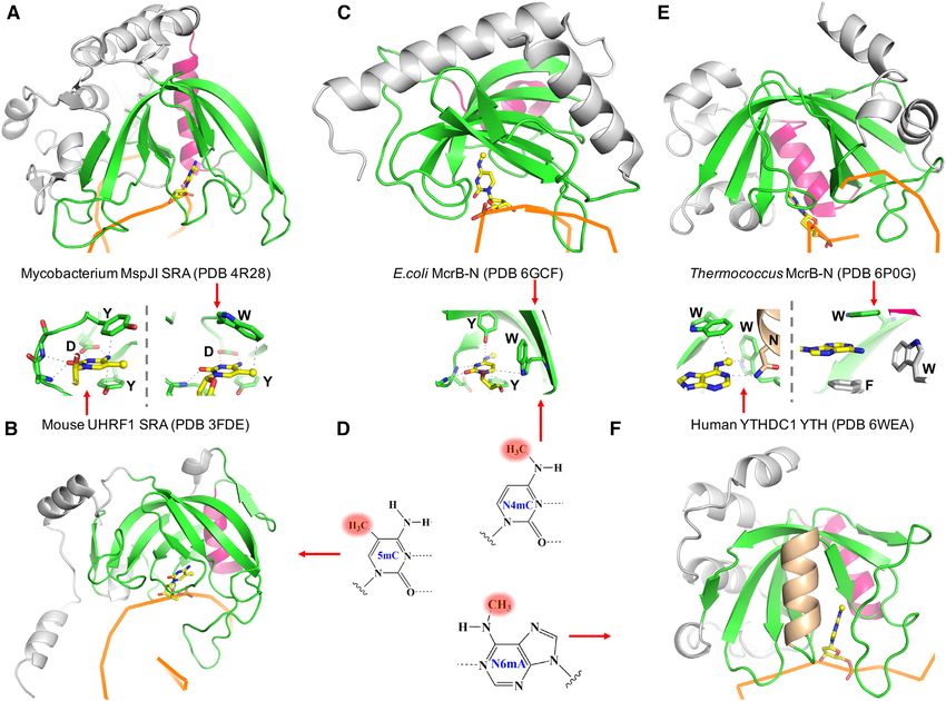
Nucleic Acids Research, 2020, Vol. 48, No. 18 10331
of TEV protease was added to cleave 6xHis tag for 15 h trations, were loaded into Monolith specific capillary tubes.
at 4◦ C while undergoing two rounds of dialysis in 3.5 kDa The signals had below 5% error among samples with re-
Snakeskin dialysis tubing (Thermo Scientific). The cleaved gards to the fluorescence and a fluorescence intensity of 500
products (with an additional N-terminal glycine) were counts using 40% power on a medium MST setting. The
passed through a subtractive HisTrap column. YTHDC1 MST data was fitted using Mo.Affinity Analysis software
interacted with the column and was eluted with a low (NanoTemper) included with the instrument.
concentration of imidazole, whereas YTHDF1 was col- Electrophoretic mobility shift assays (EMSA) were per-
lected in the flow through. The proteins were concentrated formed with the same set of samples used in the MST assays.
to 2 ml and loaded onto a Superdex S75 10/300 GL An aliquot of 5 l of complex was loaded into 6% native
equilibrated with buffer C (20 mM Tris pH 7.5, 150 mM PAGE gels, and ran at 100 V for 30 min at 4◦ C in 0.5× TBE
NaCl) with 1 mM tris(2-carboxyethyl)phosphine (TCEP). buffer. The images were collected using a BioRad Chemi-
The samples eluted as a symmetrical peak at an elution Doc imager in fluorescence mode.
volume consistent with a monomer.
Downloaded from https://academic.oup.com/nar/article/48/18/10329/5871368 by guest on 14 December 2020
For GST-tagged YTH domain of YTHDF2, the clarified
supernatant after cell lysate was passed through a gravity Crystallography
fed GST column with a volume of 10 ml at room tempera- The Art Robbins Gryphon Crystallization Robot was used
ture, washed with 170 ml of buffer A and the GST tag was to set up 0.4-l sitting drops (0.2 l of complex plus 0.2
cleaved on column with thrombin protease at room temper- l of well solution) at ∼19◦ C of the protein–DNA com-
ature for 2 h before elution. The cleaved product has no ad- plexes. A crystallization hit was observed using the 11 nt
ditional residue at the N-terminus but have an additional oligo 5 -CGCGGACTCTG-3 (A = N6mA) in a complex
five residues at the C-terminus (RPHRD). The samples were with YTH domain of YTHDC1 (10 mg/ml in 1:2 molar ra-
concentrated and loaded onto a S75 size exclusion column tio of protein to DNA) in 0.1 M Bis–Tris pH 6.0, 0.2 M am-
as described above. Supplementary Figure S1D shows ex- monium sulfate and 29% (w/v) polyethylene glycol (PEG)
amples of purified YTH domains used in the study. 3350. The crystals appeared after ∼15 days incubation at
19◦ C. The crystal shape was a consistent flat parallelogram,
and suffered from minor crystal layering (Supplementary
Binding assays of protein-nucleic acids interaction
Figure S1A).
DNA and RNA oligonucleotides containing N6mA or At molar ratios of 2:1 YTH to DNA, protein only crystals
FAM tagged oligos were synthesized by B. Baker of New appeared within 24 h with 0.1 M ammonium sulfate, 0.1–
England Biolabs, Inc., and unmodified oligos were pur- 0.15 M Bis–Tris pH 5.5 and 23–29% PEG 3350. The protein
chased from Integrated DNA Technologies. Nucleic acid only crystals were very birefringent (Supplementary Figure
samples were suspended in buffer C (without TCEP) to S1B). Because the YTH domain contains a hydrophobic
final stock concentrations of 4 mM (ss oligo) and then cage consisting of two tryptophan residues interacting with
some annealed using a thermocycler to 2 mM (ds oligo). methylated adenine (see Results), the crystals of the protein-
We used three binding assays to characterize the binding DNA complex had no UV fluorescence from tryptophan
affinity of YTHDC1 to methylated oligodeoxynucleotides: (perhaps due to quenching) and little to no birefringence,
(i) isothermal titration calorimetry (ITC), (ii) MicroScale while the protein only crystals had strong Trp UV fluores-
Thermophoresis (MST) and (iii) electrophoretic mobility cence.
shift. The second crystallization attempt used a set of three
For ITC, protein samples (100 M in buffer C) were in- oligos from 9–10 nt with varied 3 sequence. A 10 nt 5 -
jected from the syringe into sample cells containing 10 M CGCGGACTTC-3 (A = N6mA) incubated with YTH
of the 11-mer oligonucleotides in the same buffer (DNA: crystallized within 12 h at 19◦ C as heavily layered stacks
5 -CGCGGACTCTG-3 or RNA: 5 -CGCGGACUCUG- with time became circular discs. This complex (in 1:2 ratio
3 where A = N6mA), using the automated PEAQ-ITC of protein to DNA) was readily crystallizable under hun-
(Malvern Instrument Ltd). The experiments were con- dreds of conditions. The problem of heterogeneous nucle-
ducted at 25◦ C with 19 injections at 2 l each, or 13 injec- ation was resolved by generating a screen using the opti-
tions (2.9 l) if more heat was required, with a reference mal crystallization condition (0.1 M Bis–Tris pH 7.0, 0.24
setting of 10 cal/s. Binding constants were calculated by M ammonium sulfate, 23% PEG 3350) with increasing con-
fitting the data using the ITC data analysis module ‘one set centration of glycerol as an additive. Individual crystals
of sites’ supplied by the manufacturer. emerged starting at ∼10% glycerol and with an eventual sin-
MST experiments were conducted using a Mono- gle crystal per crystallization drop at 20% (Supplementary
lith NT.115 microscale thermophoresis instrument from Figure S1C).
NanoTemper Technologies. A 2-fold serial dilution of pro- Single crystals were flash frozen in liquid nitrogen by
tein was produced starting at 20 M in buffer C with 0.05% equilibrating in a cryoprotectant buffer containing the crys-
(v/v) added Tween 20 (Sigma-Aldrich). Varied protein con- tallization solution and 25% (v/v) ethylene glycol. X-ray
centrations (total of 16 samples) in 0.2 ml PCR tubes were diffraction data were collected at the SER-CAT beamline
mixed with an equal volume of the 6-carboxy-fluorescein 22ID of the Advanced Photon Source at Argonne National
(FAM)-labeled 11-mer oligonucleotide at 20 nM in buffer Laboratory. Crystallographic datasets were processed with
C and matching 0.05% Tween 20. Approximately 10 l of HKL2000 (44). Molecular replacement was performed with
the mixed samples, at half their respective starting concen- the PHENIX PHASER module (45) by using the known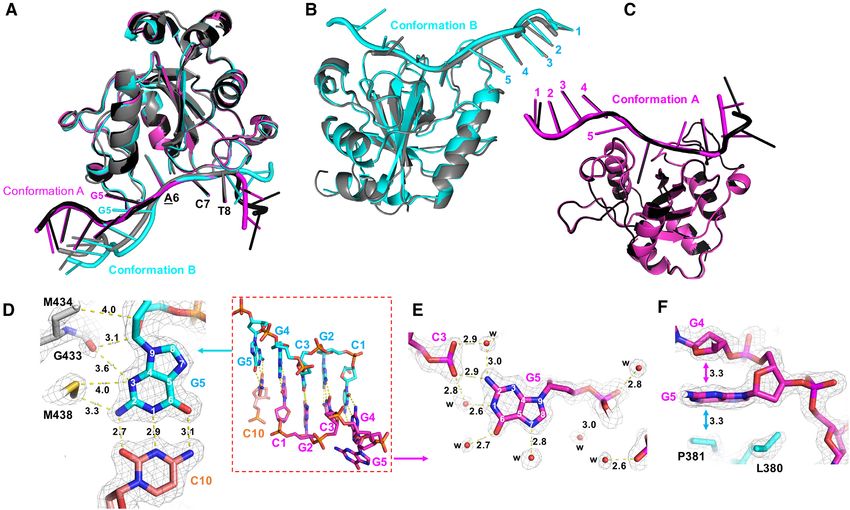
10332 Nucleic Acids Research, 2020, Vol. 48, No. 18

structure of the human YTHDC1 YTH domain (PDB ID               Overall structure of YTHDC1 YTH domain bound to methy-
4R3H) as a search model. Structure refinement was per-         lated DNA
formed with PHENIX Refine (46) with 5% randomly cho-
                                                               We next sought to understand how the YTH domain binds
sen reflections for the validation by the Rfree value. COOT
                                                               methylated DNA, and why it recognizes N6mA preferen-
(47) was used for the manual building of the structure model
                                                               tially in the context of DNA over RNA. Accordingly, we co-
and corrections between refinement rounds. DNA models
                                                               crystallized the YTH domain of YTHDC1, with the same
were built into difference density during the first several
                                                               11-nt ssDNA oligo (5 -CGCGGACTCTG-3 ) containing a
rounds of refinement for the two complex structures. Struc-
                                                               centrally located N6mA that we had used for the binding
ture quality was analyzed during PHENIX refinements and
                                                               studies. The complex crystallized in space group C2, result-
finally validated by the PDB validation server. Molecu-
                                                               ing in a structure determined to resolution of 1.59 Å (Sup-
lar graphics were generated by using PyMol (Schrödinger,
                                                               plementary Table S1). In the crystallographic asymmetric
LLC).
                                                               unit, there are two complexes (A and B) with each YTH

                                                                                                                                  Downloaded from https://academic.oup.com/nar/article/48/18/10329/5871368 by guest on 14 December 2020
                                                               domain binding a piece of ssDNA (Figure 2A). Interest-
                                                               ingly, with the 11-nt utilized for crystallization, the 5 end
RESULTS
                                                               CGCG sequence base pairs with the corresponding neigh-
YTH binding to single-strand DNA containing N6mA               boring DNA molecule, forming a 4-base pair duplex (Fig-
                                                               ure 2B). However, three nucleotides at the 3 end have poor
We used three independent methods, all of which revealed
                                                               electron density for the bases in complex A or are com-
that the binding of human YTHDC1 s YTH domain, to a
                                                               pletely disordered (no visible density) in complex B due to
11-nt single-strand (ss) oligodeoxynucleotide (DNA oligo)
                                                               the lack of direct crystallographic packing interactions.
containing a single N6mA in the context of GGACT, had
                                                                  To improve the quality of electron density for the 3 end,
higher affinity than its binding to the corresponding ssRNA
                                                               we designed a series of ssDNA oligos varying in length
of the same length and sequence (containing GGACU).
                                                               and/or sequence, and screened crystallizations for differ-
The DNA GGACT and RNA GGACU were chosen as
                                                               ent space groups with different morphology (Supplemen-
they are the equivalent DNA or RNA recognition sequence
                                                               tary Figure S1C). A complex formed with a 10-nt ssDNA
of MettL3-MettL14 (26,48). We first used ITC to quantita-
                                                               oligo (5 -CGCGGACTTC-3 ) crystallized in space group
tively measure the dissociation constants (KD ). The YTH
                                                               P21 , where we observed ordered electron density for all 10
domain bound the methylated DNA oligo with a KD of
                                                               nucleotides (Figure 2C). There are two copies of each com-
10 nM (Figure 1A). Under the same conditions, the mod-
                                                               plex (A and B), resulting in a total of four complexes within
ified RNA oligo exhibited 5-fold reduced binding affinity
                                                               the P21 asymmetric unit. The ssDNA bound by molecule
(Figure 1B). Because this observation was somewhat un-
                                                               A stacks head-to-tail with neighboring molecules on both
expected, we repeated the binding experiments with an-
                                                               ends, forming a pseudo-continuous ssDNA molecule with
other biophysical technique, MST, to measure the strength
                                                               a joint sequence of (5 -C10 -C1 G2 C3 G4 -3 ) (Figure 2D). The
of interaction between the YTH and oligos. Very similar to
                                                               DNA bound by molecule B provides the five pairing nu-
the ITC measurements, we observed a 9 nM binding affin-
                                                               cleotides (5 -CGCGG-3 ) for the joint sequence (Figure
ity for the modified DNA oligo, with 5.5× reduced affin-
                                                               2E), thus generating a continuous lattice in the crystal
ity for the corresponding RNA oligo (Figure 1C, D). The
                                                               with a 5-base pair duplex flanking the single-strand re-
same samples used for the MST assays were then used for
                                                               gion where YTH binds the methylated adenine (Figure 2F).
an electrophoretic mobility shift assay (EMSA), and again
                                                               Whereas the phosphate group at the joint of the two DNA
confirmed the enhanced affinity for the methylated DNA
                                                               molecules, connecting C10 and C1 , is of course missing, the
oligo relative to the RNA control (Figure 1E, F). Under the
                                                               five G:C base pairs form three classic Watson–Crick hydro-
same conditions, we observed no measurable binding to un-
                                                               gen bonds, respectively, that are 2.8–2.9 Å apart (Figure
modified ssDNA or ssRNA oligos, or to double-stranded
                                                               2G).
(ds)DNA or RNA/DNA hybrid oligos containing N6mA
on one strand (Supplementary Figure S2A).
                                                               Two structural conformations of YTH-bound DNA
   For comparison, we also purified the corresponding
YTH domains from YTHDF1 and YTHDF2 and mea-                    The protein components of the four complexes (A and B
sured their binding affinities to the same sets of DNA or      in both space groups) are highly similar, with root-mean-
RNA oligos by ITC (Figure 1G and Supplementary Fig-            square deviations of 0.1–0.5 Å across 135 pairs of C␣ atoms
ure S2B-D). These three proteins have distinct but overlap-    (Figure 3A). The largest deviation involves complex B in
ping binding specificities on RNA (49). The binding affini-    space group C2, where the 3 nucleotides are disordered and
ties for the methylated ssRNA by the three YTH domains         not modeled due to lack of electron density (colored grey in
were similar, in the range of 50–80 nM (Figure 1G). How-       Figure 3B). Among the four bound DNA molecules, only
ever, the three YTH domains bound to the DNA molecule          three nucleotides adopt the same conformations: N6mA at
very differently. While YTHDC1 bound DNA 5× more               position 6, and the two 3 nucleotides at positions 7 and
strongly than to RNA, YTHDF1 and YTHDF2 exhibited              8 (C7 and T8) (Figure 3A). The remaining nucleotides ex-
the opposite effect by binding DNA 1.4–2.2× weaker than        hibit varied conformations, depending on whether they are
to RNA. Significantly, YTH domain of YTHDC1 bound              involved in base pairing. The two complexes can be distin-
N6mA-containing DNA more strongly by a factor of 11 or         guished by the guanosine at position 5 (G5) and whether it
18, respectively, than did the corresponding YTH domains       is involved in base pairing (conformation B) or not (confor-
of YTHDF1 and YTHDF2.                                          mation A) (Figure 3B, C).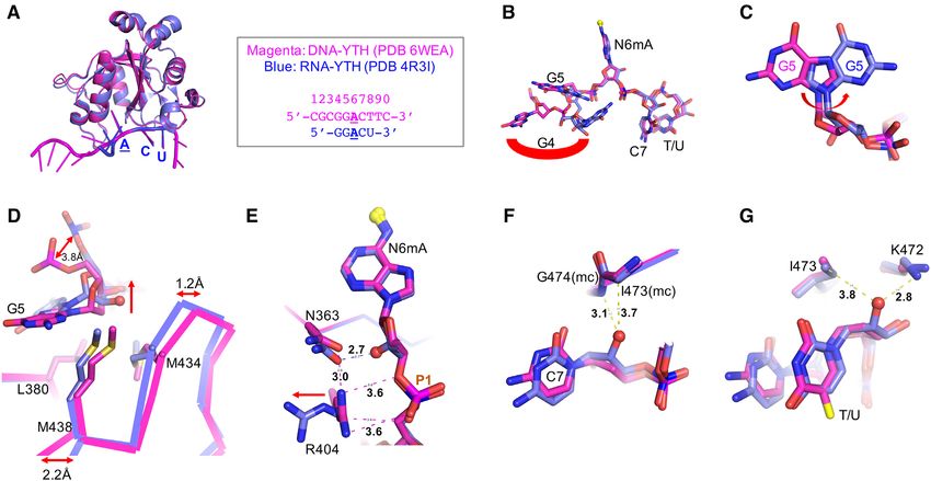
Nucleic Acids Research, 2020, Vol. 48, No. 18 10333

     A                               B                             C                                      D

                                                                    E                                     F

                                                                                                                                                     Downloaded from https://academic.oup.com/nar/article/48/18/10329/5871368 by guest on 14 December 2020
                                                                    G

Figure 1. YTH binding methylated adenine in DNA. (A, B) ITC measurements of YTHDC1 binding methylated ssDNA (panel A) or methylated ssRNA
(panel B). (C, D) MST measurements and (E, F) electrophoretic mobility shift assays of YTHDC1 binding to oligos containing a single N6mA in DNA or
RNA. (G) Summary of KD values of three YTH domains (see Supplementary Figure S2). Data represent the mean ± SD of N independent determinations
(N = 2 for ITC and N = 3 for MST).

   In conformation B, guanosine G5 pairs with cytidine C10                 pacity of polar atoms on the adenosine ring and, together
of the neighboring DNA molecule (Figure 3D). Three pro-                    with van der Waals contacts with the methyl group, defines
tein residues (Met438, Gly433 and Met434) make van der                     the specificity for the methylated adenosine in the binding
Waals contacts with N2 and N3 atoms of the guanosine G5                    pocket.
base and its ribose ring (Figure 3D). In conformation A, the                  The next nucleotide, cytidine C7, is stacked in-between
guanosine G5 base makes intra-molecular interactions with                  the side-chain of Arg475 and the following thymidine T8 of
the phosphate group bridging between nucleotides G2 and                    the same strand, both of which are solvent exposed (Fig-
C3, the N2 atom of the guanosine G5 makes a direct hy-                     ure 4E). In addition, cytidine C7 makes a direct but weak
drogen bond with one of the phosphate oxygen atoms, and                    hydrogen bond (3.2 Å) with the main-chain amide nitrogen
there are two water-mediated interactions with the same                    atom of Gly474, and a water-mediated interaction with the
phosphate group (Figure 3E). In addition, the guanosine                    side chain of Asn466 (Figure 4F). Like guanosine G5, the
ring of G5 stacks with two 5-membered rings–the ribose of                  interactions with cytidine C7 are not base-specific recogni-
the preceding guanosine G4 and the proline ring of Pro381                  tion. Interestingly, the guanidine group of Arg475 occupies
(Figure 3F). While the interactions with the guanosine G5                  the position where the N6mA would normally be located if
seen in the two conformations do not provide base specific                 it stacks with neighboring nucleotides (Figure 4G).
recognition, whether or not G5 is stacked with the 5 bases,                  We note that numerous genome-wide studies did not pro-
these interactions with YTH residues presumably enhance                    duce an agreeable consensus DNA sequence containing
the binding affinity.                                                      N6mA from mammalian cells: sequences reported include
                                                                           TTTTTAGAAGC or TACA[A/G]GA in mouse ES cells
                                                                           (9,10), [G/C]AGG[C/T] in a human genome (especially in
Interactions with N6mA-containing DNA                                      the mitochondria) (11), TGGATGGA in human glioblas-
The methylated adenosine N6mA is inserted into a hy-                       toma (13), and CT[T/C/A]ATC in human mitochondria
drophobic pocket (Figure 4A), where the base ring is                       (21). From the face value of these studies, it seems that
stacked between Trp377 on one side and Leu439 and                          the sequences immediately before and after N6mA are both
Met434 on the other side (Figure 4B). At the bottom of                     variable. In the current structures with bound ssDNA, the
the pocket lies the indole ring of Trp428, juxtaposed to the               nucleotides (G5 and C7) before and after N6mA are not en-
methyl group of N6mA (Figure 4B). The adenosine ring is                    gaged in base specific interactions with YTH. We modeled
involved in four hydrogen bonds with, respectively, the side-              the other three nucleotides (G/A/T) at position 7 and cal-
chain of Asn367 (via Ade N1), the main-chain amide ni-                     culated binding interfaces between YTH and modeled nu-
trogen atoms of Asn363 (via Ade N3), the main-chain car-                   cleotides (Supplementary Figure S3). The differences in the
bonyl oxygen atom of Ser378 (via Ade N6 amino group),                      binding interface are negligible.
and a water molecule (via Ade N7) (Figure 4C). The wa-
ter molecule is trapped at the protein-DNA interface, tetra-
                                                                           YTH–DNA phosphate interactions
hedrally coordinated with the Ade N7, the indole nitrogen
of Trp377, the hydroxyl oxygen of Thr379, and one of the                   There are direct protein–DNA phosphate contacts concen-
carboxylate oxygen atoms of Asp476 (Figure 4D). This pat-                  trated on the three phosphates 3 to the N6mA (labeled
tern of interactions fully saturates the hydrogen-bonding ca-              as P1, P2 and P3 in Figure 4H). Three positively-charged
10334 Nucleic Acids Research, 2020, Vol. 48, No. 18
Downloaded from https://academic.oup.com/nar/article/48/18/10329/5871368 by guest on 14 December 2020
Figure 2. Overall structures of YTH-DNA complexes in two space groups. (A) Two complexes of YTH-11mer DNA in space group C2 (PDB 6WE9). (B)
The 5 CGCG of two neighboring DNA molecules form 4-bp duplex. (C) Ten nucleotides of DNA used in space group P21 (PDB 6WEA), superimposed
with an omit Fo-Fc electron density map (orange) contoured at 4 above the mean by omitting the entire 10-nt DNA. (D) A long pseudo ssDNA formed
by stacking the 3 cytosine C10 to the 5 end of neighboring molecule. (E) The DNA (cyan) bound with molecule B provides the five pairing nucleotides for
the joint sequence of DNA (magenta) bound with molecule A. (F) Crystal lattice in space group P21 showing YTH (in gray) binds the methylated adenine
in the single-strand region flanked with 5-base pair duplexes. (G) Example of G4:C1 base pair superimposed with an omit Fo – Fc electron density map
(light gray) contoured at 4 above the mean. The numerical numbers are interatomic distances in Å.
residues (Arg475, Lys361 and Lys472) lie on the highly- (jawless fish), the other six vertebrate classes had clear or-
basic surface (Figure 4A), and interact directly with the thologs for all three proteins, with some positions conserved
three respective negatively-charged phosphate groups (Fig- across both classes and YTH proteins. Of the DNA con-
ure 4I-K). In addition, the main chain amide nitrogen atoms tacts in YTHDC1 YTH domain, 4/8 (50%) that contact
of Asp476 and Glu405 form hydrogen bonds with, respec- N6mA involve positions highly conserved across the three
tively, one of the non-bridging oxygen atoms of the P1 proteins in six classes (Ser362, Asn363, Asn367, Trp377,
and P2 phosphate groups (Figure 4I, J). In addition to Ser378, Thr379, Trp428 and Asp476; invariant residues in
these direct protein–DNA interactions, a complex network bold), while this is true for just 3/12 (25%) that make other
of ordered water molecules interconnects bases, phosphate DNA contacts (Lys361, Leu380, Pro381, Glu405, Gly433,
groups, and amino acids. In the case of P3, protein atoms Met434, Met438, Leu439, Asn466, Lys472, Gly474 and
might function as one of the connecting groups in a water Arg475). The invariant residues concentrated in construc-
network surrounding the charged phosphate group (Figure tion of the binding site provide direct binding to the methyl
4L). group of N6mA (Trp377 and Trp428; Figure 4B), the
trapped water molecule (Asp476; Figure 4D) and the cyti-
dine immediately 3 to N6mA (Lys361, Asn466 and Arg475;
Figure 4E–H). This suggests that recognition of the methy-
Evolutionarily-conserved interaction with N6mA
lated base and binding of the 3 cytosine are particularly
Interestingly, the residues involved in the contacts to N6mA well-conserved among YTH proteins, and are consistent
are more highly conserved than the other DNA contacts with the extensive structural recognition and binding of the
(Supplementary Table S2). While there were no obvious or- two nucleotides (N6mA6 -C7 ) described earlier in this sec-
thologs for YTHDC1, YTHDF1 or YTHDF2 in Agnatha tion.Nucleic Acids Research, 2020, Vol. 48, No. 18 10335
Downloaded from https://academic.oup.com/nar/article/48/18/10329/5871368 by guest on 14 December 2020
Figure 3. Two YTH-bound DNA conformations. (A) Superimposition of four complexes, A and B (black and dark gray) in space group C2 (PDB: 6WE9)
and A and B (magenta and cyan) in space group P21 (PDB: 6WEA). (B) Superimposition of two B complexes in space groups C2 (gray) and P21 (cyan). (C)
Superimposition of two A complexes in space groups C2 (black) and P21 (magenta). (D) Guanine G5 of complex B involves in base pairing. (E, F) Guanine
G5 of complex A involves in intra-molecular interaction and protein–DNA interaction. The omit Fo – Fc electron density map (light grey) contoured at
4 above the mean. Panels D–F are depicted from the structure (PDB: 6WEA).
Comparison between DNA and RNA bound YTH domains and does not engage in any interactions. The addition of
2 -OH to G5 in RNA moves the ribose ring more than 1
The YTH domain of YTHDC1 had been structurally char-
Å away from the protein relative to its position in DNA
acterized in complex with a 5-nt RNA (5 -GGACU-3
(Figure 5D). In DNA, three hydrophobic residues (Leu380,
where the underlined A is methylated at the N6 atom)
Met434 and Met438) encompass the deoxyribose C2 atom.
(PDB 4R3I (50)). [YTHDC1 also binds RNA containing
A loop in the RNA complex, that contains Met434 and
A methylated at the N1 atom (51) but, as this does not oc-
Met438, undergoes movement of between 1.2 Å (residue
cur normally in DNA, we have not studied it here.] Super-
prior to Met434) and 2.2 Å (residue after Met438) rela-
imposition of RNA- and DNA-bound YTH domains re-
tive to that of the DNA-bound conformation (Figure 5D).
sulted in highly similar structures, having a RMSD of just
This loop movement is significant considering the overall
0.25 Å across 130 pairs of C␣ toms (Figure 5A). For the
RMSD of 0.25 Å between the two YDH complex structures.
RNA component, the 3 ACU is superimposable with the
Furthermore, in the 1.18 Å-resolution structure of the apo-
corresponding DNA ACT, whereas the 5 guanosine (G4)
YTH domain (Supplementary Table S1), the corresponding
points in completely different directions in the two struc-
loop in the absence of DNA is less ordered and contains
tures (Figure 5B). The internal (Ade-adjacent) guanosine,
discontinuous electron density in the main-chain between
corresponding to G5 of the DNA used in our study, has a
Gly433 and Lys437, as well as in the side chains of Met434
similar backbone conformation in both structures, but the
and Met438, suggesting that the loop is stabilized by the
guanosine ring is rotated ∼180◦ C along the glycosidic bond
bound DNA substrate (Supplementary Figure S4A).
between the base and sugar group (Figure 5C). The confor-
The inclusion of a 2 -OH on the methylated deoxy-
mational changes in RNA between these two 5 guanosine
adenosine (N6mA) disrupts the Arg404–Asn363 interac-
nucleotides (G4 and G5) resulted in the loss of protein inter-
tion, which runs in parallel and makes van der Waals con-
actions described above for the DNA conformations (Fig-
tacts with the sugar-phosphate backbone in the DNA-
ure 3), presumably partly responsible for reduced affinity of
bound conformation (Figure 5E). In the absence of
the RNA binding.
DNA, the Arg404 interacts with a negatively-charged sul-
We next examined effects of the presence (ribose) or ab-
fate group (used for crystallization), which occupies an
sence (deoxyribose) of the 2 -hydroxyl group (OH) in the
equivalent position of the P1 phosphate group (Sup-
backbone sugar of the two equivalent sequences. The 2 -
plementary Figure S4A). The comparison among the
OH of guanosine G4 in RNA points toward the solvent10336 Nucleic Acids Research, 2020, Vol. 48, No. 18
Downloaded from https://academic.oup.com/nar/article/48/18/10329/5871368 by guest on 14 December 2020
Figure 4. YTH interaction with N6mA (PDB 6WEA). (A) YTH domain contains a hydrophobic pocket (circled in dashed line) next to a basic surface.
The surface charge at neutral pH is displayed as blue for positive, red for negative, and white for neutral. (B) An aromatic cage for binding N6mA. (C)
Interactions with N6mA in the pocket. (D) A trapped water molecule has tetrahedral coordination. (E) Arg475 stacks with cytosine C7. (F) Interaction
with cytosine C7. (G) Arg475 occupies the position where the N6mA would be normally located if it stacked with the neighboring nucleotide. (H) Three
phosphate groups (P1, P2 and P3) 3 to N6mA interaction with protein. (I) Arg475 interaction with the P1 phosphate group and a SO4 molecule used
in crystallization. (J) Lys361 interaction with the P2 phosphate group. (K) Lys472 interaction with the P3 phosphate group. (L) An example of a water
network surrounds phosphate groups. The 2Fo – Fc electron density map (light gray) is contoured at 2 above the mean.
three structures––YTH alone, YTH-DNA and YTH- fied adenosine (N6mA) and its following nucleotide (Sup-
RNA––suggests that the Arg404–Asn363 bridge is unique plementary Figure S5). We note that the one of the ma-
to the deoxyribose of DNA-bound form. In contrast, the jor difficulties in the structural comparisons between DNA-
conserved aromatic cage for binding the methylated adenine bound and RNA-bound forms of YTH domains is due to
base of DNA or RNA is rigid, and adopts the same confor- the shorter nucleotides observed in the RNA-bound struc-
mation with and without bound substrate (Supplementary tures: by 5-nt in YTHDC1 and 4-nt in YTHDF1. Thus,
Figure S4B). the effect of longer strands on the geometry of nucleotides
Finally, 2 -OH groups on cytidine C7 and uridine at posi- (i.e. secondary and tertiary RNA structure) surrounding
tion 8 in RNA ligands are accommodated without the need N6mA cannot be addressed in the current comparison. We
for protein conformational adjustment (Figure 5F and G). also note that our ITC data (Figure 1) do not support an en-
Taken together, the addition of 2 -OH groups in RNA re- thalpy difference between the binding modes of DNA and
sulted in three circumstances: (i) accommodation without RNA by the YTH domain of YTHDC1.
changes in the protein conformation (at C7 and U8), (ii)
disruption, unique to DNA, of intra-molecular interactions
between two protein residues (at N6mA) and (iii) repulsion Connection of YTH to bacterial (modified cytosine restric-
between protein and RNA (at G5). The differences, in the tion) McrBC
conformations of nucleotides G4, G5 (Figure 3D and F), SET and RING finger-associated (SRA) domains (Figure
and N6mA at position 6 (Figure 5E), and their varied in- 6A, B) were originally characterized in mammalian pro-
teractions with the YTH domain of YTHDC1, might ex- teomes as readers of hemi-methylated CpG sequences – that
plain the limited but significant increase in binding affinity is, sequences containing 5mC in only one strand, such as
for DNA over RNA (Figure 1). arise following DNA replication (53,54). SRA domains are
In addition, we compared YTHDC1-DNA interactions also widespread in bacterial species, often associated with
with that of YTHDF1-RNA (52). The similarity between modification-dependent restriction endonucleases ((55,56)
the two sets of interactions lies in the binding of modi- and references therein). We suggest that, like the SRA do-Nucleic Acids Research, 2020, Vol. 48, No. 18 10337
Downloaded from https://academic.oup.com/nar/article/48/18/10329/5871368 by guest on 14 December 2020
Figure 5. Comparison between DNA- and RNA-bound YTH. (A) Superimposition of complex A in space group P21 and RNA-YTH complex (PDB:
4R3I). The DNA ACT (magenta) and RNA ACU (blue) are superimposable. (B) Difference in guanine G4 binding. (C) Difference in guanine G5 binding.
(D) The effect of 2 -OH group of G5 pushes RNA molecule away. (E) The effect of 2-OH group of N6mA interrupts an Arg404-Asn363 interaction. (F, G)
The 2-OH groups of cytosine C7 and uracil U8 have no visible impact on YTH binding.
main itself, the YTH domain may also have bacterial ori- with about the same affinity to either double-stranded or
gins. McrBC (modified cytosine restriction) is an antiphage single-stranded DNA containing N6mA, with an increased
defense system of E. coli, which specifically cleaves in- affinity relative to its binding to unmodified or 5mC-
vading DNA that is methylated in specific ways (57–59). containing oligos. In contrast, it shows little affinity for
The McrBC restriction system consists of two distinct sub- RNA substrates, whether they are methylated or unmethy-
units: McrB, comprising an N-terminal DNA binding do- lated. Looking at these various modification-dependent
main (McrB-N) and a C-terminal AAA+ ATPase domain binding domains together, they share a common structural
for GTP hydrolysis, and McrC, harboring an endonucle- feature - a twisted anti-parallel -sheet that forms an arch-
ase domain (60). The McrB-N DNA binding domain rec- like structure, with a variable number of helices packed
ognizes DNA containing modified cytosine residues (61) against the outer surface (Figure 6). The inner surface of the
and flips out of the duplex various modified cytosines in- arch, where DNA is bound, contains an aromatic pocket
cluding N4mC, 5mC and 5-hydroxymethylcytosine (5hmC) which defines the binding site of the modified nucleotide
(58,59) (Figure 6C). This base flipping is independent of (Figure 6), which is flipped out from the DNA duplex if
the sequence context of the target modified base. Inter- the substrates are double-strand DNA. A common helix lies
estingly McrB-N binds cytosine derivatives in the order in the back of the arch (colored red in Figure 6), perhaps
of descending affinity of N4mC > 5mC > 5hmC (59). functioning as a gatekeeper to control the access to the aro-
There is no mammalian protein domain currently known matic cage. The YTH domain of YTHDC1 contains an ad-
to bind N4mC; but MettL15 introduces N4mC in rRNA ditional helix in front of the arch (colored brown in Figure
(62). However, the DUF55 domain of human thymocyte 6F). The unique features for each domain lie in the num-
nuclear protein 1 (THYN1; also known as Thy28) shares a ber and length of their -strands. Some strands are as long
structural fold with the YTH domain (63,64), and has been as 20-residues and curved, responsible for the arch-like ap-
suggested to bind DNA containing modified cytosine (65), pearance. A longer strand can be disrupted into two shorter
while YTHDF2 binds 5mC in RNA (66). We note that the strands linked by an inserted bulged segment. We consider
methyl groups of N4mC and N6mA share their location on the SRA and YTH domains as diverse members of a larger
the exocyclic amino groups of cytosine (at N4) and adenine family of DNA/RNA modification readers.
(at N6) (Figure 6D), and the MTases responsible for these
two types of amino (N4-cytosine and N6-adenine) methy-
DISCUSSION
lation belong to the same MTase family group (4,25).
Recently, the structure of the McrB-N domain from the Here we characterized, for the first time, the in vitro bind-
archaeon Thermococcus gammatolerans revealed a strik- ing activity of the YTH domain of human YTHDC1 as
ing similarity to that of the eukaryotic YTH domain (67) preferentially targeting DNA, relative to RNA. YTH binds
(Figure 6E). This YTH-like DNA binding domain binds N6mA in the context of ssDNA, an activity comparable to10338 Nucleic Acids Research, 2020, Vol. 48, No. 18
Downloaded from https://academic.oup.com/nar/article/48/18/10329/5871368 by guest on 14 December 2020
Figure 6. The 5mC-binding SRA domain and N6mA-binding YTH domain are members of a larger family of modified nucleotide binding proteins
associated with bacterial modification-dependent endonucleases. The conserved -strands are in green and one conserved helix (colored in red) is behind
the arch. The other ␣-helices are in gray. DNA strands are in ribbon with the flipped base in stick presentation. (A) MspJI SRA domain (with 5mC).
(B) mouse UHRF1 SRA domain (with 5mC). (C) E. coli McrB-N DNA binding domain (with N4mC). (D) Three kinds of methylated DNA bases. (E)
Thermococcus McrB-N in complex with a piece of dsDNA (with Ade). We note that a 19-bp duplex DNA was used for co-crystallization with Thermococcus
McrB-N (67): a piece of 6-bp was observed that aligned tail-to-end in the crystal lattice and left no space for the missing two thirds of remaining oligo
duplex. There are discrepancies of bases in the structure deposited in the PDB (6P0G) and that described in the publication (67): 5mC vs C on one strand
and T versus C on the other strand. Nevertheless, the flipped base is an unmodified adenine. (F) Human YTHDC1 YTH domain (with N6mA) has a
unique helix (colored in brown) in front of the arch. The modified bases are bound in a cage formed by 2–3 aromatic residues.
that found in the human enzymatic activities of the MettL3– pair. Furthermore, the existence of these transient non-B
14 MTase complex and the demethylase Alkbh1, as they DNA structures is prolonged under physiological or biolog-
each act on damaged or unpaired DNA (26,37). Additional ical stresses. For example, the accumulation of replication-
study will be required to address whether YTHDC1 is in- associated ssDNA gaps has been observed in tumor cells
volved in the recognition of DNA adenine methylation in (69–71). Accumulation of R-loops, a specific DNA–RNA
vivo and, if so, its impact on chromatin organization. hybrid with an unpaired ssDNA strand formed during tran-
However, the following considerations suggest the hy- scription, is associated with disease in the context of cel-
pothesis that genomic N6mA is associated with maintain- lular stress (72,73), and N6mA-associated R-loop accumu-
ing genome stability. First, upon ultraviolet irradiation, the lates during the cell cycle (43). Resolution of the four-way
human MTase complex MettL3-L14 is recruited within 2 Holliday junction, the central intermediate of recombina-
min to the damaged sites, and MettL3 methylation activity tion, results in two dsDNA helices linked by a ssDNA re-
is required for the DNA repair (31). Second, while DNA gion containing a single base gap (74) (Supplementary Fig-
sequences are normally base paired in a canonical dou- ure S6B); and the gapped DNA is anisotropically bent (75).
ble helix, transient local unwinding of dsDNA occurs dur- Perhaps cells respond to the inherent topological stress that
ing the processes of transcription (forming a transcriptional arises from non-B DNA by coating ssDNA with protective
bubble (68); Supplementary Figure S6A), replication (such protein complexes, that include the stress-induced writer-
as the single-strand regions between Okazaki fragments in reader-eraser of N6mA-containing ssDNA. This would be
the legging strand synthesis), recombination, and DNA re- consistent with the requirement of MettL3 methylation ac-Nucleic Acids Research, 2020, Vol. 48, No. 18 10339
tivity for UV-induced DNA repair (31). A single-nucleotide- ACKNOWLEDGEMENTS
resolution sequencing study revealed N6mA clusters associ-
We thank Dr Tao Wu of Baylor College of Medicine for
ated with single-strand DNA binding protein on the human
discussion.
mitochondrial genome (12).
Authors contributions: C.B.W. performed protein purifica-
The appearance of ssDNA has been observed in E.
tion, DNA binding assays and crystallization. J.R.H. per-
coli as well as mammalian cells. For example, ssDNA can
formed X-ray data collection and structure determination.
be induced by stress-induced DNA duplex destabilization
J.Z. helped with protein purification. M.T.B. made the ini-
(SIDD) in E. coli (76), and ssDNA is a common feature of
tial suggestion for testing YTH binding methylated DNA
the mammalian genome potentially involved in gene reg-
and provided expression constructs. R.M.B. participated in
ulation (77). In E. coli, heterologous site-specific adenine
discussion and performed analysis on sequence conserva-
methylation can induce the SOS DNA repair genes due to
tion and assisted in preparing the manuscript. X.Z. and
the action of the modification-specific endonuclease Mrr
X.C. organized and designed the scope of the study.
(modified DNA rejection and restriction) (78). In Caulobac-
Downloaded from https://academic.oup.com/nar/article/48/18/10329/5871368 by guest on 14 December 2020
ter crescentus, the cell cycle-regulated DNA adenine MTase
(CcrM) binds DNA by strand-separation of dsDNA and FUNDING
creates a bubble at its recognition site (79) (Supplementary
Figure S6C). CcrM is active on both dsDNA and ssDNA U.S. National Institutes of Health (NIH) [R35GM134744
as well as mismatches within or immediately outside of the to X.C., R01GM126412 to M.T.B.]; Cancer Prevention and
recognition sequence (80,81). Research Institute of Texas (CPRIT) [RR160029 to X.C.
Finally, we note that many nucleic acid-modifying en- who is a CPRIT Scholar in Cancer Research]. Funding for
zymes are able to modify both DNA and RNA ((82) and open access charge: MD Anderson Cancer Center.
references therein). This group of enzymes include mem- Conflict of interest statement. M.T.B. is a cofounder of
bers of the AlkB family, involved in the direct reversal of EpiCypher. Others declare no competing interests.
alkylation damage to both DNA and RNA (83), and mem-
bers of the Apobec family of cytidine deaminases (84). Tet2, REFERENCES
one of the ten-eleven translocation proteins initially discov-
ered as DNA 5mC dioxygenases (85), mediates oxidation of 1. Ehrlich,M., Gama-Sosa,M.A., Carreira,L.H., Ljungdahl,L.G.,
Kuo,K.C. and Gehrke,C.W. (1985) DNA methylation in thermophilic
5mC in mRNA (86,87). Murine MettL4 was reported to be bacteria: N4-methylcytosine, 5-methylcytosine, and
responsible for N6mA deposition in genic elements, corre- N6-methyladenine. Nucleic Acids Res., 13, 1399–1412.
sponding with transcriptional silencing (10). Interestingly, 2. Sanchez-Romero,M.A., Cota,I. and Casadesus,J. (2015) DNA
recombinant human MettL4 expressed in HEK293T cells methylation in bacteria: from the methyl group to the methylome.
Curr. Opin. Microbiol., 25, 9–16.
has in vitro enzymatic activity on mitochondrial DNA (21), 3. Posfai,J., Bhagwat,A.S., Posfai,G. and Roberts,R.J. (1989) Predictive
whereas recombinant human MettL4 expressed in E. coli motifs derived from cytosine methyltransferases. Nucleic Acids Res.,
has RNA MTase activity (29). The former study showed 17, 2421–2435.
that MettL4 localizes with mitochondria in all tested tis- 4. Malone,T., Blumenthal,R.M. and Cheng,X. (1995) Structure-guided
sues (21) and the latter study found mainly nuclear local- analysis reveals nine sequence motifs conserved among DNA
amino-methyltransferases, and suggests a catalytic mechanism for
ization of an exogenously introduced MettL4, and failed these enzymes. J. Mol. Biol., 253, 618–632.
to identify appreciable levels of N6mA in mitochondrial 5. Cheng,X. (1995) Structure and function of DNA methyltransferases.
DNA (29). The difference among these various studies illus- Annu. Rev. Biophys. Biomol. Struct., 24, 293–318.
trates the complex nature of DNA vs. RNA adenine methy- 6. Bestor,T., Laudano,A., Mattaliano,R. and Ingram,V. (1988) Cloning
and sequencing of a cDNA encoding DNA methyltransferase of
lation in mammalian genomes, and the accumulation of mouse cells. The carboxyl-terminal domain of the mammalian
N6mA in DNA and/or RNA might reflect diverse cellu- enzymes is related to bacterial restriction methyltransferases. J. Mol.
lar and mitochondrial stress responses under different lab- Biol., 203, 971–983.
oratory conditions. The 5-fold difference in affinity, of the 7. Okano,M., Xie,S. and Li,E. (1998) Cloning and characterization of a
YTHDC1 YTH domain preference for DNA (Figure 1), family of novel mammalian DNA (cytosine-5) methyltransferases.
Nat. Genet., 19, 219–220.
therefore takes nothing away from the significance of YTH 8. Wyatt,G.R. (1951) Recognition and estimation of 5-methylcytosine in
protein activities on RNA but suggests an additional layer nucleic acids. Biochem. J., 48, 581–584.
of complexity involving mammalian DNA adenine methy- 9. Wu,T.P., Wang,T., Seetin,M.G., Lai,Y., Zhu,S., Lin,K., Liu,Y.,
lation and its potential role in maintaining genome stability. Byrum,S.D., Mackintosh,S.G., Zhong,M. et al. (2016) DNA
methylation on N(6)-adenine in mammalian embryonic stem cells.
Nature, 532, 329–333.
10. Kweon,S.M., Chen,Y., Moon,E., Kvederaviciute,K., Klimasauskas,S.
DATA AVAILABILITY and Feldman,D.E. (2019) An adversarial DNA
N(6)-methyladenine-sensor network preserves polycomb silencing.
The X-ray structures (coordinates and structure factor files) Mol. Cell, 74, 1138–1147.
of YTH domain with or without bound DNA have been 11. Xiao,C.L., Zhu,S., He,M., Chen,D., Zhang,Q., Chen,Y., Yu,G.,
submitted to PDB under accession number 6WEA (10- Liu,J., Xie,S.Q., Luo,F. et al. (2018) N(6)-Methyladenine DNA
modification in the human genome. Mol. Cell, 71, 306–318.
mer), 6WE9 (11-mer) and 6WE8 (no DNA). 12. Koh,C.W.Q., Goh,Y.T., Toh,J.D.W., Neo,S.P., Ng,S.B., Gunaratne,J.,
Gao,Y.G., Quake,S.R., Burkholder,W.F. and Goh,W.S.S. (2018)
Single-nucleotide-resolution sequencing of human
SUPPLEMENTARY DATA N6-methyldeoxyadenosine reveals strand-asymmetric clusters
associated with SSBP1 on the mitochondrial genome. Nucleic Acids
Supplementary Data are available at NAR Online. Res., 46, 11659–11670.10340 Nucleic Acids Research, 2020, Vol. 48, No. 18
13. Xie,Q., Wu,T.P., Gimple,R.C., Li,Z., Prager,B.C., Wu,Q., Yu,Y., 32. Wang,W., Xu,L., Hu,L., Chong,J., He,C. and Wang,D. (2017)
Wang,P., Wang,Y., Gorkin,D.U. et al. (2018) N(6)-methyladenine Epigenetic DNA modification N(6)-methyladenine causes
DNA modification in glioblastoma. Cell, 175, 1228–1243. Site-Specific RNA polymerase II transcriptional pausing. J. Am.
14. Ratel,D., Ravanat,J.L., Charles,M.P., Platet,N., Breuillaud,L., Chem. Soc., 139, 14436–14442.
Lunardi,J., Berger,F. and Wion,D. (2006) Undetectable levels of 33. Flusberg,B.A., Webster,D.R., Lee,J.H., Travers,K.J., Olivares,E.C.,
N6-methyl adenine in mouse DNA: Cloning and analysis of Clark,T.A., Korlach,J. and Turner,S.W. (2010) Direct detection of
PRED28, a gene coding for a putative mammalian DNA adenine DNA methylation during single-molecule, real-time sequencing. Nat.
methyltransferase. FEBS Lett., 580, 3179–3184. Methods, 7, 461–465.
15. Schiffers,S., Ebert,C., Rahimoff,R., Kosmatchev,O., Steinbacher,J., 34. Valinluck,V., Liu,P., Burdzy,A., Ryu,J. and Sowers,L.C. (2002)
Bohne,A.V., Spada,F., Michalakis,S., Nickelsen,J., Muller,M. et al. Influence of local duplex stability and N6-methyladenine on uracil
(2017) Quantitative LC-MS provides no evidence for m(6) dA or m(4) recognition by mismatch-specific uracil-DNA glycosylase (Mug).
dC in the genome of mouse Embryonic stem cells and tissues. Chem. Res. Toxicol., 15, 1595–1601.
Angewandte Chemie, 56, 11268–11271. 35. Lopez,C.M., Lloyd,A.J., Leonard,K. and Wilkinson,M.J. (2012)
16. Liu,B., Liu,X., Lai,W. and Wang,H. (2017) Metabolically generated Differential effect of three base modifications on DNA
stable isotope-labeled deoxynucleoside code for tracing DNA thermostability revealed by high resolution melting. Anal. Chem., 84,
Downloaded from https://academic.oup.com/nar/article/48/18/10329/5871368 by guest on 14 December 2020
N(6)-methyladenine in human cells. Anal. Chem., 89, 6202–6209. 7336–7342.
17. Musheev,M.U., Baumgartner,A., Krebs,L. and Niehrs,C. (2020) The 36. Song,Q.X., Ding,Z.D., Liu,J.H., Li,Y. and Wang,H.J. (2013)
origin of genomic N(6)-methyl-deoxyadenosine in mammalian cells. Theoretical study on the binding mechanism between
Nat. Chem. Biol., 16, 630–634. N6-methyladenine and natural DNA bases. J. Mol. Model., 19,
18. Liu,X., Lai,W., Li,Y., Chen,S., Liu,B., Zhang,N., Mo,J., Lyu,C., 1089–1098.
Zheng,J., Du,Y.R. et al. (2020) N(6)-methyladenine is incorporated 37. Zhang,M., Yang,S., Nelakanti,R., Zhao,W., Liu,G., Li,Z., Liu,X.,
into mammalian genome by DNA polymerase. Cell Res., Wu,T., Xiao,A. and Li,H. (2020) Mammalian ALKBH1 serves as an
doi:10.1038/s41422-020-0317-6. N(6)-mA demethylase of unpairing DNA. Cell Res., 30, 197–210.
19. Woodcock,C.B., Yu,D., Zhang,X. and Cheng,X. (2019) Human 38. Patil,D.P., Pickering,B.F. and Jaffrey,S.R. (2018) Reading m(6)A in
HemK2/KMT9/N6AMT1 is an active protein methyltransferase, but the transcriptome: m(6)A-binding proteins. Trends Cell Biol., 28,
does not act on DNA in vitro, in the presence of Trm112. Cell 113–127.
Discov., 5, 50. 39. Liao,S., Sun,H. and Xu,C. (2018) YTH domain: a family of
20. Li,W., Shi,Y., Zhang,T., Ye,J. and Ding,J. (2019) Structural insight N(6)-methyladenosine (m(6)A) readers. Genomics Proteomics
into human N6amt1-Trm112 complex functioning as a protein Bioinformatics, 16, 99–107.
methyltransferase. Cell Discov., 5, 51. 40. Tong,J., Flavell,R.A. and Li,H.B. (2018) RNA m(6)A modification
21. Hao,Z., Wu,T., Cui,X., Zhu,P., Tan,C., Dou,X., Hsu,K.W., Lin,Y.T., and its function in diseases. Front Med., 12, 481–489.
Peng,P.H., Zhang,L.S. et al. (2020) N(6)-deoxyadenosine methylation 41. Nayler,O., Hartmann,A.M. and Stamm,S. (2000) The ER repeat
in mammalian mitochondrial DNA. Mol. Cell, 78, 382–395. protein YT521-B localizes to a novel subnuclear compartment. J. Cell
22. Figaro,S., Scrima,N., Buckingham,R.H. and Heurgue-Hamard,V. Biol., 150, 949–962.
(2008) HemK2 protein, encoded on human chromosome 21, 42. Xiao,W., Adhikari,S., Dahal,U., Chen,Y.S., Hao,Y.J., Sun,B.F.,
methylates translation termination factor eRF1. FEBS Lett., 582, Sun,H.Y., Li,A., Ping,X.L., Lai,W.Y. et al. (2016) Nuclear m(6)A
2352–2356. reader YTHDC1 regulates mRNA splicing. Mol. Cell, 61, 507–519.
23. Metzger,E., Wang,S., Urban,S., Willmann,D., Schmidt,A., 43. Abakir,A., Giles,T.C., Cristini,A., Foster,J.M., Dai,N., Starczak,M.,
Offermann,A., Allen,A., Sum,M., Obier,N., Cottard,F. et al. (2019) Rubio-Roldan,A., Li,M., Eleftheriou,M., Crutchley,J. et al. (2020)
KMT9 monomethylates histone H4 lysine 12 and controls N(6)-methyladenosine regulates the stability of RNA:DNA hybrids in
proliferation of prostate cancer cells. Nat. Struct. Mol. Biol., 26, human cells. Nat. Genet., 52, 48–55.
361–371. 44. Otwinowski,Z., Borek,D., Majewski,W. and Minor,W. (2003)
24. Bujnicki,J.M., Feder,M., Radlinska,M. and Blumenthal,R.M. (2002) Multiparametric scaling of diffraction intensities. Acta Crystallogr. A,
Structure prediction and phylogenetic analysis of a functionally 59, 228–234.
diverse family of proteins homologous to the MT-A70 subunit of the 45. McCoy,A.J., Grosse-Kunstleve,R.W., Adams,P.D., Winn,M.D.,
human mRNA:m(6)A methyltransferase. J. Mol. Evol., 55, 431–444. Storoni,L.C. and Read,R.J. (2007) Phaser crystallographic software.
25. Woodcock,C.B., Horton,J.R., Zhang,X., Blumenthal,R.M. and J. Appl. Crystallogr., 40, 658–674.
Cheng,X. (2020) Beta class amino methyltransferases from bacteria 46. Headd,J.J., Echols,N., Afonine,P.V., Grosse-Kunstleve,R.W.,
to humans: evolution and structural consequences. Nucleic Acids Chen,V.B., Moriarty,N.W., Richardson,D.C., Richardson,J.S. and
Res., doi:10.1093/nar/gkaa446. Adams,P.D. (2012) Use of knowledge-based restraints in
26. Woodcock,C.B., Yu,D., Hajian,T., Li,J., Huang,Y., Dai,N., phenix.refine to improve macromolecular refinement at low
Correa,I.R. Jr, Wu,T., Vedadi,M., Zhang,X. et al. (2019) Human resolution. Acta Crystallogr. D. Biol. Crystallogr., 68, 381–390.
MettL3-MettL14 complex is a sequence-specific DNA adenine 47. Emsley,P. and Cowtan,K. (2004) Coot: model-building tools for
methyltransferase active on single-strand and unpaired DNA in vitro. molecular graphics. Acta Crystallogr. D. Biol. Crystallogr., 60,
Cell Discov., 5, 63. 2126–2132.
27. Frye,M., Harada,B.T., Behm,M. and He,C. (2018) RNA 48. Schibler,U., Kelley,D.E. and Perry,R.P. (1977) Comparison of
modifications modulate gene expression during development. methylated sequences in messenger RNA and heterogeneous nuclear
Science, 361, 1346–1349. RNA from mouse L cells. J. Mol. Biol., 115, 695–714.
28. Liu,J., Dou,X., Chen,C., Chen,C., Liu,C., Xu,M.M., Zhao,S., 49. Arguello,A.E., Leach,R.W. and Kleiner,R.E. (2019) In vitro selection
Shen,B., Gao,Y., Han,D. et al. (2020) N (6)-methyladenosine of with a site-specifically modified RNA library reveals the binding
chromosome-associated regulatory RNA regulates chromatin state preferences of N(6)-methyladenosine reader proteins. Biochemistry,
and transcription. Science, 367, 580–586. 58, 3386–3395.
29. Chen,H., Gu,L., Orellana,E.A., Wang,Y., Guo,J., Liu,Q., Wang,L., 50. Xu,C., Wang,X., Liu,K., Roundtree,I.A., Tempel,W., Li,Y., Lu,Z.,
Shen,Z., Wu,H., Gregory,R.I. et al. (2020) METTL4 is an snRNA He,C. and Min,J. (2014) Structural basis for selective binding of m6A
m(6)Am methyltransferase that regulates RNA splicing. Cell Res., 30, RNA by the YTHDC1 YTH domain. Nat. Chem. Biol., 10, 927–929.
544–547. 51. Dai,X., Wang,T., Gonzalez,G. and Wang,Y. (2018) Identification of
30. Wang,S., Song,Y., Wang,Y., Li,X., Fu,B., Liu,Y., Wang,J., Wei,L., YTH domain-containing proteins as the readers for
Tian,T. and Zhou,X. (2017) The m(6)A methylation perturbs the N1-methyladenosine in RNA. Anal. Chem., 90, 6380–6384.
Hoogsteen pairing-guided incorporation of an oxidized nucleotide. 52. Xu,C., Liu,K., Ahmed,H., Loppnau,P., Schapira,M. and Min,J.
Chem. Sci., 8, 6380–6388. (2015) Structural basis for the discriminative recognition of
31. Xiang,Y., Laurent,B., Hsu,C.H., Nachtergaele,S., Lu,Z., Sheng,W., N6-Methyladenosine RNA by the human YT521-B Homology
Xu,C., Chen,H., Ouyang,J., Wang,S. et al. (2017) RNA m(6)A domain family of proteins. J. Biol. Chem., 290, 24902–24913.
methylation regulates the ultraviolet-induced DNA damage response.
Nature, 543, 573–576.You can also read