Towards the Next Generation of Retinal Neuroprosthesis: Visual Computation with Spikes
←
→
Page content transcription
If your browser does not render page correctly, please read the page content below
Towards the Next Generation of Retinal Neuroprosthesis: Visual Computation with Spikes

                                                     Zhaofei Yua,b , Jian K. Liuc,∗, Shanshan Jiaa,b , Yichen Zhanga,b , Yajing Zhenga,b , Yonghong Tiana,b , Tiejun Huanga,b
                                                  a NationalEngineering Laboratory for Video Technology, School of Electronics Engineering and Computer Science, Peking University, Beijing, China
                                                                                                     b Peng Cheng Laboratory, Shenzhen, China
                                                           c Centre for Systems Neuroscience, Department of Neuroscience, Psychology and Behaviour, University of Leicester, Leicester, UK

                                            Abstract
                                            Neuroprosthesis, as one type of precision medicine device, is aiming for manipulating neuronal signals of the brain in a closed-loop
                                            fashion, together with receiving stimulus from the environment and controlling some part of our brain/body. In terms of vision,
arXiv:2001.04064v1 [q-bio.NC] 13 Jan 2020

                                            incoming information can be processed by the brain in millisecond interval. The retina computes visual scenes and then sends its
                                            output as neuronal spikes to the cortex for further computation. Therefore, the neuronal signal of interest for retinal neuroprosthesis
                                            is spike. Closed-loop computation in neuroprosthesis includes two stages: encoding stimulus to neuronal signal, and decoding it
                                            into stimulus. Here we review some of the recent progress about visual computation models that use spikes for analyzing natural
                                            scenes, including static images and dynamic movies. We hypothesize that for a better understanding of computational principles
                                            in the retina, one needs a hypercircuit view of the retina, in which different functional network motifs revealed in the cortex
                                            neuronal network should be taken into consideration for the retina. Different building blocks of the retina, including a diversity
                                            of cell types and synaptic connections, either chemical synapses or electrical synapses (gap junctions), make the retina an ideal
                                            neuronal network to adapt the computational techniques developed in artificial intelligence for modeling of encoding/decoding
                                            visual scenes. Altogether, one needs a systems approach of visual computation with spikes to advance the next generation of retinal
                                            neuroprosthesis as an artificial visual system.
                                            Keywords: Visual Coding, Retina, Neuroprosthesis, Brain-machine Interface, Artificial Intelligence, Deep Learning, Spiking
                                            Neural Network, Probabilistic Graphical Model

                                            1. Introduction                                                                    Motor neuroprosthesis has a long history with intensive stud-
                                                                                                                            ies, in particular with the recent techniques where cortical neu-
                                               The concept of precision medicine has been proposed for a                    ronal spikes can be well recorded and used to control neuro-
                                            few years. Mostly, it has been referred to customize healthcare                 prosthesis [6]. In term of sensory neuroprosthesis, cochlear im-
                                            to individual patient. Nowadays, the advancements of artifi-                    plants are severed as the most widely used neuroprosthesis, and
                                            cial intelligence techniques, including both hardware and soft-                 have a fair good performance for helping hearing loss, although
                                            ware/algorithm, make the process of healthcare more precise                     there are many remaining questions about how to improve its
                                            for each individual patient such that the communication be-                     performance in a noisy environment and its effect on neuronal
                                            tween healthcare device/service and patient is specifically de-                 activity of downstream auditory cortex [11, 12]. However, in
                                            signed and justified.                                                           contrast to the intensive computational modeling of cochlear
                                               Neuroprosthesis is such a precise medicine device. As a way                  implants [11], retinal neuroprosthesis is much less well studied,
                                            of therapy besides traditional pharmacological treatment, it usu-               and has a much worse performance for restoring the eyesight,
                                            ally has a direct interaction with neuronal activity, in particular,            although a few types of retinal neuroprosthesis are being used
                                            neuronal spikes, for each individual brain [1, 2, 3, 4, 5, 6, 7, 8,             in clinical trials [13, 14].
                                            9]. It consists of a series of devices that could substitute some
                                            part of our body and/or brain, such as motor, sensory and cogni-                   The retina consists of three layers of neurons with pho-
                                            tive modality that has been damaged. As the brain is the central                toreceptors, bipolar cells, and ganglion cells together with in-
                                            hub to control and exchange information used by our motor,                      hibitory horizontal and amacrine cells in between. Photorecep-
                                            sensory and cognitive behavior, the performance of neuropros-                   tors receive the incoming light signal encoded natural environ-
                                            thesis has to rely on how to better analyze the neuronal signal                 ment and transform it into the electrical activity that is modu-
                                            used by neuroprosthesis. Therefore, besides the development of                  lated by horizontal cells. Then these activities are sent to bipo-
                                            neuroprosthesis hardware, better algorithms are the core feature                lar cells and amacrine cells to further processing. In the end,
                                            of neuroprosthesis for better performance [6, 10, 11].                          all of these signals go to the output side of the retina, where
                                                                                                                            retinal ganglion cells, as the only output neurons, produce a se-
                                                                                                                            quence of action potentials or spikes, which are transmitted via
                                               ∗ Correspondingauthor.                                                       the optic nerve to various downstream brain regions. Essen-
                                                Email address: jian.liu@leicester.ac.uk (Jian K. Liu )                      tially, all the visual information about our environment, both in
                                            Preprint submitted to                                                                                                                      January 14, 2020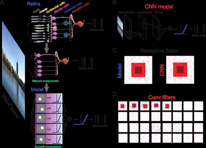
space and time, is encoded by these spatiotemporal patterns of            sual feature. In particular, we outline three views of the retinal
spikes from ganglion cells.                                               neuronal circuit as feedforward, recurrent and winner-take-all
   Many types of eye diseases are caused by neuronal degener-             network structures. For each of these three viewpoints, we pro-
ation of photoreceptors, whereas the outputs of the retina, gan-          vide some evidence and recent results that fit into the proposed
glion cells, remain healthy. One type of therapy would be to              framework.
develop an advanced retinal prosthesis to directly stimulate gan-            In Sec. 4, feature-based modeling approach is discussed,
glion cells with an array of electrodes. Retinal neuroprosthesis          where the models of encoding and decoding visual scenes based
also has a relatively long history of research [15]. However,             on feature extraction by the retina are reviewed. For encod-
much effort is dedicated to material design of retinal neuropros-         ing, we first summarize biophysical models that directly ana-
thesis hardware [13, 14, 15, 16, 17, 18]. Recently, it has been           lyze and fit neuronal spikes to obtain some neuronal properties
suggested that employing better neural coding algorithms were             such as receptive field of the neuron. Then we review some en-
able to improve the performance of retinal neuroprosthesis [10],          coding models based on artificial neural networks (ANNs) that
where it was shown that on top of neuroprosthesis, reconstruc-            use recent state-of-the-art machine learning techniques to ad-
tion of visual scenes can be significantly improved by adding             dress complex natural scenes. For decoding, however, one has
an encoder converting input images into spiking codes used by             to rely on statistical and machine learning models aiming for
retinal ganglion cells, then using these codes to drive transduc-         reconstruction of visual scenes from neuronal spikes. We re-
ers, such as electrodes, optogenetic stimulators, or other com-           view some of these decoders with an emphasis on how they can
ponents for vision restoration.                                           be used for retinal neuroprosthesis to get a better performance
   Therefore, one needs better computational models to advance            for both static images and dynamical videos.
the performance of retinal neuroprosthesis. Comparing to other               In Sec. 5, sampling-based modeling approach is discussed,
neuroprostheses where stimulus signals are relatively simple,             where we give an overview of the retinal circuitry in which vi-
retinal neuroprostheses deal with dynamical visual scenes in              sual computation can be implemented by probabilistic graph
space and time with higher order correlations. Low perfor-                models and spiking neuronal networks, such that different func-
mance is mainly due to a major difficulty that there is no clear          tional networks can conduct visual computations observed in
understanding of how ganglion cells encode rich visual scenes.            the retina. We first introduce the basis of neural computation
Much of our knowledge has been documented through exper-                  with spikes. Some modeling frameworks about neuronal spikes
iments with simple artificial stimuli, such as white noise im-            and spiking neural networks (SNNs) are discussed with a sam-
ages, bars, and gratings, etc. It remains unclear how our retina          pling perspective. We then propose that studying of the retinal
processes complex natural images with its neuronal underpin-              computation should go beyond the classical description of dy-
nings. In recent years, artificial intelligence has seen remark-          namics of neurons and neural networks by taking into account
able progress in analyzing complex visual scenes, including               probabilistic inference. We review some of the recent results
natural images and movies. Thus, now it is possible to develop            about how to implement probabilistic inference with SNNs.
novel functional artificial intelligence models to study the en-          Traditionally, these approaches are applied to theoretical stud-
coding and decoding of natural scenes by analyzing retinal gan-           ies of the visual cortex. Here we demonstrate that how one can
glion cell spiking responses.                                             use these similar computational approaches for the retinal com-
   In this paper, we review some of the recent progress on this           putation.
topic. Most of the studies on visual coding can be roughly clas-             Finally, Sec.5 concludes the paper with discussion for some
sified into two streams. The first and traditional stream can be          possible research directions in the future.
named as feature-based modeling approach, where visual fea-
tures or filters can be aligned with some biophysical properties,         2. Visual computation in neuronal circuit of the retina
such as receptive field, of the retinal neurons. The second and
relative new stream can be named as sampling-based modeling                  Fig. 1 shows a typical setup of the retinal neuronal circuit.
approach, where statistics of visual scenes, such as pixels, are          Roughly, there are three layers of networks consisted of a few
formulated by some probabilistic models. We review some of                types of neurons. Following the information flow of optical vi-
the core ideas emerged from both approaches for analysis of vi-           sual scenes, photoreceptors convert the light with a wide spec-
sual scenes with the utility of neural spikes with the aim for the        trum of intensities, from dim to bright, and colors, from red,
next generation of retinal neuroprosthesis where computational            green to blue, into electrical signals that are then modulated by
modelling plays an essential role.                                        inhibitory horizontal cells. Next, these signals are transferred
   The organization of this review is as follows.                         to excitatory bipolar cells that carry out complex computations.
   Sec. 2 gives an introduction of the biological underpinnings           The outputs of bipolar cells are mostly viewed as graded sig-
of the retina with a focus on its inner neuronal circuit. We em-          nals, however, the recent evidence suggests that bipolar cells
phasize that the retinal circuit carries out rich computations that       could generate some fast spiking events [19]. Then, inhibitory
are beyond the dynamics of single cells of the retina.                    amacrine cells modulate these outputs in different ways to make
   In Sec. 3, in contrast to the view that the retina is a simple         computations more efficient, specific and diverse [20]. At the
neural network, we hypothesize that the retina is highly com-             final stage of the retina, the signals pass to the ganglion cells
plex and comparable to some aspects of the cortex with differ-            for final processing. In the end, ganglion cells send their spikes
ent network motifs for specialized computations to extract vi-            to the thalamus and cortex for higher cognition.
                                                                      2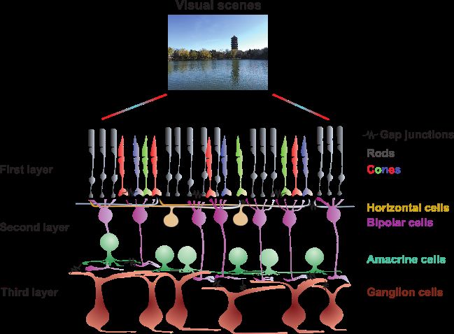
Fig. 1. Illustration of the retinal neuronal circuit. Visual scenes are converted by photoreceptors at the first layer, where rods encode
the dim light, and cones encode color. Then the signals, after modulated by horizontal cells, send to bipolar cells at the second
layer. The outputs are sent to the third layer consisted of amacrine cells and ganglion cells for further processing. Final signals of
the retina are the spikes of ganglion cells transferred to the cortex. Besides chemical synapses between cells, massive gap junctions
exist between different and same types of cells, e.g. ganglion-ganglion cells.

   Each type of neuron in the retina has a large variation of               The retinal ganglion cells are the only output of the retina,
morphology, for example, it has been suggested that in the               but their activities are tightly coupled and highly interactive
mouse retina, there are about 14 types of bipolar cells [21, 22],        with the rest of the retina. These interactions not only make
40 types of amacrine cells [23], and 30 types of ganglion                the retinal circuitry complicated in its structure, but also make
cells [24]. Besides neurons, one unique feature of any neu-              the underlying computation much richer for visual processing.
ronal circuitry is the connections between neurons. Typically,           Therefore, the retina should be considered smarter than what
connections between neurons in the retina are formed by vari-            scientists believed [34]. These observations lead us to rethink
ous types of chemical synapses. However, there are a massive             the functional and structural properties of the retina. Given such
number of electrical synaptic connections, or gap junctions,             a complexity of neurons and neuronal circuits in the retina, we
between different types of cells and within the same type of             propose that the computations of visual scenes carried by the
cells [25, 26, 27, 28]. It remains unclear what is the functional        retina need to go beyond the view that the retina is just like a
role of these gap junctions [25]. We hypothesize that gap junc-          feedforward network making the information go through. Like
tions have a functional role of recurrent connections to enhance         the cortical cortex, the retina also has lateral inhibition and re-
visual computation in the retina, which will be discussed in later       current connections (e.g. gap junctions), which make the retina
sections.                                                                inherit various motifs of neural networks for specific compu-
                                                                         tation of extracting different features of visual scenes, just like
   In the field of the retinal research, most of the studies are
                                                                         the visual processing occurred in the visual cortex [35, 36, 37].
based on the traditional view that neurons in the retina have
static receptive fields that are considered as spatiotemporal fil-
ters to extract local features from visual scenes. We also know             It should be noted that compared to the visual cortex, the de-
that the retina has many levels of complexities in the infor-            tailed understanding of computation and function of the retina
mation processing, from photoreceptors, bipolar cells, to gan-           for visual processing has just emerged in recent several decades.
glion cells. In addition, the functional role of modulation of in-       Nowadays, the retinal computation of visual scenes by its neu-
hibitory horizontal and amacrine cells are still unclear [20, 29].       rons and neuronal circuits is also refined at many different lev-
Perhaps, the only relative well-understood example is the com-           els, for details, see recent reviews on neuroscience advance-
putation of direction selectivity in the retina [30, 31, 32, 33].        ments on the retina [20, 21, 25, 26, 27, 28, 29, 34].
                                                                     3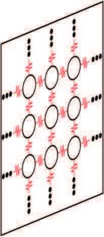
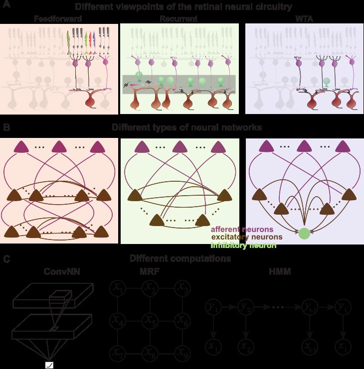
3. Computational framework for the retina                                important for the retina. The formation of recurrent connec-
                                                                         tions in the retina is mainly produced by a massive number
   Different pieces of neuroscience experimental evidence from           of gap junctions as in Fig. 2(A). Unlike chemical synapses,
the retinal circuit seems to be hard to unify from the viewpoint         gap junctions are bidirectional or symmetric. Within and be-
of biology [38]. Here we instead hypothesize that one has to             tween all types of cells in the retina, gap junctions are used to
study the computation carried out by the retinal circuit with a          form short connections between neighboring cells. However,
combination of diverse neural network structure motifs. Such             the functional role of these gap junctions remains unclear [25].
an as-yet-to-emerge computational framework could benefit our               From the computational viewpoint, recurrent connections
understanding of visual computation by utilizing emerged ma-             formed by gap junctions make the retinal circuit like a prob-
chine learning techniques in recent years [39]. When looking             abilistic graphical model (PGM) of undirected Markov ran-
at the complete overview of the retinal neuronal circuitry as            dom field (MRF) as in Fig. 2(B-C). PGM provides a powerful
in Fig. 1, it seems rather complicated. After extracting some            formalism for multivariate statistical modeling by combining
features of network structures, there are some simple network            graph theory and probability theory [48]. It has been widely
motifs emerged. Here we only focus on three types of network             used in computer vision and computational neuroscience. In
structures: feedforward, recurrent and winner-take-all networks          contrast to MRF, there is another type of PGM that is mainly
as illustrated in Fig. 2, and hypothesize that they play different       referred to Bayesian network, in which the connections have
functional roles in visual computation of the retina. However,           a direction between nodes. Fig. 2(C) shows one type of
the retina is more than a hybrid of these three network motifs,          Bayesian network, termed hidden Markov model (HMM). In
but consists of multiple types of networks to form a hypercir-           recent years, much effort has been dedicated to implement these
cuit [38], where more computational features can be extracted            PGMs by SNNs, which setup an insightful connection between
with the advancement of experimental and computational tech-             artificial machine computation by PGMs and neural computa-
niques. Such a hypercircuit view provides the biological basis           tion observed in the brain, as well as visual computation in the
for a potential unified framework of the retinal computation,            retina.
although how these different networks work together more effi-
ciently for visual computation is still an open question.
                                                                         3.3. Winner-take-all network
3.1. Feedforward network                                                    Finally, we hypothesize that the retinal circuit has a compu-
   Feedforward network is the most classical view of the retina          tational network unit as winner-take-all (WTA) motif. In the
as the direction of the visual information flow as in Fig. 2(A-          cortical cortex, WTA circuit has been suggested as a powerful
B). Feedforward information flow of the light goes through               computational network motif to implement normalization [49],
the retina by three major types of cells, photoreceptors, bipo-          visual attention [50], classification [51], and others [52].
lar cells and ganglion cells. The other two types of inhibitory             In the retina, there are two types of inhibitory neurons sit-
cells play a modulation role, which has been ignored simply in           ting in the first two layers. Horizontal cells target photorecep-
this viewpoint. The biological basis of this view can be seen            tors and relay the light information to bipolar cells. Amacrine
from the fovea where excitatory cells play a major role, but in-         cells modulate the signals between bipolar cell terminals and
hibitions are little [40]. In the fovea, there is a direct cascade       ganglion cell dendrites. In both types of cells, there are some
processing from photoreceptors, to bipolar cells, and then gan-          specific subtypes that are wide-filed or polyaxonal such that
glion cells as outputs.                                                  they spread action potentials over a long distance greater than 1
   The advantage of feedforward network has been demon-                  mm [38]. From the computational viewpoint, this hypercircuit
strated by the advancement of ANNs in recent years, in par-              feature of the retina plays a similar functional role as a WTA
ticular, some breakthroughs have been made in the framework              network motif. The recent study shows that MRF can be im-
of deep convolutional neural networks (CNNs) [39]. A simple              plemented by a network of WTA circuit, which suggest that the
CNN with three layers as in the retina is shown in Fig. 2(C),            WTA could be the minimal unit of probabilistic inference for
where a convolutional filter plays a role of the receptive field         visual computation [53].
of retinal cell. A cascade processing of visual inputs is com-
puted by the receptive field of each individual neuron in the            3.4. Rich computation with network motifs
retina. The pooling of computation from the previous layer                  Above we briefly reviewed the retinal circuitry and pointed
passes to a neuron in the next layer. Recent studies highlight           out three basic neural network motifs that play a role as units for
the similarity between the structure of CNNs and retinal neural          complex computations conducted in the retina. However, there
circuitry [41, 42], which will be discussed in later sections.           are more different network motifs suggested in cortical micro-
                                                                         circuits [37], and these motifs are also suggested to involved
3.2. Recurrent network                                                   in the retinal computation to form the retinal hypecircuit [38].
   Recurrent network has been seen as a major type in the cor-           Such a hypercircuit view of the retina makes most of the meth-
tical cortex. The dynamics of recurrent network [43, 44, 45],            ods, which are developed for studying visual processing in the
together with the diversity of synaptic dynamics and plastici-           cortex, transfer to investigate the retinal computation that em-
ties [46, 47], are important for understanding the brain’s func-         bed rich dynamics beyond the traditional view of the retina [34].
tion. Here we hypothesize that recurrent connections are also            In particular, quite a few visual functions have been found to be
                                                                     4
Fig. 2. Illustration of different computational network motifs. (A) Part of retinal circuity shows different network motif as
feedforward, recurrent and winner-take-all (WTA) subnetwork. (B) Abstract representation of different types of neural networks
used by modeling, where stimulus is first represented by the activities of afferent neurons, and then feed into a network of excitatory
and/or inhibitory neurons for computation. Shadowed networks indicate the same motifs. (C) Abstract computation specifically
used by certain typical ANNs, such as convolutional neural networks (ConvNNs), Markov random fields (MRFs) and hidden
Markov models (HMMs). Note ANNs can use one or mixed computational network motifs shown in (B).

implemented by some certain types of network mechanisms in               mulated by various types of probabilistic models, where dif-
the retina, see Ref. [34] for a detailed discussion.                     ferent types of network motifs can implement certain compu-
   Recent computational advancements in the field of ANN                 tations [57]. Not only in higher part of the cortex, but also
make many breakthroughs on visual tasks. For instance, deep              in the visual cortex, there are numerous computational tech-
CNN is a hierarchical network modeling of visual computa-                niques in Bayesian models suitable for visual processing of the
tion from the retina to inferior temporal part of the cortex [54].       retina [56].
These feature-based models take advantages of the receptive                 However, these two approaches are not completely separate,
field to capture visual features. However, CNN models suffer a           and in fact, there are more close interactions between them [55].
few disadvantages for visual computation, for instance, the ar-          We will explain these ideas by using the retina as a model sys-
chitecture of CNN is largely lacking design principles, which            tem in below sections: feature-based approach will be discussed
may be enhanced by biological neural networks in the brain,              in Sec.4, and sampling-based approach will be discussed in
including the retina [55].                                               Sec.5.

   On the other hands, it has been suggested that one needs a
                                                                         4. Encoding and decoding models of the retina
hierarchical Bayesian inference framework to understand vi-
sual computation [56]. In such a sampling-based modeling                   Neural coding is one of the central questions for systems neu-
approach, statistical computation of visual scenes can be for-           roscience [58, 59, 60]. In particular, for visual coding, it is to
                                                                     5
understand how visual scenes are represented by neuronal spik- some parts of receptive field; 2-layer linear-nonlinear network
ing activities, and in turn, how to decode neuronal spiking activ- model [74], where a cascade process is implemented by 2-layer
ities to represent the given visual information. The retina serves LN models; spike-triggered non-negative matrix factorization
as a useful system to study these questions. (STNMF) model [75], where the orthogonality constraint used
in spike-triggered covariance is relaxed to obtain a sets of non-
4.1. Biophysical encoding model orthogonal subunits shown as the bipolar cells in the retina. It
For understanding the encoding principles of the retina, quite has been further shown that STNMF can recover various bio-
a few models are developed based on biophysical properties of physical properties of upstream bipolar cells, including spa-
neurons and neuronal circuits in the retina, which have been re- tial receptive fields, temporal filters, transferring nonlinearities,
viewed recently [63]. Here we briefly review some approaches. synaptic connection weights from bipolar cells to ganglion cell.
The starting point for looking at retinal neuronal computa- In addition, a subset of spikes contributed by each bipolar cell
tion is to find the receptive fields (RFs) of neurons. The clas- can also be teased apart from the whole spike train of one gan-
sical approach to mapping the neuronal RF is to patch a sin- glion cell [61].
gle cell and then vary the size of a light spot to obtain the RF
structure as a difference-of-Gaussian filter with center excita- 4.2. ANN-based encoding model
tion and surround inhibition. Later on, a systematic experi- In recent years, ANNs, such as deep CNNs and probabilistic
mental method was developed by using multielectrode array to graph models, make some breakthroughs for numerous prac-
record a population of retinal ganglion cells, in which one can tical tasks related to system identification of visual informa-
manipulate light stimulation with various types of optical im- tion [39]. For instance, with a large set of visual images col-
ages, including simple bars, spots, gratings, white noise, and lected and well-labeled by specific tags, ANNs can outperform
complex well-controlled images and movies. In particular, one the human-level performance for object recognition and classi-
can analyze the spike trains of individual neurons when record- fication [39]. Various techniques have been developed to visu-
ing a large population at one time with white noise stimulus. alize the features of images learned by CNN. However, end-
A simple reverse correlation technique, termed spike-triggered to-end learning of complex natural images makes CNN not
average (STA) [64], can obtain the RF of every recorded gan- very interpretable for the underlying network structure compo-
glion cell. An extension of STA to covariance analysis, termed nents [77, 78].
spike-triggered covariance, serves as a powerful tool for ana- Inspired by experimental observation in neuroscience [79,
lyzing the 2nd order dynamics of the retinal neurons [65, 66]. 55], a typical deep CNN has a hierarchical architecture with
With the receptive field mapped from each neuron, a simple many layers [80]. Out of these layers, there are some layers
and useful analysis is based on a linear-nonlinear (LN) model having a bank of convolutional filters, such that each convolu-
to simulate the cascade processing of light information. There tional filter is served as a feature detector to extract important
are two stages in the LN model [67, 68]. The first stage is a property of images [81, 82]. Therefore, after training with a
linear spatiotemporal filter encoding the way of integrating in- large set of images, these convolutional filters play a functional
puts, which represents the sensitive area of the cell, i.e., the role as neurons in our retina and other visual systems to en-
characteristic of the receptive field. The second stage is a non- code complex statistical properties of natural images [59]. The
linear transformation to convert the output of the linear filter to shapes of these filters are sparse and localized, and like recep-
a firing rate. Both properties of the LN model can be easily es- tive fields of visual neurons.
timated from the spikes with white noise stimulus [66]. Other- Therefore, it is not trivial to use similar the ANN-based ap-
wise, for complicated stimulus signals rather than white noise, proache to investigate the central question of neuronal cod-
one has to use other methods, such as maximum likelihood es- ing in neuroscience [83, 54]. In particular, for visual coding,
timation [67] and maximally informative dimensions [69], to it has been widely accepted that the ventral visual pathway
estimate the model components when there are enough data. in the brain is a path starting from the retina, lateral genicu-
Until now, quite a few models are developed to refine the late nucleus, then layered visual cortex to reach inferior tem-
building blocks of LN model to advance the model to be more poral part of the cortex. This visual pathway has been sug-
powerful, such as linear-nonlinear Poisson model [65], where gested as the “what pathway” for recognition and identifica-
after nonlinear operation, a Poisson process is used to deter- tion of visual objects. When CNN is used to model experi-
mine whether a spike would be generated; generalized lin- mental neuroscience data recorded in neurons of inferior tem-
ear model [70], where several more components are included, poral cortex in monkeys, neuronal response can be predicted
such as a spike history filter for adaptation, and a coupling very well [84, 85, 86, 54]. Therefore, it is possible to relate the
filter for influence of nearby neurons. Recently, the models biological underpinnings of visual processing in the brain with
with a few components of subunits to mimic upstream non- those network structure components used in CNN. However,
linear components are emphasized, such as nonlinear input it is not straightforward to interpret this relationship since the
model [71], where a few upstream nonlinear filters are included pathway from the retina to inferior temporal cortex is compli-
with the assumption that the input of the neuron is correlated; cated [54]. One possible and easier way is to use CNN to model
spike-triggered covariance model [72, 73, 66], where covari- the early visual system of the brain, in particular, the retina as
ance of spike-triggered ensemble is analyzed with eigenvector introduced above, in which neuronal organization is relatively
analysis to obtain a sequences of filters as a combination of simple.
6Fig. 3. Encoding visual scenes by simplified biophysical model with CNN approach. (A) Simplification of a retinal circuitry to a
biophysical model. (Top) Feedforward network represented as part of retina circuitry receiving incoming visual scenes and sending
out spike trains from ganglion cells. (Middle) Minimal network with one ganglion cell and five bipolar cells. (Bottom) Biophysical
model with five subunits representing five bipolar cells, where each has a linear filter as the receptive field, and a nonlinearity. The
outputs of five subunits are pooled and rectified by another output nonlinearity. The final output can be sampled to give a spike
train. (B) Representative CNN model trained with images as input and spikes as output. Here there are two convolutional layers and
one dense layer. (C) After training, the CNN model shows the same receptive field as the biophysical model for modeled ganglion
cell. (D) Convolutional filters after training resemble the receptive fields used by the biophysical model of bipolar cells in (A). (A)
is adapted from Ref. [61]. (B-D) are adapted from Ref. [41, 62].
Indeed, a few studies take this approach by using CNNs and the retina. Indeed, it is found that CNNs can learn their inter-
their variations to earlier visual systems in the brain, such as nal structure components to match the biological neurons of the
the retina [41, 42, 87, 88, 62], V1 [89, 90, 91, 92, 93], and retina [42, 62], as illustrated in Fig. 3 (D).
V2 [94]. Most of these studies are driven by the goal that the Given that the retina has a relatively clear and simple cir-
better performance of neural response can be archived by us- cuit, and the eyes have (almost) no feedback connection from
ing either feedforward and recurrent neural networks (or both). the cortical cortex, it is a suitable model system as a feedfor-
These new approaches increase the complexity level of sys- ward neural network, similar to the principle of CNN. Certainly,
tem identification, compared to conventional linear/nonlinear the contribution from the inhibitory neurons, such as horizon-
models [71]. Some of these studies also try to look into the tal cells and amacrine cells, play a role for the function of the
detail of network component after learning to see if and how retina. In this sense, the potential neural networks with lateral
it is comparable to the biological structure of neuronal net- inhibition and/or recurrent units are desirable [87, 95].
works [41, 42, 93].
4.3. Decoding visual scenes from retinal spikes
Fig. 3 shows a typical setup of CNN modeling approach for From the viewpoint of retinal neuroprosthesis, an ideal en-
the retina. To understand the fine structure of the receptive field coder model is able to deliver precise stimulation to electrodes
in the retinal circuit, this is important to understand the filters with given visual scenes. For this, one has to close the loop
leaned by CNNs. In contrast to the studies where a population to find an ideal decoder model that can readout and reconstruct
of retinal ganglion cells are used [42, 93, 95], one can sim- stimulus of visual scenes from neuronal responses.
plify the model from a complicated retinal circuit to a simple Reconstruction of visual scenes has been studied over many
network model as in Fig. 3(A), which makes the modeling eas- years. The neuronal signals of interest can be fMRI hu-
ier to refine the structure components at the single cell level of man brain activities [96, 97, 98, 99], neuronal spikes in the
7Image RGC population RGC spikes Decoder Decoded image
Neuron #
Spike-Image Decoder
1s
Spike-Image Decoder
End Spike-Image converter Image-Image autoencoder End
RGC spikes Dense Dense Dense Intermediate image Image
(# RGC) (512) (# Pixel)
Conv filter
Neuron #
Fig. 4. Decoding visual scenes from neuronal spikes. (Top) Workflow of decoding visual scenes. Here a salamander swimming
video was presented to a salamander retina to get a population of ganglion cells fired with a sequence of spikes. A population of
spike trains is used to train a decoder, spike-image decoder, to reconstruct the same video. Receptive fields of ganglion cells are
mapped onto the image. Each colored circle is an outline of receptive field. (Bottom) Spike-image decoder is an end-to-end decoder
with two stages: spike-image converter used to map a population of spikes to a pixel-level preliminary image, and image-image
autoencoder for mapping every pixel to the target pixels in the desired images. Note that there is no unique architecture of spike-
image decoder, and the state-of-art model could be adopted and optimized. Exact preliminary images depend on the loss functions
used for training. Details of the decoding process can be found in Ref. [76] and online (see Sec. 5).
retina [100, 101, 102, 103] and lateral geniculate nucleus [104], used to map spikes of every ganglion cell to images at the pixel
neuronal calcium imaging data in V1 [105]. However, the de- level. After that, one can apply autoencoder deep learning neu-
coding performance of current methods is rather low for natural ral network to transfer/enhance spike-based images to original
scenes, either static natural images or dynamical movies. One stimulus images. Essentially, this approach has two stages with
particularly interesting example of the movies reconstructed one as spike-image converter and the other as image-image au-
from fMRI data can be found from Ref. [98]. toencoder. Most of the previous studies focused on the first
For the retinal neuroprosthesis, one would expect to decode stage, which is the traditional decoder to be optimized by some
visual scenes by using spiking responses of a population of gan- statistical models and/or ANN-based models in either linear or
glion cells. Decoding of visual scenes is possible when there nonlinear fashion [96, 97, 98, 99, 100, 101, 102, 103, 104]. A
are enough retinal ganglion cells available as shown in a recent recent study trained a separate CNN autoencoder as the second
study with simulated retinal ganglion cells [101]. However, it is stage to enhance the quality of images [101]. Instead, we found
unclear whether one can use experimental data to achieve this a better quality can be achieved by the end-to-end training pro-
aim. One can name this decoding approach as a spike-image cess with both stages of spike-to-image converter and image-to-
decoder that performs an end-to-end training process from neu- image autoencoder together. However, the detailed architecture
ronal spikes to visual scenes. of networks used in these two stages could be optimized to an
even better quality with other possible deep learning neural net-
Recently, we developed such a decoder with a model of deep
works.
learning neural network that can achieve a much better reso-
lution than previous studies for reconstructing natural visual
scenes, including both static images and dynamic videos, from 5. Modeling the retina with SNNs and PGMs
spike trains of a population of retinal ganglion cells recorded
simultaneously [76]. SNNs are thought as the third generation of ANN models,
The workflow of the spike-image decoder is illustrated in which use neuronal spikes for computation as in the brain [108].
Fig. 4. With a setup of multi-electrode array, a large population Together with neuronal and synaptic states, the importance of
of the retinal ganglion cells can be recorded simultaneously, and spike timing is also considered in SNNs. It has been proved
their spikes can be extracted. Then, a spike-image converter is that SNNs are computationally more powerful than other ANNs
8A Photoreceptor net B MRF WTA
xj
xj z lj
zj1 z 2j
zjL
zi1 z i2
xi zik ziK
xi
Gap junction
C Input CNN Output
ge s
Ima tor ge
isy cep Ima
No otore ised
Ph no
De
D MRF x3
x2
x1 x6
x5
x4 x9
x8 et ge
al N Ima
x7 eur
ge ent
N ised
Ima urr De
no
oisy Rec
N
Fig. 5. Implementation of noise reduction computation with the retinal photoreceptors, probabilistic graph model and spiking
neural network. (A) A network of rod photoreceptors connected by gap junctions. (B) A graph of Markov random field (MRF)
represented by a network of spiking neurons with subnetworks as winner-take-all (WTA) circuits. Each variable of MRF is rep-
resented by one WTA neural network. (C) Noisy images can be denoised by photoreceptor network, then enhanced by CNN. (D)
Noisy images can be denoised by MRF implemented by a recurrent spiking neuron network without enhancement. (A) and (C) are
adapted from Ref. [106]. (B) is adapted from Ref. [53]. (D) is adapted from Ref. [107].
with the same number of neurons [108]. In recent years, SNNs demonstrated by the classical Hodgkin-Huxley model [120].
have been widely studied in a number of research areas [109, However, the computational principles used in the brain seem
110, 111]. In particular, recent studies show that SNNs can be to go beyond this viewpoint [57].
combined with a deep architecture of multiple layers to obtain There is an increasing volume of neuroscience evidence that
the similar or better performance as ANNs [112, 113, 114, 115, humans and monkeys (other animals as well) can represent
116]. In addition, the spiking feature of SNNs is particularly probabilities and implement probabilistic computation [121,
important for the next generation of neuromorphic computer 122, 123], and the viewpoint of the probabilistic brain is in-
chips [117, 118]. creasingly recognized [124]. Therefore, the network of spiking
The computational capability of a single neuron is limited. neurons has been used to implement probabilistic inference at
However, when a population of neurons are connected together the neural circuit level [124]. The combination of SNNs with
to form a network, their computational ability can be greatly probabilistic computation shows an increasing research interest
expanded. In terms of the language of graphs [119], a SNN can for both understanding the principles of brain computation and
be denoted as a graph G = (V, E), of which V represents the set solving practical problems with these brain-inspired principles.
of neurons and E ⊂ V × V represents the set of synapses. Given Traditionally, probabilistic inference studied in the frame-
this equivalence between graphs and neural networks, a dif- work of PGM is a combination model of probability theory
ferent approach termed probabilistic graphical models (PGMs), and graph theory. The core idea of PGMs is taking advan-
has also been intensively studied over recent years. The idea of tage of a graph to represent the joint distribution among a set
PGM is in that, traditionally, both ANNs and SNNs are doing of variables, of which each node corresponds to a variable and
modeling as a deterministic dynamical system, which has been each edge corresponds to a direct probabilistic interaction be-
9tween two variables. With the benefit of a graph structure, a larity at the network level between a network of photoreceptors
complex distribution over a high-dimensional space can be fac- connected by gap junctions (Fig. 5(A)), a Markov random field
torized into a product of low-dimensional local potential func- model (Fig. 5(B)), and an implementation of MRF by a net-
tions. PGMs can be divided into directed graphical models, work of spiking neurons consisted of clusters of winner-take-
such as Bayesian network, and undirected graphical models, all microcircuits (Fig. 5(B)). As illustrated in Fig. 2, massive
such as Markov random fields. Bayesian networks can rep- gap junctions play a functional role of recurrent connections be-
resent causality between variables, so they are often used to tween retinal neurons. A recent study shows that a network of
model the process of cognition and perception. While Markov rod photoreceptors with gap junctions can denoise images that
random fields can represent a joint distribution by a product of can be further enhanced by an additional CNN as in Fig. 5(C).
local potential functions. It is found that this CNN with photoreceptors included, com-
Implementing PGMs by SNNs is to explain how neuronal paring to other traditional CNNs, can achieve state-of-the-art
spikes can implement probabilistic inference. Inference in performance for denoising [106]. Similarly, PGM has also been
SNNs includes two main questions. 1.) Probabilistic coding: used to denoise images [167]. Recently, it is shown that PGMs
how neural activities of a single cell or a population of cells can be implemented by SNNs for various types of computa-
(like membrane potential and spikes) encode probability dis- tions [53, 164, 168, 169, 170, 171], thus, when using SNNs for
tribution. 2.) Probabilistic inference: how the dynamics of denoising, similar performance can be achieved [107], as illus-
a network of spiking neurons approximate the inference with trated in Fig. 5(D).
probabilistic coding. Probabilistic graphical models have been intensively stud-
Obviously, probabilistic coding is the precondition of prob- ied and used for visual coding, but mostly for the cortical pro-
abilistic inference. According to the way of expressing prob- cess [56]. Here these results suggest that one can study visual
ability, probabilistic codes can be divided into three basic computation in the retina by combing several approaches into
types: 1) those that encode the probability of each variable a systematical framework, including classical PGMs, nontrivial
in each state, such as probability code [125], log-probability retinal circuit structures, in particular, gap junctions, and recent
code [126, 127], and log-likelihood ratio code [128, 129], 2) efforts about the implementation of PGMs by SNNs. Future
those that encode the parameters of a distribution, such as prob- work is needed to study this framework with more inspirations
abilistic population code that takes advantage of neural vari- from the rich network structure of the retina, including recur-
ability [130, 131, 132], that is, neural activities in response to rent neural network, winner-take-all circuit, and feedforward
a constant stimulus have a large variability, which suggests that neural network, even other ubiquitous motifs of cortical micro-
population activities of neurons can encode distributions auto- circuits [37].
matically, 3) those that consider neural activities as sampling
from a distribution [133, 134], which has been suggested by Discussion
numerous experiments [135, 136, 137, 138].
According to these coding principles, there are different ways Neuroprosthesis is a promising medical device within the
to implement inference with a network of neurons: 1) imple- framework of precision medicine. With directly talking to the
menting inference with neural dynamics that has equations sim- brain of each individual patient, neuroprosthesis needs to be ad-
ilar to the inference equations of some probabilistic graphical vanced with better computational algorithms for neuronal sig-
models over the time course [126, 127, 129, 139, 140, 141]. nal, besides better hardware designs. For the computational ca-
This approach is mainly suitable for small-scale SNNs; 2) pability of the retinal neuroprosthesis, the major difficulty is in
inference with neural variational approximations that is suit- that one has to track the complexity of spatiotemporal visual
able to describe the dynamics of a large-scale SNN directly scenes.
[53, 56, 142, 143, 144, 145, 146, 147, 148, 149]; 3) inference In contrast to other neuroprostheses, where incoming signals
with probabilistic population coding and some neural plausible are in a low dimensional space, such as moving trajectory of
operations, including summation, multiplication, linear com- body arms/legs in 3D space, or auditory signal in a 1D fre-
bination and normalization [150, 151, 152, 153, 154]; 4) in- quency space, our visual scenes are more complex with infor-
ference with neural sampling over time where the noise, such mation in a spatiotemporal fashion. Recent advancements of
as stochastic neural response found in experimental observa- computer vision make some breakthroughs for analyzing these
tions [155, 156], is the key for neural sampling and infer- complex natural scenes, which make a wave of artificial intelli-
ence [157, 158, 159, 160, 161]. Similarly, one can do the sam- gence up to a high attitude than ever before.
pling by using a large number of neurons to sample from a dis- On the other hand, with experimental advancements in neu-
tribution at the same time [154, 162, 163, 164], as it is found roscience, one can collect a large population of neurons si-
that the states of neurons in some areas of the brain follow spe- multaneously. In particular, in the retina, a population of
cial distributions [165, 166]. spike trains from hundreds of retinal ganglion cells can be
Although the above studies are mostly conducted in an ab- obtained with well-controlled visual scenes, such as images
stract way for neural computation of the cortex, including the and movies [172]. The newest technique can record several
visual cortex. Here we suggest that these computational tech- thousands of neurons simultaneously [173, 174, 175]. This
niques can be transferred to study the retinal computation. opens the gate for studying the encoding and decoding of visual
Fig. 5 shows some examples in the retina where there is a simi- scenes by using enough spikes to achieve a superb resolution.
10Out of the current approaches for retinal neuroprosthesis, the different tasks, of well-trained models are still far from human
implants with electrodes are the mainstream and have been used performance [55]. Sampling-based modelling with neuronal
in clinical trials. However, there are very limited computational spikes emerges as a new approach, which takes advantage of
models embedded into the retinal prosthesis [10, 13, 176]. With many factors of the neuronal system of the brain [57], such as
an encoder embedded, it is possible to process incoming visual noise at the level of single neurons and synapses [161, 158, 52].
scenes to better trigger ganglion cells [10, 13]. The benefit of With the generic benefit of pixel representation of visual scenes,
decoding models is to justify the spiking patterns produced by sampling models can be naturally used for various types of vi-
the targeted downstream neurons. Ideally, electrical stimulation sual computations [167]. However, the efficiency of learning
should be able to close to those desired patterns of retinal neu- algorithms of sampling model is still far from the flexibility
ral activity in a prosthesis. To compare the similarity between of the neuron system of the brain [184]. Nevertheless, these
spiking patterns, the traditional way focuses on how to compute two approaches could be combined by utilizing both advantages
the distance between two spike trains in general [177, 178], and of feature and sampling for visual computation. For this, one
in the context of the retinal prosthesis [179]. Another way of needs to consider the retina as a neuronal network where visual
doing this is to using decoding models for the purpose of better computation can be done by different functional network struc-
performance of neuroprosthesis [180, 101, 10]. Ideally, sim- tures. The future work is needed to combine various network
ilar to the other neuroprostheses, where a closed-loop device motifs into a hybrid network, in which different visual informa-
can be employed to decode neuronal signal to control stimu- tion can be extracted, processed, and computed. Such hybrid or
lus, the signal delivered by a retinal prosthesis should be able to hypercircuit networks have been explored only in very recent
reconstruct the original stimuli, i.e., dynamic visual scenes pro- years so far, in particular, WTA network motif has been shown
jected into the retina. Thus, one can use a decoding model to as a functional module in more complex hypercircuit network
reconstruct visual scenes from the spiking patterns of the retinal model for various types of computations [52, 53, 111, 185].
ganglion cells [10, 101]. Such a direct measure of the precision One expects that there will be more studies align this line in
of spiking patterns with the given decoding model could play future.
a functional role of controlling electrical stimulation patterns The modelling framework mentioned in this paper is not lim-
generated by the retinal neuroprosthesis, which is the goal of a ited to the application of the retina, but could be used to other
better and adjustable neuroprosthesis. visual systems in the brain, and to other artificial visual sys-
Here we only focused on the computational modelling issue tems. The main feature of these algorithms is to make use of
of one type of retinal neuroprosthsis with electrodes embedded. neural spikes. Advancements of recent artificial intelligence
Certainly, for retinal neuroprosthsis, as an engineering system, computing align with the development of the next generation
there are many parallel difficult issues, such as advanced mate- of neuromorphic chips and devices, where the new data for-
rials, power designing, communication efficiency, and other re- mat is processed as spikes or events [186, 187, 188, 189, 190].
lated hardware issues, which have been covered by many well- Therefore, the methods can be applied for neuromorphic visual
written reviews [16, 18, 13, 15]. One should note that there cameras with spike or event signals as well. One can use these
are different types of visual implants, including those with light computational retinal models to simulate a population of spikes
retinal stimulation such as optogenetics and chemical photo- for encoding and decoding of any given visual scenes, includ-
switches, as well as implants in other parts, beyond the retina, of ing static natural images, dynamic videos, even real-time videos
the brain. The computational issues raised in this paper are also captured by standard frame-based camera [76]. Taken neuro-
relevant to the general visual prosthesis. Besides these artificial morphic hardware and event/spiking computing algorithm to-
visual implants, another line of researches focuses on retinal re- gether, the next generation of computational vision can develop
pair by biological manipulation of stem cells, such as induced a better system for artificial vision beyond the purpose of reti-
pluripotent stem cells [181, 182, 183], where understanding the nal neuroprosthesis. Therefore, we believe that rich interactions
computational mechanisms of biological neurons and neuronal between artificial intelligence, computer vision, meromorphic
circuits is more relevant to encoding visual scenes. For which, computing, neuroscience, bioengineering, and medicine, will
the potential decoding models may need more efforts to include be important for advancing our understanding of the brain, and
the biological principles found in the retina [34]. developing the next generation of retinal neuroprosthesis for ar-
Taken together with all these advancements of neuroscience tificial vision system. The algorithm part of the artificial eye, in-
experiment and prosthesis engineering, now it is time to ad- cluding encoding and decoding models of natural visual scenes,
vance our understanding of visual coding by using the retinal will be in particular crucial for such a systems-level approach.
spiking data and ANN-based models to get better computa-
tional algorithms for improving the performance of retinal neu-
roprosthesis. Here we reviewed some of the recent progress on
Data availability
developing novel functional artificial intelligence models for vi-
sual computation. Feature-based modelling approach, such as
deep CNN, has made significant progresses on analysis of com- Data presented in Fig. 4 are publicly available online:
plex visual scenes. For some particular visual tasks, these mod- Retinal experimental data demonstrated are available at
els can outperform the human [39]. However, the efficiency, dx.doi.org/10.5061/dryad.4ch10. Reconstruction examples are
generalization ability, and adaption or transfer learning between available at https://sites.google.com/site/jiankliu.
11References [23] M. Helmstaedter, K. L. Briggman, S. C. Turaga, V. Jain, H. S. Seung,
W. Denk, Connectomic reconstruction of the inner plexiform layer in the
mouse retina, Nature 500 (7461) (2013) 168–174.
References [24] T. Baden, P. Berens, K. Franke, M. R. Rosón, M. Bethge, T. Euler,
The functional diversity of retinal ganglion cells in the mouse, Nature
[1] J. K. Chapin, K. A. Moxon, R. S. Markowitz, M. A. Nicolelis, Real- 529 (7586) (2016) 345.
time control of a robot arm using simultaneously recorded neurons in [25] S. A. Bloomfield, B. Völgyi, The diverse functional roles and regulation
the motor cortex, Nature Neuroscience 2 (7) (1999) 664. of neuronal gap junctions in the retina, Nature Reviews Neuroscience
[2] D. M. Taylor, S. I. H. Tillery, A. B. Schwartz, Direct cortical control of 10 (7) (2009) 495.
3 D neuroprosthetic devices, Science 296 (5574) (2002) 1829–1832. [26] W. N. Grimes, A. Songco-Aguas, F. Rieke, Parallel processing of rod
[3] S. Musallam, B. D. Corneil, B. Greger, H. Scherberger, R. A. Ander- and cone signals: Retinal function and human perception, Annual Re-
sen, Cognitive control signals for neural prosthetics, Science 305 (5681) view of Vision Science 4 (2018) 123–141.
(2004) 258–262. [27] J. O’Brien, S. A. Bloomfield, Plasticity of retinal gap junctions: Roles
[4] C. T. Moritz, S. I. Perlmutter, E. E. Fetz, Direct control of paralysed in synaptic physiology and disease, Annual Review of Vision Science 4
muscles by cortical neurons, Nature 456 (7222) (2008) 639. (2018) 79–100.
[5] M. Velliste, S. Perel, M. C. Spalding, A. S. Whitford, A. B. Schwartz, [28] M. Rivlin-Etzion, W. N. Grimes, F. Rieke, Flexible neural hardware
Cortical control of a prosthetic arm for self-feeding, Nature 453 (7198) supports dynamic computations in retina, Trends in Neurosciences 41
(2008) 1098. (2018) 224–237.
[6] V. Gilja, P. Nuyujukian, C. A. Chestek, J. P. Cunningham, M. Y. Byron, [29] J. B. Demb, J. H. Singer, Functional circuitry of the retina, Annual Re-
J. M. Fan, M. M. Churchland, M. T. Kaufman, J. C. Kao, S. I. Ryu, et al., view of Vision Science 1 (2015) 263–289.
A high-performance neural prosthesis enabled by control algorithm de- [30] K. L. Briggman, M. Helmstaedter, W. Denk, Wiring specificity in the
sign, Nature Neuroscience 15 (12) (2012) 1752. direction-selectivity circuit of the retina, Nature 471 (7337) (2011) 183–
[7] L. R. Hochberg, D. Bacher, B. Jarosiewicz, N. Y. Masse, J. D. Simeral, 188.
J. Vogel, S. Haddadin, J. Liu, S. S. Cash, P. van der Smagt, et al., Reach [31] W. Wei, M. B. Feller, Organization and development of direction-
and grasp by people with tetraplegia using a neurally controlled robotic selective circuits in the retina, Trends in Neurosciences 34 (12) (2011)
arm, Nature 485 (7398) (2012) 372. 638–645.
[8] J. L. Collinger, B. Wodlinger, J. E. Downey, W. Wang, E. C. Tyler- [32] C. Zhang, A. L. Kolodkin, R. O. Wong, R. E. James, Establishing wiring
Kabara, D. J. Weber, A. J. McMorland, M. Velliste, M. L. Boninger, specificity in visual system circuits: from the retina to the brain, Annual
A. B. Schwartz, High-performance neuroprosthetic control by an indi- Review of Neuroscience 40 (2017) 395–424.
vidual with tetraplegia, The Lancet 381 (9866) (2013) 557–564. [33] A. S. Mauss, A. Vlasits, A. Borst, M. Feller, Visual circuits for direction
[9] M. M. Shanechi, A. L. Orsborn, H. G. Moorman, S. Gowda, S. Dangi, selectivity, Annual Review of Neuroscience 40 (2017) 211–230.
J. M. Carmena, Rapid control and feedback rates enhance neuropros- [34] T. Gollisch, M. Meister, Eye smarter than scientists believed: Neural
thetic control, Nature Communications 8 (2017) 13825. computations in circuits of the retina, Neuron 65 (2) (2010) 150–164.
[10] S. Nirenberg, C. Pandarinath, Retinal prosthetic strategy with the capac- [35] S. Navlakha, Z. Bar-Joseph, A. Barth, Network design and the brain.,
ity to restore normal vision, Proceedings of the National Academy of Trends in Cognitive Sciences 22 (2017) 64–78.
Sciences 109 (37) (2012) 15012–15017. [36] R. J. Douglas, K. A. Martin, Neuronal circuits of the neocortex, Annual
[11] B. Seeber, I. Bruce, The history and future of neural modelling for Review of Neuroscience 27 (2004) 419–451.
cochlear implants, Network: Computation in Neural Systems 27 (2-3) [37] O. Braganza, H. Beck, The circuit motif as a conceptual tool for multi-
(2016) 53–66. level neuroscience, Trends in Neurosciences 41 (3) (2018) 128–136.
[12] L. A. Johnson, C. C. Della Santina, X. Wang, Representations of time- [38] F. S. Werblin, The retinal hypercircuit: a repeating synaptic interactive
varying cochlear implant stimulation in auditory cortex of awake mar- motif underlying visual function, The Journal of physiology 589 (15)
mosets (callithrix jacchus), Journal of Neuroscience 37 (29) (2017) (2011) 3691–3702.
7008–7022. [39] Y. Lecun, Y. Bengio, G. Hinton, Deep learning, Nature 521 (7553)
[13] L. Yue, J. D. Weiland, B. Roska, M. S. Humayun, Retinal stimulation (2015) 436–444.
strategies to restore vision: Fundamentals and systems, Progress in reti- [40] R. Sinha, M. Hoon, J. Baudin, H. Okawa, R. O. Wong, F. Rieke, Cellular
nal and eye research 53 (2016) 21–47. and circuit mechanisms shaping the perceptual properties of the primate
[14] S. Ha, M. L. Khraiche, A. Akinin, Y. Jing, S. Damle, Y. Kuang, fovea, Cell 168 (3) (2017) 413–426.
S. Bauchner, Y.-H. Lo, W. R. Freeman, G. A. Silva, et al., Towards high- [41] Q. Yan, Z. Yu, F. Chen, J. K. Liu, Revealing structure components of
resolution retinal prostheses with direct optical addressing and inductive the retina by deep learning networks, arXiv preprint arXiv:1711.02837
telemetry, Journal of neural engineering 13 (5) (2016) 056008. (2017).
[15] R. K. Shepherd, M. N. Shivdasani, D. A. Nayagam, C. E. Williams, P. J. [42] N. Maheswaranathan, L. T. McIntosh, D. B. Kastner, J. Melander,
Blamey, Visual prostheses for the blind, Trends in biotechnology 31 (10) L. Brezovec, A. Nayebi, J. Wang, S. Ganguli, S. A. Baccus, Deep learn-
(2013) 562–571. ing models reveal internal structure and diverse computations in the
[16] D. Ghezzi, Retinal prostheses: progress toward the next generation im- retina under natural scenes, bioRxiv (2018) 340943.
plants, Frontiers in Neuroscience 9 (2015) 290. [43] N. Brunel, Dynamics of networks of randomly connected excitatory and
[17] J. Tang, N. Qin, Y. Chong, Y. Diao, Z. Wang, T. Xue, M. Jiang, J. Zhang, inhibitory spiking neurons, Journal of Physiology - Paris 94 (5-6) (2000)
G. Zheng, et al., Nanowire arrays restore vision in blind mice, Nature 445–63.
Communications 9 (1) (2018) 786. [44] J. K. Liu, D. V. Buonomano, Embedding multiple trajectories in simu-
[18] G. Goetz, D. Palanker, Electronic approaches to restoration of sight, Re- lated recurrent neural networks in a self-organizing manner, Journal of
ports on Progress in Physics 79 (9) (2016) 096701. Neuroscience 29 (42) (2009) 13172–81.
[19] T. Baden, F. Esposti, A. Nikolaev, L. Lagnado, Spikes in retinal bipolar [45] J. K. Liu, Learning rule of homeostatic synaptic scaling: Presynaptic
cells phase-lock to visual stimuli with millisecond precision, Current dependent or not, Neural computation 23 (12) (2011) 3145–3161.
Biology 21 (22) (2011) 1859–1869. [46] V. Zampini, J. K. Liu, M. A. Diana, P. P. Maldonado, N. Brunel,
[20] P. D. Jadzinsky, S. A. Baccus, Transformation of visual signals by in- S. Dieudonné, Mechanisms and functional roles of glutamatergic
hibitory interneurons in retinal circuits, Annual Review of Neuroscience synapse diversity in a cerebellar circuit, eLife 5 (2016) e15872.
36 (2013) 403–28. [47] G. Bi, M. Poo, Synaptic modification by correlated activity: Hebb’s pos-
[21] T. Euler, S. Haverkamp, T. Schubert, T. Baden, Retinal bipolar cells: ele- tulate revisited, Annual Review of Neuroscience 24 (2001) 139–66.
mentary building blocks of vision, Nature Reviews Neuroscience 15 (8) [48] D. Koller, N. Friedman, Probabilistic graphical models: principles and
(2014) 507. techniques, MIT press, 2009.
[22] K. Franke, P. Berens, T. Schubert, M. Bethge, T. Euler, T. Baden, In- [49] M. Carandini, D. J. Heeger, Normalization as a canonical neural compu-
hibition decorrelates visual feature representations in the inner retina, tation, Nature Reviews Neuroscience 13 (1) (2012) 51–62.
Nature 542 (7642) (2017) 439.
12You can also read